International PhD Program in Neuropharmacology
University of Catania
Role of Dopamine D3 receptor in the regulation of memory
related genes
Agata Grazia D’Amico
XXVI cycle
Coordinator: Prof. Salvatore Salomone Tutor: Prof.Velia D’Agata
Table of contents
General Introduction………..……5
Hippocampal neurofibromin and amyloid precursor protein expression in dopamine D3 receptor knock-out mice following passive avoidance conditioning Abstract………...……… ………..………..9
Introduction……….10
Materials and Methods Animals……….11
Passive avoidance test………..12
Measurement of D3R, NF1 and APP levels by quantitative real time PCR...13
Western blot analysis...15
Tissue preparation for immunohistochemical staining...16
Immunohistochemical analysis...16
Statistical analysis...17
Results Cognitive performance of WT and D3R-/- mice in the passive avoidance test...17
D3R expression in the hippocampus of WT mice after the acquisition of passive avoidance trial ...18
Hippocampal D3R immunolocalization in WT mice subjected to PA conditioning ...20
NF1 and APP expression in the hippocampus of WT and D3R-/- mice after the acquisition of passive avoidance trial...21
Discussion...24
Increased hippocampal CREB phosphorylation in dopamine D3 receptor knockout mice
following passive avoidance conditioning
Abstract...27
Introduction...28
Materials and Methods Animals...30
Passive avoidance test...30
Western blot analysis...32
Statistical analysis...33
Results Enhanced phosphorylation of p-CREB in hippocampus of D3R-/- mice trained with passive avoidance test...33
MAPKs activation in the hippocampus of WT and of D3R-/- mice after passive avoidance training...34
D3R-/- mice exhibit increased phosphorylation of Akt which is not dependent on the acquisition of the passive avoidance trial...36
Discussion...38
Acknowledgements...40
Dopamine D3 receptor deletion increases tissue plasminogen activator (tPA) activity in prefrontal cortex and hippocampus Abstract...41
Introduction...42
Experimental Procedures Animals...44
Quantitative real time polymerase chain reaction ...45
Western blot analysis...46
Statistical analysis ...48
Results Tissue plasminogen activator and brain derived neutrophic factor mRNA expression levels are increased in the prefrontal cortex and hippocampus of D3R-/- mice...49
Enhanced tPA expression in D3R-/- mice brain correlates with increased plasmin/plasminogen and mature BDNF/proBDNF protein ratios ...49
D3R deletion increases tPA and mature BDNF immunoreactivity in the prefrontal cortex and hippocampus ...52
D3R-/- mice exhibit increased basal phosphorylation of Akt, DARPP-32 and GSK-3β ...54
Opposite effects of D3R deletion on CREB phosphorylation at Ser133 and Ser129 residues ...56 Discussion...58 Conclusions...61 Acknowledgements...61 General discussion...62 Conclusions...68
General introduction
Dopamine (DA) is the predominant catecholamine neurotransmitter in the brain. The major population of dopaminergic neurons in the brain are synthesized by mesencephalic neurons in the substantia nigra (SN) and ventral tegmental area (VTA). DA neurons originate in these nuclei and project to the striatum, cortex, limbic system and hypothalamus. Through these pathways, DA affects many physiological functions, such as the control of coordinated movements and hormone secretion, as well as motivated and emotional behaviors and cognition (Carlsson, 2001; Gainetdinov and Caron, 2003; Beaulieu and Gainetdinov, 2011; Tritsch and Sabatini, 2012).
The physiological actions of dopamine are mediated by five distinct receptor subtypes but closely related G protein-coupled receptors (GPCRs) that are divided into two major groups: the D1-class dopamine receptors (D1R and D5R) (Tiberi et al.,1991) and D2-class of dopamine
receptors (D2R, D3Rand D4R) (Andersen et al., 1990; Niznik and Van Tol, 1992; Sibley and
Monsma, 1992; Sokoloff et al., 1992; Civelli et al., 1993; Vallone et al., 2000). This classification is generally based on their structural, biochemical properties, on their ability to modulate cAMP production and the differences in their pharmacological properties. It is known that D1-like receptors activate the Gs family of G proteins to stimulate intracellular cAMP
production by adenylate cyclase (AC) (Dearry et al., 1990; Zhou et al.,1990; Grandy et al.,1991; Sunahara et al.,1991), while the D2-like receptors coupled to the Gi family of G proteins induce
inhibition of AC (Bunzow et al.,1988; Dal Toso et al.,1989; Sokoloff et al., 1990; VanTol et al.,1991; Neve et al., 2004). In contrast to the D1-class dopamine receptors, D2 and D3
dopamine receptors are expressed both postsynaptically on dopamine target cells and presynaptically on dopaminergic neurons (Sokoloff et al., 2006; Rondou et al., 2010) (Missale et al., 1998; Sibley et al., 1999).
D3R are involved in reward and reinforcement mechanisms. A growing body of
pharmacological evidences (Gainetdinov et al., 1996; Zapata and Shippenberg, 2002) and genetic studies in D3 dopamine receptor knockout mice (Sibley, 1999) suggest that D3
thereby complementing the D2 autoreceptor’s role in regulating of synthesis and phasic release
of dopamine (De Mei et al., 2009). D3R seem to influence some specific aspects of cognitive
functions that are mediated by hippocampal areas (Missale et al., 1998; Sibley, 1999; Sokoloff et al., 2006).
In the first study we investigated the role of the D3R during acquisition of a behavioral task of
associative memory and we underpinned the molecular events that contribute to the enhanced cognitive performance of D3 knockout mice (Micale et al., 2010).
The rational of our work was based on evidence indicating that D3-/- mice exhibit enhanced
cognitive performance in the single trial step-trough passive avoidance (PA) task as compared to wild type mice (Micale et al., 2010) and data suggesting a functional connection among D3R,
neurofibromin (NF1) and amyloid precursor protein (APP), two genes related to cognitive function (Donarum et al., 2006; Castorina et al., 2011).
The NF1 gene encodes for a large cytoplasmic tumor suppressor protein called neurofibromin, its loss results in constitutive activation of the Ras signalling pathway (Lau et al., 2000), which has been implicated both in neuronal activity and synaptic plasticity (Costa et al., 2002). APP plays an important role in brain development, memory and synaptic plasticity (Nalivaeva and Turner, 2013). Dysfunctions in the metabolic processes of APP are widely hypothesized to underlie Alzheimer’s disease (AD) (Selkoe, 2002). Indeed amyloid β-peptide (Aβ), derived by proteolytic processing of the membrane glycoprotein amyloid precursor protein (APP), lead to the formation of Aβ plaques, synapse dysfunction/loss, neuronal loss, and overall brain atrophy, which cause decline of cognitive abilities (Selkoe, 2002).
Our hypothesis was that D3R expression could be influenced by acquisition of PA task and it
was involved in transcriptional regulation of NF1 and APP.
To address this question we evaluated the mRNA and protein expression levels of D3Rs, NF1
and APP genes in the hippocampus of both wild-type (WT) and D3R-/- mice performing the PA
task. Our results indicated that acquisition of behavioral task leads to increased expression levels of D3Rs and NF1, but not of APP in hippocampus of WT mice. Since NF1 does not
change in its expression in D3R-/- mice, we suggested that D3R might be involved in
trascriptional hippocampal regulation of NF1 gene.
It’s well documented that under dopaminergic tone the two classes of dopaminergic receptors activate different signaling pathways, which can lead to activation of different transcription factors, like CREB, which in turn induce transcription of genes resulting in proteins underline consolidation of memory (Beaulieu and Gainetdinov, 2011). CREB is a transcription factors which plays a critical role in many function including neurogenesis, neural survival, differentiation, synaptic plasticity (Carlezon et al., 2005; Mioduszewska et al., 2003; Silva et la., 1998). The analysis of the intracellular signal transduction pathway that leads from stimulation of dopamine receptors to CREB phosphorylation is an important step toward elucidating the relationship between molecular adaptations and behavioral consequences (Dudman et al., 2003). Since the involvement of D3R in hippocampal CREB activation of mice during PA conditioning
has not investigated yet, we attempted to clarify the role of this receptor in regulating the activity of CREB during PA conditioning, and to underly the signaling pathways involved in D3R-induced CREB activation.
To address this question, we evaluated the posphorylation levels of CREB in hippocampus of WT and D3-/- mice performing PA task, and subsequently we evaluated the
phosphorylation/activation levels of different protein kinase involved in CREB activation, like MAPKs. Our results showed increased phosphorylation levels of CREB and ERK in D3-/- mice
as compared to WT, while JNK and p-38 don’t showed changes in their activation. Serine/threonine kinase Akt is also involved in CREB activation, thus we have also analyzed the phosphorylation levels of Akt in threonine 308 residue. Results reveal that its phosphorylation levels increased only in D3-/- mice, while the acquisition of behavioral paradigm don’t affected
its activation. Data suggest that D3Rs could modulate hippocampal CREB activation probably
through phosphorylation of ERK.
Several studies show that dopaminergic receptors can modulate CREB activation through different downstream substrates, like DARPP-32 (32-kDa dopamine and cAMP-regulated
D2-like receptors may also trigger a cAMP-indipendent pathway and it has been proposed that
D3R participate in D2R cAMP-independent pathway by enhancing D2R-mediated Akt (Thr 308)
phosphoryation (Beaulieu et al., 2007). In particular when DARPP-32 is phosphorylated by PKA on Th34, inhibiting the activity of PP-1, acts in synergic manner with different protein kinases to increase the phosphorylation levels of various downstream effector proteins, among these CREB (Greengard et al., 1999). To the other hand, dephosphorylation of Akt following dopamine binding to D2-like receptor leads to a reduction of kinase activity and a concomitant
activation of its substrates glycogen synthase kinase (GSK-3β), which is negatively regulated by Akt, and this results in inhibitory effects on CREB activation (Beaulieu et al., 2005; 2007; Cross et al., 1995). Furthermore, is well known that CREB phosphorylation leads to activation of CREB responsive genes, among these BDNF (Brain-derived neurotrophic factor) or the enzyme involved in its proteolytic processing such as tPA (tissue plasminogen activator) which catalyzes the conversion of plasminogen in plasmin and leads to converting pro-BDNF in mature BDNF (Boneva and Yamashima, 2012; Ohlsson et al., 1993; Benito and Barco, 2010). However there are not evidences as regard role of D3Rs in basal transcriptional regulation of
tPA.
In the third work we hypothesized that the dopamine D3Rs can influenced baseline tPA activity
in prefrontal cortex and hippocampus, two brain region involved in learning and memory process, by modulation of Akt/CREB signaling cascade.
Results revealed that both in prefrontal cortex and hippocampus of D3-/- mice, tPA, mBDNF and
the expression ratio of plasmin/plasminogeno both mRNA and protein expression levels were significantly increased as compared to the WT mice. Concurrently basal Akt phosphorylation at Thr308, DARPP-32 at Thr34 and GSK3β at Ser9 were significantly increased both in the prefrontal cortex and hippocampus of D3-/- mice. These data suggesting that the increased tPA
Hippocampal neurofibromin and amyloid precursor protein expression in dopamine D3
receptor knock-out mice following passive avoidance conditioning D’Amico A.G.1,2, Castorina A.1, Leggio G.M.2, Drago F.2, D’Agata V.1,2
1Department of Bio-Medical Sciences, Section of Anatomy and Histology; 2Department of
Clinical and Molecular Biomedicine, Section of Pharmacology and Biochemistry, University of Catania, Italy
Abstract
Passive avoidance (PA) conditioning is a fear motivated task able to initiate a cascade of altered gene expression within the hippocampus, a structure critical to learning and memory. We have previously shown that neurofibromin (NF1) and amyloid precursor protein (APP), two genes implicated in cognitive function, are differentially expressed in brain of dopamine D3 receptor
knock-out mice (D3R-/-), suggesting that the receptor might have a role in their trascriptional
regulation. Here in this study, we hypothesized that during acquisition of PA conditioning the expression of NF1 and APP genes could be influenced by D3Rs. To address this issue, we
analyzed the expression of NF1 and APP in the hippocampus of both wild-type (WT) and D3R
-/-mice subjected to the single trial step-through PA paradigm. Our finding demonstrated that (1) D3R-/- mice exhibit increased cognitive performance as compared to wild-type (WT) mice in the
step-through PA trial; (2) acquisition of PA increased D3R and NF1, but not APP expression in
WT mice hippocampus; (3) PA-driven NF1 induction in WT was abrogated in D3R-/- mice and
finally that (4) the heightened basal APP expression observed in naive D3R-/- mice was totally
reversed by acquisition of PA. In conclusion, the present finding show for the first time that both D3R and NF1 genes are upregulated following PA conditioning and suggest that
hippocampal D3Rs might be relevant to NF1 transcriptional regulation in the hippocampus.
Keywords : dopamine D3 receptor, neurofibromin, amyloid precursor protein, passive
Introduction
Dopamine (DA) is a neurotransmitter with a broad array of effects in the central nervous system. The actions of DA are mediated by five distinct G-protein coupled receptors grouped into two subclasses: D1-like (D1R and D5R) and D2-like (D2R, D3R, and D4R), based on their
structural and pharmacological properties (Missale et al., 1998; Karasinska et al., 2005). The D3R, cloned by Sokoloff, (1990), is an autoreceptor mainly distributed within limbic areas, as
well as in brain regions critical to learning and memory, such as the hippocampus (Levant, 1998; Castorina et al., 2011)
Involvement of hippocampal D2-like receptors in mnemonic processes has been attentioned by
several research groups in human (Kaasinen et al., 2000; 2002) and rodents (Laszy et al., 2005; Izquierdo et al., 2006; Micale et al., 2010). Furthermore, it has been suggested that disturbances in hippocampal DAergic systems cause memory impairment (Gasbarri et al., 1996). However, whether hippocampal expression of D3R is influenced by acquisition of a fear-motivated task
has still not been evaluated.
Passive avoidance (PA) conditioning is a fear-motivated task able to trigger altered gene expression within the hippocampus (Izquierdo et al., 2006; Izquierdo et al., 2000; D’Agata and Cavallaro, 2003). Recently, we have shown that D3R-/- mice exhibit changes in the expression of
two genes related to cognitive function, namely NF1 and APP (Castorina et al., 2011; Costa et al., 2002; Marcello et al., 2008).
The NF1 gene encodes neurofibromin, a large protein with Ras GTPase activity (De Schepper et al., 2006; Guilding et al., 2007). Neurofibromin works by inhibiting excessive accumulation of the protein Ras, responsible for the increased GABA-mediated inhibition of hippocampal synaptic transmission. As such, a nonfunctional NF1 gene may ultimately lead to increased GABA activity and consequently learning deficits (Costa et al., 2002; Guilding et al., 2007). Amyloid precursor protein (APP) is a type 1 membrane glycoprotein distributed in the central and peripheral nervous system (Selkoe, 2002; Seabrook and Rosahl, 1999). APP is alternatively processed by three different proteases, α- β- and γ secretases, to produce either non-amyloidogenic or non-amyloidogenic Aβ fragments. These fragments may aggregate and lead to
deposition of senile plaques in the cortex and hippocampus, a hallmark of Alzheimer’s disease (AD) (Moran et al., 1995; Senechal et al., 2008; DeGiorgio et al., 2002; Suh and Checler, 2002). However, despite the pathological significance of APP in AD, its well-known involvement in physiological neuronal function, such as synapse formation, axonal and dendritic outgrowth, suggest that APP may have important implications in signal transduction (De Strooper and Annaert, 2000) and memory (Marcello et al., 2008; Seabrook and Rosahl, 1999; De Strooper and Annaert, 2000). To support this, it has been reported that mice deficient in APP show a decline in memory performance which is associated with a loss of synaptic markers, further implying that APP may be critical for synaptic function and for the neuroplastic events that accompany a learning task (Dawson et al., 1999; Conboy et al., 2005).
Previously we have shown that expression levels of NF1 and APP are modified in various brain regions, including the hippocampus, of D3R-/- mice, suggesting that the receptor might be
implicated in the transcriptional regulation of these two memory-related genes (Castorina et al., 2011).
Herein this study, we hypothesized that during acquisition of PA the expression of NF1 and APP could be influenced by D3Rs. We demonstrated that (1) D3R-/- mice exhibited increased
cognitive performance as compared to wild type (WT) mice in the step-through PA task; (2) that acquisition of PA was associated with increased D3R and NF1, but not APP expression in the
hippocampus of WT mice and finally (3) that D3R is required for PA-driven NF1 induction.
Materials and Methods
Animals
All experiments were carried out on D3R-/- and WT mice (male mice 8-12 weeks old). The
animals were housed four per cage and fed with standard laboratory food and allowed free access to water ad libitum, in an air-conditioned room with a 12 h light-dark cycle. All the experimental procedures were performed during the light cycle (between 10 a.m. and 2 p.m.).
generated by a backcrossing strategy. The genotypes of the D3R mutant and WT mice were
identified by a PCR method by using two pairs of primers flanking either exon 3 of the wild-type D3R or the PGK (phosphoglycerate kinase 1 gene promoter) cassette of the mutated gene
(Accili et al., 1996). All animals were used only once in the experiments, which were carried out according to the European Community Council Directive 86/609/EEC. Efforts were made to minimize animal suffering and to reduce the number of animals used. The rationale, design and methods of this study were approved by the Ethical Committee for Animal Research, University of Catania.
Passive avoidance test
The single trial step-through passive avoidance test was performed as previously described (Venault et al., 1986; Shirayama et al., 2002). using a passive avoidance apparatus (San Diego Instruments, Inc., San Diego, CA, USA). The apparatus was divided into two compartments by a retractable door: a lit safe compartment and a darkened shock compartment.
The experiment was carried out on male homozygous D3R-/- (n=56) and WT mice (n=62). Each
strain of animals was divided in four groups. The first group (naive, n=14 for D3R-/- and n=17
for WTs, respectively) was maintained in the home cage. The rest of the animals experienced a 2-day behavioral training. On the first day, animals were handled by the experimenter for 2 min and then placed into the safe compartment and allowed to explore both chambers of the apparatus for 3 min. The second day, in the training trial, the second group of animals (termed ‘conditioned animals’, CA; n=17 for WT and n=14 for D3R-/-, respectively) were placed in the
safe compartment with the door closed. After 2 min of acclimatization the light was turned on, the door opened and the animal was allowed to enter the dark compartment. After the mouse stepped completely with all four paws into the dark compartment, the door was closed, and a mild inescapable foot shock (0.5 mA, 2 s duration) was delivered from the grid floor. Following the shock, the mouse was removed and returned to its home cage. A third group of animals (termed ‘conditioned stimulus-trained animals’, CSTA; n=14 for each genotype) were placed into the safe compartment. After 2 min of acclimatization the light was turned on, the door
opened and the animal allowed to enter the dark compartment. After the mouse stepped into the dark compartment, the door was closed but no foot shock was delivered from the grid floor. Then mice returned to their home cage. The fourth group of animals (termed ‘unconditioned stimulus-trained animals’, USTA; n=14 for each genotype) were placed in one of the two dark compartments. They were allowed to move freely to both compartments. After 2 min of acclimatization they received an inescapable foot shock (0.5 mA, 2 s duration) and then returned to their home cage.
Six hours later, animals from each of the four experimental groups (n=6, except for naive and CA WTs, n=9) were sacrificed by cervical dislocation, hippocampi were rapidly dissected and stored at -80°C until use.
Twenty-four hours after the training trial, the remaining animals from each of the four experimental groups (n=8) performed the retention test. The animals were placed in the safe compartment with the door closed. After 2 min of acclimatization the light was turned on, the door opened and the animal was allowed to enter the dark compartment. The latency to enter the dark compartment was recorded and used as the measure of retention. Mice avoiding the dark compartment for >300 s were considered to have a step-through latency of 300 s (D’Agata and Cavallaro, 2003; Venault et al., 1986).
Measurement of D3R, NF1 and APP levels by quantitative real time PCR
Hippocampal total RNA extracts from D3R-/- (n=3 for each experimental group) and WT (n=3
for each experimental group) mice were isolated by 1 ml TRIzol reagent (Invitrogen) and 0.2 ml chloroform and precipitated with 0.5 ml isopropanol. Pellet was washed with 75% ethanol and air dried. Single stranded cDNAs were synthesized by incubating total RNA (5µg) with SuperScript III RNase H-reverse transcriptase (200 U/μl) (Invitrogen); Oligo-(dT)20 primer (100
nM) (Invitrogen); 1 mM dNTP mix (Invitrogen), dithiothreitol (DTT, 0.1 M), Recombinant RNase-inhibitor (40 U/μl) at 42°C for 1 h in a final volume of 20 μl. Reaction was terminated by incubation of samples at 70°C for 10 min.
Aliquots of cDNA (400 ng) from WT and D3R-/- mice hippocampi and external standards at
known amounts (purified PCR products, ranging from 102 to 108 copies) were amplified in
parallel reactions, using primer pairs indicated in table 1. mRNA levels of the reference gene, 18S ribosomial subunit, were measured in each amplification. Each PCR reaction contained 0.5 μM primers, 1.6 mM MgCl2+, 1X Light Cycler-FastStart DNA Master SYBR Green I (Roche
Diagnostic). Amplifications were performed using the Light Cycler 1.5 instrument (Roche Diagnostic) with the following program setting : (I) cDNA denaturation (1 cycle: 95°C for 10 min); (II) quantification (45 cycles: 95°C for 10 s, 60°C for 30 s, 72°C for 7 s); (III) melting curve analysis (1 cycle: 95°C for 0 s, 65°C for 15 s, 95°C for 0 s); (IV) cooling (1 cycle: 40°C for 30 s). Quantification was obtained by comparing the fluorescence emitted by PCR products at unknown concentration with the fluorescence emitted by external standards at known concentration. For this analysis, fluorescence values, measured in the log-linear phase of amplification, were estimated with the second derivative maximum method using Light Cycler Data Analysis software. PCR products specificity was evaluated by melting curve analysis. To assess the different expression levels we analyzed the mean fold change values of each sample, calculated using the comparative Ct method (Schmittgen and Livak, 2008). The Ct represents the number of cycles needed to detect a fluorescence above a specific threshold level and it is inversely correlated to the amount of nucleic acids template present in the reaction. The ΔCt was calculated by normalizing the mean Ct of each sample to the mean Ct of the reference gene measured in the same experimental conditions. For the quantification of each gene we considered the naive WT mice group as the positive sample (calibrator sample). The ΔΔCt of each sample was then calculated by subtracting calibrator ΔCt to sample ΔCt. The formula 2–
ΔΔCt was used to calculate fold changes. Baseline measurements for each calibrator sample were
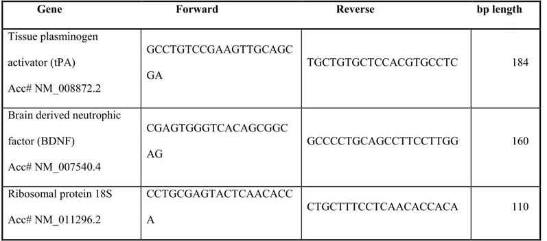
Table 1: Primer sequences.
Forward and reverse primers were selected from the 50 and 30 region of each gene mRNA. The expected length of each PCR amplification product is indicated in the right column
Gene Forward Reverse bp length
NF1Acc#NM_01089 7.2 TTCGATACACTTGCGGAAAC CACATTGGCAAGAGCCATAG 114 APPAcc# NM_007471 GGTTCTGGGCTGACAAACAT CAGTTTTTGATGGCGGACTT 102 Dopamine D3 receptor Acc# NM_007877 GGGGTGACTGTCCTGGTCTA AAGCCAGGTCTGATGCTGAT 110 Ribosomal protein 18S Acc# NM_011296.2 GAGGATGAGGTGGAACGTGT GGACCTGGCTGTATTTTCCA 115
Western blot analysis
Crude extracts from WT (n=3 for each experimental group) and D3R-/- (n=3 for each
experimental group) mice hippocampi were prepared by homogenizing samples in a buffer containing 20 mM Tris (pH 7.4), 2 mM EDTA, 0.5 mM EGTA; 50 mM mercaptoethanol, 0.32 mM sucrose and a protease inhibitor cocktail (Roche Diagnostics) using a Teflon-glass homogenizer and then sonicated twice for 20 sec using an ultrasonic probe, followed by centrifugation at 10.000 g for 10 min at 4 °C. Protein concentrations were determined by the Quant-iT Protein Assay Kit (Invitrogen). Sample proteins (30 μg) were diluted in 2X Laemmli buffer (Invitrogen, Carlsbad, CA, USA), heated at 70°C for 10 min and then separated on a Biorad Criterion XT 4-15% Bis-tris gel (Invitrogen) by electrophoresis and then transferred to a nitrocellulose membrane (Invitrogen). Blots were blocked using the Odyssey Blocking Buffer (Li-Cor Biosciences). Immunoblot analysis was performed by using a rabbit polyclonal
Biotechnology Inc), a rabbit polyclonal antibody raised against amino acids 676-695 of APP of human origin (A8717, Sigma), a rabbit polyclonal antibody raised against peptide mapping within the C-terminus of neurofibromin of human origin (sc-67, Santa Cruz Biotechnology Inc) and a rabbit polyclonal antibody raised against amino acids 210-444 of β-tubulin of human origin (sc-9104, Santa Cruz Biotechnology Inc). All primary antibodies were diluted 1:200, while the secondary antibody (goat anti-rabbit IRDye 800nm, cat #827-06905; Li-Cor Biosciences) was used at 1:20000. Blots were scanned with an Odissey Infrared Imaging System (Odyssey). Densitometric analyses of Western blot signals were performed at non-saturating exposures and analyzed using the ImageJ software (NIH, Bethesda, MD; available at http://rsb.info.nih.gov/ij/index.html). Values were normalized to β-tubulin, which served as loading control. No signal was detected when the primary antibody was omitted (data not shown).
Tissue preparation for immunohistochemical staining
Brains from decapitated mice (naive WT, n=3; CA WT, n=3) were removed and stored for at least 24 h in 4% formaldehyde at 4°C before dehydration and embedding in paraffin. Ten-micrometer-thick sections were cut, mounted on glass slides, kept overnight at 37°C, and then at room temperature until use. Prior to immunohistochemical staining, the sections were dewaxed in xylene and rehydrated through graded alcohols. They were then rinsed in 0.1M Tris–HCl buffered saline (TBS, pH 7.4) and treated with 3% hydrogen peroxide (H2O2) in PBS for 10 min
to reduce endogenous peroxidase activity.
Immunohistochemical analysis
Immunohistochemical analysis was performed in accordance with the standard ABC method. To reduce nonspecific staining, sections were treated with 5% bovine serum albumin (BSA) and 3% goat serum in TBS for 1 h. Sections were then incubated with a rabbit polyclonal antibody raised against amino acids 1-50 of D3R of human origin (sc-9114, Santa Cruz
1% BSA, and 0.25% Triton X-100. After several rinses in TBS, the sections were incubated with a 1:200 diluted biotinylated goat anti-rabbit IgG for 1 h at room temperature. To visualize the immunoreaction sites in tissues, the sections were then rinsed and treated with reagents from an ABC Kit for 1 h at room temperature. The sections were rinsed in TBS and incubated with 0.025% 3,3-diaminobenzidine (DAB) plus 0.33% H2O2 in TBS for 10 min. Then, Tris buffer
was added to stop the DAB reaction. The stained sections were dehydrated through graded alcohols, cleared in xylene, and covered with neutral balsam. All sections were examined and images were taken with a light microscope (Axiovert, Carl Zeiss Inc) equipped with a digital color camera. The images were further processed using Adobe Photoshop software.
Statistical analysis
One-way analysis of variance (ANOVA) was used to compare differences among three or more groups followed by Tukey post-hoc test to evaluate statistical significances. A level of p<0.05 was accepted as indicative of significant difference.
Results
Cognitive performance of WT and D3R-/- mice in the passive avoidance test
Mice were trained using a behavioral protocol, the single trial step-through passive avoidance test, known to require hippocampus-dependent learning (Venault et al., 1996). In these experiments, conditioned animal (CA) were trained to avoid moving from the lighted to the darkened section of a conditioning chamber by delivering a foot-shock when they entered the darkened section. Control mice included untrained (naive) animals, and animals exposed to the unconditioned (USTA) or the conditioned (CSTA) stimulus. To verify that the trained mice in fact learned the passive avoidance (PA) task, learning was assessed in a comparable group of animals by evaluating the latency of step-through in the retention test. Twenty four hours after the one-trial training period, only CA in either genotype learned to associate stepping through
CSTA WT mice, # p<0.05 vs USTA WT mice, §§§ p<0.001 vs naïve CSTA and USTA D3-/- mice) (D’Agata and Cavallaro, 2003). Furthermore, CA D3R-/- mice exhibited a better
behavioral response as compared to CA WTs in the retention test (Fig. 1) (++ p<0.01 vs CA WT, One-Way ANOVA followed by Tukey-Kramer post-hoc test).
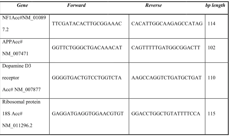
Figure 1. Cognitive response of WT and D3R-/- animals in the passive-avoidance paradigm
Cognitive response of WT and D3R-/- animals in the passive avoidance paradigm. Conditioned
animals (CA) were trained to avoid moving from the lighted to darkened section of a conditioning chamber by the delivery of the foot shock when they entered the darkened section. Control mice included untrained (naive) animals, and animals exposed to the conditioned (CSTA) or unconditioned (USTA) stimulus. The values (time in seconds taken for re-entering the dark box measured in the retention test performed 24 h after the learning trial) are the means ± S.E.M. of WT (n=8 per group) and D3R-/- mice (n=8 per group) (*** p<0.001 vs naïve and
CSTA WT mice, # p<0.05 vs USTA WT mice, §§§ p<0.001 vs naïve CSTA and USTA D3
-/-mice, ++ p<0.01 vs CA WT -/-mice, One-Way ANOVA followed by Tukey-Kramer post-hoc test).
D3R expression in the hippocampus of WT mice after the acquisition of passive avoidance trial
To evaluate whether acquisition of PA influenced hippocampal D3R expression in WT mice, we
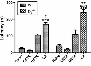
an interval of time sufficient to observe changes at protein level. To exclude the potential involvement of non-learning based, state-dependent changes such as arousal or stress factors which could affect gene or protein expression, CSTA mice (mice that were subjected to the same experimental procedure as trained animals with the exception that they did not receive the associative stimulus, i.e. the inescapable footshock) and USTA mice (mice that received only the inescapable footshock stimulus) were included as further control groups. Comparative analyses with control groups demonstrated that acquisition of PA in CA WT animals significantly increased D3R expression both at mRNA (F3,23=41.89, ***p<0.001 vs naïve, CSTA
and USTA) and protein levels (F3,11=12.83, ***p<0.001 vs naïve, CSTA and USTA) (Fig. 2
A-C).
Figure 2. D3R mRNA and protein expression in the hippocampus of WT mice after
acquisition of the passive avoidance trial
(A-C) Quantitative real-time PCR and Western blot analyses showing increased D3R mRNA
and protein expression in the hippocampus of WT mice 6 h after the acquisition of the passive avoidance trial (CA) with respect to naïve, CSTA and USTA animals. (A) Results are presented
animals (CA n=3) ± S.E.M. Relative fold changes of D3R expression were normalized to the
endogenous ribosomal protein 18S (housekeeping gene) and then calculated using the comparative ΔCt method. Baseline expression levels of the control group were set to 1. Experiments were performed four times independently, each run in duplicate. (B) Representative immunoblots containing 30μg of tissue homogenates (n=3 hippocampi per group) were incubated using a rabbit polyclonal D3R antibody and scanned with an Odyssey
Infrared Imaging System, as described in Materials and Methods section. (C) Bar graph showing bands intensity ratios normalized to β-tubulin which were obtained using the ImageJ software and are expressed as mean ± S.E.M from at least three independent determinations. *** p<0.001 vs Naïve, CSTA and USTA WT mice, as determined by One-Way ANOVA followed by Tukey-Kramer post-hoc test.
Hippocampal D3R immunolocalization in WT mice subjected to PA conditioning
To determine hippocampal D3R distribution before and after acquisition of PA conditioning
immunohistochemical analyses were carried out in brain sections of both naive and CA WT mice. Naive mice sections served as control. As shown in Fig. 3, no evident changes in the distribution of D3Rs between the two mice groups were apparent in the hippocampal regions
examined (CA1, CA2, CA3 and dentate gyrus, respectively). However, the weak D3R signal
intensity observed in hippocampal regions of naive WT mice was remarkably increased by PA acquisition in CA1-CA3 fields, but not in the dentate gyrus of CA mice (Fig. 3).
Figure 3. D3R distribution in the hippocampus of naive and trained WT mice.
Representative photomicrograhs showing D3R immunoreactivity in specific hippocampal brain
regions (CA1-CA2-CA3 and dentate-gyrus, respectively) of WT mice before (naïve) and after acquisition (CA) of the passive avoidance task. No apparent changes in receptor distribution are visible between the two groups. However, the weak D3R positiveness in naive animals is clearly
increased following the conditioning trial almost in every hippocampal region examined (CA1, CA2 and CA3 fields), except the dentate gyrus. Scale bar = 40μm. Images were taken from different brain sections of naive and CA WT animals and examined under a light microscope (Axiovert, Carl Zeiss Inc) equipped with a digital color camera.
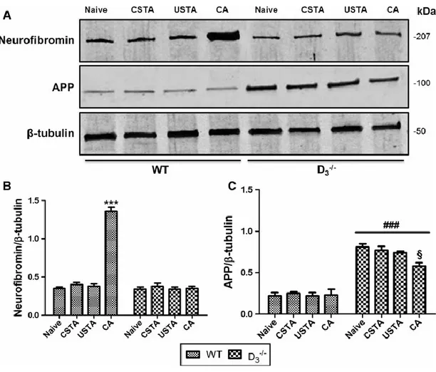
NF1 and APP expression in the hippocampus of WT and D3R-/- mice after the acquisition of
passive avoidance trial
To establish whether acquisition of PA differentially influenced NF1 and APP mRNA and protein expression levels either in the presence or absence of D3Rs, quantitative real-time PCR
and Western blot analyses were performed in hippocampi of both WT and D3R-/- mice from the
four experimental groups (detailed in Materials and Methods section).
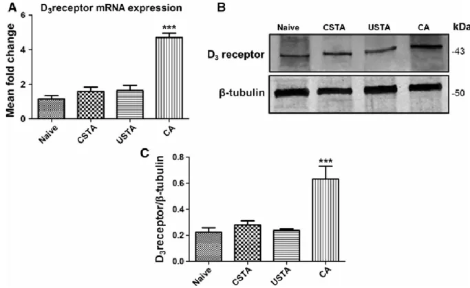
We found that NF1 mRNA expression, as well as its gene product neurofibromin, were significantly upregulated in the hippocampus of CA WTs in comparison with naive, CSTA or USTA mice (F7,47 = 17.4 ***p<0.001 vs naive, CSTA and USTA mRNA levels; F7,23= 113.4
***p<0.001 vs naive, CSTA and USTA protein levels, respectively) (Fig. 4 and 5). Interestingly, PA-driven increase in gene and protein expression was completely abrogated in D3R-/- mice (Fig. 4 and 5), suggesting that D3R might be necessary for the transcriptional
regulation of NF1.
In contrast to NF1, neither APP mRNA nor protein levels were affected by acquisition of the PA trial in WT mice, whereas the heightened basal expression observed in naive (Castorina et al., 2011) CSTA or USTA D3R-/- mice was significantly reduced by acquisition of the avoidance
Figure 4. NF1 and APP mRNA expression in the hippocampus of WT and D3R-/- mice
after acquisition of the passive avoidance trial
Data obtained for quantitative real-time PCR analyses showing NF1 and APP mRNA expression in the hippocampus of WT mice 6h after the acquisition of the passive avoidance trial (CA) as compared to naïve, CSTA or USTA mice. For more details on experimental groups refer to the corresponding “Materials and Methods” subsection. Results are presented as mean fold changes of WTs (n=3 per group) and D3R-/- (n=3 per group) ± S.E.M. Relative fold
changes of either NF1 (A) or APP (B) genes were normalized to the endogenous ribosomal protein 18S (housekeeping gene) and then calculated using the comparative Ct method. Baseline expression levels of the control group (Naive WT) were set to 1. Experiments were performed four times independently, each run in duplicate. *** p<0.001 vs Naïve, CSTA and USTA WT mice, ### p<0.001 vs WT mice, §§§ p<0.001 vs Naïve, CSTA and USTA D3R
Figure 5. Neurofibromin and APP protein expression in the hippocampus of WT and
D3R-/- mice after acquisition of the passive avoidance trial
(A) Representative immunoblots containing 30μg of tissue homogenate (n=3 hippocampi per group) were incubated using rabbit polyclonal antibodies raised against both neurofibromin and APP and scanned with an Odyssey Infrared Imaging System, as described in the corresponding “Materials and Methods” subsection.
(B-C) Bar graphs showing relative bands intensities normalized to β-tubulin were obtained using the ImageJ software and are expressed as mean ± S.E.M. *** p<0.001 vs Naïve, CSTA and USTA WT mice, ### p<0.001 vs WT mice; § p<0.05 vs Naïve D3R-/- mice, as determined
Discussion
The rationale of the present study was based on previous evidence indicating that D3R-/- mice
exhibit enhanced cognitive performance in the single trial step-through passive avoidance (PA) task as compared to WTs (Micale et al., 2010) and on our recent observation showing that expression levels of both NF1 and APP genes are modified in various brain regions, including the hippocampus, of mice lacking D3R (Castorina et al., 2011). Since PA is known to initiate a
cascade of altered gene expression in the hippocampus (Izquierdo et al., 2000), we first hypothesized that D3R expression might be affected by acquisition of the trial and subsequently,
that receptor could be involved in the transcriptional regulation of these two memory-related genes.
As shown in Fig. 1, results obtained from the PA behavioral paradigm are in agreement with previous data showing that the genetic inactivation of D3R ameliorates the learning processes of
rodents subjected to several experimental cognitive paradigms. The mechanisms underlying the enhanced cognitive performance are not fully understood, even though the involvement of this receptor in the control of AChergic transmission has been suggested (Glickstein et al., 2005; Lacroix et al., 2006; Millan et al., 2007). However, the potential interaction with AChergic systems may be just part of a bigger puzzle, since the involvement of endocannabinoid/endovanilloid systems have also been proposed (Micale et al., 2010). Therefore, it is likely that acquisition of a memory-related task involves co-activation of a multitude of systems, which thereby initiates a broad array of transcriptional changes in genes associated with maintenance of synaptic function and/or neuronal remodelling. In the present study we have focused our attention on NF1 and APP genes, both of which have been shown to play a significant role in cognition and memory performance (Costa et al., 2002; De Schepper et al., 2006; Guilding et al., 2007; Dawson et al., 1999; Conboy et al., 2005). Converging data obtained through qPCR, Western blot and immunohistochemistry revealed that hippocampal D3R expression and immunoreactivity are significantly increased following acquisition of PA
(Fig. 2 and 3), suggesting the hippocampal DA levels might be increased soon after the learning event and consistent with D3R autoreceptor function (Collo et al., 2012). Interestingly, NF1 but
not APP expression mirrored PA-driven increase in D3R mRNA and protein levels (Fig. 2-5),
which was abrogated in trained D3R-/- mice, supporting a role of the receptor on gene
transcriptional activity, at least in the hippocampus.
Previous studies have indicated that the NF1 gene acts as an inhibitory regulator of Ras, involved both in GABA-mediated inhibition of hippocampal synaptic transmission (Costa et al., 2002) and in the activation of signaling cascades that regulate neuronal outgrowth during both early- and late-phase LTP (Guilding et al., 2007). These evidences, together with our finding, suggest that NF1 expression might be under the control of D3R to exert either
facilitatory/inhibitory actions on synaptic function following acquisition of the cognitive task. As opposite to NF1 data, APP expression was unchanged in WT mice following PA acquisition, but was significantly increased in naive D3R-/- mice and totally reversed after the acquisition of
the behavioral task (Fig. 4 and 5). This result is consistent with our previous evidence showing that APP levels are thoroughly augmented in several brain regions of mice lacking D3R
(Castorina et al., 2011), even though it does not explain why expression levels were significantly reduced by acquisition of the avoidance task (Fig. 4 and 5). Unfortunately, a plausible explanation for the latter result could not be attributed directly to D3R, although it is
possible that genetic inactivation of the receptor has profound effects on PA-driven regulation of APP expression, possibly through the involvement of alternative molecular mechanisms. In agreement with this hypothesis, a study performed using NF1 knock-out mice proposed that APP and neurofibromin form a binding complex that interacts with D3Rs and that their
dysfunctional cellular trafficking due to the primary gene defect might explain the cognitive deficits observed in these murine models (Donarum et al., 2006). It is therefore possible that the imbalanced APP expression observed in D3R-/- mice both before and after the training trial might
involve the disrupted interaction between the NF1/APP complex and the receptor, although it remains unclear whether it could have repercussions of gene transcriptional activity. However, our study was limited to the evaluation of gene expression profile during the acquisition of PA, a specific fear conditioning paradigm. Using different behavioral tests beside fear-associated
ones should be warranted to enhance the conclusiveness of these finding with respect to associative memory.
In conclusion, the present study provides novel insights to better comprehend the relevance of hippocampal D3R in the transcriptional regulation of NF1 and APP genes following the
acquisition of the PA task.
Acknowledgements
These experiments were supported by the international PhD program in Neuropharmacology, University of Catania, Medical School. We thank Mr P. Asero for his technical support.
Increased hippocampal CREB phosphorylation in dopamine D3 receptor knockout mice
following passive avoidance conditioning
Agata Grazia D’Amico1, Soraya Scuderi1, Gian Marco Leggio2, Alessandro Castorina1, Filippo
Drago2 , Velia D’Agata1*
1 Section of Anatomy and Histology, Department of Bio-Medical Sciences,2 Section of
Pharmacology and Biochemistry, Department of Clinical and Molecular Biomedicine, University of Catania, Italy
Abstract
Dopamine D3 receptors (D3Rs) are implicated in synaptic plasticity and memory processes.
Previously we have shown that D3Rs mediate inhibitory effects on learning, since D3R knockout
(D3-/-) mice display enhanced performance in the passive avoidance task (PA). Formation of
new memories is known to require de novo synthesis of proteins related to synaptic function through the activation of signalling pathways including the mitogen-activated protein kinases (MAPKs) and activation of the nuclear transcription factor cAMP response element binding protein (CREB). However, there are no clear indications regarding the specific involvement of D3Rs in the activation of these signalling cascades after acquisition of PA. Therefore, in this
study we assessed whether phosphorylation levels of several MAPKs, Akt and CREB were differentially affected by PA in both wild-type (WT) and D3-/- mice hippocampi. Animals were
divided in naive, unconditioned stimulus trained, conditioned stimulus trained and conditioned animals. Phosphorylation of extracellular signal-regulated kinase 1/2 (ERK 1/2), c-Jun-N-terminal kinase (JNK) and p38, as well as of Akt and CREB were determined. Acquisition of PA significantly increased pCREB levels both in WT and D3-/- mice. The extent of PA-driven
increase in pCREB levels was significantly higher in mice lacking D3Rs. Similarly, pERK 1/2
was further augmented in trained D3-/- mice as compared to trained WTs, whereas JNK and p38
phosphorylation was not affected neither by PA nor by genetic background. Finally, Akt activation was observed in D3-/- mice, but not in response to PA. In conclusion, these data
supports the notion that D3Rs might modulate CREB phosphorylation after acquisition of PA,
probably via activation of ERK signaling.
Keywords : dopamine D3 receptor, cAMP/CREB signalling, passive avoidance
Introduction
Dopamine (DA) is a monoamine neurotransmitter involved in regulation of multiple functions in the central nervous system (CNS) and periphery, including locomotion, endocrine regulation, emotional behaviours and cognition (Carlsson, 2001; Gainetdinov and Caron, 2003; Zhou and Palmiter, 1995; Beaulieu and Gainetdinov, 2011). DA system is part of a network that plays very important roles in cognitive function, in which multiple DA receptor subtypes contribute to different aspects of learning and memory. It has been shown that DA functions are mediated by two different classes of GPCRs (Beaulieu et al., 2005), D1-like (D1R and D5R) and D2-like
(D2R, D3R and D4R), based on their structural and pharmacological properties (Missale et al.,
1998; Karasinska et al., 2005). Although evidence suggests that, a the five DA receptors, the D1R plays a dominant role in modulating synaptic plasticity and memory process, other DA
receptor subtypes including the D3 receptor (D3R) seem to be involved in cognitive functions
(El-Ghundi et al., 2007). In fact, many studies have demonstrated that the D3R could be a
critical modulator of normal DAergic function, and consequently cognition (Nakajima et al., 2013) and that it is also involved in synaptic plasticity and memory processes (Laszy et al., 2005) and in regulation of gene expression (Castorina et al., 2011). D3R exhibits sustained high
affinity for DA, suggesting that D3R in vivo may be occupied by endogenous DA for extended
periods of time, leading to its high spontaneous activation (Richtand et al., 2001; Vanhauwe et al., 2000).
In last decade many findings showed that mice genetically deficient in D3R show regular
emotional behavior (Chourbaji et al., 2008) and increased cognitive flexibility in the attentional set-shifting task and enhanced cognitive performance in the single trial step-through passive
avoidance (PA) task as compared to WTs mice (Glickstein et al., 2005; Micale et al., 2010; D'Amico et al., 2013).
The cognitive effects mediated by D3Rs may be attributable to activation of the
cAMP-PKA/CREB signaling in the hippocampus (Nakajima et al., 2013). CREB is a nuclear transcription factor which is thought to play a key role in learning and memory process (Xing et al., 2010). CREB-dependent signaling associated with memory impairment is well-documented in literature; for example studies have shown that loss of CREB signalling in rodent hippocampus is implicated in age-related spatial memory impairment (Brightwell et al., 2004; Mouravlev et al., 2006). CREB mediated gene expression is dependent on its phosphorylation at Ser133 via a number of signaling pathways that involve the activation of protein kinases, among these the MAPKs (Josselyn and Nguyen, 2005). Activation of MAPK, including the extracellular signal-regulated kinase (ERK), c-Jun-N-Terminal kinase (JNK), and p38 transmits signals from a variety of extracellular stimuli to the nucleus (Schaeffer et al., 1999). The ERK signaling has been implicated in hippocampal synaptic plasticity, spatial learning and memory (English et al., 1997; Blum et al., 1999; Selcher et al., 1999) and recent findings also suggest that JNK and p38 might play a role in synaptic plasticity (Brust et al., 2007; Brust et al., 2008). Moreover, in vivo studies revealed that D2 class receptors also exert their action in a cAMP-independent manner by promoting the dephosphorylation-inactivation of serine/threonine kinase Akt on its regulatory Thr308 residue (Beaulieu et al., 2004).
The serine/threonine kinase Akt is also involved in the activation of CREB (Beaulieu et al., Accili et al., 1996). It is clear that D3Rs may influence cognition by regulating CREB signalling
in the hippocampus (Xing et al., 2010), however no data exist about this relationship during the formation of associative learning. Given the involvement of D3Rs in memory processes and in
regulating the activity/phosphorylation of several signalling molecules, we used D3-/- and WT
mice to further elucidate the relative contribution of this receptor in regulating CREB phosphorylation following the acquisition of PA, as well as the major underlying molecular mechanism involved.
Our data points to D3Rs as negative regulators of CREB activity in the hippocampus, as a
targeted D3R deletion further reinforces PA-driven increases in the phosphorylation state of this
transcription factor, probably through the involvement of ERK-CREB signalling.
Materials and Methods
Animals
All experiments were carried out on D3R-/- and WT mice (male mice 8-12 weeks old). The
animals were housed four per cage and fed with standard laboratory food and allowed free access to water ad libitum, in an air-conditioned room with a 12 h light-dark cycle. All the experimental procedures were performed during the light cycle (between 10 a.m. and 2 p.m.). D3R-/- mice used in these experiments were 5th-8th generation of congenic C57BL/6J mice, and
generated by a backcrossing strategy. The genotypes of the D3R mutant and WT mice were
evaluated by PCR analysis by using two pairs of primers flanking either exon 3 of the wild-type D3R or the PGK (phosphoglycerate kinase 1 gene promoter) cassette of the mutated gene (Accili
et al., 1996). Each strain was divided into four groups, as previously described by D’Agata and Cavallaro (2003) and sacrificed by cervical dislocation six hours after the training trial, hippocampi were rapidly dissected and stored at -80°C until use (D'Amico et al., 2013). All animals were used only once in the experiments, which were carried out according to the European Community Council Directive 86/609/EEC. Efforts were made to minimize animal suffering and to reduce the number of animals used. The rationale, design and methods of this study were approved by the Ethical Committee for Animal Research, University of Catania.
Passive avoidance test
The single trial step-through passive avoidance test was performed as previously described by D’Amico et al. (2013) using a passive avoidance apparatus (San Diego Instruments, Inc., San Diego, CA, USA). The apparatus was divided into two compartments by a retractable door: a lit safe compartment and a darkened shock compartment.
The experiment was carried out on male homozygous D3R-/- and WT mice. Each strain of
animals was divided in four groups. The first group (naive) was maintained in the home cage. The rest of the animals experienced a 2-day behavioral training. On the first day, animals were handled by the experimenter for 2 min and then placed into the safe compartment and allowed to explore both chambers of the apparatus for 3 min. The second day, in the training trial, the second group of animals (termed ‘conditioned animals’, CA) were placed in the safe compartment with the door closed. After 2 min of acclimatization the light was turned on, the door opened and the animal was allowed to enter the dark compartment. After the mouse stepped completely with all four paws into the dark compartment, the door was closed, and a mild inescapable foot shock (0.5 mA, 2 s duration) was delivered from the grid floor. Following the shock, the mouse was removed and returned to its home cage. A third group of animals (termed ‘conditioned stimulus-trained animals’, CSTA) were placed into the safe compartment. After 2 min of acclimatization the light was turned on, the door opened and the animal allowed to enter the dark compartment. After the mouse stepped into the dark compartment, the door was closed but no foot shock was delivered from the grid floor. Then mice returned to their home cage. The fourth group of animals (termed ‘unconditioned stimulus-trained animals’, USTA) were placed in one of the two dark compartments. They were allowed to move freely to both compartments. After 2 min of acclimatization they received an inescapable foot shock (0.5 mA, 2 s duration) and then returned to their home cage.
Six hours later, animals from each of the four experimental groups were sacrificed by cervical dislocation, hippocampi were rapidly dissected and stored at -80°C until use. These hippocampi were used in our experimental protocols. Twenty-four hours after the training trial, the remaining animals from each of the four experimental groups performed the retention test. The animals were placed in the safe compartment with the door closed. After 2 min of acclimatization the light was turned on, the door opened and the animal was allowed to enter the dark compartment. The latency to enter the dark compartment was recorded and used as the measure of retention. Mice avoiding the dark compartment for >300 s were considered to have a
Western blot analysis
Crude extracts of WT (n=3 for each experimental group) and D3R-/- (n=3 for each experimental
group) mice hippocampi were dissected according to the mouse brain atlas (Paxinos et al. 2001) and prepared by homogenizing samples in a buffer containing 20 mM Tris (pH 7.4), 2 mM EDTA, 0.5 mM EGTA; 50 mM mercaptoethanol, 0.32 mM sucrose and a protease inhibitor cocktail (Roche Diagnostics) and phosphatase inhibitor (PhosSTOP, Roche Diagnostic) using a Teflon-glass homogenizer and then sonicated twice for 20 sec using an ultrasonic probe, followed by centrifugation at 10.000 g for 10 min at 4 °C. Protein concentrations were determined by the Quant-IT Protein Assay Kit (Invitrogen). Then, protein samples were separated by electrophoresis as previously described (Scuderi et al., 2013). Briefly, sample proteins (40 μg) were diluted in 2X Laemmli buffer (Invitrogen, Carlsbad, CA, USA), heated at 70°C for 10 min and then separated on a Biorad Criterion XT 4-15% Bis-tris gel (Invitrogen) by electrophoresis and then transferred to a nitrocellulose membrane (Invitrogen). Blots were blocked using the Odyssey Blocking Buffer (Li-Cor Biosciences). Immunoblot analysis was performed by using a rabbit phospho-CREB (AB3442, Millipore, 1:500), rabbit anti-CREB (04-218, Millipore, 1:1000), mouse ERK1/2 monoclonal antibody (sc-135900, Santa Cruz, 1:200), rabbit p-ERK1/2 (sc-16982, Santa Cruz, 1:200), mouse total Akt (C67E7, Cell Signaling, 1:1000), mouse phspho-Akt (C31E5E, Cell Signaling, 1:1000), rabbit p-p38 17852-R, Santa Cruz, 1:200), rabbit p38α/β 7149, Santa Cruz, 1:200), mouse p-JNK (sc-6254, Santa Cruz, 1:200), rabbit JNK (sc-571, Santa Cruz 1:200). The secondary antibody goat anti-rabbit IRDye 800CW, (cat #926-32211; Li-Cor Biosciences), goat anti-mouse IRDye 680CW, (cat#926-68020D; Li-Cor Biosciences) was used at 1:20000, 1:30000 respectively. Blots were scanned with an Odissey Infrared Imaging System (Odyssey). Densitometric analyses of Western blot signals were performed at non-saturating exposures and analyzed using the ImageJ software (NIH, Bethesda, MD; available at http://rsb.info.nih.gov/ij/index.html). Values were normalized to β-tubulin, which served as loading control. To assess phosphorylation / activity state we normalized the expression of phospho-proteins over pan-proteins. No signal was detected when the primary antibody was omitted (data not shown).
Statistical analysis
Data are reported as mean ± S.E.M. One-way analysis of variance (ANOVA) was used to compare differences among groups and statistical significance was assessed by Tukey-Kramer post-hoc test. The level of significance for all statistical tests was p ≤ 0.05.
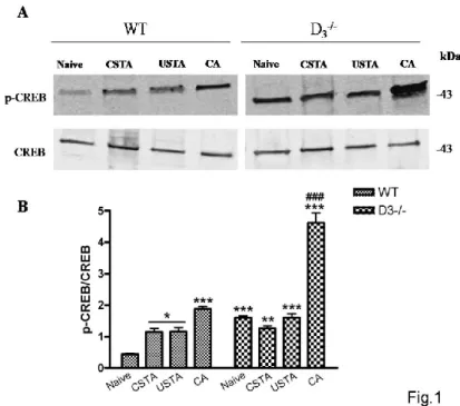
Results
Enhanced phosphorylation of p-CREB in hippocampus of D3R-/- mice trained with passive
avoidance test
In order to evaluate whether cognitive response of WT and D3R-/- animals in the passive
avoidance paradigm affected CREB activation we performed Western blot analyses in WT and D3R-/- animals that underwent a passive avoidance task according to a protocol described in our
previous work (D'Amico er al., 2013). As seen in Fig. 1, expression levels of p-CREB were significantly increased following passive avoidance conditioning in WT mice (CA) as compared to control groups (F7,23=74,89, *p<0.05 or ***p<0.001 vs Naïve WT). Moreover, the degree of
hippocampal CREB phosphorylation was significantly higher in D3R-/- conditioned animals with
respect to either untrained WT or D3R-/- mice (Naïve animals) (***p<0.001 vs Naïve WT,
###p<0.001 vs Naïve D3R-/- ; Fig. 1 A-B). This result suggests that learning-dependent
Figure 1. CREB phosphorylation in the hippocampus of WT and D3-/- mice subjected to
the passive avoidance task.
(A) Representative immunoblots obtained using 40μg of homogenates from WT and D3-/- mice
hippocampus in conditioned animals (CA) and in control mice that included untrained (naive) animals, and animals exposed to the conditioned (CSTA) or unconditioned (USTA) stimulus. (B) The bar graph shows the results of three independent experiments. Protein levels are expressed as arbitrary units obtained after normalization to β-tubulin, which was used as loading control. Data are expressed as mean ± S.E.M. *p<0.05, **p<0.01 or ***p<0.001 vs Naïve WT, ### p<0.001 vs Naïve, CSTA and USTA D3-/- as determined by One-Way ANOVA followed by
Tukey post-hoc test.
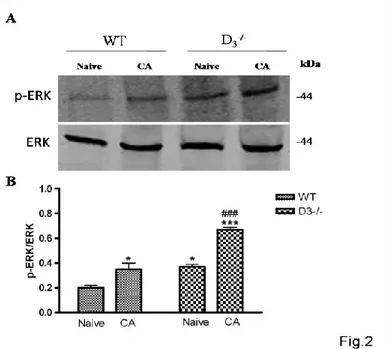
MAPKs activation in the hippocampus of WT and of D3-/- mice after passive avoidance training
The involvement of D3Rs in the regulation of signaling pathways related to the formation of
new associative memories was evaluated by performing Western blot analyses on ERK, which is known to be involved in hippocampal synaptic plasticity and memory formation (English and Sweatt, 1997; Blum et al., 1999). As shown in Fig. 2 there was a significant activation of ERK in the hippocampus of either in WT mice subjected to the step-through PA and heightened activity was also identified in DR-/- naïve when compared to respective control groups
(F3,15=37,91, *p<0.05 or ***p<0.001 vs Naïve WT). Interestingly, the degree of ERK activation
was significantly higher in the hippocampus of D3R-/- conditioned animals as compared to the
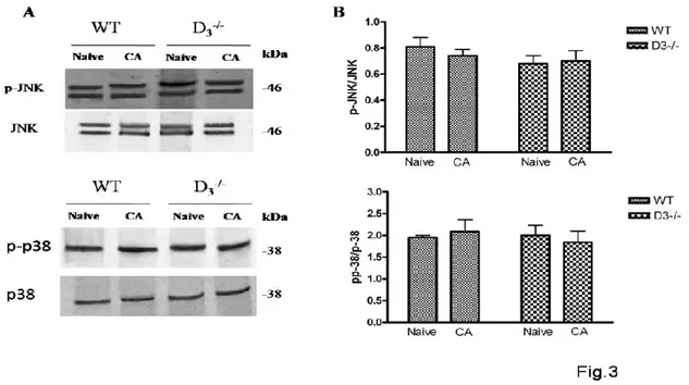
D3R-/- naïve mice (###p<0.001 vs Naïve D3R-/- ; Fig. 2 A-B). Since that other two MAPKs, JNK
and p38, are involved in synaptic plasticity (Brust et al., 2007; Moult et al., 2008), to further investigate the roles of D3R in MAPK signaling in associative memory formation, we also
examined JNK and p38 phosphorylation levels in the hippocampus of WT and D3R-/- mice after
PA conditioning. As shown in Fig. 3, no significant changes in activity levels could be observed neither for p-JNK (F3,11=0,8620) nor p-p38 (F3,11=0,2253) in both WT and D3R-/-
animals compared to baseline (Naïve) (Fig.3), suggesting that activation of MAPKs after PA training is specific for ERK.
Figure 2. Hippocampal MAPK activaty is specific for ERK in WT and D3-/- mice after PA
acquisition
(A) Representative immunoblots obtained using 40μg of homogenates from WT and D3-/- mice
hippocampus.
(B) The bar graph shows the results on three independent experiments. Protein levels are
Figure 3. MAPK activation in the hippocampus of WT and D3-/- mice after PA training.
(A) Representative immunoblots obtained using 40μg of homogenates from WT and D3-/- mice
hippocampus.
(B) The bar graph shows the results on three independent experiments. Protein levels are
expressed as arbitrary units obtained after normalization to β-tubulin, which was used as loading control. Data are expressed as mean ± S.E.M. and determined by One-Way ANOVA followed by Tukey post-hoc test.
D3R-/- mice exhibit increased phosphorylation of Akt which is not dependent on the acquisition
of the passive avoidance trial
D2-class receptor (D2R,D3R,D4R) couple to Gαi/0, thus downregulating cAMP production and PKA activity (English and Sweatt, 1997; Blum et al., 1999). Previous study have demonstrated that prolonged stimulation of D2 class receptor leads to specific dephosphorylation/inactivation of the serine/threonine kinase Akt on its regulatory threonine 308 (Thr308) residue (Beaulieu et al., 2004). Therefore, in the attempt to estabilish the relative contribution of D3Rs on Akt
activation after PA conditioning, we compared the phosphorylation levels of Akt at its Thr308 residue in WT and D3R-/- mice both in basal condition or after acquisition of the task. Results
hippocampus of naïve D3R-/- mice, confirming recently published data in literature (Castorina et
al., 2013) and in CA animals as compared to WT (F3,15=25,90, **p<0.01 or ***p<0.001 vs
Naïve and CA WT, Fig.4). This result suggests that Akt activation might be correlated to the absence of the receptor and not to this specific type of conditioning.
Figure 4. AKT phosphorylation at the Thr308 residue in the hippocampus of WT and D3-/- mice after PA training.
(A) Representative immunoblots obtained using 40μg of homogenates from WT and D3-/- mice
hippocampus.
(B) The bar graph shows the results on three independent experiments. Protein levels are
expressed as arbitrary units obtained after normalization to β-tubulin, which was used as loading control. Data are expressed as mean ± S.E.M. **p<0.01 or ***p<0.001 vs Naïve WT, as determined by One-Way ANOVA followed by Tukey post-hoc test.
Discussion
Learning and memory are integrative brain functions based on neural plasticity, which involve a variety of molecules such as neurotransmitters, neurotrophins, and their receptors as well as second messengers and protein kinases (Kudo et al., 2004). Futhermore, acquisition and consolidation of new memories depend upon induction of gene expression and subsequent protein synthesis (Davis and Squire, 1984). The DAergic system has been implicated in cognitive function through animal and human research, including studies of molecular genetics and neuroimaging (Bäckman et al., 2006; Cole et al., 2012). Unlike D2R receptors, D3Rs can be
stimulated by tonic DA levels in the brain due to their high affinity for DA (Sokoloff et al., 1990) and may attenuate any effects of DA fluctuation related to phasic DA release. Accordingly, small changes in the number or function of D3Rmay lead to dramatic effects on
synaptic transmission, suggesting that this receptor could be implicated in cognition (Nakajima et al., 2013). Numerous animal studies have explored the cognitive effects of pharmacological interventions targeting D3R. In recent years, the performance of D3R-/- mice was tested using
various cognitive tasks, such as the two-choice perceptual discrimination test, social novelty discrimination, the step-through passive-avoidance test, T-maze and the delayed alternation test, thus showing that this receptor subtype might contribute to ameliorate certain aspects of cognitive performance, including associative learning (Chourbaji et al., 2008; Glickstein et al., 2005; Micale et al., 2010; D'Amico et al., 2013; Xing et al., 2010; Watson et al., 2012). Furthermore a recent study showed that D3R-regulated CREB signalling in the hippocampus
may be involved in age-associated cognitive alterations (Xing et al., 2010), and other findings showed a close relationship between behavioural performance and CREB phosphorylation in hippocampus after contextual fear and PA conditioning (Kudo et al., 2004). In particular the transcription factor CREB is required for hippocampus-depenent long term potentiation (LTP) (Balschun et al., 2003) and many papers suggest that several memory-associated signaling molecules are related to phosphorylation of CREB (Dubynina and Dolotov, 2009).
Several studies have suggested that D3R activation might mediate negative effects on cognitive