R E S E A R C H A R T I C L E
Open Access
The density and spatial tissue distribution
of CD8
+
and CD163
+
immune cells predict
response and outcome in melanoma
patients receiving MAPK inhibitors
Daniela Massi
1, Eliana Rulli
2, Mara Cossa
3,4, Barbara Valeri
4, Monica Rodolfo
3, Barbara Merelli
5, Francesco De Logu
6,
Romina Nassini
6, Michele Del Vecchio
7, Lorenza Di Guardo
7, Roberta De Penni
8, Michele Guida
9,
Vanna Chiarion Sileni
10, Anna Maria Di Giacomo
11, Marco Tucci
12, Marcella Occelli
13, Francesca Portelli
1,
Viviana Vallacchi
3, Francesca Consoli
14, Pietro Quaglino
15, Paola Queirolo
16, Gianna Baroni
1,
Fabrizio Carnevale-Schianca
17, Laura Cattaneo
18, Alessandro Minisini
19, Giuseppe Palmieri
20, Licia Rivoltini
3*,
Mario Mandalà
5and on behalf of the Italian Melanoma Intergroup
Abstract
Background: Clinical response to MAPK inhibitors in metastatic melanoma patients is heterogeneous for reasons still needing to be elucidated. As the patient immune activity contributes to treatment clinical benefit, the pre-existing level of immunity at tumor site may provide biomarkers of disease outcome to therapy. Here we investigated whether assessing the density and spatial tissue distribution of key immune cells in the tumor microenvironment could identify patients predisposed to respond to MAPK inhibitors.
Methods: Pretreatment tumor biopsies from a total of 213 patients (158 for the training set and 55 for the validation set) treated with BRAF or BRAF/MEK inhibitors within the Italian Melanoma Intergroup were stained with selected immune markers (CD8, CD163,β-catenin, PD-L1, PD-L2). Results, obtained by blinded immunohistochemical scoring and digital image analysis, were correlated with clinical response and outcome by multivariate logistic models on response to treatment and clinical outcome, adjusted for American Joint Committee on Cancer stage, performance status, lactate dehydrogenase and treatment received.
Results: Patients with high intratumoral, but not peritumoral, CD8+T cells and concomitantly low CD163+myeloid cells displayed higher probability of response (OR 9.91, 95% CI 2.23–44.0, p = 0.003) and longer overall survival (HR 0.34, 95% CI 0.16–0.72, p = 0.005) compared to those with intratumoral low CD8+T cells and high CD163+myeloid cells. The latter phenotype was instead associated with a shorter progression free survival (p = 0.010). In contrast, L1 and PD-L2 did not correlate with clinical outcome while tumorβ-catenin overexpression showed association with lower probability of response (OR 0.48, 95% CI 0.21–1.06, p = 0.068).
(Continued on next page)
© The Author(s). 2019 Open Access This article is distributed under the terms of the Creative Commons Attribution 4.0 International License (http://creativecommons.org/licenses/by/4.0/), which permits unrestricted use, distribution, and reproduction in any medium, provided you give appropriate credit to the original author(s) and the source, provide a link to the Creative Commons license, and indicate if changes were made. The Creative Commons Public Domain Dedication waiver (http://creativecommons.org/publicdomain/zero/1.0/) applies to the data made available in this article, unless otherwise stated. * Correspondence:[email protected]
3Unit of Immunotherapy of Human Tumors, Fondazione IRCCS Istituto
Nazionale dei Tumori, Milan, Italy
Full list of author information is available at the end of the article
on January 17, 2021 by guest. Protected by copyright.
(Continued from previous page)
Conclusions: Analysis of the spatially constrained distribution of CD8+and CD163+cells, representative of the opposite circuits of antitumor vs protumor immunity, respectively, may assist in identifying melanoma patients with improved response and better outcome upon treatment with MAPK inhibitors. These data underline the role of endogenous immune microenvironment in predisposing metastatic melanoma patients to benefit from therapies targeting driver-oncogenic pathways.
Keywords: Myeloid cells, T lymphocytes, Microenvironment, Melanoma prognosis
Introduction
Approximately 40–50% of metastatic melanoma patients (MPs) harbor point mutations inBRAF, over 95% of which are at V600 inBRAF exon 15 [1]. The discovery of this mu-tation provided the genetic basis for the development of BRAF inhibitors (BRAFi) for the treatment of melanoma. Clinical efficacy of this class of drugs was initially demon-strated by their use in mono-therapy in patients withBRAF
V600
-mutant melanoma. In two prospective randomized clinical trials BRAFi showed a better response rate, pro-gression free survival (PFS) and overall survival (OS) than chemotherapy [2, 3]. However, responses were temporally limited, mainly because of acquired resistance. Improve-ment of efficacy and tolerability was attained with dual MAPK pathway inhibition by adding a MEK inhibitor (MEKi) to a BRAFi as reported in phase 3 randomized studies [4–6]. Therefore, BRAFi/MEKi combination has been recommended as a standard therapy for advanced BRAF V600
-mutated melanoma, being associated with a median PFS and OS of 12 months and 24–36 months, re-spectively [4–6]. Albeit the problem of overcoming primary and acquired resistance still needs to be faced for thera-peutic amelioration, about 30–35% of patients are alive at 5 years indicating the onset of long-term tumor control [7]. The identification of biomarkers that predict durable bene-fit in patients with BRAFV600-mutated melanoma would provide essential tools for better treatment personalization. Beside the effect on the biological target and pathway, there is strong evidence that the therapeutic efficacy of BRAFi and MEKi relies on additional factors involved in tumor-host interactions and preclinical data show that onco-genic BRAF contributes to immune evasion, as targeting this mutation may increase melanoma immunogenicity [8].
Several genomic mechanisms of intrinsic or acquired tumor resistance to MAPKi therapies have been reported, including BRAFV600 amplification and single nucleotide variants in NRAS, KRAS, MEK1/2, PTEN, CDKN2A and DUSP4 [9]. A study comparing the genomic features of complete responders (CR) versus fast progressors (PD) in patients treated with BRAFi/MEKi showed higher rates of MITF amplification and TP53 mutation in PD, whereas NF1 deletion and deleterious mutations were more com-mon in CR [10]. Nevertheless, gene signatures of CD8 T ef-fector cells, cytolytic T-cells, antigen presentation and NK
cells were significantly enriched in CR tumors [10]. Indeed, several evidences support a key role of tumor immunity in the therapeutic efficacy of MAPKi. LEF1 down-expression andβ-catenin induction, which reduce T cells and CD103+ dendritic cells tumor infiltrate via inhibition of CCL4 secre-tion [11], have been reported to promote acquired resist-ance to BRAFi and MEKi [12]. A rapid accrual of activated CD8+ T cells in tumor microenvironment is instead trig-gered by BRAFi administration at early time points [13], in association with clinical benefit [14]. Preclinical studies linked this effect to the upregulation of HLA molecule ex-pression in tumor cells, favoring increased antigen presen-tation and activation of antitumor T cells, together with the downregulation of certain immunosuppressive factors such as PD-L1, IL1, IL8, NT5E, and VEGFA [15]. On the other hand, non responding patients are featured by the accrual in the tumor site and peripheral blood of myeloid immunosup-pressive cell elements and macrophages [16], again pointing to immunity as a key player to MAPKi therapeutic activity.
Based on these data, we designed a study aimed at identi-fying essential tissue immune biomarkers able to capture the immune contexture of tumor microenvironment that could potentiate or contrast the clinical efficacy of MAPKi.
Materials and methods
Patient characteristics
The cohort of the training set (n = 158) was identified by inspecting the electronic databases of all metastatic MPs treated at Italian Melanoma Intergroup (IMI) centers from June 2011 to February 2017. We retrieved data concerning clinical outcome and MAPKi treatment from patients enrolled in compassionate, expanded access use protocols or therapeutic use of BRAFi with or without MEKi since 2011. The local Ethics Committees approved the study protocol. The study was conducted in compli-ance with the World Medical Association Declaration of Helsinki. Patients enrolled in the study were treated with vemurafenib or vemurafenib and cobimetinib within the therapeutic and expanded access use according to clin-ical practice. For patients included in the vemurafenib compassionate program use, the inclusion criteria were an Eastern Cooperative Oncology Group performance status (ECOG-PS) 0–2, as well as normal hepatic (serum bilirubin < 1.5 mg/dl), renal (serum creatinine < 1.5 mg/
Massiet al. Journal for ImmunoTherapy of Cancer (2019) 7:308 Page 2 of 13
on January 17, 2021 by guest. Protected by copyright.
http://jitc.bmj.com/
dl) and bone marrow (leucocyte count > 4000/1 l, plate-let count > 100,000/1 l) functions. For the other patients, exclusion criteria were a rapid deteriorating medical con-dition, with severe liver or renal failure, QTc > 500 mS and ECOG-PS 4. Information on age, gender, histopath-ology, and surgical and medical treatment were retrieved for each patient, as well as data on tumor objective response rate (ORR), PFS and OS. Data on treatment and survival were collected prospectively. Medical records and/or review of pathology material confirmed accuracy in histopathological classification. Tumor stage was assessed according to the American Joint Committee on Cancer (AJCC) TNM (Tumor, Node, Metastasis) staging system classification (VII edition). Clinical response to BRAFi/MEKi was assessed by RECIST v1.1 criteria [17].
Patients of the validation set (n = 55) were instead treated at the Istituto Nazionale dei Tumori of Milan, with BRAFi according to the MO25515 trial (multicenter phase II study on first−/second-line Vemurafenib;ClinicalTrials. gov, NCT01307397) [18] (n = 35) or BRAFi/MEKi by
clin-ical practice (n = 20). Similarly to the training cohort ECOG-PS was 0–2, normal hepatic (serum bilirubin < 1.5 mg/dl), renal (serum creatinine < 1.5 mg/dl) and bone marrow (leucocyte count > 4000/1 l, platelet count > 100, 000/1 l) functions was required to receive targeted ther-apy. Information on demographics was retrieved for each patient, as well as data on PFS and OS. Data on treatment and survival were collected prospectively.
Tissue samples
Formalin fixed paraffin-embedded (FFPE) tissue sections, 4μm in thickness, were stained with hematoxylin and eosin and centrally reviewed to confirm the histopathological diagnosis and to assess pathology tissue quality control. Immunohistochemistry
Representative 4-μm thick FFPE tissue sections of pre-treatment melanoma samples were selected for immuno-histochemical analysis. Sections were incubated with the following primary antibodies: CD8 (rabbit monoclonal CONFIRM, clone SP57 ready to use; Ventana Medical Sys-tems, Tucson, AZ), CD163 (mouse monoclonal, clone 10D6, dilution 1:100, Novocastra Laboratories Ltd., New-castle, UK),β-catenin (mouse monoclonal, clone 14 ready to use, Ventana Medical Systems, Tucson, AZ), PD-L1 (rabbit monoclonal, clone E1L3N, dilution 1:50, Cell Signal-ing, Danvers, USA) and PD-L2 (rabbit monoclonal, clone D7U8C, dilution 1:50, Cell Signaling, Danvers, USA) on a Ventana BenchMark ULTRA immunostainer (Ventana Medical Systems, Tucson, AZ). The staining procedure cluded pretreatment with cell conditioner 1 followed by in-cubation with the different antibodies. For all antibodies, the signal was developed with the Universal Red Detection Kit (Ventana Medical Systems, Tucson, AZ). Sections were
then counterstained with hematoxylin. Tissue sections of tonsil were used as positive control. As negative controls, mouse IgG1 isotype control was used for β-catenin and CD163 while rabbit IgG isotype control was used for CD8, PD-L1 and PD-L2, respectively. The control sections were treated in parallel with the samples.
Immunohistochemical scoring was performed in a blinded fashion by experienced melanoma pathologists (DM, MC, BV). Stained sections were initially assessed at low magnification to select the areas with highest density of positive immune cells at peritumoral and intratumoral location. Assessment of CD8+ T lympho-cytes and CD163+ macrophages score density was com-pared with evaluation obtained by image analysis. Evaluation of tumoral β-catenin and PD-L1 was per-formed as previously described [19, 20]. PD-L2 expres-sion was evaluated on tumor cells. Assessment of the training set was centralized in the University of Florence, while the validation set was evaluated at the Istituto Nazionale dei Tumori of Milan, according to shared standard operating procedures.
Digital image acquisition and analysis
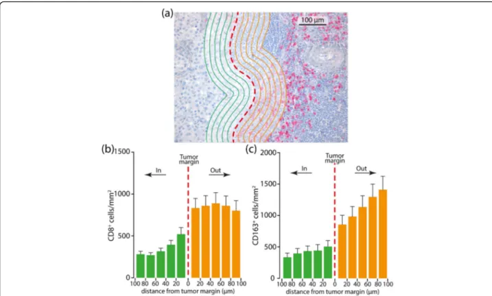
Tissue sections stained with CD8 and CD163 antibodies were digitally scanned at an absolute magnification of X200 using D-Sight platform (A. Menarini Diagnostic, Florence, Italy). An algorithm was designed based on pat-tern recognition that quantified CD8+ and CD163+ cells within two tumor compartments: the invasive tumor mar-gin (stromal-tumor edge) and inside the tumor paren-chyma (tumor center). Image analysis based on RGB (red, green, blue) spectra was used to detect all cells by counter-staining with hematoxylin (blue) and fast red. The number of fast red CD8+and CD163+ cells was calculated in five different high-power magnification fields of 10− 3mm2. The algorithm calculated the density of CD8+ and CD163+ cells/mm2. The total number of CD8+ and CD163+cells was then calculated as the mean of each high-power magnification field. CD8 and CD163 expres-sion was determined using two read-outs that were inde-pendent of each other to account for tumor heterogeneity. The immune cell density (CD8+ and CD163+ cells) at the peritumoral area was further explored in order to generate a cell density histogram. The peritumoral com-partment was defined as the region centered on the border separating the host tissue from the malignant nests, with an extent of 500μm. To analyze further the spatial distribution of CD8+ and CD163+ cells in the peritumoral area, an algorithm was designed to create thick bands (1 mm2) 20μm internal and external to the tumor margin. Then, the distribution of the CD8+ and CD163+cells related to the tumor margin was identified in consecutive 20μm steps (distance classes) within 100μm (Fig.1).
on January 17, 2021 by guest. Protected by copyright.
Statistical analysis
PFS was defined as the time from the date of start treat-ment to the date of progression or death from any cause whichever comes first. Patients who did not progress or die at the date of analysis were censored at their last disease assessment date. OS was defined as the time from the date of start treatment to the date of death from any cause. Overall response rate (ORR) was defined as the proportion of patients with a complete or partial response to treatment. Survival curves were estimated with the Kaplan-Meier method. PFS and OS were analyzed by means of Cox regression model and results were expressed as hazard ratios (HR) with their 95% confidence intervals (95% CI). ORR was analyzed by means of logistic regression models and expressed as odds ratios (OR) with their 95% CI. All multivariate models included as covari-ates immunohistochemical variables, AJCC stage, per-formance status, lactate dehydrogenase (LDH) and the treatment received (BRAFi+MEKi vs BRAFi alone). OS multivariable models included also the subsequent treat-ment (immunotherapy vs no immunotherapy).
PD-L1 and PD-L2 were tested as a continuous or as a di-chotomous variable using 5% as cut-off. Score density of CD8+ T cells and CD163+ macrophages in intratumoral
and peritumoral location was assessed as follows: 0, absent; 1+, mild (< 10%); 2+, moderate (10–50%); 3+, marked (50– 100%) and their density was evaluated as dichotomous variable as high (2+, 3+) versus low (0, 1+).β-catenin was tested as a continuous or as a dichotomous variable using the median value as cut-off. CD8+ T cells and CD163+ macrophages were also analyzed in combination, categoriz-ing patients in three groups: group 1, high CD8+ T cells and low CD163+macrophages; group 2, high CD8+T cells and high CD163+ macrophages/low CD8+ T cells and low CD163+macrophages; group 3, low CD8+T cells and high CD163+ macrophages. CD8+ T cells and immunohisto-chemical PD-L1 overexpression were combined in three groups as follow: group 1, PD-L1≥ 5% and low CD8+ T cells; group 2, PD-L1≥ 5% and high CD8+T cells/PD-L1 < 5% and low CD8+ T cells; group 3, PD-L1 < 5% and high CD8+T cells. Combiningβ-catenin expression and CD8+T cells, patients were categorized in three groups: group 1, low CD8+ T cells and β-catenin overexpressed; group 2, high CD8+ T cells and β-catenin overexpressed/low CD8+ T cells and β-catenin not overexpressed; group 3, high CD8+T cells andβ-catenin not overexpressed.
Chi square test was used to assess associations between PD-L1, PD-L2, β-catenin, CD8+ and CD163+ status and
Fig. 1 Representative metastatic melanoma tissue with analysis mark-up (a). Panel A illustrates a CD8 stain; red dashed line is the invasive tumor margin. CD8+and CD163+cells are counted within the invasive margin, 100
μm inside and 100 μm outside the tumor as identified with green and orange lines spaced 20μm. b, c CD8+and CD163+cells
’ density is binned according to the distance from the margin and a 20 μm bin histogram is generated. The middle of the histogram if the tumor boundary (red dashed line), to the left is inside tumor (green bars) and to the right is outside tumor (orange bars)
Massiet al. Journal for ImmunoTherapy of Cancer (2019) 7:308 Page 4 of 13
on January 17, 2021 by guest. Protected by copyright.
http://jitc.bmj.com/
other clinical and pathological features. The Kruskal-Wallis test was used to analyze the association between the cell count and the density score in CD8+T cells and CD163+macrophages.
To test the robustness of the results, an independent series of metastatic MPs was analyzed separately as valid-ation cohort. The validvalid-ation cohort included metastatic MPs who received BRAFi or BRAFi plus MEKi at the Isti-tuto Nazionale dei Tumori of Milan; their inclusion and ex-clusion criteria were the same as those for the training set.
All tests were two sided and the statistical significance was set at < 0.05 for each analysis. Statistical analyses were carried out using SAS version 9.4 (SAS Institute, Cary, NC) and R language environment for statistical computing (open source,www.r-project.orgversion 3.4.3).
Results
Patients and treatments
Demographic and clinical characteristics of the training set included are summarized in Additional file 1: Table S1. 158 patients were enrolled in the training set; 60 % of patients were male, and the median age at diagnosis of metastatic disease was 59 years (Q1-Q3: 47.7–70.7). All patients had metastatic disease, 60% with M1c dis-ease (95 patients). One hundred and thirty-six patients (86%) and 22 patients (14%) received MAPKi as 1st- or 2nd-line therapy, respectively. Ninety-four patients (60%) received BRAFi as a monotherapy, while 64 pa-tients (40%) received BRAFi+MEKi. The most frequent subsequent lines of treatment were immunotherapy and chemotherapy in 25 and 17% of patients, respectively. Approximately 56% of patients were not treated with further treatments because of rapid progressive disease.
Patients of the validation set were comparable to the training set cohort for demographics and clinical param-eters. Thirty patients (55%) were male; all patients had metastatic disease and 55% with M1c disease (30 patients). Thirty-five patients (64%) received BRAFi as a monotherapy, while 20 patients (36%) received BRAFi+-MEKi. Twelve patients (22%) received immunotherapy as a subsequent line of therapy.
Immunohistochemicalβ-catenin, PD-L1, PD-L2, CD8 and
CD163 expression in melanoma samples
A panel of representative immune markers was tested by immunohistochemistry on melanoma biopsies from the training set, including PD-L1 and PD-L2 (included as sur-rogates of inflammed tumors and tumor immune escape), β-catenin (chosen as tumor pathway driving immune-suppressed microenvironment), CD8 (as marker of antitu-mor effector T cells) and CD163 (recapitulating tuantitu-mor as-sociated myeloid cells including macrophages). Immune markers expression was evaluated in the last available metastatic sample before starting MAPKi therapy in 122
patients (Fig. 2, Additional file 2: Fig. S1 and S2). In the remaining cases, biomarkers were evaluated in the pri-mary melanoma samples due to unavailability of meta-static tissue. The median interval between metameta-static biopsies and treatment starting was 3 months (range 1–6 months). PD-L1 expression on the tumor cell membrane was negative in 82 patients (57%), positive in 63 patients (43%) and technically not evaluable (NE) in 15 patients, while PD-L2 was negative in 126 patients (89%), positive in 15 (11%) patients and NE in 18 patients. The median expression of β-catenin was 60% (interquartile range (IQR): 20–80, NE, in 9 patients), 0 (IQR: 0–0, N NE A: 11 patients) and 10 (IQR: 0–80, NE: 9 patients) for membranous, nuclear and cytoplasmic expression, re-spectively. These values were used as cut-offs to analyze β-catenin as dichotomous variable.
The expression of PD-L1 was associated with high intratumoral CD163+ macrophages (p = 0.008) and high peritumoral CD163+ cells (p = 0.032), conversely PD-L1 expression was not associated neither with intratumoral nor with peritumoral CD8+ T-cell melanomas (Add-itional file1: Table S2).
Density and spatial distribution of the above men-tioned immune markers were then divided in discrete categories, and their prevalence is reported in Additional file1: Table S3 and Additional file2: Figure S3.
The impact of tissue biomarkers on ORR
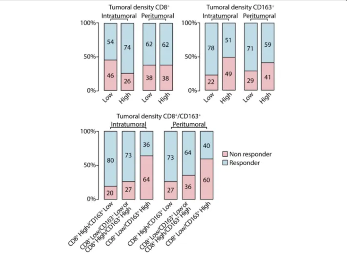
Treatment response was available for 156 patients and included 26 (16.7%) complete responses; 73 (46.8%) par-tial responses; 25 (16.0%) stable disease and 32 (20.5%) progressive disease. The distribution of responder pa-tients according to intra and peritumoral CD8+ T-cell and CD163+macrophages density is reported in Fig.3.
Results of multivariate logistic models on response to treatment, adjusted for AJCC stage, performance status, LDH and treatment received (BRAFi+MEKi vs BRAFi), are reported in Additional file1: Table S4. Metastatic MPs with high intratumoral CD8+ T-cell count (OR 2.15 95% CI 0.93–4.98, p = 0.074) had a higher probability of re-sponse to treatment, while those with membranous β-catenin overexpression > 60% (OR 0.48, 95% CI 0.21–1.06, p = 0.068) showed a lower probability of response. Meta-static MPs with high intratumoral CD163+ count (OR 0.28, 95% CI 0.12–0.65, p = 0.003) had a statistically sig-nificant lower probability of response, while the same pro-file (high CD163+ macrophages) in the peritumoral space did not reach any statistical difference (p = 0.136) (Add-itional file 1: Table S4). The rate of CR was 24% vs 4% among patients with high CD8+/low CD163+ immuno-phenotype, respectively (p = 0.04).
Furthermore, a statistically significant higher probabil-ity of response was observed in patients with β-catenin negative and high intratumoral CD8+ T-cell count
on January 17, 2021 by guest. Protected by copyright.
compared to those with β-catenin overexpression and low intratumoral CD8+ melanomas (Additional file 1: Table S4).
Interestingly, when patients were analyzed according to the combined evaluation of the intratumoral and peri-tumoral density of CD8+ and CD163+ cells, a higher probability of response was observed in patients with high intratumoral, but not peritumoral, CD8+ T cells and low CD163+ macrophages compared to those with low intratumoral CD8+ T cells and high intratumoral CD163+ macrophages (OR 9.91, 95% CI 2.23–44.0, p = 0.003) (Additional file1: Table S4).
The impact of tissue biomarkers on PFS and OS
At a median follow-up of 34 months, 121 (78.1%) patients had progressed and 109 (69.0%) had died. Overall, 126 (79.7%) patients progressed or died. The median PFS and OS were 8.3 (IQR: 4.6–19.2) and 13.7 (IQR: 6.1–38.6) months, respectively.
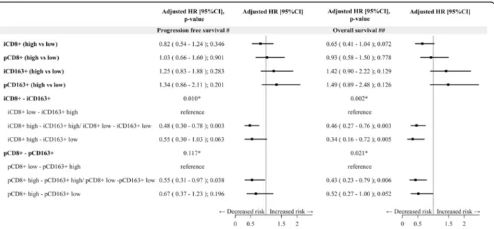
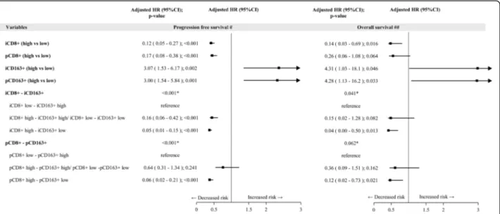
Results of multivariate analysis, both for PFS and OS are reported in Fig. 4 and Additional file 1: Table S5. At multivariate assessment, a shorter PFS was observed in pa-tients with intratumoral, but not peritumoral, low CD8+T cells and high CD163+ macrophages (p = 0.010) (Fig. 4, Additional file1: Table S5). At multivariate analysis, after
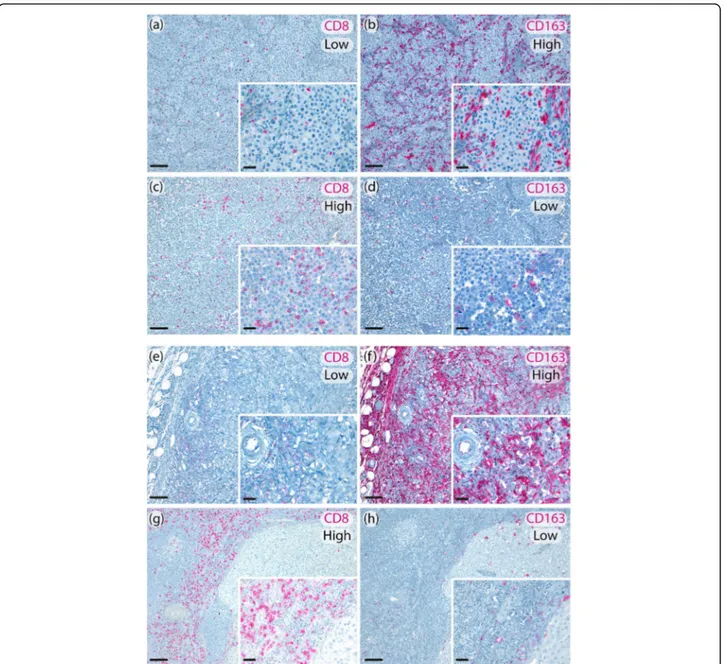
Fig. 2 Representative metastatic melanoma tissues showing intratumoral low CD8+/ high CD163+expression (a, b); intratumoral high CD8+/low
CD163+expression (c, d). (original magnification 10x, scale bar 100μm, insert 40x, scale bar 20 μm); peritumoral low CD8+/high CD163+
expression (e, f); peritumoral high CD8+/low CD163+expression (g, h). (original magnification 10x, scale bar 100μm, insert 40x, scale bar 20 μm)
Massiet al. Journal for ImmunoTherapy of Cancer (2019) 7:308 Page 6 of 13
on January 17, 2021 by guest. Protected by copyright.
http://jitc.bmj.com/
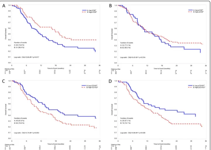
adjusting for stage, LDH, PS, treatment received (BRAFi+-MEKi vs BRAFi), subsequent immunotherapy (yes/no), metastatic MPs with high intratumoral, but not peritu-moral, CD8+ T cells density showed a barely detectable statistically significant difference in terms of OS (HR 0.65, 95% CI 0.41–1.04, p = 0.072) (Fig. 4). Notably, patients with high intratumoral, but not peritumoral, CD8+ T cells and low intratumoral CD163+macrophages (HR 0.34, 95% CI 0.16–0.72, p = 0.005) had a longer OS compared to those with intratumoral low CD8+ T cells and high CD163+ macrophages. Figure 5 and Fig. 6 show Kaplan-Meier curves for OS according to CD8+T cell and CD163+ macrophages alone or in combination, respectively. Validation cohort: impact of tissue biomarkers on PFS and OS
Patients of the validation case set (n = 55) were enrolled and evaluated independently at the Istituto Nazionale dei Tumori of Milan. Patient demographic and clinical characteristics are summarized in Add-itional file 1: Table S6.
At a median follow-up of 41.5 months, 45 (81.8%) pa-tients had progressed and 12 (21.8%) had died. Overall, 45 (78.2%) patients progressed or died. The median PFS was 9.3 (IQR: 5.8–48.0), while the median OS was not reached.
Results of multivariate analysis for PFS and OS are re-ported in Fig.7. At multivariate assessment, after adjust-ing for stage, treatment received (BRAFi+MEKi vs BRAFi), a shorter PFS was observed in patients with intratumoral low CD8+T cells and high CD163+ macro-phages (p < 0.001 and p = 0.002 for CD8+
and CD163+, respectively) (Fig.7). Regarding OS, at multivariate ana-lysis, after adjusting for stage, treatment received (BRA-Fi+MEKi vs BRAFi) and subsequent immunotherapy (yes vs no), metastatic MPs with high intratumoral, but not peritumoral, CD8+ T cells density showed a statisti-cally significant better prognosis (HR 0.14, 95% CI 0.03– 0.69, p = 0.016 for intratumoral and HR 0.26, 95% CI 0.06–1.08, p = 0.064 for peritumoral CD8+
T cells) (Fig.
7). Notably, patients with high intratumoral CD8+ T cells and low intratumoral CD163+ macrophages (HR 0.04, 95% CI 0.00–0.50, p = 0.013) had a longer OS
Fig. 3 Response to treatment according to intratumoral and peritumoral density CD8+T cells and CD163+macrophages. Low: score = 0, 1+; High: score = 2+,3+; non responder: patients who have experienced a stable or progressive disease as best response to therapy; responder: patients who have experienced a complete or partial response as best response to therapy
on January 17, 2021 by guest. Protected by copyright.
compared to those with intratumoral low CD8+ T cells and high CD163+macrophages (Fig.7).
Discussion
Increasing evidence indicates that response and long-term outcome to treatment with MAPKi in melanoma patients is influenced by clinical prognostic parameters related mostly to tumor burden and aggressiveness features. While the initial clinical response to MAPKi primarily re-lies on the loss of kinase activity of ERK, subsequent adap-tive events appear to be mediated by the intervening action of immune cells. Accordingly, strategies to improve long-term responses to MAPKi necessarily require a bet-ter understanding of the diverse cellular patbet-terns of the complex tissue microenvironment (TME). In this timely context of clinical and translational research, the three most striking findings of this study are: 1) metastatic MPs with absent/low infiltration of CD8+ T cells and a high density of CD163+ macrophages at intratumoral, but not peritumoral location, had a statistically significant shorter OS compared to those with high density CD8+T cells and absent/low density of CD163+macrophages. 2) metastatic MPs with absent/low intratumoral CD8+T cells and high intratumoral CD163+macrophages showed a nearly statis-tically significant shorter PFS compared to those with the opposite profile, while the same profile (low CD8+T cells/ high CD163+ macrophages) in the peritumoral space did not exhibit any tendency; 3) the response rate of patients with high intratumor CD163+ macrophages was lower than those with absent or low CD163+intratumor infiltra-tion, while the response rate was not affected by changes
in the peritumoral CD163+ macrophages. Thus, both the density and distribution of CD163+macrophages seem to determine the biological and clinical events associated with ORR. One of the major issues in exploiting MAPKi for metastatic melanoma patients lies in the interpatient degree and duration of response: some patients progress upon treatment, while others achieve complete response, and the remainder is somewhere in between. Hence, there is a clinical need to identify biomarkers that can allow ac-curate identification of the best treatment approach in the individual patient with BRAF-mutated melanoma. Identi-fying biomarkers correlated with a higher probability of response and a longer PFS could be clinically and transla-tionally relevant for two main reasons: i) in symptomatic patients or in those who are candidate to a neoadjuvant approach the probability and the degree of response could be important to identify patients who can draw a remark-able and sustained response to treatment, which in turns correlates with a good prognosis; ii) several studies showed that the high and prolonged response correlates with better outcome. The rate of CR is indeed a surrogate biomarker strongly correlated with long term outcome in several prospective studies investigating the efficacy of tar-geted therapy in melanoma [21,22].
For this reason we assessed the rate of patients who achieved a complete response to targeted therapy according to the investigated biomarkers in the TME. In our series, the rate of CR was significantly increased in MPs with high CD8+
/low CD163+ versus those with low CD8+/high CD163+immunophenotype. Our study, by identifying sim-ple and reliable biomarkers correlated with response and
Fig. 4 Forest plot on progression free survival and overall survival -Multivariable Cox regression model– Impact of tissue biomarkers on progression free survival and overall survival. Legend:#Adjusted for Stage, LDH, PS, treatment (BRAFi+MEKi vs BRAFi);##Adjusted for Stage, LDH,
PS, treatment (BRAFi+MEKi vs BRAFi), subsequent Immunotherapy (yes/no); i: intratumoral; p: peritumoral; CD8+/ CD163+low: score 0,1+, high:
score 2+,3 +
Massiet al. Journal for ImmunoTherapy of Cancer (2019) 7:308 Page 8 of 13
on January 17, 2021 by guest. Protected by copyright.
http://jitc.bmj.com/
longer PFS, could be translationally and clinically rele-vant. Reproducible biomarker measurements are essen-tial, particularly for long-term projects with valuable patient samples.
Our results showed an uneven spatial distribution of im-mune cells in the intra- and peritumoral space, and allowed to combine these cellular biomarkers in biosigna-tures with opposing roles, favoring or disfavoring response and better prognosis of metastatic MPs treated with BRAFi/MEKi, [13, 14, 23]. We suggest that none of the biomarkers taken individually is able to predict the long-term outcome of patients receiving MAPKi. Only the com-bination of multiple markers is therefore able to reflect the complexity of the TME and to predict the outcome of pa-tients. Furthermore, our findings support the hypothesis that a more hostile TME at baseline is associated with a worse ORR and outcome in BRAFV600-mutated metastatic MPs receiving MAPKi. However, in our cohort, tumor overexpression of PD-L1 or β-catenin in association with intratumoral or peritumoral CD8+ T lymphocytes or CD163+ was not an independent prognostic factor at
multivariate analysis. Consistently with our previous study, we found that a statistically significant higher probability of response was observed in metastatic MPs with β-catenin negative and high intratumoral CD8+ T-cell count com-pared to those with β-catenin overexpression and low intratumoral CD8+melanomas [19]. Nevertheless, we pre-viously reported a better OS in metastatic MPs with high density of CD8+T lymphocytes and no overexpression of β-catenin, than those with no CD8+
T lymphocytes and overexpression of β-catenin [19]. Incorporating the evalu-ation of both CD8+T cells and CD163+ macrophages at-tenuates the predictive power of β-catenin in identifying MAPKi-treated metastatic MPs with better outcome. The key role of CD8+T cells recruited into the tumor compart-ment is underlined by the adoptive T cell transfer proto-cols developed in melanoma that have consistently yielded high and durable clinical response in selected pa-tients [24]. However, our data further support the implication of CD163+ cells in dominant inhibitory pathways in melanoma, implying that the presence of protumor and immunosuppressive myeloid cells as
Fig. 5 Kaplan-Meier curves for overall survival according to intratumoral CD8+T cells (a), peritumoral CD8+T cells (b), intratumoral
CD163+macrophages (c), peritumoral CD163+macrophages (d). Low: score = 0, 1+; High: score = 2+,3+; iCD8+: intratumoral CD8+; pCD8+:
peritumoral CD8+; iCD163+: intratumoral CD163+; pCD163+: peritumoral CD163+
on January 17, 2021 by guest. Protected by copyright.
shutting down their function in TME ultimately favors tumor outgrowth. Our original contribution definitively includes macrophages in this scenario, where conflicting data have been reported so far [25].
The observation in human tumor biopsies from 10 patients treated with vemurafenib or a combination of dab-rafenib and trametinib that treatments increased macro-phages [26,27], suggests that macrophages are recruited to the tumor site by BRAFi/MEKi treatment, and that target-ing macrophages in combination with BRAFi/MEKi may affect patient response. Tumor-promoting M2 macro-phages can contribute to tolerance to MAPK inhibition, and their accumulation within tumors during treatment strongly correlates with an aggressive phenotype in different
melanoma models, through different mechanisms, includ-ing VEGF and TNF-alpha secretion. The M2 macrophage phenotype, promoted by IL-4, IL-13, IL-10 and M-CSF, ap-pears to contribute to immune suppression through the production of IL-10 and TGF-β [28]. Present findings are in line with the protumor function of M2 CD163+ macro-phages that in combination CD8+T cells represent predict-ive prognostic biosignatures in BRAFV600-mutated patients receiving MAPKi. However, they point on the key predict-ive role of M2 macrophage level outside and, more import-antly, inside the tumor at baseline, before the treatment initiation.
This study presents some strengths: i) patients have been enrolled and treated homogeneously in IMI centers; ii) the
Fig. 7 Forest plot on progression free survival and overall survival in the validation cohort. Multivariable Cox regression model– Impact of tissue biomarkers on progression free survival and overall survival.#Adjusted for Stage, treatment (BRAFi+MEKi vs BRAFi);##Adjusted for Stage, treatment
(BRAFi+MEKi vs BRAFi), subsequent Immunotherapy (yes/no); i: intratumoral; p: peritumoral; CD8+/ CD163+low: score 0,1+, high: score 2+,3 +
Fig. 6 Kaplan-Meier curves for overall survival according to the combination of intratumoral (a) and peritumoral (b) CD8+T cells and CD163+
macrophages. Low: score = 0, 1+; High: score = 2+,3+; iCD8+: intratumoral CD8+; pCD8+: peritumoral CD8+; iCD163+: intratumoral CD163+;
pCD163+: peritumoral CD163+
Massiet al. Journal for ImmunoTherapy of Cancer (2019) 7:308 Page 10 of 13
on January 17, 2021 by guest. Protected by copyright.
http://jitc.bmj.com/
majority of enrolled and investigated metastatic MPs were (122/158, 77%) in latest metastatic samples, thus reducing the potential discordance between primary and metastatic samples and to better reflect the actual immune biological status of the patient cohort; iii) semi-automated counting upon digital image acquisition, which allows unbiased and rapid quantification of the immune infiltrate in immuno-stained tissue sections and minimizes significant user errors due to categorical rankings was adopted; IV) since pro-spective clinical trials have demonstrated that single-agent BRAFi and BRAFi+MEKi have different response rates, PFS, and OS, we addressed this potential bias by accounting for the difference in treatments in the multivariate model, V) our findings were validated in an independent patient cohort, strictly following the Remark checklist [29]. How-ever, we are aware of the study limitations, including: i) the retrospective nature of the analysis of prospective collected cohorts of patients, ii) overall, the time schedule for disease assessment was similar but not absolutely overlapping in all patients; iii) complex highly pigmented or necrotic meta-static melanoma tissues in which macrophages overlap or fuse together with pigmented melanoma cells forming densely packed layers of cells were seldom present. Al-though careful correlation with cell morphology and ac-curate identification of viable representative tumor areas were performed, this may represent a confounding factor that was addressed by optical microscopic evaluation. An-other point is worthy to be underlined: in our cohort of metastatic lymph nodes, scoring evaluation did not differ from the other metastatic sites and positivity for the se-lected markers was evaluated within the tumor (intratu-morally) as well as at the interface between the tumor and immune stroma (peritumorally). Nevertheless, the im-munologic environment in the lymph node is peculiar and the crosstalk between specific subsets of lymphocytes and macrophages in different anatomical lymph node com-partments may likely yield biological insights not globally applicable to other metastatic sites.
In our study, the main comparison was between the ex-treme categories high CD8+/low CD163+ and low CD8+/ high CD163+, and the results of the categories in between (both low or both high) were instrumental only to confirm the trend of the risk in the three analyzed groups. The threshold for statistical significance was set at 0.05, and no adjustment for multiple tests was planned. The purpose of our study was to evaluate the impact of a limited number of biomarkers on prognosis, and these biomarkers should be prospectively validated in large clinical studies. Never-theless, the robustness of our results was tested by includ-ing a validation cohort.
Conclusions
Our findings indicate that a specific preexisting profile of T and macrophage distribution inside and outside
melanoma dictates the level of resistance to MAPKi. Our results could have important implications for clin-ical therapeutic strategies. Since patients with absent/ low intratumoral infiltration of CD8+ cells and high intratumoral CD163+ cells have a statistically significant lower ORR and shorter OS, they should deserve a differ-ent therapeutic strategy. Whether the hostile immune microenvironment induced by accumulated macro-phages can be overcome by either inhibiting macrophage polarization to a M2 phenotype or targeting the inflam-matory signaling promoted by NF-kB with IkB kinase in-hibitors is currently unknown. Additional strategies can include the colony-stimulating factor (CSF)-1R inhibitor PLX3397 that has been shown to reduce myeloid cell in-filtration and enhance adoptive cell transfer immuno-therapy in BRAFV600E-driven melanoma genesis in mice [30]. Our findings along with other translational studies support the proposal to design new ad hoc prospective clinical trials in order to improve long-term survival of advanced MPs receiving MAPKi. In addition, the present study further underlines that a better understanding of the mechanisms that control the recruitment of immune cells in the TME and their distribution in the intra- and peritumoral space is essential to devise better thera-peutic options in metastatic MPs, and particularly in those undergoing treatment with MAPKi.
Supplementary information
Supplementary information accompanies this paper athttps://doi.org/10. 1186/s40425-019-0797-4.
Additional file 1: Table S1. Patients’ characteristics. Table S2. Associations. Table S3. Tissue biomarkers– combination. Table S4. Multivariable logistic model - Response rate. Table S5. Multivariable Cox regression model– Survival. Table S6. Validation cohort. Table S7. Validation cohort - Multivariable Cox regression model– Impact of tissue biomarkers on progression free survival and overall survival– validation cohort.
Additional file 2: Figure S1. Immunohistochemistry with anti-PD-L1 antibody shows positivity in more than 5% of tumor cells at membranous level (A). Immunohistochemistry with anti-PD-L2 antibody shows negative tumor cells with internal positive control (B). (original magnification 10x, scale bar 100μm, inset 40x, scale bar 20 μm). Figure S2. Immunohisto-chemicalβ-catenin expression in metastatic melanoma tissues. At subcel-lular level, immunoreactivity is observed in the cytoplasm and scattered nuclei (A) and membrane and nuclear (B). (original magnification 10x, scale bar 100μm, inset 40x, scale bar 20 μm). Figure S3. Distribution pat-terns and density of intratumoral and peritumoral CD8+T and CD163+ cells in the training cohort. Low: score = 0, 1+; High: score = 2+,3 + .
Abbreviations
AJCC:American Joint Committee on Cancer; BRAFi: BRAF inhibitors; CR: complete response; ECOG-PS: Eastern Cooperative Oncology Group performance status; FFPE: Formalin fixed paraffin-embedded; HR: hazard ratio; IMI: Italian Melanoma Intergroup; LDH: lactate dehydrogenase; MAPKi: MAPK inhibitors; MPs: melanoma patients; NE: not evaluable; OR: odd ratio; ORR: Overall response rate; OS: overall survival; PD: progressing disease; PFS: progression free survival; RGB: red, green, blue.; TME: tumor
microenvironment; TNM: Tumor, Node, Metastasis
Acknowledgments
The authors thank Sara Simi and Fabio Galli for their technical assistance.
on January 17, 2021 by guest. Protected by copyright.
Ethics approval and consent to partecipate The study was approved by local Ethics Committees.
Authors’ contributions
Conception and design: MM and DM. Development of methodology: MM, DM, ER, LR, MR. Acquisition of data (managed patients, provided samples, techinical analysis of tumour samples): DM, ER, MC, BV, MR, BM, MDV, LDG, FDL, RDP, RN, MG, VCS, AMDG, MT, MO, FP, VV, FC, PQuaglino, PQueirolo, GB, FC-S, LC, AM, GP, LR. Analysis and interpretation of data (e.g., statistical ana-lysis, biostatistics): MMM, DM, ER, LR, MR. Writing the manuscript: MM, DM, ER, MR, LR. Review, and/or revision of the manuscript: DM, ER, MC, BV, MR, BM, MDV, LDG, FDL, RDP, RN, MG, VCS, AMDG, MT, MO, FP, VV, FC, PQ, PQ, GB, FC-S, LC, AM, GP, LR. All authors read and approved the final manuscript.
Funding
This work is sponsored by Azienda Socio Sanitaria Papa Giovanni XXIII, Bergamo, Italy and was supported by a Roche S.p.A. grant, by the Associazione Italiana per la Ricerca sul Cancro (AIRC) Special Program Innovative Tools for Cancer Risk Assessment and early Diagnosis 5X1000 (no. 12162 to LR), and by the Cariplo Foundation (2015–0911 to VV).
Availability of data and materials Not applicable.
Consent for publication Not applicable.
Competing interests
The authors declare that they have no competing interests.
Author details
1Section of Pathological Anatomy, Department of Health Sciences, University
of Florence, Florence, Italy.2Department of Oncology, Istituto di Ricerche Farmacologiche Mario Negri IRCCS, Milan, Italy.3Unit of Immunotherapy of
Human Tumors, Fondazione IRCCS Istituto Nazionale dei Tumori, Milan, Italy.
4Department of Pathology, Fondazione IRCCS Istituto Nazionale dei Tumori,
Milan, Italy.5Unit of Medical Oncology, Department of Oncology and Hematology, Papa Giovanni XXIII Hospital, Bergamo, Italy.6Unit of Clinical
Pharmacology and Oncology, Department of Health Sciences, University of Florence, Florence, Italy.7Unit of Oncology, Fondazione IRCCS Istituto
Nazionale dei Tumori, Milan, Italy.8Department of Oncology, Hematology, and Respiratory Diseases, University Hospital of Modena, Modena, Italy.
9Department of Medical Oncology and Molecular Genetics Laboratory, IRCCS
Istituto Tumori Giovanni Paolo II, Bari, Italy.10Melanoma and Esophageal
Cancer Unit, Istituto Oncologico Veneto-IRCCS, Department of Medical Oncology, Padua, Italy.11Medical Oncology and Immunotherapy, Center for
Immuno-Oncology, University Hospital of Siena, Istituto Toscano Tumori, Siena, Italy.12Medical Oncology Unit, Department of Biomedical Sciences
and Human Oncology, University of Bari‘Aldo Moro’, Bari, Italy.13Azienda Ospedaliera Santa Croce e Carle di Cuneo SC Oncologia, Cuneo, Italy.
14Department of Oncology, ASST Spedali Civili, Brescia, Italy.15Department of
Medical Sciences, Section of Dermatology, University of Turin, Turin, Italy.
16
Unit of Medical Oncology, Ospedale Policlinico San Martino, Genoa, Italy.
17Medical Oncology Candiolo Cancer Institute-FPO, IRCCS, Candiolo, Italy. 18Division of Pathological Anatomy, Papa Giovanni XXIII Hospital, Bergamo,
Italy.19Department of Oncology, Azienda Sanitaria Universitaria Integrata di
Udine, Udine, Italy.20Unit of Cancer Genetics, Institute of Biomolecular Chemistry, National Research Council, Sassari, Italy.
Received: 19 June 2019 Accepted: 30 October 2019
References
1. Davies H, Bignell GR, Cox C, Stephens P, Edkins S, Clegg S, et al. Mutations of the BRAF gene in human cancer. Nature. 2002;417(6892):949–54. 2. Chapman PB, Hauschild A, Robert C, Haanen JB, Ascierto P, Larkin J, et al.
Improved survival with vemurafenib in melanoma with BRAF V600E mutation. N Engl J Med. 2011;364(26):2507–16.
3. Hauschild A, Grob JJ, Demidov LV, Jouary T, Gutzmer R, Millward M, et al. Dabrafenib in BRAF-mutated metastatic melanoma: a multicentre, open-label, phase 3 randomised controlled trial. Lancet. 2012;380(9839):358–65.
4. Long GV, Stroyakovskiy D, Gogas H, Levchenko E, de Braud F, Larkin J, et al. Dabrafenib and trametinib versus dabrafenib and placebo for Val600 BRAF-mutant melanoma: a multicentre, double-blind, phase 3 randomised controlled trial. Lancet. 2015;386(9992):444–51.
5. Ascierto PA, McArthur GA, Dreno B, Atkinson V, Liszkay G, Di Giacomo AM, et al. Cobimetinib combined with vemurafenib in advanced BRAF(V600)-mutant melanoma (coBRIM): updated efficacy results from a randomised, double-blind, phase 3 trial. Lancet Oncol. 2016;17(9):1248–60.
6. Dummer R, Ascierto PA, Gogas HJ, Arance A, Mandala M, Liszkay G, et al. Encorafenib plus binimetinib versus vemurafenib or encorafenib in patients with BRAF-mutant melanoma (COLUMBUS): a multicentre, open-label, randomised phase 3 trial. Lancet Oncol. 2018;19(5):603–15.
7. Long GV, Eroglu Z, Infante J, Patel S, Daud A, Johnson DB, et al. Long-term outcomes in patients with BRAF V600-mutant metastatic melanoma who received Dabrafenib combined with Trametinib. J Clin Oncol. 2018;36(7): 667–73.
8. Mandalà M, De Logu F, Merelli B, Nassini R, Massi D. Immunomodulating property of MAPK inhibitors: from translational knowledge to clinical implementation. Lab Investig. 2017;97(2):166–75.
9. Winder M, Virós A. Mechanisms of drug resistance in melanoma. Handb Exp Pharmacol. 2018;249:91–108.
10. Yan Y, Robert C, Larkin J, Ascierto PA, Dreno B, Maio M, et al. Genomic features of complete responders (CR) versus fast progressors (PD) in patients with BRAFV600-mutated metastatic melanoma treated with cobimetinib + vemurafenib or vemurafenib alone. Clin Cancer Res. 2019; 25(11):3239–46.
11. Spranger S, Bao R, Gajewski TF. Melanoma-intrinsic beta-catenin signalling prevents anti-tumour immunity. Nature. 2015;523(7559):231–5.
12. Hugo W, Shi H, Sun L, Piva M, Song C, Kong X, et al. Non-genomic and immune evolution of melanoma acquiring MAPKi resistance. Cell. 2015; 162(6):1271–85.
13. Frederick DT, Piris A, Cogdill AP, Cooper ZA, Lezcano C, Ferrone CR, et al. BRAF inhibition is associated with enhanced melanoma antigen expression and a more favorable tumor microenvironment in patients with metastatic melanoma. Clin Cancer Res. 2013;19(5):1225–31.
14. Wilmott JS, Long GV, Howle JR, Haydu LE, Sharma RN, Thompson JF, et al. Selective BRAF inhibitors induce marked T-cell infiltration into human metastatic melanoma. Clin Cancer Res. 2012;18(5):1386–94.
15. Liu L, Mayes PA, Eastman S, Shi H, Yadavilli S, Zhang T, et al. The BRAF and MEK inhibitors Dabrafenib and Trametinib: effects on immune function and in combination with Immunomodulatory antibodies targeting PD-1, PD-L1, and CTLA-4. Clin Cancer Res. 2015;21(7):1639–51.
16. Steinberg SM, Shabaneh TB, Zhang P, Martyanov V, Li Z, Malik BT, et al. Myeloid cells that impair immunotherapy are restored in melanomas which acquire resistance to BRAF inhibitors. Cancer Res. 2017;77(7):1599–610. 17. Eisenhauer EA, Therasse P, Bogaerts J, Schwartz LH, Sargent D, Ford R, et al.
New response evaluation criteria in solid tumours: revised RECIST guideline (version 1.1). Eur J Cancer. 2009;45(2):228–47.
18. Larkin J, Del Vecchio M, Ascierto PA, Krajsova I, Schachter J, Neyns B, et al. Vemurafenib in patients with BRAF(V600) mutated metastatic melanoma: an open-label, multicentre, safety study. Lancet Oncol. 2014;15(4):436–44. 19. Massi D, Romano E, Rulli E, Merelli B, Nassini R, De Logu F, et al. Baseline
beta-catenin, programmed death-ligand 1 expression and tumour-infiltrating lymphocytes predict response and poor prognosis in BRAF inhibitor-treated melanoma patients. Eur J Cancer. 2017;78:70–81. 20. Massi D, Brusa D, Merelli B, Falcone C, Xue G, Carobbio A, et al. The status
of PD-L1 and tumor-infiltrating immune cells predict resistance and poor prognosis in BRAFi-treated melanoma patients harboring mutant BRAFV600. Ann Oncol. 2015;26(9):1980–7.
21. Schadendorf D, Long GV, Stroiakovski D, Karaszewska B, Hauschild A, Levchenko E, et al. Three-year pooled analysis of factors associated with clinical outcomes across dabrafenib and trametinib combination therapy phase 3 randomised trials. Eur J Cancer. 2017;82:45–55.
22. Osgood C, Mulkey F, Mishra-Kalyani PS, Lemery S, Ward A, Keegan P, et al. U.S. Food and Drug Administration, Silver Spring, MD FDA analysis of depth of response (DpR) and survival across 10 randomized controlled trials in patients with previously untreated unresectable or metastatic melanoma (UMM) by therapy type. J Clin Oncol 2019; 37 Suppl: 9508.
23. Wargo JA, Cooper ZA, Flaherty KT. Universes collide: combining immunotherapy with targeted therapy for cancer. Cancer Discov. 2014; 4(12):1377–86.
Massiet al. Journal for ImmunoTherapy of Cancer (2019) 7:308 Page 12 of 13
on January 17, 2021 by guest. Protected by copyright.
http://jitc.bmj.com/
24. Rosenberg SA, Yang JC, Sherry RM, Kammula US, Hughes MS, Phan GQ, et al. Durable complete responses in heavily pretreated patients with metastatic melanoma using T-cell transfer immunotherapy. Clin Cancer Res. 2011;17(13):4550–7.
25. Ladanyi A. Prognostic and predictive significance of immune cells infiltrating cutaneous melanoma. Pigment Cell Melanoma Res. 2015;28(5):490–500. 26. Smith MP, Sanchez-Laorden B, O'Brien K, Brunton H, Ferguson J, Young H,
et al. The immune microenvironment confers resistance to MAPK pathway inhibitors through macrophage-derived TNFalpha. Cancer Discov. 2014; 4(10):1214–29.
27. Smith MP, Wellbrock C. Molecular pathways: maintaining MAPK inhibitor sensitivity by targeting nonmutational tolerance. Clin Cancer Res. 2016; 22(24):5966–70.
28. Mantovani A, Marchesi F, Malesci A, Laghi L, Allavena P. Tumour-associated macrophages as treatment targets in oncology. Nat Rev Clin Oncol. 2017; 14(7):399–416.
29. Mallett S, Timmer A, Sauerbrei W, Altman DG. Reporting of prognostic studies of tumour markers: a review of published articles in relation to REMARK guidelines. Br J Cancer. 2010;102(1):173–80.
30. Zhu Y, Knolhoff BL, Meyer MA, Nywening TM, West BL, Luo J, et al. CSF1/ CSF1R blockade reprograms tumor-infiltrating macrophages and improves response to T-cell checkpoint immunotherapy in pancreatic cancer models. Cancer Res. 2014;74(18):5057–69.
Publisher’s Note
Springer Nature remains neutral with regard to jurisdictional claims in published maps and institutional affiliations.
on January 17, 2021 by guest. Protected by copyright.