Pathology of disappearing bone disease:
a case report with immunohistochemical study
U. E. Pazzaglia
1, L. Andrini
1, M. Bonato
2, M. Leutner
21Clinica Ortopedica, 2a FacoltaÁ di Medicine e Chirurgia, UniversitaÁ di Pavia, Varese, Italy 2Servizio di Anatomia e Istologia Patologica, Ospedale Multinazionale, Varese, Italy
Accepted: 1 February 1997
Summary. A case of disappearing bone disease of
the proximal femur is reported with
histopatholo-gical and immunohistochemical studies. There was
a densely packed cellular tissue, positive to
en-dothelial antibodies, in areas of massive bone
de-struction. A more differentiated vascular tissue was
present where trabecular cancellous or cortical
bone was preserved with only focal zones of
ac-celerated bone remodelling. The self-limited course
correlates well with two phases of evolution of the
histopathological lesions with neoplastic-like
pro-liferation of endothelial cells corresponding to the
rapid and massive bone destruction, and a later
differentiation of the cells in mature vascular
structures, but still with accelerated bone
resorp-tion which is partly compensated by apposiresorp-tional
activity.
ReÂsumeÂ. Nous deÂcrivons un cas d'osteÂolyse
mas-sive idiopathique localiseÂe aÁ l'extremiteÁ proximale
du feÂmur ainsi qu'une eÂtude histopathologique et
immunohistochimique. LaÁ ouÁ il y a une plus grande
destruction osseuse on observe un tissu avec des
cellules en amas, ayant une reÂaction positive aux
anticorps endotheliaux. Par contre il y a une plus
grande diffeÂrenciation vasculaire la ouÁ l'os
spon-gieux et cortical eÁtait preserve avec seulement des
petites zones ayant un remodelage osseux acceÂleÂreÂ.
L'eÂvolution autolimitante de cette pathologie est
mise en correÂlation avec deux phases eÂvolutives des
leÂsions histopathologiques. En premier la
pro-liferation des cellules endotheliales neÂoplasiques
correspond aÁ une eÂnorme et rapide destruction de
l'os, ensuite une differentiation successive des
cel-lules en structures vasculaires matures, tandis que
la resorption osseuse, est compenseÁe partiellement
par une neÂoapposition.
Introduction
Several reviews of disappearing bone disease have
been published [1, 5, 7, 8, 9, 13, 14, 16]. The bone
lesions consist of thin walled vessels, like
capil-laries, filled with blood cells in the marrow spaces
and in cortical bone [7]. The mechanism leading to
massive osteolysis and the replacement of bone by
vascular connective tissue is uncertain, particularly
since few osteoclasts have been reported in the
bone [2, 6, 10, 12].
Case report
A farmer, aged 55, had pain in the right hip for 6 months; radiographs showed a lytic area in the subtrochanteric region (Fig. 1a). He refused operation, but 3 days later sustained a pathological fracture (Fig. 1b). His general condition was good and laboratory investigations, including the alkaline phospha-tase, were normal.
The fracture was treated with a dynamic hip screw plate, the lytic area being curetted and filled with acrylic cement. The histology showed bone haemangioma without malignant change.
One month later, he had acute pain; radiographs showed displacement of the cement mass, and extension of the lesion proximally and distally (Fig. 1c). Bone destruction was rapid and 14 days later, there was further lysis (Fig. 1d) which would not be expected in an haemangioma.
Reprint request to: U. E. Pazzaglia, Clinica Ortopedica, Ospe-dale F. Del Ponte, p.zza Biroldi, I-21100 Varese, Italy
Orthopaedics
Investigations showed no primary neoplasm in the extra-skeletal organs, his general condition remained good and laboratory tests were normal. Because of the clinical course the proximal femur was resected and replaced with a prosthe-sis.
After 4 years, he was free of symptoms and radiographs showed no sign of recurrence of the lesion.
Histopathology
The resected femur was split in the frontal plane after removal of the plate and screws, decalcified in a solution of hydro-chloric and acetic acid, and embedded in paraffin. Sections were stained with haematoxylin-eosin and studied im-munohistochemically with the avidin-biotin-peroxidase com-plex technique [11]. The following markers were used: for endothelium factor VIII, CD3 1, Ulex; for histiocytes, KP1; for T-lymphocytes, CD3, UCHL1, and for B-lymphocytes, L26, 4KB5 (Table 1).
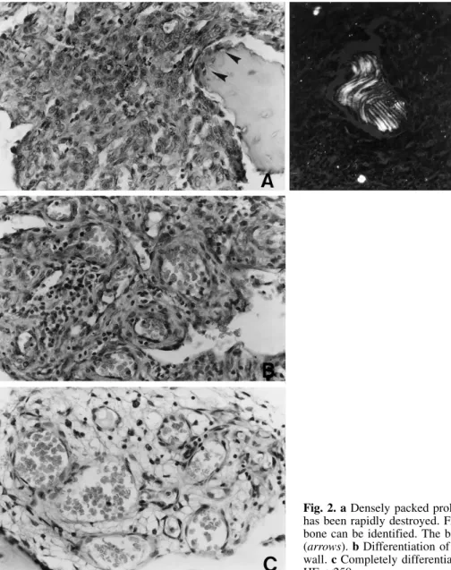
Two main histopathological appearances were seen: (1) a densely packed cellular tissue was present in zones of the specimen where there was massive bone destruction (MOZ = massive osteolytic zones); this was confirmed by remnants of the original lamellar bone of the cortex, presenting a moth-eaten surface completely surrounded by pathological tissue (Fig. 2a). The latter was characterised by cells with large nuclei and scanty cytoplasm, as well as capillary-like lumens. (2) zones where cancellous trabeculae or cortical bone were preserved (BSZ = bone structured zones) which presented with medullary spaces or large lacunae of the cortex occupied by vascular tissue formed by flattened or focally plump endothe-lial cells. These merged with a network of anastomosing thin-walled capillaries with irregular and often cystic spaces, and venous structures (Fig. 2b, c). Lymphoid aggregates and Fig. 1. a Radiograph showing a subtrochanteric lytic area with
poorly defined edges. Pain had been present for 6 months. b Pathological fracture through the lytic area. c One month after operation, the device has failed and the cement is
displaced. d Proximal and distal extension of the lesion during 2 weeks. e Four years after excision of the proximal femur and replacement with a prosthesis. An incomplete cortex has formed around the stem
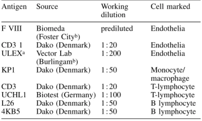
Table 1. Immunohistochemical markers
Antigen Source Working
dilution Cell marked F VIII Biomeda
(Foster Cityb) prediluted Endothelia
CD3 1 Dako (Denmark) 1:20 Endothelia ULEXa Vector Lab
(Burlingamb) 1:200 Endothelia
KP1 Dako (Denmark) 1:50 Monocyte/ macrophage CD3 Dako (Denmark) 1:20 T-lymphocyte UCHL1 Biotest (Germany) 1:100 T-lymphocyte L26 Dako (Denmark) 1:50 B lymphocyte 4KB5 Dako (Denmark) 1:50 B lymphocyte
aLectin
bUSA
Table 2. Distribution of cellular immunoreactivity in relation to zones in the specimen and cell morphology
Markers
F VIII CD3 1 ULEX CD3 UCHL1 L26 4KB5 KP1
MOZ Densely-packed vascular tissue + + + ± ± ± ± ±
Lymphoid infiltrate ± ± ± + + ± ± ±
Osteoclasts ± ± ± ± ± ± ± +
BSZ Differentiated vascular tissue + + + ± ± ± ± ±
Lymphoid infiltrate ± ± ± + + ± ± ±
Osteoclasts ± ± ± ± ± ± ± +
MOZ = massive osteolytic zones BSZ = bone structured zones
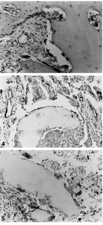
perivascular plasma cells infiltrated the stroma (Fig. 2b). Focal areas of accelerated bone remodelling were present with many osteoclasts lining the trabecular surface and cortical lacunae associated with wide appositional fronts (Fig. 3).
The compact tissue of the MOZ and the more differentiated vascular tissue of BSZ were both positive for the endothelial markers factor VIII, CD31 and Ulex (Fig. 4). Lymphoid aggregates and perivascular infiltrate reacted to markers CD3 and HCHL1 suggesting that T-lymphocytes were prevalent in the lymphoid component.
Osteoclasts (Fig. 5) and interstitial monocytes were KP1 positive (Table 2).
Discussion
The extensive bone destruction in disappearing
bone disease is related to the angioma- or
lymph-angioma-like tissue reported in most studies [2, 6].
In a few cases, vascular proliferation also involved
the skin and soft tissues overlying the bony lesion
[5, 8, 9, 15]. The mechanism leading to the
mas-sive bone resorption is still not explained because
increased osteoclastic activity has not been
de-scribed [6, 10, 12].
Our case shows new histopathological features
which have not been previously described.
Den-sely packed cellular tissue was associated with the
massive osteolysis. The moth-eaten appearance of
the surface of remnants of cortical bone which are
scattered in this tissue is peculiar to osteoclastic
bone resorption. These features indicate that there
must have been an earlier phase of intense
osteo-clastic activity. The immunohistochemical study
confirmed the endothelial nature of this
neoplastic-Fig. 2. a Densely packed proliferating cells in an area where bone has been rapidly destroyed. Fragments of original cortical lamellar bone can be identified. The bony surface is attacked by cells (arrows). b Differentiation of vessels with a multi-layered cellular wall. c Completely differentiated thin-walled capillary-like vessels. HE, ´250
like proliferation which showed the same reaction
to antibodies factor VIII, CD31 and Ulex as the
more differentiated vascular tissue of BSZ. In this
tissue, increased remodelling was limited to focal
areas, and the number of osteoclasts or rate of
resorption is not sufficient to produce the bone loss
of the MOZ.
The lymphoid infiltrate of T-lymphocytes seems
peculiar to disappearing bone disease and suggests
an immunological cell-mediated mechanism of
bone destruction. The production of lymphokines
by T-lymphocytes could induce recruitment of
activated monocyte and macrophage elements, and
of osteoclasts. On the contrary, the usual
angio-matous lesions of bone are formed by thin-walled
vessels without perivascular cellularity and
in-flammatory infiltration. They typically show a
slowly progressive lysis, and not the rapid and
massive resorption of disappearing bone disease.
In our case, it was possible to correlate the rapid
radiographic progress of the lytic lesion with the
histopathological findings. Bone resorption was
massive in the areas of solid tissue which had
completely replaced the original cortical bone,
where the vascular tissue was more differentiated
while the process of accelerated bone remodelling
was prevalent.
The vascular proliferation does not have
ma-lignant characteristics which is in keeping with the
self-limiting course of disappearing bone disease
[3, 4]. The reported histological findings make it
possible to suggest an evolving type of disease, a
first phase characterised by neoplastic-like
pro-liferation of endothelial cells corresponding to the
massive bone destruction and a second phase with
better differentiated vascular structures, where
bone resorption is still accelerated but partly
compensated by appositional activity.
In several reports, osteoclasts have not been
observed on bony surfaces in spite of the
radio-graphic appearance of massive osteolysis [6, 10,
12]. Hypotheses have been advanced to explain the
role of mononuclear perivascular cells and
hydro-lytic enzyme activation as due to hypoxia and low
pH stimulated by the slowed blood flow in the
capillary-like network [10]. This contrasts with the
view that bone resorption is carried out by
osteo-clasts. If the evolving pattern we have suggested is
correct, the latter histological features can be
ex-plained because the examination was carried out
when the disease was already in a quiescent phase
with exhaustion of resorption and bone
remodel-ling becoming normal. In this case only
thin-wal-led well differentiated vessels remain in the
me-dullary and lacunar spaces in the bone.
Conse-Fig. 5. Resorption of a fragment of cortical bone by numerous KP1-positive osteoclasts. Vascular proliferative tissue sur-rounds the bone. ABC-peroxidase, DAB-haematoxylin, ´400 Fig. 3. Area of accelerated bone remodelling with numerous osteoclasts and a front of osteoblastic apposition (arrows). The marrow spaces are occupied by incompletely differentiated vascular tissue. HE, ´250
Fig. 4. Vascular channels with a CD31 endothelial layer. ABC-peroxidase, DAB-haematoxylin, ´200
quently, it is possible that the disease might be
successfully treated with radiotherapy [3] or
cy-totoxic drugs in the first phase, but no effect could
be expected in the second phase.
References
1. Brauch HE (1945) Acute spontaneous absorption of bone: report of a case involving a clavicle and a scapula. J Bone Joint Surg 27: 706±710
2. Cannon SR (1968) Massive osteolysis. A review of seven cases. J Bone Joint Surg [Br] 68: 24±28
3. Dunbar SR, Rosenberg, A, Mankin H, Rosenthal D, Suit HD (1993) Gohram's massive osteolysis: the role of ra-diation therapy and review of the literature. J Radiat Oncol Biol Phys 26: 491±497
4. Edwards WJ Jr, Thompson RC Jr, Varsa LW (1983) Lymphangiomatosis and massive osteolysis of the cervical spine: a case report and a review of the literature. Clin Orthop 177: 222±229
5. Fornasier VL (1970) Haemangiomatosis with massive osteolysis. J Bone Joint Surg [Br] 52: 444±451
6. Gorham LW, Wright AW, Shultz HH, Maxon FC Jr (1954) Disappearing bones: a rare form of massive osteolysis. Report of two cases, one with autopsy findings. Am J Med 17: 674±682
7. Gorham LW, Stout AP (1955) Massive osteolysis: its re-lation to hemangiomatosis. J Bone Joint Surg [Am] 37: 985±1004
8. Halliday FA, Dahlin DL, Pugh DG, Young HH (1964) Massive osteolysis and angiomatosis. Radiology 82: 637±644
9. Hambach R, Pujam J, Maly V (1958) Massive osteolysis due to hemangiomatosis. Report of a case of Gorham's disease with autopsy. Radiology 71: 43±47
10. Heyden G, Kindblau LG, Nielsen M (1977) Disappearing bone disease. J Bone Joint Surg [Am] 59: 57±61 11. Hsu SM, Raine L, Fanger H (1980) Use of
avidin-biotin-peroxidase complex (ABC) in immunoavidin-biotin-peroxidase techni-ques. J Histochem Cytochem 25: 577±580
12. Johnson PM, Mc Clure JG (1958) Observations on mas-sive osteolysis: a review of the literature and report of a case. Radiology 71: 28± 42
13. Jones GB, Midgley RL, Smith GS (1958) Massive os-teolysis disappearing bone. J Bone Joint Surg [Br] 40: 494±501
14. Kery L, Wouters HW (1970) Massive osteolysis: report of two cases. J Bone Joint Surg [Br] 52: 452±459
15. Pazzaglia UE, Mora R, Ceciliani L (1987) Lymphangio-matosis of the arm with massive osteolysis. A case report. Int Orthop 11: 367±369
16. Sage MR, Allen PW (1974) Massive osteolysis: report of a case. J Bone Joint Surg [Br] 56: 130±135