Female Urology
Role of Diffusion-Weighted Magnetic
Resonance Imaging in the Diagnosis
of Bladder Pain Syndrome/Interstitial
Cystitis
Porru Daniele, Regina Cesare, Oworae Howardson Bright, Fiorello Nicolo, Gardella Barbara,
Manzoni Federica, Klersy Catherine, Sala Maria Gabriella, La Fianza Alfredo,
Ballerini Daniela, Preda Lorenzo, Simeone Claudio, Spinillo Arsenio, and Jallous Hussein
OBJECTIVE To evaluate whether DW-MRI can contribute to noninvasive diagnosis of BPS/IC. The agree-ment between two raters (2 radiologists involved in the study) was also evaluated, the relevance of the "operator-dependent" factor defined.
PATIENTS AND METHODS
Twenty-two female patients with a diagnosis of BPS-IC were recruited and performed DW-MRI. The same investigation was also performed in 20 patients with pelvic gynecological diseases and no BPS-IC.
RESULTS A significant difference was found between BPS-IC and no-BPS-IC since 17 out of 22 subjects of thefirst group were positive, compared to 3 out of 20 no-IC subjects, with a P value of .001 to highlight the statistical significance. The sensitivity of the exam was 77%, while the specificity was 85%. There was good agreement between the 2 raters in the evaluation of MRI results. CONCLUSION DW-MRI helps to obtain a noninvasive diagnosis of BPS/IC, by providing useful information on
the choice of which patients may be more appropriately submitted to cystoscopy and bladder biopsy. UROLOGY 141: 55−59, 2020. © 2020 Elsevier Inc.
B
ladder Pain Syndrome/Interstitial Cystitis (BPS-IC) is a clinical condition characterized by the sen-sation of pain, a sense of pressure and discomfort perceived as correlated with the bladder associated with at least 1 urinary symptom, usually urinary frequency and urgency lasting more than 6 months, with repeatedly neg-ative urine culture.1Thefirst epidemiological study was performed by Oravisto in Finland analyzing patients diagnosed in a population of 1million people recruited into the study.2Prevalence was 18/100,000 in female population, 10.6/100,000 including both female and male patients, since 10% of the entire patient population were male. The diagnosis is made by combining clinical features with cystoscopy and
hydrodistension, which results in a severity progressing from 1 to 3, and with histologic examination of bladder biopsy, which allows to obtain a descriptive index of the disease.1
In the context of chronic pelvic pain it is possible to define chronic pelvic pain syndrome, which may be associ-ated with urologic, gynecologic, physiatric, neurologic symp-toms. In the urologic field, pain can be perceived as associated with the testicle, epididymis, prostate, vagina, vulva, bladder and, in the latter case, the interstitial cystitis described according to the criteria stated by the European Association for the Study of Interstitial Cystitis (ESSIC), shared by the scientific community, can be included.
Some studies have evaluated the introduction of nonin-vasive imaging techniques in the diagnostic procedures, specifically MRI. In 2016, Towner et al3analyzed the use of MRI with intravesical contrast to assess increased blad-der permeability, observe the correlation with symptoms, and stratify and monitor patients’ disease progress. Char-lanes et al4 sought to determine whether patients with BPS/IC revealed bladder hyperintense signals with diffu-sion weighted-magnetic resonance imaging (DW-MRI).
PATIENTS AND METHODS
Twenty-two female patients with a diagnosis of BPS-IC, made by means of clinical features indicating BPS-IC underwent
Conflict of interests: The authors declare that there are no conflicts of interest. Funding: This research received no specific grant from any funding agency in the public, commercial, or not-for-profit sectors.
From the Urology Department, Fondazione IRCCS Policlinico San Matteo, Pavia, Italy; the Gynecologic and Obstetric Clinic, Fondazione IRCCS Policlinico San Matteo, Pavia, Italy; the Servizio di Epidemiologia Clinica e Biometria, Direzione Scientifica, Fondazione IRCCS Policlinico San Matteo, Pavia, Italy; the Radiology Unit, Fondazione IRCCS Policlinico San Matteo, Pavia, Italy; and the Urology Unit, Department of Med-ical and SurgMed-ical Specialties, RadiologMed-ical Science, and Public Health, ASST Spedali Civ-ili Hospital, University of Brescia, Brescia, Italy
Address correspondence to: Porru Daniele M.D., Urology Department, Fondazione IRCCS Policlinico San Matteo, Viale Golgi 19, Pavia 27100, Italy E-mail:
Submitted: January 27, 2020, accepted (with revisions): March 11, 2020
55
https://doi.org/10.1016/j.urology.2020.03.019
cystoscopy, hydrodistension, and bladder biopsy. Between Janu-ary 2018 and March 2019, they were recruited and performed DW-MRI. The same investigation was also conducted in a group of 20 patients with benign pelvic gynecologic diseases and no BPS-IC, who were selected as controls to compare the radiologi-calfindings observed in the bladder wall study.
The exclusion criteria were: age<18 years
confounding diseases (eg, pudendal neuropathy, etc.) according to ESSIC criteria5
Recurrent urinary tract infections Pregnancy and breastfeeding Claustrophobia
The following parameters were evaluated: date of cystoscopy under sedation with hydrodistension and bladder biopsy includ-ing detrusor muscle, disease duration evaluated by means of the interval elapsed between the diagnostic procedure and the date of MRI, the degree of signal intensity, the sites where MRI signal intensity was detected in the bladder, the results of the SF-36 questionnaire including the following domains: physical activity, physical pain, general health, vitality, social activities, limitation of emotional role, mental health.
Validation studies showed that SF-36 has discriminating abilities between the normal population and patients with psychiatric dis-eases or physical disorders and allows to discriminate between groups of populations with severe medical conditions and groups with moderate disease. The index called Physical Component Summary (PCS) concerns the physical state, while the Mental Component Summary (MCS) index measures the mental state.
Pelvic DW-MRI of BPS-IC patients and controls were prospec-tively examined by 2 radiologists who were unaware of the medical diagnosis and MRI indication. For each case, the radiologist exam-ined T2-weighted multiplanar images, DWI trace acquired with b-value of 800, 1000 or 1,200 s/mm2. The signal intensity of the blad-der wall in DW-MRI sequences was classified into: high signal intensity of the bladder wall, moderate signal, or no signal.
The primary objective of this prospective study is to evaluate whether DW-MRI can contribute to a noninvasive diagnosis of interstitial cystitis/bladder pain syndrome (BPS/IC).
Secondary objectives are
a) to evaluate if there is a link between the duration of the dis-ease and the DW-MRI signal.
b) Evaluate the agreement between the raters (the 2 radiologists involved in the study) to define the relevance of the "opera-tor-dependent" factor.
c) Observe if there is an association between deterioration in quality of life (assessed with the Short Form 36 [SF-36]) ques-tionnaire and signal intensity (which may be absent, moder-ate, or high).
Statistical Analysis
The associations between the qualitative variables were evalu-ated with the Fisher test; the chi-square test was used as a test for the linear trend of the proportions for the evaluation of the MRI signal intensity in the 2 different groups of patients.
The difference in the risk of disease assessment (with the respective 95% confidence interval) between the group of patients reporting a magnetic resonance signal and the group with no signal was also calculated.
Continuous variables were compared between the 2 groups with the nonparametric Mann-Whitney test, while comparisons between 2 groups (high, moderate signal, and no signal) were performed with the Kruskal-Wallis test.
The concordance between the evaluations carried out with MRI and with cystoscopy was evaluated, and also correspon-dence between signal evaluations of 2 different raters was calcu-lated with the Cohen kappa coefficient, with 95% confidence interval.
All tests are 2-sided. The significance level was set at alpha = 0.05. Statistical analyses were performed with the STATA software version 15.0 (StataCorp, College Station, TX).
RESULTS
InTable 1, wefind the global descriptive statistics of age and dis-ease duration for the whole sample and for each group: patients with IC vs no-IC. The median age is 49.97 years with an inter-quartile range (betweenfirst and third quartiles, or between 25th percentile and 75th percentile) which corresponds to p25 = 35.50 and p75 = 64.50. The median age of the group with IC corresponds to 45.73 years, while that of no-IC women was 53.71 years, with P value .1989, it can be observed that there is no significant age difference between the 2 groups. Three patients with BPS/IC were excluded because lost to follow-up, in 1 case a mass of uncertain origin was accidentally found (which represented an exclusion criterion), 2 patients could not be evaluated due to incomplete bladder filling, inadequate for MRI evaluation.
Table 2describes the presence of signal in the anterior blad-der wall at DW-MRI (Fig. 1), the signal could be detected in 15 patients out of 22, which corresponds to 68.18% of patients with BPS-IC, compared to 3 out of 20 patients, that is 15% positivity in the control group. The P value = .005 confirms the statistical significance of this difference.
The right wall of the bladder has shown a prevalence of posi-tive signal, although slightly less marked than that observed on the anterior wall (Supplementary Table 1). On the other hand, the difference between no-IC patients with no signal, 18 out of 20, compared to 2 no-IC patient with signal present, is signifi-cantly more evident. This suggests a high specificity of the exam, since with absent signal (Fig. 2) there is a high possibility that a
Table 1. Age and disease duration of IC and no-IC patients
Total N. 42
No IC
n = 20 n = 22IC P Value
Age 49.97 y (35.50-64.50) 53.71 y (43-68) 45.73 y (32-61) .1989 Disease duration 30.31 (9-23) months _______ 30.31 (9-23)
bladder wall involvement of the disease is not identifiable, the incidence being calculated at 90%.
The data of the left lateral wall of the bladder are different from those obtained on the right wall, the percentage of DW-MRI positive signal in the no-IC group was similar to that of IC group. The results of the signal in the posterior wall reflect the trend recorded in the previous analyses, but signal positivity was statistically not significant in IC group (P value = .263). Looking at the average duration of the disease, that is the time between
symptom onset and the date of cystoscopy with hydrodistension and biopsy, in relation to the intensity of the signal, it can be seen that disease duration has no particular influence on the intensity of the signal. In fact, patients with a mean duration of 43.3 months had no signal, compared to those with a shorter duration of 20.7 months in patients with moderate signal and 32.6 months in patients with intense signal, the differences were not statistically significant (P = ,82); however, the results could have been different in a larger number of patients.
Supplementary Table 2reports the data showing the impact of the disease on physical and mental health respectively. The categories of physical activity, limitations to the physical role, and physical pain for the first domain are reported in Supple-mentary Table 3.Supplementary Table 4reports data on social activities, limitations of emotional role, mental health.
Primary endpoint- There is a clear difference of DW-MRI between IC group and no-IC group since 17 out of 22 IC cases were positive, compared to 3 out of 20 no-IC cases, with a P value of .001 to highlight the statistical significance. The sensi-tivity of the exam varies between 59% and 68% according to the site of its signal, while the specificity is between 85% and 90%.
Table 2. Comparison between patients with IC and no-IC patients based on the location of the signal detected on DW-MRI in anterior bladder wall
Signal DW-MRI Anterior
Bladder Wall no IC IC Total Signal absent 17 85% 7 31.82% 24 Signal present 3 15% 15 68.18% 18 Total 20 22 42 P value = .005.
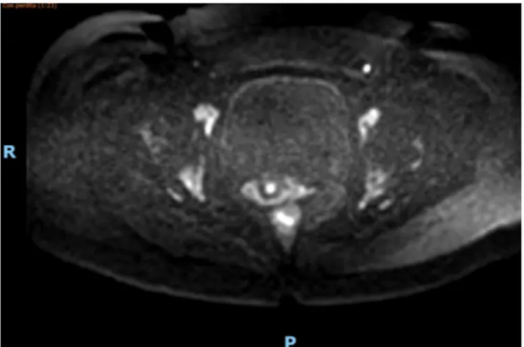
Figure 1. DW-MRI. Signal present in the anterior bladder wall in IC patient. (Color version available online.)
The risk difference (RD) is calculated on these results, mean-ing that if a subject has a positive signal at DW-MRI he is likely to have a diagnosis of BPS/IC, there seems to be a risk difference between exposed (exposed risk = 0.81) and not exposed (unex-posed risk = 0.17) being equivalent to RD = 0.64, with P value = .0008.
Supplementary Table 3reports the results of the scores obtained from responses of patients to questionnaires, with an interquartile range, for 3 degrees of magnetic resonance signal: absent, moderate, and high. The scores are rather low both for the Physical Compo-nent Summary and for the Mental CompoCompo-nent Summary com-pared to the average values in relation to sex and age, indicating the impact of the disease on the quality of life.
Secondary endpoints-Supplementary Table 5shows the cor-relation of average duration of the disease (months) with the intensity of signal, and it shows no significant association between these 2 data.
The 2 radiologists are defined as "raters," blind to the diagno-sis, who evaluated the MRI. There is good agreement between them in the evaluation of anterior and posterior walls ( Supple-mentary Table 6), K = 0.695 e K = 0.640, and fair agreement in right and left lateral walls, with K = 0.263 e K = 0.397. No sig-nificant correlation resulted between DW-MRI signal and dis-ease-related negative impact on Physical (Supplementary Table 7) and Mental Health Index of quality of life (Supplementary Table 8), measured with SF-36.
DISCUSSION
Unlike a recent study by another author4 on DW-MRI, thefirst in IC/BPS patients, we carried out our study using the reports of 2 different radiologists who were unaware of the diagnosis, therefore blind, the correlation between the 2 radiologists was evaluated together with the degree of operator-dependency of the exam.
Patients on average had been diagnosed 30 months after symptom onset, and they had started treatment once the diagnosis was made. The global analysis of the absence or presence of a signal on the bladder walls shows a high specificity, it also highlights a high positive predictive value, since it is likely that if a patient has a positive mag-netic resonance signal she actually has interstitial cystitis. These data are similar to the results reported by the Char-lanes group.4
The interpretation of the RD leads to the conclusion that among subjects who present a positive DW-MRI sig-nal the incidence of cases of interstitial cystitis is higher, and there is a statistically significant difference compared to patients who do not have a high DW-MRI signal. Hav-ing a positive signal is therefore suggestive of interstitial cystitis.
Patients were analyzed based on the type of signal detected: absent, moderate, and high. It should be noted that in the anterior bladder wall, when gathering patients with moderate and high signal in a single group, that is signal present, 17 patients have no signal in 20 no-IC patients, while 15 out of 22 IC patients have signal pres-ent (Table 2).
A statistically significant trend (P value = .001) is noticeable, which shows that the more intense the signal,
the more likely is that it is related to an inflamed bladder wall, and vice versa. In fact, in IC group there are 5 patients with absent signal, 4 with moderate signal and 11 with high signal intensity, compared to 17 patients with absent signal, 2 with moderate signal and 1 with high sig-nal intensity of the no-IC group. In follow-up studies of the pelvis, it has been suggested that when performing DW-MRI study the bladderfilling should be standardized and antiperistaltic drugs should be administered to avoid misinterpretation and bowel motion artifacts. In the female pelvis6in fact it is likely that the distension of the bladder wall due tofilling can alter the quality and inten-sity of the signal. DW-MRI is a noninvasive investigation, its features have recently been expanded to applications related to pelvic imaging, including diagnostic investiga-tion of pelvic tumours.7-9 DW-MRI could also provide information in the evaluation of treatment response com-paring to other sequences in muscle-invasive bladder can-cer.10 Similarly, it could provide useful information in treatment response of BPS/IC in terms of signal intensity.
Our study had a prospective design, the pain score was available when DW-MRI was performed. However, as in the study by Charlanes,4the bladder volume during the MRI was not standardized, and this may represent a limi-tation.
Two different radiologists, blind to the diagnosis, inde-pendent of beach other carried out the DW-MRI analysis, and they classified the report in 3 subjective categories (no signal, moderate, and high signal).
The analysis of the bladder wall signal with DW-MRI, reflecting the inflammatory bladder wall tissue, could help to divide BPS/IC into subclasses. Besides, the definition of an apparent diffusion coefficient (ADC) cutoff, and an analysis of its variation with treatment would represent a further future step of the study.
CONCLUSION
The approach assessing the bladder using DW-MRI in IC/ BPS patients may provide an additional diagnostic evalua-tion that could be used to stratify the wide spectrum of chronic pelvic pain, help to select patients to be assessed performing more invasive assessments, such as cystoscopy with hydrodistension and bladder biopsy, it could also prove particularly useful in clinical trials and for monitor-ing the efficacy of therapy.
The study has been approved by the appropriate institu-tional and/or nainstitu-tional research ethics committee and has been performed in accordance with the ethical standards as laid down in the 1964 Declaration of Helsinki and its later amendments or comparable ethical standards.
SUPPLEMENTARY MATERIALS
Supplementary material associated with this article can be found in the online version athttps://doi.org/10.1016/ j.urology.2020.03.019.
References
1. van de Merwe JP. Nordling J, Bouchelouche P, et al. Diagnostic cri-teria, classification, and nomenclature for painful bladder syndrome/ interstitial cystitis: an ESSIC proposal. Eur Urol2008. doi:10.1016/j. eururo.2007.09.019.
2. Oravisto KJ. Epidemiology of interstitial cystitis. Ann Chir Gynaecol Fenn. 1975;69:75–77.
3. Towner RA, Wisniewski AB, Wu DH, et al. A feasibility study to deter-mine whether clinical contrast enhanced magnetic resonance imaging can detect increased bladder permeability in patients with interstitial cys-titis. J Urol. 2016.https://doi.org/10.1016/j.juro.2015.08.077.
4. Charlanes A, Boudghene F, Chesnel C, et al. Diffusion-weighted magnetic resonance imaging: a new tool for the diagnosis of bladder pain syndrome/interstitial cystitis. Urol Int. 2019;102:109–112.
5. van de Merwe J, Nordling J, Bouchelouche P, et al. Diagnostic crite-ria, classification, and nomenclature for painful bladder syndrome/ interstitial cystitis: an ESSIC proposal. Eur Urol. 2008;53:60–67. Epub 2007 Sep 20.
6. Thoeny HC, Forstner R, De Keyzer F. Genitourinary applications of diffusion-weighted MRI imaging in the Pelvis. Radiology. 2012;263:326–342.
7. Fujii S, Matsusue E, Kigawa J, et al. Diagnostic accuracy of the apparent diffusion coefficient in differentiating benign from malig-nant uterine endometrial cavity lesions: initial results. Eur Radiol. 2008;18:384–389.
8. Takeuchi M, Matsuzaki K, Nishitani H. Diffusion-weighted mag-netic resonance imaging of endometrial cancer: differentiation from benign endometrial lesions and preoperative assessment of myome-trial invasion. Acta Radiol Stockh Swed 1987. 2009;50:947–953.
9. Takeuchi M, Sasaki S, Ito M, et al. Urinary bladder cancer: diffu-sion-weighted MR imaging—accuracy for diagnosing T stage and estimating histologic grade. Radiology. 2009;251:112–121.
10. Yoshida S, Koga F, Kawakami S, et al. Initial experience of diffu-sion-weighted magnetic resonance imaging to assess therapeutic response to induction chemoradiotherapy against muscle-invasive bladder cancer. Urology. 2010;75:387–391.