High Risk for Breast Cancer
(HIBCRIT Study):
Interim Results
1
Francesco Sardanelli, MD Franca Podo, DrSci Giuliano D’Agnolo, PhD Arduino Verdecchia, DrSci Mariano Santaquilani, Tech Eng Renato Musumeci, MD Giovanna Trecate, MD Siranoush Manoukian, MD Sandro Morassut, MD Clelia de Giacomi, MD Massimo Federico, MD Laura Cortesi, MD Stefano Corcione, MD Stefano Cirillo, MD Vincenzo Marra, MD
For the High Breast Cancer Risk Italian Trial (HIBCRIT)
Purpose: To prospectively compare clinical breast examination
(CBE), mammography, ultrasonography (US), and con-trast material– enhanced magnetic resonance (MR) imag-ing for screenimag-ing women at genetic-familial high risk for breast cancer and report interim results, with pathologic findings as standard.
Materials and Methods:
Institutional review board of each center approved the research; informed written consent was obtained. CBE, mammography, US, and MR imaging were performed for yearly screening of BRCA1 or BRCA2 mutation carriers, first-degree relatives of BRCA1 or BRCA2 mutation carri-ers, or women enrolled because of a strong family history of breast or ovarian cancer (three or more events in first-or second-degree relatives in either maternal first-or pater-nal line; these included breast cancer in women younger than 60 years, ovarian cancer at any age, and male breast cancer at any age).
Results: Two hundred seventy-eight women (mean age, 46 years⫾
12 [standard deviation]) were enrolled. Breast cancer was found in 11 of 278 women at first round and seven of 99 at second round (14 invasive, four intraductal; eight were ⱕ10 mm in diameter). Detection rate per year was 4.8% (18 of 377) overall; 4.3% (11 of 258) in BRCA1 or BRCA2 mutation carriers and first-degree relatives of BRCA1 or BRCA2 mutation carriers versus 5.9% (seven of 119) in women enrolled because of strong family history; and 5.3% (nine of 169) in women with previous personal breast and/or ovarian cancer versus 4.3% (nine of 208) in those without. In six (33%) of 18 patients, cancer was detected only with MR imaging. Sensitivity was as follows: CBE, 50% (95% confidence interval [CI]: 29%, 71%); mammography, 59% (95% CI: 36%, 78%); US, 65% (95% CI: 41%, 83%); and MR imaging, 94% (95% CI: 82%, 99%). Positive predictive value was as follows: CBE, 82% (95% CI: 52%, 95%); mammography, 77% (95% CI: 50%, 92%); US, 65% (95% CI: 41%, 83%); and MR imaging, 63% (95% CI: 43%, 79%).
Conclusion: Addition of MR imaging to the screening regimen for high-risk women may enable detection of otherwise unsus-pected breast cancers.
娀 RSNA, 2007
1From the University of Milan School of Medicine,
De-partment of Medical and Surgical Sciences, Unit of Radi-ology, IRCCS Policlinico San Donato, Via Morandi 30, 20097 San Donato Milanese, Milan, Italy (F.S.); and De-partment of Cell Biology and Neurosciences, Istituto Su-periore di Sanita`, Rome, Italy (F.P.). The remaining au-thors and their affiliations are listed at the end of this article. From the 2004 RSNA Annual Meeting. Received December 5, 2005; revision requested January 25, 2006; revision received February 28; accepted April 3; final ver-sion accepted June 30. The HIBCRIT study was supported by the Italian Ministry of Health (Ricerca Finalizzata 1% 98/JT/T) and Istituto Superiore di Sanita` (ricerca corrente C3A3/2004) and coordinated by the Istituto Superiore di Sanita`, Rome, Italy. Address correspondence to F.S. (e-mail: [email protected] ). 姝 RSNA, 2007
ORIGINAL
T
he cumulative lifetime risk of breast cancer is greatly different for women with a hereditary pre-disposition for this disease from that for women in the general population. Al-though breast cancer affects a range of as many as one in seven to one in 11 women in Western countries (1,2), spo-radic disease accounts for about 85%– 95% of the occurrences. The remaining 5%–15% of breast cancers are clus-tered in families with high breast cancer incidence (3–5). For women in the lat-ter group, an inherited predisposition may result in an early onset of the dis-ease (typically in the premenopausal pe-riod) and in a lifetime risk greater than 50%– 60% (3–7).About 50% of the occurrences in high-risk families can be explained by the dominant autosomal inheritance of the deleterious BRCA1 or BRCA2 muta-tions on the 17q21 or the 13q12 chro-mosomal location, respectively, with in-complete penetrance (6,8,9). BRCA1 mutations are associated with an in-creased risk of ovarian cancer, whereas BRCA2 mutations may be associated with male breast cancer. Breast cancers in very young women (younger than 35 years) are frequently associated with
BRCA1 mutations (10). Moreover,
breast cancers in BRCA1 mutation car-riers are more frequently high grade and estrogen receptor negative com-pared with sporadic cancers (11,12), and they are associated with less favor-able survival (13,14).
The strategy for surveillance of high-risk women is different from that for surveillance of the general popula-tion (eg, annual mammography for women older than 40 years in the United States and biannual mammogra-phy for women older than 50 years in many European countries). Screening for high-risk women should begin at a younger age and with no more than a 1-year interval between examinations. As reported in a recent review (15), researchers determined that contrast material– enhanced magnetic resonance (MR) imaging is highly sensitive for identification of breast cancer in a large spectrum of clinical situations. Because of lower values for specificity and posi-tive predicposi-tive value (PPV), with con-trast-enhanced MR imaging, however, unnecessary breast biopsy has been re-ported because of false-positive findings (16,17), especially in premenopausal women (18,19) and also in high-risk women (20).
Findings from two retrospective (21,22) and five prospective studies (23–27) indicated that contrast-enhanced MR imaging is useful in the screening of high-risk women. These single- and multicenter studies differed in the num-ber of diagnostic modalities used for comparative evaluation, in enrollment criteria, in selected age groups, and in levels of cumulative lifetime risk of breast cancer. The purpose of our
mul-ticenter trial was to prospectively com-pare the sensitivity of clinical breast ex-amination (CBE), mammography, ultra-sonography (US), and contrast-enhanced MR imaging for the screening of women at genetic-familial high risk for breast cancer and to report our interim re-sults, with pathologic findings as the ref-erence standard.
Materials and Methods Study Design and Population
An open prospective nonrandomized multicenter comparative trial was de-signed (7), and the institutional review board of each center approved the re-search. Informed written consent was obtained from each subject enrolled in the study. We planned to enroll asymp-tomatic subjects at high risk for breast cancer, women at least 25 years of age and men at least 50 years of age, who were (a) carriers of BRCA1 or BRCA2 mutations, as demonstrated with ge-netic testing, or subjects with personal unknown mutational status but who were first-degree relatives of BRCA1 or BRCA2 mutation carriers or (b) sub-jects with a strong family history of breast or ovarian cancer who had three or more events of breast or ovarian can-cer in first- or second-degree relatives in either the maternal or the paternal line. These three or more events could have included female breast cancer in relatives younger than 60 years, ovarian cancer in relatives at any age, or male breast cancer in relatives at any age.
The occurrence of previous breast cancer and/or ovarian cancer in the
Published online before print
10.1148/radiol.2423051965
Radiology 2007; 242:698 –715
Abbreviations:
BI-RADS⫽ Breast Imaging Reporting and Data System
CBE⫽ clinical breast examination
PPV⫽ positive predictive value
Author contributions:
The list of author contributions is at the end of this arti-cle.
Authors stated no financial relationship to disclose. Advances in Knowledge
䡲 By using a multimodality surveil-lance approach that included con-trast-enhanced MR imaging, the detection rate of breast cancer in asymptomatic high-risk women was 4.8% in our study. 䡲 In women enrolled only on the
basis of well-defined criteria for strong family history of breast and/or ovarian cancer, the detec-tion rate was not significantly dif-ferent from that of those enrolled for positive BRCA1 or BRCA2 ge-netic testing (or for being first-degree relatives of subjects with positive BRCA1 or BRCA2 genetic testing).
䡲 Contrast-enhanced MR imaging showed a high sensitivity (94%; 33% of patients with cancer de-tected only with contrast-en-hanced MR imaging), with an ac-ceptable positive predictive value (63%) that was close to that of US (65%); the addition of contrast-enhanced MR imaging to the screening regimen of high-risk women may enable the detection of otherwise unsuspected breast cancers.
䡲 Contrast-enhanced MR imaging showed a high sensitivity in the detection of multiple malignant foci in the same breast in high-risk women with invasive cancers.
subject to be enrolled could contribute to the number of events needed to meet the second criterion defined previously. A multifocal unilateral cancer was re-garded as one event. A multicentric uni-lateral cancer was regarded as two events. An ipsilateral metachronous breast cancer was regarded as a new event if it was located at least at 5 cm from the previous cancer. Bilateral syn-chronous breast cancers were regarded as two different events.
Subjects with a history of personal breast cancer were included (28), pro-vided that at least one breast was not completely excised. Exclusion criteria were pregnancy, breast-feeding, cur-rent chemotherapy, terminal illness, and contraindications to MR imaging. History of surveillance imaging prior to entry into the study was not specifically recorded. For each enrolled patient, we planned two annual rounds of assess-ment with CBE, mammography, US, and contrast-enhanced MR imaging and at least a 1-year follow-up with CBE, mammography, and US. At each screen-ing event, each of these tests was per-formed by an independent physician who was unaware of the results ob-tained by the other three colleagues.
Centers were asked to plan the in-dependent examinations for each screen-ing event in the same day for as many enrolled subjects as possible. When we considered the need for planning con-trast-enhanced MR imaging during the 2nd week of the menstrual cycle in pre-menopausal women and logistic prob-lems (eg, different locations for mam-mography, US, and MR imaging facili-ties; members of the same family preferred to have the examination on the same day), a maximum time of 1 month for performance of the four diag-nostic modalities was allowed. In some instances, justified by particular condi-tions, the central unit accepted a maxi-mum delay of 2 months.
A national health institution (Istituto Superiore di Sanita`, Rome, Italy) was responsible for multicenter project de-sign, central coordination, and data management and funding (central unit). This institution was not directly in-volved in the enrollment or in the
per-formance of genetic testing, CBE, mam-mography, breast US, and contrast-en-hanced MR imaging. Seventeen centers in 14 Italian towns participated. The first center was activated in June 2000, and the 17th center was activated in November 2003; four institutes for can-cer research and treatment, 12 univer-sity hospitals, and one general hospital were included. Quality assessment pro-grams were regularly performed in the radiology departments of these institu-tions. At each center, examinations were performed, and findings from the examinations were interpreted with the supervision of a radiologist who had at least 10 years of experience in breast imaging; moreover, other physicians were involved in the trial at each cen-ter, and the centers and names of those involved appear at the end of this article.
During the first phase of the study (from June 2000 to December 2002), the enrollment was limited to the first criterion described before; preliminary results for this phase, which pertained to the first eight patients in whom breast cancer was detected, were re-ported during an international work-shop in early 2002 (7). In January 2003, enrollment on the basis of the second criterion was also opened. The general plan of the trial was aimed at enrolling approximately 500 subjects in about 6 years, with about 50% fulfilling the first criterion and about 50% fulfilling the second criterion. For each of the three diagnostic imaging modalities (mam-mography, US, and contrast-enhanced MR imaging), performed in nonran-domized order, the results of local pro-spective independent readings are in-cluded in the data presented here. Each reader was aware of the high-risk condi-tion of the women but was blinded to the results of the other three diagnostic modalities. During the second round, the readers were aware of the results from the first annual round.
Genetic Testing
Genetic testing for identification of dele-terious BRCA1 and BRCA2 mutations included the use of individual or
com-bined procedures, such as direct se-quencing (29–31), protein truncation testing (29), single-strand conformation polymorphism (32), or mutation screen-ing with denaturscreen-ing chromatography (33).
Clinical Breast Examination
Thirty-nine physicians performed CBE in the 17 centers. Findings of each ex-amination were coded as normal, sug-gestive of benign disease, indetermi-nate, suspicious for malignant disease, or highly suggestive of malignant dis-ease. Palpable nodules that were judged as benign or indeterminate but were clearly benign at mammography and/or US (eg, cysts, calcified fibroadenomas) were not considered in the current re-port. In the present analysis, for each patient who had cancer during the trial, the cancer was considered palpable if at least one of the pathologically demon-strated malignant foci was coded at CBE as indeterminate, suspicious for malig-nant disease, or highly suggestive of ma-lignant disease. Clinical findings were marked on a diagram and correlated with imaging findings after reporting.
Mammographic Examination
Bilateral two-view mammography (uni-lateral in women who previously had undergone one-breast mastectomy) was performed with screen-film units with a rotating anode, a 0.3– 0.1-mm focus, a focus-film distance of 55 cm or greater, homogeneous breast compression, a mobile grid, and automatic exposure control. Dedicated daylight processing was performed. Standard mediolateral oblique and craniocaudal projections were acquired. Further dedicated mam-mograms (ie, magnification, spot com-pression, or other additional views) were obtained when necessary.
When an enrolled subject under-went a previous mammographic exami-nation performed within the 2 months preceding enrollment, the mammogram from that examination was acquired as part of the study after a quality check; if mammography was performed more than 2 months before the enrollment, the first round of complete
multimodal-ity assessment was planned 12 months after the previous mammogram was ob-tained.
Images from mammographic exami-nations were evaluated by one reader. Evaluations were performed by radiolo-gists with at least 10 years of experience in breast imaging, and findings were classified by using the five-point scale of the Breast Imaging Reporting and Data System (BI-RADS) (34). Thirty-nine ra-diologists interpreted the mammograms in the 17 centers. The density pattern was classified according to two catego-ries: breasts with more than 50% of the breast occupied by fibroglandular den-sity as a mean of the two mammo-graphic views and breasts with 50% or
less of the breast occupied by fibroglan-dular density as a mean of the two mam-mographic views.
US Examination
US examinations were performed by ra-diologists with at least 10 years of expe-rience in breast imaging. Both breasts were systematically examined by using vertical, horizontal, radial, and antira-dial scans obtained by a radiologist who reported the findings of the examination by using the five-point BI-RADS scale (35). Forty-one physicians performed and interpreted the US breast examina-tions in the 17 centers. Transducers with a frequency of 10 MHz or greater
were used in 14 centers, transducers with a frequency of 7.5 MHz or greater were used in two centers, and only a transducer with a frequency of 7.5 MHz was used in one center; transverse res-olution of 0.5 mm or less and lateral-transverse resolution of 1 mm or less were used.
Contrast-enhanced MR Imaging
Contrast-enhanced MR imaging was performed at 1.5 T (12 centers) or 1.0 T (four centers), whereas at one center both field strengths were used because of the change of equipment during the trial. The MR units, equipped with ac-tively shielded gradients of 15 mT/m or greater, were purchased from three manufacturers (GE Medical Systems, Milwaukee, Wis; Philips Medical Sys-tems, Best, the Netherlands; or Sie-mens Medical Solutions, Erlangen, Ger-many). Dedicated synchronous breast coils were used for bilateral studies (or unilateral studies when a mastectomy previously had been performed) with the patient in the prone position. The examination was planned on the 7th– 14th day of the menstrual cycle in pre-menopausal women but without sched-uling limitations in postmenopausal women.
A long venous catheter access was obtained by using a plastic cannula in a cubital vein. After localizing scout views were obtained, a contrast-enhanced dy-namic three-dimensional T1-weighted spoiled gradient-echo sequence was performed in the transverse or coronal plane. The parameters were as follows: repetition time, 13 msec or less; flip angle, 20°–30°; partition thickness, 3 mm or smaller; intersection gap, none; number of partitions, 40 –128 (to cover both breasts entirely); and acquisition time, not longer than 120 seconds. The echo time was chosen to prevent fat-water signal opposition at 1.0 or 1.5 T (36). The field of view and image matrix were combined to obtain a pixel size of 1.4⫻ 1.4 mm or less.
An unenhanced sequence was per-formed before contrast agent injection. Then, 0.1 mmol/kg of one of the com-mercially available 0.5 mol/L two-Table 1
Scoring System with Combination of Morphologic and Dynamic Parameters for Evaluation of Breast Lesions on Contrast-enhanced MR Images
Criterion Score
Shape
Round, oval, lobular 0
Linear, ductal, segmental, dendritic, spiculated 1
Margins Regular 0 Irregular 1 Kinetic pattern Initial enhancement* ⬍50% 0 ⱖ50 and ⬍100% 1 ⱖ100% 2 Postinitial enhancement Continuous† 0 Plateau‡ 1 Washout§ 2 Enhancement pattern Homogeneous 0 Inhomogeneous 1 Rim 2 Source.—References 38, 39.
Note.—Total score interpretation and translation into BI-RADS categories 1–5 is as follows: no detectable enhancement⫽ no
enhancing focus⫽ BI-RADS 1, score 0–2 ⫽ benign ⫽ BI-RADS 2, score 3 ⫽ probably benign ⫽ BI-RADS 3, score 4–5 ⫽
suspicious⫽ BI-RADS 4, and score 6–8 ⫽ highly suggestive of malignancy ⫽ BI-RADS 5.
* Initial peak signal intensity within the first 3 minutes after contrast agent administration, relative to the signal intensity at unenhanced imaging.
†Continuous signal intensity is defined as when a progressive signal intensity increase is found during the 4th, 5th, and 6th
minutes and the signal intensity at the 6th minute is greater than 10% of that of the initial peak signal intensity obtained during the 1st, 2nd, and 3rd minutes.
‡Plateau is defined as when the postinitial signal intensity during the 4th, 5th, and 6th minutes remains within the range of
⫾10% of that of the initial peak signal intensity obtained during the 1st, 2nd, and 3rd minutes.
§Washout is defined as when a progressive signal intensity decrease is found during the 4th, 5th, and 6th minutes and the
signal intensity at the 6th minute is less than 10% of that of the initial peak signal intensity obtained during the 1st, 2nd, and 3rd minutes.
compartment gadolinium chelates (ga-dopentetate dimeglumine, Magnevist, Schering, Berlin, Germany; gadoterate meglumine, Dotarem, Guerbet, Paris, France; gadodiamide, Omniscan, Am-ersham Health, Oslo, Norway; or gado-teridol, ProHance, Bracco, Milan, Italy) was intravenously administered at the rate of 2 mL/sec by using an automatic injector. Injection was followed by flush-ing with 20 mL of saline. Takflush-ing into account that the central lines of the k-space, mainly devoted to image contrast resolution, were acquired in the central third of the sequence time duration, the contrast agent injection was started at the same time as the first contrast-en-hanced sequence was started. The num-ber of contrast-enhanced sequences, typically five with a temporal resolution of 90 seconds, was sufficient to obtain dynamic information during at least the first 6 minutes after contrast agent in-jection.
Temporal subtraction (enhanced
minus unenhanced images) was always
performed for the first, second, and last contrast-enhanced sequences, and a
maximum intensity projection
algo-rithm was applied for the first and the second contrast-enhanced sequences. Signal intensity–time and percentage of enhancement–time dynamic curves were obtained for targeted small regions of in-terest positioned on the areas of homoge-neous maximal enhancement within the enhancing lesion (37). Source and pro-cessed images were electronically stored on compact discs. Twenty-eight radiolo-gists interpreted the contrast-enhanced MR images from breast examinations in the 17 centers.
Morphologic and dynamic parame-ters were combined according to the criteria first defined by Fischer et al (38) and tested by Baum et al (39) in 1031 breasts. In the study by Baum et al, sensitivity and specificity were both 92%. This eight-level scoring system was modified into a five-point scale ac-cording to the BI-RADS (40) classifica-tion (Table 1).
Work-up of Lesions Suspicious for Malignancy and Pathologic Standard of Reference
An integration across the modalities was performed at each center after each physician had reported findings for CBE, mammography, US, and contrast-enhanced MR imaging. In patients who had a palpable breast nodule that was indeterminate or suspicious for malig-nancy or an imaging-detected lesion classified as BI-RADS 4 or 5 with one or more than one of the three modalities, fine-needle aspiration or core-needle (14-gauge) biopsy was performed. When the lesion was detectable at US (at the first look before contrast-enhanced MR imaging or at the second look after con-trast-enhanced MR imaging), the biopsy
was performed with US guidance.
When the lesion was mammographi-cally detectable and not detectable at US, the biopsy was performed with ste-reotactic guidance. When the lesion was detectable at only contrast-en-hanced MR imaging (which included a
Table 2
Multimodality Surveillance of 278 Women at High Risk of Breast Cancer: Enrollment and Detection Rate per Year
Population Characteristics Enrolled Detection Rate per Year*
Enrollment
BRCA1 mutation carriers 98 (35.3) 7/142 (4.9)†
BRCA2 mutation carriers 68 (24.5) 4/103 (3.9)†
First-degree relatives of BRCA1 or BRCA2 mutation carriers 9 (3.2) 0/13 (0)
Overall no. of BRCA1 or BRCA2 mutation carriers and first-degree relatives of BRCA1 or BRCA2 mutation carriers 175 (62.9) 11/258 (4.3)‡
Strong family history of breast cancer and/or ovarian cancer§ 103 (37.1) 7/119 (5.9)‡
Overall no. of women who underwent screening 278 (100) 18/377 (4.8)
Previous vs no previous breast cancer and/or ovarian cancer
Women with previous personal breast cancer and/or ovarian cancer㛳 123 (44.2) 9/169 (5.3)#
Women without previous personal breast cancer and/or ovarian cancer 155 (55.8) 9/208 (4.3)#
Note.—Data are numbers of women, and numbers in parentheses are percentages. All 278 enrolled women underwent first-round multimodality evaluation, whereas only 99 of them underwent second-round evaluation (44 BRCA1 mutation carriers, 35 BRCA2 mutation carriers, four first-degree relatives of a BRCA1 mutation carrier, no first-degree relatives of a BRCA2 mutation carrier, and 16 women enrolled on the basis of a strong family history of breast cancer and/or ovarian cancer).
* The detection rate per year was calculated for each subset and overall as the ratio between the number of patients who were affected by breast cancer during the first or second round and the number of women who underwent screening at the first round plus the number of women who underwent screening at the second round.
†The difference in the detection rate between BRCA1 mutation carriers and BRCA2 mutation carriers was not significant (2test).
‡The difference in the detection rate between BRCA1 or BRCA2 mutation carriers or first-degree relatives of a BRCA1 or BRCA2 mutation carrier and women enrolled on the basis of a strong family
history of breast cancer and/or ovarian cancer was not significant (2test).
§Subjects with a strong family history of breast cancer and/or ovarian cancer had first- or second-degree relatives in either the maternal or the paternal line who had at least three female breast
cancers in those younger than 60 years, at least three female breast cancers in those younger than 60 years and/or ovarian cancer at any age, or at least three female breast cancers in those younger than 60 years and/or male breast cancer at any age.
㛳Of 123 enrolled patients with previous breast cancer and/or ovarian cancer, 104 had a previous breast cancer, 14 had a previous ovarian cancer, and five had a previous breast cancer and ovarian
cancer; seven of these patients with previous breast and/or ovarian cancer had had an inconclusive genetic test (ie, negative for BRCA1 or BRCA2 mutations [BRCAX status]).
#The difference in the detection rate between women with a previous personal breast cancer and/or ovarian cancer versus women without a previous personal breast cancer and/or ovarian cancer
second-look US examination with a negative image), biopsy and presurgi-cal lopresurgi-calization were performed with contrast-enhanced MR imaging guid-ance.
For any finding classified as BI-RADS 3 (probably benign) at each imag-ing modality without a higher classifica-tion (ie, BI-RADS 4 or 5) at other diag-nostic modalities in the same round, a short-term (within 4 months) follow-up with the same modality with which the lesion was detected was planned. If the finding was confirmed (ie, a lesion with at least a BI-RADS 3 classification and was not reduced in diameter), an imag-ing-guided biopsy was performed ac-cording to the protocol defined previ-ously. When the report for the fine-nee-dle or core biopsy indicated that the lesion was suspicious for malignant dis-ease or highly suggestive of malignancy, the patient always underwent surgery. When the findings in the report for the fine-needle or core biopsy suggested a diagnosis of lobular neoplasia (lobular carcinoma in situ), atypical ductal hy-perplasia, radial scar, papilloma, or papillomatosis, the patient underwent surgery, and pathologic examination of the surgical specimen was performed. For all the lesions classified as BI-RADS 4 or 5 at any imaging modality, the pa-tient underwent surgery, and pathologic examination was performed, even though the fine-needle or core biopsy yielded a negative, benign, or probably benign re-sult.
All nonpalpable lesions were preop-eratively localized by using a hook wire or a charcoal suspension, with the
guid-ance of the imaging method used for biopsy.
At the 17 centers, 19 pathologists who had 9 –35 years of experience with breast pathology classified the patho-logic specimens according to the 1981 World Health Organization breast can-cer classification (41). The maximal di-ameter of each malignant lesion was re-corded. For invasive malignant lesions, axillary nodal status was explored with axillary dissection or sentinel lymph node biopsy, depending on the prefer-ence of each center involved in the study.
Statistical Analysis
Overall detection rate per year was cal-culated as the ratio between the number of patients with pathologically proved breast cancer and the sum of screening events at the first and the second rounds (278 ⫹ 99 ⫽ 377). The detection rate for each round was calculated as the ratio between the number of patients with pathologically proved breast can-cer at each round and the number of women who underwent screening in the same round. The detection rate per year for subsets of enrolled women was calculated as the ratio between the number of patients who had a patholog-ically proved breast cancer in the subset of women and the number of screening events in the members of the subset of women in the analysis.
Sensitivity of each of the four modal-ities was calculated as the ratio between the number of true-positive findings (pathologically proved breast cancer de-tected by using each modality) and the
number of true-positive findings plus false-negative findings (pathologically proved breast cancer examined with the same modality but not correctly diag-nosed [ie, BI-RADS categories 1–3]). For each modality, the false-negative findings were defined as breast cancers detected by using one, two, or three of the remaining modalities and confirmed at pathologic examination.
PPV of each of the four modalities was calculated as the ratio between true-positive findings (defined as previ-ously mentioned) and the number of true-positive findings plus false-positive findings (findings that needed to be ex-plored with invasive procedures that de-fined a benign cytologic or pathologic diagnosis). Invasive procedures in-cluded fine-needle aspiration, core-nee-dle biopsy, and surgical biopsy after im-aging-guided localization (one patient opted for bilateral prophylactic mastec-tomy, which revealed a mammographic finding as false-positive).
In the evaluation of multiple malig-nant foci in the same breast, the total number of malignant foci demon-strated at pathologic examination was used as the standard of reference. The capability of each modality for depic-tion of multiple malignant foci was evaluated without considering the dif-ference between multifocal and multi-centric cancers.
For both sensitivity and PPV, 95% confidence intervals were calculated from the exact binomial distribution.
Differences in the mean age of the enrolled women or patients and in the mean pathologically determined diame-ter of the tumors were tested by using the two-tailed Mann-Whitney U test. Differences in detection rate, percent-age of patients with invasive cancer, and percentage of patients with axillary
nodal metastatic involvement were
tested by using the2test or the Fisher exact test, when the former was not applicable—that is, for small samples (fewer than 20 subjects sampled)— or when one or more of the so-called ex-pected values was smaller than five (42). Statistical calculations were per-formed by using a software package (SPSS, version 6.0; SPSS, Chicago, Ill). Figure 1
Figure 1: Flow diagram of the
multicenter surveillance of women at high risk for breast cancer by using contrast-enhanced MR imaging, mammography, US, and CBE.
Table 3 Characteristics of 18 Patients with Breast Cancer according to Multimodality Surveillance of 278 High-Risk Women Data Enrollment Basis Previous Breast Cancer Round at Detection CBE* Mammography US BI-RADS Category Contrast- enhanced MR Imaging BI-RADS Category Pathologic Examination Axillary Nodal Involvement § Side and Histologic Finding Age (y) Fibroglandular Density BI-RADS Category Side and Type Histologic Grade of Cell Differentiation † Diameter (mm) ‡ Patient No./ Age at Entry (y) 1/69 BRCA1 mutation carrier Left, IDC 55 2 Nonpalpable ⱕ 50% 1 1 5 Right, multifocal DCIS 3 3 Axillary
dissection not performed
2/53 BRCA1 mutation carrier None ND 2 Palpable ⱕ 50% 5 5 5 Left, IDC 3 18 N⫹ , 19/32 3/50 BRCA1 mutation carrier Left, medullary cancer 47 2 Nonpalpable ⱕ 50% 4 4 5 Right, IDC; left, multifocal DCIS 3 19 right, 5left N⫺ , 0/38, right axilla 㛳 4/35 BRCA2 mutation carrier None ND 1 Palpable ⬎ 50% 2 4 4 Left, ILC and LCIS 23 5 N⫺ , 0/18 5/52 BRCA1 mutation carrier Left, IDC 33 1 Nonpalpable ⱕ 50% 1 2 4 Right, IDC 2 6 N⫺ , 0/1, SNB 6/61 Strong family history of breast and/or ovarian cancer, BRCAX # Left, multifocal ILC** 44 1 Nonpalpable ⬎ 50% 1 Not performed 5 Left, bifocal IDC and ILC 16 N⫹ , 9/15 7/60 BRCA1 mutation carrier Left, IDC 49 2 Palpable ⱕ 50% 4 4 Not performed †† Right, IDC 3 13 N⫺ , 0/1, SNB 8/47 BRCA1 mutation carrier None ND 2 Palpable ⬎ 50% 4 5 Indeterminate ‡‡ Right, IDC 3 13 N⫺ , 0/1, SNB 9/53 BRCA2 mutation carrier None ND 1 Palpable ⬎ 50% 4 4 4 Left, multifocal ILC and DCIS 11 5 N⫺ , 0/1, SNB 10/70 BRCA2 mutation carrier Left, IDC 56 1 Nonpalpable ⬎ 50% 1 1 4 Left, DCIS and LCIS 1 4 Axillary
dissection not performed
11/37 BRCA2 mutation carrier None ND 2 Palpable ⱕ 50% 5 5 5 Right, multicentric IDC 32 2 N⫺ , 0/12 12/44 Strong family history of breast and/or ovarian cancer, BRCAX # None ND 2 Palpable ⬎ 50% 4 4 5 Left, atypical invasive medullar 32 2 N⫺ , 0/2, SNB
13/40 Strong family history of breast and/or ovarian cancer None ND 1 Nonpalpable ⬎ 50% 3 3 4 Right, multicentric DCIS 3 5 Axillary
dissection not performed
14/45 BRCA1 mutation carrier Left and right, not specified 38 1 Nonpalpable ⱕ 50% 3 1 5 Right, microinvasive DCIS 2 7 Axillary
dissection not performed
15/56 Strong family history of breast and/or ovarian cancer None ND 1 Nonpalpable ⬎ 50% 4 4 2 Left, bifocal ILC 2 9 N⫺ , 0/2, SNB 16/52 Strong family history of breast and/or ovarian cancer Right, IDC 50 1 Nonpalpable ⱕ 50% 5 2 5 Right, bifocal IDC 21 0 ... §§ 17/39 Strong family history of breast and/or ovarian cancer Left, IDC 38 1 Palpable ⬎ 50% 4 4 5 Bilateral, multicentric IDC 3 6 right, 15 left N⫺ , 0/18, right axilla 㛳㛳 18/57 Strong family history of breast and/or ovarian cancer None ND 1 Palpable ND ... ## 4 4 Right, IDC 3 18 N⫹ , 4/17 Statistics*** Mean age ND ND 45.6 ND ND ND 3.2 3.3 4.4 ND 2.4 13.3 ND Standard deviation ND ND 8.0 ND ND ND 1.5 1.4 0.8 ND 0.8 8.2 ND Median ND ND 47.0 ND ND ND 4.0 4.0 5.0 ND 3.0 13.0 ND Note.—No patient had a previous ovarian cancer. Nine patients had a previous breast cancer. DCIS ⫽ Ductal carcinoma in situ, IDC ⫽ invasive ductal carcinoma, ILC ⫽ invasive lobular carcinoma, LCIS ⫽ lobular carcinoma in situ, ND ⫽ no data, SNB ⫽ sentinel node biopsy. * Palpable nodules judged as benign or for which mammographic and/or US findings (eg, cysts, fibroadenomas) were clearly benign were not considered. †Grades were 1 ⫽ high, 2 ⫽ intermediate, and 3 ⫽ low. ‡In case of multiple foci, the diameter of the largest focus was reported; in case of bilateral cancers, in the calculation of mean, standard deviation, and median of maximal diameter, the largest of the two cancers was included. §N⫺⫽ absence of axillary nodes positive for metastasis, N⫹⫽ presence of one or more axillary nodes positive for metastasis. Numbers are the ratios of number of positive nodes/number of nodes dissected. 㛳Axillary dissection and SNB were not performed for the left multifocal DCIS. #BRCAX means that the genetic test results were inconclusive in a woman with a strong family history of breast cancer and/or ovarian cancer. ** This patient had also had previous ductal carcinoma in situ in the left breast at 51 years old. †† MR imaging was not performed because of the patient’s anxiety. ‡‡ MR imaging findings were indeterminate because of movement artifacts. §§ The patient had previously undergone breast-conserving surgery of the right breast including axillary dissection for an IDC; consequently, for the new right bifocal IDC, the right axilla was not reexplored. 㛳㛳Because of previous breast-conserving intervention including axillary dissection on the left side for IDC, the left axilla was not reexplored. ## The patient had breast implants, and mammography, with dedicated breast positioning, was not performed because of marked discomfort during compres sion. *** The mean age at entry was 51.1 years, with a standard deviation of ⫾ 10.2 and a median age of 52.0 years.
A difference with P⬍ .05 was consid-ered significant.
Results
Screening Population
The present interim analysis is based on the data available in March 2004 at the central data management unit in regard to the first round for the first 278 en-rolled subjects and the second round for 99 subjects (ie, the first 99 subjects who underwent assessment in the second round), for a total of 377 screening events. The screening population (Table 2) consisted of 278 women (mean age, 46.0 years⫾ 12.0 [standard deviation]; range, 25–79 years) enrolled by 17 cen-ters. All 278 women underwent assess-ment in the first round and, until March 2004, 99 women participated in the sec-ond round (35.6%). One hundred sev-enty-five (63%) of 278 women were en-rolled because they were BRCA1 or BRCA2 mutation carriers or because they were first-degree relatives of BRCA1 or BRCA2 mutation carriers, whereas 103 (37%) were enrolled on the basis of family history, according to the criteria stated before; seven of them, all with a previous breast and/or ovarian cancer, had an inconclusive ge-netic test (BRCAX status).
The mean ages of patients enrolled by using the two criteria stated previously were 45.8 years⫾ 12.0 (175 women en-rolled because they were BRCA1 or BRCA2 mutation carriers or first-degree relatives of BRCA1 or BRCA2 mutation carriers) and 46.5 years ⫾ 12.0 (103 women enrolled on the basis of the family history), and there was no significant dif-ference (Mann-Whitney U test) in mean age between them. In particular, the mean age of BRCA1 mutation carriers or first-degree relatives of BRCA1 mutation carriers was 45.7 years⫾ 11.9, whereas the mean age for the analogous BRCA2 group was 45.9 years⫾ 12.3, and there was no significant difference (Mann-Whitney U test) in mean age between the groups. Of 278 women who underwent screening, 123 (44.2%) had had previous breast and/or ovarian cancer and 155 (55.8%) had not had these conditions;
the mean ages at entry were 50.4 years⫾ 11.5 for those who had had previous can-cer and 42.6 years⫾ 11.3 for those who had not had previous cancer; the differ-ence between the groups was significant (P ⬍ .001, Mann-Whitney U test). The mean number of enrolled women per center was 16.3 (range, 4 – 62). The dis-tribution among the centers was as fol-lows: nine centers, four to 10 enrolled women; five centers, 11–30 enrolled women; and three centers, more than 30 enrolled women. For each of the 377 screening events, four examinations (CBE, mammography, US, and contrast-enhanced MR imaging) were performed on the same day (n⫽ 347), during 1 week (n⫽ 1), during 2 weeks (n ⫽ 15), during 3 weeks (n⫽ 2), during 4 weeks (n ⫽ 2), during 5 weeks (n⫽ 1), during 6 weeks (n ⫽ 2), during 7 weeks (n ⫽ 2), and during 8 weeks (n⫽ 5). All the MR exam-inations of fertile women were performed on the 7th–14th day of the menstrual cy-cle. A flow diagram of the entire study is presented in Figure 1.
Cancer Detection
Of 278 women who underwent screen-ing at the first round, we found 11 pa-tients with cancer (detection rate, 4.0%), whereas of 99 women who un-derwent screening at the second round, we found seven patients with cancer (detection rate, 7.1%), and the differ-ence in rates was not significant (2 test). The overall detection rate was 4.8% (18 of 377). The detection rates for enrollment criteria and presence or absence of personal history of breast or ovarian cancer are presented in Table 2.
The overall maximal pathologically
determined mean diameter was 13.3 mm⫾ 8.2, and eight of 18 patients had tumors 10 mm or smaller in diameter. Fourteen patients had at least one inva-sive cancer and four patients had only in situ cancers; considering the largest tu-mor for each patient in both groups, the mean diameter was 15.8 mm⫾ 7.6 for the invasive cancers and 4.8 mm⫾ 1.7 for the in situ cancers. Nine (50%) of 18 patients had multifocal, multicentric, or bilateral breast cancers. When we con-sidered the most aggressive lesion in the nine patients with multifocal, multicen-tric, or bilateral cancers, we observed a percentage of patients with invasive cancers of 78% (14 of 18) (Table 3).
No interval cancer was reported. The sensitivity of contrast-enhanced MR imaging, mammography, and US is shown in Table 4. Mammography, which was not performed in one pa-tient, aided in the diagnosis of a cancer in five (63%) of eight patients with 50% or less of the breast occupied by fi-broglandular density and in five (56%) of nine patients with more than 50% of the breast occupied by fibroglandular density, and the difference was not sig-nificant (Fisher exact text); the only false-negative finding at contrast-en-hanced MR imaging was in a breast with fibroglandular density that extended into more than 50% of the breast on the mammograms. Examples of a three-mo-dality true-positive finding and of a find-ing that was true-positive only at con-trast-enhanced MR imaging are shown in Figures 2 and 3. The false-negative finding at contrast-enhanced MR imag-ing is shown in Figure 4.
Of 18 patients in whom breast
can-Table 4
Patient-based Sensitivity and PPV of Contrast-enhanced MR Imaging, Mammography, US, and CBE in Multimodality Surveillance for Breast Cancer in 278 High-Risk Women
Modality Sensitivity (%) PPV (%) Value 95% CI Value 95% CI Contrast-enhanced MR imaging 93.8 (15/16) 71.7, 98.9 62.5 (15/24) 42.7, 78.8 Mammography 58.8 (10/17) 36.0, 78.4 76.9 (10/13) 49.7, 91.8 US 64.7 (11/17) 41.3, 82.7 64.7 (11/17) 41.3, 82.7 CBE 50.0 (9/18) 29.0, 71.0 81.8 (9/11) 52.3, 94.9
cer was detected, 11 had been enrolled on the basis of genetic testing and seven had been enrolled only on the basis of a strong family history of breast cancer and/or ovarian cancer. No significant difference was observed between the mean age of the seven BRCA1 muta-tion carriers with breast cancer (53.7 years⫾ 8.3) and that of the four BRCA2 mutation carriers (48.8 years ⫾ 16.3)
(Mann-Whitney U test), with a mean age of patients with BRCA1 and BRCA2 of 51.9 years⫾ 11.3 (Table 5). In the 18 patients who presently had breast can-cer, nine (50%) had palpable tumors and nine (50%) had nonpalpable tu-mors (Tables 6, 7).
In the 18 patients who had breast cancers, six (33%) cancers were de-tected only with contrast-enhanced MR
imaging. None of the cancers were de-tected only with CBE, only with mam-mography, or only with US (Table 8). Detection of Multiple Malignant Foci in Patients with Invasive Cancers
When we considered the six patients with invasive cancers and multiple malignant foci per breast, we ob-served seven breasts with multiple Figure 2
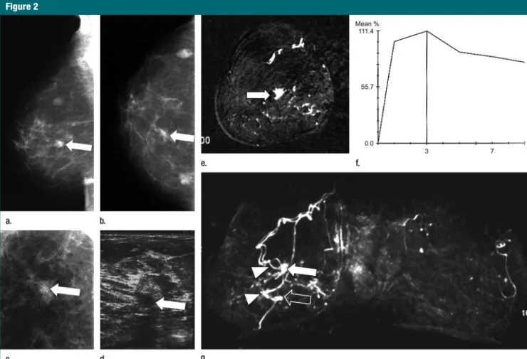
Figure 2: Asymptomatic 52-year-old woman with a strong family history of breast cancer that included a personal previous invasive ductal carcinoma of the right
breast at 50 years of age had no suspicious finding at CBE. (a) Mediolateral oblique, (b) craniocaudal, and (c) magnified craniocaudal mammographic views of the right breast show a suspicious opacity with irregular borders associated with microcalcifications in central location (arrow on a and b). Less than 50% of the breast was occu-pied by fibroglandular density. Microcalcifications were confirmed with magnified spot compression (arrow on c). At first-look (ie, prospective) US examination (10-MHz probe), not shown, no suspicious findings were detected. (d) Second-look US scan (10-(10-MHz probe) at final assessment showed hypoechoic suspicious mass (ar-row) at same location as mammographic finding. (e) Coronal-subtracted (first enhanced minus unenhanced) contrast-enhanced T1-weighted gradient-echo MR image
(repetition time msec/echo time msec, 11/4.8; flip angle, 25°; section thickness, 1 mm; matrix, 192⫻ 384; field of view, 192 ⫻ 384 mm) obtained with 0.1 mmol/kg
gadoterate meglumine. (f) Percentage of enhancement–time dynamic curve for enhancing focus shown on c. (g) Coronal maximum intensity projection of both breasts shows spiculated enhancing focus in central location (arrow on e) associated with a strong initial enhancement with postinitial washout, visible on f. On the maximum intensity projection, the central lesion is well depicted (white arrow), whereas a second smaller enhancing focus 3 cm below (open arrow) is visible (percentage of en-hancement–time dynamic curve with an initial enhancement greater than 100% and postinitial washout not shown) but was not detected at mammography and US. Note the prominent vascular supply of both lesions (arrowheads on g). Pathologic findings at mastectomy revealed bifocal invasive ductal carcinoma of 10 mm in diameter for one lesion (white arrow) and of 6 mm in diameter for the other (open arrow) of both lesions. At mammography, two other small masses were observed in both projections (a, b). They were known benign nodules that were unchanged in comparison with previous mammograms.
foci. At CBE, the diagnosis of generi-cally multiple palpable nodules was that they were suspicious for malig-nancy in one breast; the wrong diag-nosis of unifocal cancer was deter-mined in two breasts, and the diagno-sis of all malignant lesions was missed in four. At mammography, underesti-mation of the number of malignant foci in one breast occurred, the wrong diagnosis of unifocal cancer was deter-mined in four breasts, and the diagno-sis of all malignant lesions was missed in two breasts. US, which was not per-formed in one patient with unilateral bifocal cancer, aided in the identifica-tion of the exact number of malignant foci in two breasts, contributed to un-derestimation of the number of malig-nant foci in one breast, and led to the wrong diagnosis of unifocal cancer in three breasts. At contrast-enhanced MR imaging, the number of malignant foci was correctly identified in four breasts and was underestimated in one breast, and the wrong diagnosis of unifocal cancer was determined in one breast. Moreover, contrast-enhanced MR imaging was the only technique that depicted a second focus of inva-sive lobular carcinoma, although it failed in the characterization of both foci as malignant (Table 3, patient 15). Thus, the percentage of breasts with an exact detection of the number of malignant foci was 0% (zero of seven) for CBE and mammography, 33% (two of six) for US, and 71% (five of seven) for contrast-enhanced MR imaging. False-Positive Diagnoses and PPVs A total of 15 invasive procedures were performed in 13 patients without breast cancer who had a mean age of 43.9 years⫾ 10.3; five of them were enrolled because they were BRCA1 mutation carriers, one was enrolled as a first-degree relative of a BRCA1 mutation carrier, and seven were enrolled be-cause of a strong family history of breast and/or ovarian cancer. Only two of them had a previous personal history of breast cancer. The following 20 false-positive findings led to invasive diagnostic procedures: two at CBE, three at mam-mography, six at US, and nine at
contrast-Figure 3
Figure 3: Asymptomatic 69-year-old BRCA1 carrier with previous left mastectomy for invasive ductal carcinoma at 55
yearsoldhadnosuspiciousfindingatCBEofrightbreast.(a)Mediolateraland(b)craniocaudalmammographicviewsof rightbreastwithoutanydetectedsuspiciousabnormality;lessthan50%ofbreastwasoccupiedbyfibroglandulardensity. NosuspiciousfindingwasdetectedatUS(notshown).(c)Transverseand(d)lateralmaximumintensityprojectionsof subtracted(firstenhancedminusunenhanced)coronalT1-weightedgradient-echocontrast-enhancedMRimages(8.9/ 4.3,25°flipangle)afteradministrationof0.1mmol/kggadopentetatedimeglumine.Twosmallenhancingsuspiciousfoci (solidarrowsoncandd)arevisibleonthelateralquadrantsoftherightbreast.(e)Transversefastspin-echoMRimaging– guided(204/20,90°flipangle)biopsyofoneofthetwofociconfirmedpresenceofamalignantlesion.Arrowshowsthe amagneticneedle,withthetipatthesiteoftheenhancingfocus.Pathologicexaminationofthebreastaftermastectomy demonstratedmultiplefociofductalcarcinomainsitu(maximumdiameter,3mm);twowerelocatedwhereMRimaging showedenhancinglesions.Intheinnerposteriorpartofthebreast,thetransverseMRsubtractedmaximumintensitypro-jectionimageshowsenhancementcausedbythesuperimpositionofinternalmammaryvessels(openarrowonc).
enhanced MR imaging. Because three invasive diagnostic procedures were determined with a concordant false-positive finding at two modalities and one invasive diagnostic procedure was determined with a concordant false-positive finding at three modalities, 20 false-positive findings led to only 15 invasive diagnostic procedures. These
15 invasive procedures were
US-guided fine-needle aspiration or
core-needle biopsy (n ⫽ 11), US-guided
fine-needle aspiration and local exci-sion (n ⫽ 2), fine-needle aspiration with mammographic stereotactic guid-ance (n⫽ 1), and mastectomy (n ⫽ 1).
The woman who underwent mastec-tomy decided to undergo bilateral pro-phylactic mastectomy, which did not reveal any malignant lesions at patho-logic examination; thus, one false-pos-itive finding occurred at mammogra-phy. Of the 11 US-guided procedures, seven were performed as the result of second-look US findings detected only with contrast-enhanced MR imaging. The cytologic or pathologic benign diag-nosis obtained with these 15 invasive procedures included one or two of the following findings: single or multiple fi-broadenomas (n⫽ 4), papilloma (n ⫽ 2), adenosis or fibroadenosis (n⫽ 2),
intraparenchymal lymph node (n⫽ 1), and fibrocystic changes or other benign findings (n⫽ 8). On this basis, we calcu-lated the PPVs (Table 4).
Discussion
The detection rate for breast cancer in high-risk women (in our study, 4.8%) was more than 10-fold that obtained in the screening of a general female popu-lation. The first-screening detection rate recently reported by Caines et al (43) by using mammography in the gen-eral female population was 0.37% for women aged 40 – 49 years and 0.58% Figure 4
Figure 4: Asymptomatic 56-year-old woman with a strong family history of breast cancer. (a) Craniocaudal mammographic view shows asymmetric opacity (arrow) in
left breast (fibroglandular density greater than 50%). (b) US scan shows hypoechoic mass (arrow) of 3 mm in diameter with partly irregular borders. Both mammogram and US scan were judged as suggestive of malignancy. (c, e) Two contrast-enhanced coronal subtracted (first enhanced minus unenhanced) T1-weighted gradient-echo
MR images (8/4, 25° flip angle, 2.5-mm section thickness; 128⫻ 256 matrix; 175 ⫻ 350-mm field of view) obtained after administration of 0.1 mmol/kg gadopentetate
dimeglumine. Each shows a round enhancing lesion (arrow). (d, f) Percentage of enhancement–time dynamic curves show continuous increase. Both enhancing lesions were judged as benign at MR imaging. Pathologic examination revealed two foci of invasive lobular carcinoma of 3 and 9 mm in diameter.
for women aged 50 –59 years. We be-lieve our high detection rate was caused by not only the high disease prevalence in the population but also the multimo-dality approach, which included con-trast-enhanced MR imaging. In fact, in the trial by Brekelmans et al (44) in which patients underwent yearly screen-ing mammography and CBE every 6 months, the detection rate in BRCA1 and BRCA2 mutation carriers was 3.3%.
More important, we found no signif-icant difference in detection rate in BRCA1 and BRCA2 mutation carriers or first-degree relatives of BRCA1 or BRCA2 mutation carriers compared with that in women enrolled only on the basis of a strong family history of breast cancer and/or ovarian cancer. The com-parison between the cancers detected in the two groups of women enrolled according to these two selection criteria revealed no significant differences in terms of patients’ ages, tumoral patho-logic features, and axillary nodal in-volvement. This lack of a significant dif-ference is due to the strict criteria we adopted for including women on the ba-sis of only the family history and it also explains our high detection rate (4.8%). This detection rate is identical to that obtained in the study by Warner et al (26), who included only BRCA1 and BRCA2 mutation carriers, with 39% of women with personal previous breast cancers; that percentage was 44% in our study.
In breast cancer screening pro-grams for the general female popula-tion, women with a previous personal history of breast cancer are frequently excluded. This approach is under dis-cussion (28) because no consensus ex-ists about the duration and frequency of follow-up in women treated for breast cancer or about the schedule of exami-nations, particularly those at 5 years af-ter the primary treatment (45). In our study, nine (50%) of 18 patients who had a newly diagnosed breast cancer were women with a previous personal history of breast cancer, and the detec-tion rate was 5.3%, which was not sig-nificantly different from the detection rate (4.3%) found in women without a
previous personal breast cancer and/or ovarian cancer.
In women with familial-genetic pre-disposition to the disease, the risk of developing a contralateral breast cancer or an ovarian cancer within 5 years af-ter a previous tumoral event is esti-mated to be between 30% and 60% (4,46). Moreover, the inclusion of women with previous personal breast cancer and/or ovarian cancer is the
probable reason for our relatively
higher mean age at entry for the
screen-ing population (46 years) when we com-pared that age with the age in two other studies in which women with a personal history of previous breast cancer were excluded (25,27). In both of the studies, the mean or median age was 40 years. On the other hand, in the study by Warner et al (26), the percentage of women with a personal history of breast cancer was 39%, which is close to our percentage of 44%, and the reported mean age was 47 years.
Of the 11 patients genetically tested
Table 5
Comparison between 11 BRCA1 and BRCA2 Mutation Carriers and Seven Women Enrolled on the Basis of a Strong Family History of Breast Cancer and/or Ovarian Cancer in 278 High-Risk Women
Clinical Data Group A Group B
Patient age (y) 51.9⫾ 11.3 49.9⫾ 8.8
Diameter at pathologic examination (mm) 14.1⫾ 9.4 12.1⫾ 6.4
Invasive breast cancers* 9 (82) 5 (71)
Pathologic grade of cell differentiation 2.4⫾ 0.8 2.5⫾ 0.7
Multifocal, multicentric, or bilateral cancers* 4 (37) 5 (71)
Axillary nodal involvement* 1 (9) 2 (29)
Note.—Group A included 11 women who were BRCA1 and BRCA2 mutation carriers who had cancer, and group B included
seven women with a strong family history of breast cancer and/or ovarian cancer who had cancer. Data are the mean⫾
standard deviation except where otherwise specified. The Fisher exact test was used for comparisons between percentages, and the Mann-Whitney U test was used for comparisons between patients’ ages, between tumoral diameters at pathologic examinations, and between pathologic grades of cell differentiation. Differences between group A and group B were not significant.
* Data are numbers of women, and numbers in parentheses are percentages.
Table 6
Comparison between Nine Patients with Palpable Cancers and Nine Patients with Nonpalpable Cancers Detected in Multimodality Screening in 278 Women at Genetic-Familial High Risk for Breast Cancer
Clinical Data Group A Group B
Patient age (y) 47.2⫾ 9.1 55.0⫾ 10.2
Diameter at pathologic examination (mm) 19.0⫾ 6.9 7.7⫾ 4.8
Invasive breast cancers* 9/9 (100) 6/9 (67)
Pathologic grade of cell differentiation 2.7⫾ 0.7 2.1⫾ 0.8
Multifocal, multicentric, or bilateral cancers* 3/9 (33) 6/9 (67)
Axillary nodal involvement* 2/8 (25) 1/10 (10)
Note.—Group A included nine patients with palpable cancers, and group B included nine patients with nonpalpable cancers.
Data are the mean⫾ standard deviation except where otherwise specified. Of the nine patients with palpable cancers, one
with marked anxiety did not undergo MR imaging, one had indeterminate results of MR imaging because of movement artifacts, and one with breast implants did not undergo successful mammography because of marked discomfort during compression. Of the nine patients with nonpalpable cancers, one did not undergo US for unspecified reasons. The Fisher exact test was used for comparisons between percentages, and the Mann-Whitney U test was used for comparisons between patients’ ages, between tumoral diameters at pathologic examination, and between pathologic grades of cell differentiation.
Differences between group A and group B were not significant except for diameter at pathologic examination (P⬍ .002).
in whom breast cancer was detected, seven were BRCA1 mutation carriers and four were BRCA2 mutation carri-ers. The relatively advanced mean age of the BRCA1 and BRCA2 mutation car-riers (53.7 years⫾ 8.3 and 48.8 years ⫾ 16.3, respectively) and the higher age (even though not significantly different) of the BRCA1 group can be explained by the presence of patients with a personal history of previous breast cancer in both groups: five (71%) of seven in the BRCA1 mutation carrier group and one (25%) of four in the BRCA2 mutation carrier group.
The mean tumoral diameter at pathologic examination was 13.7 mm⫾ 8.6, and eight (44%) of 18 patients had tumors of 10 mm or smaller in diame-ter; the percentage of patients with tu-mors of that diameter was similar to the
percentage reported by other authors, and the percentages ranged from 43% to 55% (25–27). This result appears to be interesting if we consider that the tumor volume doubling time of invasive cancers is shorter in BRCA1 or BRCA2 mutation carriers than it is in noncarri-ers, as recently reported (ie, 45 days for carriers and 84 days for noncarriers) (47).
The high percentage (78%) of inva-sive cancers in our study is in agree-ment with data in previous studies; in those studies, the researchers reported a percentage from 73% (26) to 88% (25). Our percentage of cancers with a pathologic grade of cell differentiation of 3 (10 of 18, 56%) is slightly lower than that reported in two previous stud-ies about high-risk women; in those studies, this percentage was 66% (27)
and 67% (24). Interestingly, Kriege et al (25), who included a large proportion of women with a lower risk of breast can-cer (starting from only a 15% of lifetime risk) in their study, reported a percent-age of grade 3 cancers of only 37%.
On the other hand, of 13 patients with invasive cancers in whom the axil-lary nodal status was explored, we had only three patients with nodal cancer involvement (23%), confirming the pos-sibility of an early diagnosis in terms of nodal involvement in high-risk women, as already found in other studies in which the percentages of invasive can-cers with known nodal status and meta-static involvement ranged from 0% (24) to 21% (25). For comparison, we should consider that several screening studies of high-risk women by using mammography without contrast-enhanced MR imaging have shown an incidence of positive nodes of 30%– 45% (44,48,49).
The absence of interval cancer in our series must be related to the small number of screening events per woman (only 1.4) considered in this report and to a limited follow-up. The higher detec-tion rate at the second round, which was 7.1% and was greater but not sig-nificantly higher than 4.0% at the first round, confirms that in high-risk women we should adjust some conceptual tools used in mammographic screening of the general female population. For in-stance, the typical difference between “prevalent” cancers of the first screen-ing and “incident” cancers of subse-quent screenings was not observed in our study. Similar results were reported by Warner et al (26), who described a detection rate of 5.5% at the first round and 5.1% at the second round. This finding is probably related to the high speed of tumoral growth of breast can-cer in high-risk women (26,47).
Our study findings indicate that con-trast-enhanced MR imaging may enable the detection of unsuspected breast cancers by using other diagnostic mo-dalities in women at genetic-familial high risk, as already reported in other studies (23–27). In our study, the high sensitivity of 94% for contrast-en-hanced MR imaging could be related to the lower number of screening events Table 7
Patient-based Sensitivity of Contrast-enhanced MR Imaging, Mammography, and US for Palpable and Nonpalpable Breast Cancers in Multimodality Screening in 278 Women at Genetic-Familial High Risk for Breast Cancer
Modality
Sensitivity for Palpable Cancers (%)
Sensitivity for Nonpalpable Cancers (%)
Contrast-enhanced MR imaging 100.0 (7/7) 88.9 (8/9)
Mammography 87.5 (7/8) 33.3 (3/9)
US 100.0 (9/9) 25.0 (2/8)
Note.—Numbers in parentheses were used to calculate the percentages.
Table 8
Comparison between 12 Patients with Tumors Detected with a Combination of CBE, Mammography, and/or US and Six Patients with Tumors Detected Only with Contrast-enhanced MR Imaging in 278 High-Risk Women
Clinical Data Group A Group B
Patient age (y) 48.6⫾ 8.2 56.2⫾ 12.5
Diameter at pathologic examination (mm) 18.0⫾ 7.3 5.2⫾ 1.5
Invasive cancers* 11 (92) 3 (50)
Pathologic grade of cell differentiation 2.5⫾ 0.7 2.0⫾ 1.0
Multifocal, multicentric, or bilateral cancers* 6 (50) 3 (50)
Axillary nodal involvement* 2 (17) 1 (17)
Note.—Group A included 12 patients with tumors detected with a combination of CBE, mammography, and US, and group B included six patients with tumors detected only with contrast-enhanced MR imaging. For group A, of the 12 tumors, 11 were
true-positive and one was false-negative at MR imaging. Data are the mean⫾ standard deviation except where otherwise
specified. The Fisher exact test was used for comparisons between percentages, and the Mann-Whitney U test was used for comparisons between ages, between tumoral diameters at pathologic examination, and between pathologic grades of cell differentiation. The differences between group A and group B were not significant except for diameter at pathologic
examination (P⬍ .001). For invasive cancers, P ⫽ .078.
per woman, which was 1.4 without in-terval cancers, when we compared these values with those in the study by Kriege et al (sensitivity, 71%; screening events per woman, 2.7; interval can-cers, four) (25). In both the study by Warner et al (26) and the Magnetic Res-onance Imaging in Breast Screening study by Leach et al (27), the research-ers reported a sensitivity of 77% for contrast-enhanced MR imaging. Warner et al reported 1.9 screening events per woman with only one interval cancer, and Leach et al reported 3.0 screening events per woman with two interval cancers. Kuhl et al (24), however, re-ported a sensitivity of 100% and 1.9 screening events per woman without in-terval cancers. On the other hand, if the indeterminate MR result caused by arti-facts from the patient’s movements is considered false-negative, we obtained a sensitivity of 88% (15 of 17). In this patient, however, contrast-enhanced MR imaging was not repeated because we had a concordant positive result with CBE, mammography, and US.
We did not observe a higher prev-alence of palpable cancers at the first round: The percentage of palpable cancers was 36% (four of 11) at the first round and 57% (four of seven) at the second round. Palpability was clearly related to the mean tumoral diameter (19.0 mm⫾ 6.9 for nine
pal-pable cancers vs 7.7 mm ⫾ 4.8 for
nine nonpalpable cancers), and this high percentage of palpable cancers could be related to the rapid tumoral growth in these patients (47). We con-firm the insufficient sensitivity of CBE in high-risk women, as already re-ported (24–26).
The range of sensitivity values (88%–100%) for palpable cancers among the three imaging modalities in our study compared with that for nonpal-pable cancers (33% for mammography, 25% for US, and 89% for contrast-en-hanced MR imaging) shows that the ad-vantage of contrast-enhanced MR imag-ing is in the detection of small nonpal-pable cancers. It should be emphasized that the mean age of patients in whom cancers were detected with only con-trast-enhanced MR imaging was 56.2
years⫾ 12.5, and this age is relatively more advanced, even though it is not significantly higher than the mean age of
48.6 years ⫾ 8.2 in women in whom
diagnosis was determined with a combi-nation of CBE, mammography, and US. In other words, we do not have evi-dence to advocate a gain in sensitivity by using contrast-enhanced MR imaging only in younger premenopausal high-risk women and not in older postmeno-pausal high-risk women. Also, the breast density pattern does not seem to play a key role, as shown by the similar low sensitivity for mammography in both breasts with fibroglandular density of 50% or less (five of eight, 63%) and breasts with fibroglandular density greater than 50% (five of nine, 56%). Moreover, the possible overdiagnosis because of the high sensitivity of con-trast-enhanced MR imaging is counter-balanced by the fact that five of six pa-tients in whom breast cancer was diag-nosed during the study only with contrast-enhanced MR imaging had an invasive or multifocal, multicentric, or bilateral cancer.
The PPV of 63% for contrast-en-hanced MR imaging in our experience is very close to that of 64% reported by Kuhl et al (24) and higher than that of 46% reported by Warner et al (26). Our performance can be related to the use of a model of interpretation of contrast-enhanced MR images in which both morphologic and dynamic parameters are integrated in a simple score (38,39), which can be easily translated into one of the BI-RADS categories of 1–5. The key point is that, with this scoring sys-tem, a round, oval, or lobular homoge-neously enhancing lesion with regular margins, less than 100% initial en-hancement, and a continuous or plateau postinitial enhancement (or with an ini-tial enhancement of 100% or greater and a continuous postinitial enhance-ment) is considered benign. We paid a trade-off for this criterion: The only false-negative finding at contrast-en-hanced MR imaging was an invasive lob-ular carcinoma that was diagnosed as benign. Sensitivity, however, remained as high as 94%, and a good balance with PPV (63%) was obtained. Nevertheless,
it is important to realize that our results (in particular the PPV of 63% for con-trast-enhanced MR imaging in a screen-ing settscreen-ing) were obtained in a very high-risk population and that the same protocol in a lower-risk population could have a lower performance.
Our PPV for mammography (77%) is within the range of values in similar studies: from 30% (24) to 89% (26). We observed a higher PPV for US (65%) if one compares the PPV with that of 29% from one previous study (26) and also a higher PPV for CBE (82%) if one compares the PPV with those of 18% (25) and 50% (26) from two previous studies.
The first general limitation of our study was the nonrandomized study de-sign. We do not know whether an inten-sive screening protocol (including con-trast-enhanced MR imaging) is able to provide an effect in terms of saved years of life in such high-risk women. Such a question could be answered with ran-domized trials, although we agree with the ethical concerns recently raised about this possibility (50).
The second important limitation was the small number of screening events per woman considered in the study (99 women for the first and sec-ond rounds and 179 women for only the first round, for a total of 377 screening events and a mean of 1.4 screening events per woman). We should con-sider, however, that very few interval cancers were reported in prospective studies in which contrast-enhanced MR imaging was used for the screening of high-risk women: a total of seven inter-val cancers versus 104 cancers detected during the trials for 2956 women en-rolled and 2.7 screening events per woman (24–27).
The third limitation was that paired statistical analysis of the diagnostic formance of the modalities was not formed. This comparison will be per-formed at the end of the trial.
In conclusion, our interim experi-ence suggests that contrast-enhanced MR imaging is highly sensitive (94%) for detection of breast cancer in multimo-dality yearly surveillance—which also includes CBE, mammography, and US—