School of Graduate Studies
“Scienze del Farmaco e delle Sostanze Bioattive”
Università di Pisa
March 2013
Gap Junctions in Mammalian Photoreceptors:
Functional Impact and Modulation
Sabrina Asteriti
Supervisor: Prof. Maria Claudia Gargini
Dip. di Farmacia
Co-supervisor: Dr. Lorenzo Cangiano
Dip. di Ricerca Traslaz. e delle Nuove Tecn. in Med. e Chir.
Abstract
7
1 Introduction
9
1.1 Morphology of the Retina 13
1.2 Early Retinal Processing of Visual Signals 22
1.2.1 Phototransduction in Rods and Cones 22
1.2.2 Rod and Cone Signal Pathways 28
1.3 Gap Junctions and their Role in the Retina 32
1.3.1 Connexin 36 is the Major Isoform in the Retina 34 1.3.2 Light Adaptation and Gating of Gap Junctional Coupling 36 1.3.3 Endogenous Neuromodulators Coordinate Network Adaptation 38 1.3.4 Dopamine in the Regulation of Gap Junctions 40 1.3.5 Intracellular Signaling Pathways and Junctional Coupling 42 1.4.4 State of the Art and Open Questions in Rod-Cone Coupling 44
2 Materials and Methods
47
2.1 Electrophysiology 47
2.1.1 Dissection, Slicing and Extracellular Solutions 47 2.1.2 Perforated Patch-Clamp Recordings from Mouse Retinal Neurons 48
2.1.3 Light Stimulation Protocols 50
2.2 Mouse Genotyping 53
2.3 Modeling 54
2.4 Whole-Cell Recordings from AII Amacrines in Rat (at the University of Bergen) 56
3 Results
61
3.1 Rods and Cones Recorded in Mouse Retinal Slices 61 3.2 Cones Display Rod-like Light Sensitivity and Response Recovery 64 3.3 Sealing on Cones Triggers a Spontaneous Increase in Their Coupling to Rods 64 3.4 A Rod-Saturating Light Background Isolates the Pure Cone Component 67
3.5 Cone Spectral Type and Rod Coupling 69
3.6 Dim and Bright Flash Responses Come From Separate Compartments 71 3.7 Blocking Gap Junctions Reverts Cones to Their Intrinsic Phenotype 72
3.8 Mixed S/M cones dominate the mouse retina 74
3.9 Coupling is also Expressed at Body Temperature 75 3.10 Cone Light Responses in the Connexin36-KO Mouse 76
3.12 Is Rod-Cone Coupling Modulated by Dopamine? 79
4 Discussion
83
4.1 The Functional Latitude of Rod-Cone Coupling 83
4.2 Possible Mechanisms Driving Coupling Run-up 84
4.3 Dynamic Regulation of Rod-Cone Coupling 86
4.4 Conclusions 87
5 Acknowledgements
89
Science may set limits to knowledge, but should not set limits to imagination.
Bertrand Russell (1872–1970)
Material in this thesis is reproduced with permission from:
Cangiano L, Asteriti S, Cervetto L, Gargini C (2012). The photovoltage of rods and cones in the dark–adapted mouse retina. J Physiol 590, 3841–3855.
or will be included in:
Asteriti S, Gargini C, Cervetto L, Cangiano L. The regulatory latitude and functional impact of rod coupling in mouse cones. In preparation.
Abstract
Rod and cone photoreceptors form gap junctional contacts (GJs), which provide an alternative route for rod signals when light saturates the primary high-gain rod pathway. Indirect evidence suggests that in mammals, as in lower vertebrates, rod-cone coupling is dynamically regulated by light and circadian rhythmicity through endogenous neuromodulators such as dopamine (DA). However, the only direct tests done so far, in macaque, found coupling to be static. Moreover, recordings from the postsynaptic cone bipolar cell, in mouse, suggest that this route may give only a minor contribution to rod signaling.
In my thesis I investigated the functional impact and regulatory latitude of rod-cone coupling by recording, with perforated patch clamp, from mouse cones in an in vitro retinal slice preparation. In the process, I optimized the techniques required to gain intracellular access to these small and challenging neurons. I dissected rod input in the photovoltage of wild type mouse cones by exploiting differences in light sensitivity, kinetics of recovery from bright flashes, and relative spectral preference to green (G) over ultraviolet (UV) light. Most cones expressed rod-like features, including: (1) responses to dim flashes, (2) slow plateaus in response to moderately bright flashes and a transient suppression of dim flash responses, (3) long recovery of dim flash responses and slow plateaus after rod-saturating backgrounds, (4) preference for dim G over dim UV flashes, irrespective of the intrinsic spectral preference of the cone determined with rod-saturating pre-flashes. Dim and bright flash responses had different reversal potentials, consistent with an origin in separate electrotonic compartments. The role of GJs was confirmed pharmacologically. Cones dramatically increased their coupling to rods within minutes after seal formation, revealing mechanisms for rapid plastic change, triggered in my experiments by a perturbation of the intracellular milieu. In fully coupled cones the overall junctional conductance could exceed the light-sensitive conductance. In contrast to wild type animals, in connexin isoform 36 (Cx36) knockout mice cones did not appear to be able to couple to rods, supporting a key role for Cx36 in rod-cone GJs. In disagreement with indirect data, but similarly to what observed in single macaque cones, I found evidence that would rule out a role of the dopaminergic system in the regulation of rod-cone coupling.
My work provides the first direct and conclusive evidence for rod-cone coupling in the mouse retina, an emerging model for studies of early visual processing in health and disease. This coupling is not hardwired but can be rapidly up-regulated, revealing that junctional contacts are adequate for it to play an important role in rod visual signaling and, potentially, also in the biochemical interaction between photoreceptors. The cellular mechanisms leading to a spontaneous coupling increase during patch recordings need to be investigated to reconcile the lack of DAergic modulation in single cell recordings with other indirect evidence.
1 Introduction
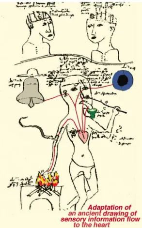
From the dawn of history humans wondered why they could distinguish light from darkness and why everything in nature appears in color. However, if early humans had obviously recognized the eye as the organ of vision, they did not appreciate the complexity of visual perception by the brain. The eye has been the subject of conflicting interpretations since antiquity. Aristotle, in the IV century B.C., rejecting the Platonic ‘extramission’ theory of vision, advocated for a theory of ‘intromission’ by which the eye received rays rather than directed them outward. He proposed that visual sensation passed from the eye to the heart, which at that time was considered as the center of sensation and psychic function (the brain was thought to merely be a cooling device) (Jung, 1984) (Fig. 1-1). In the XIII century A.D., St. Thomas D’Aquino, sanctioning the aristotelic axiom,
wrote Nihil est in intellectu quod non prius fuerit in sensu (Latin for "nothing in
Figure 1-1. Aristotelian concept of five senses
projecting to the heart, either directly or via the “sensus communis” in the anterior part of the head (lower figure). The upper drawing shows the four (Galen’s and Avicennas’s) or five (Albertus Magnus’s) brain compartments (reproduced from: Webvision, webvision.med.utah.edu; from: Jung, 1984).
the intellect without first being in the senses"). The Aristotelian concept of five senses projecting to the heart continued into the Middle Ages.
In the 2nd century A.D., Galen chose the extramission theory because it
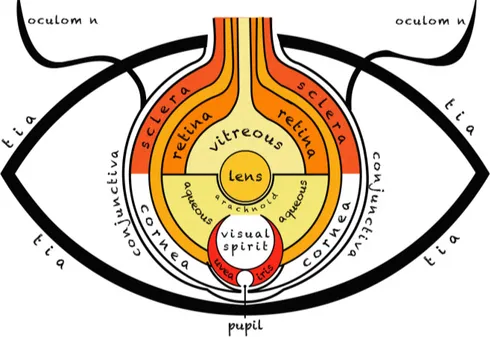
corresponded well with his image of sight as a function of an optical pneuma, flowing forth from the brain to the eyes through hollow optic nerves. Further, Galen defined many of the fundamental features of the anatomy and physiology of the eye, which remained as a reference until the 17th century. Benefiting from
the work of the anatomists who dissected in Alexandria, such as Rufus of Ephesus, he described the retina, cornea, iris, uvea, tear ducts, and eyelids, as well as two fluids he called the vitreous and aqueous humors. He noted some of the peculiar features of sight such as binocular vision. According to the Galenic conception, the photoreceptive element in the eye was a central crystalline lens, while the retina, which abounded in blood vessels, was an organ of nutrition (Fig. 1-2). The eye was the subject of special interest in medieval Islamic medicine and philosophy. Influenced by their readings of Galen, the majority of Islamic scholars, such as al-Kindi and Hunain ibn Ishaq (Johannitius) in the 9th
century, favored the extramission theory of sight. The latter, in such works as
Figure 1-2. The eye according to Hunain ibn Ishaq, from a manuscript dated circa 1200 (adapted
‘Ten Treatises on the Eye’ and the ‘Book of the Questions on the Eye’, elaborated on the series of tunics behind the vitreous humor, paying special attention to the retina, whose role was to nourish the vitreous humor and conduct the visual spirit outward through the hollow optic nerve. He further reinforced the centrality of the crystalline lens (Fig. 1-2). Islamic physicians actively debated many details of the eye, among them, the number of tunics and, most importantly, the theory of extramission. In the early 10th century two Islamic
scholars reasonably concluded that light affected the eye and not vice versa. While, Avicenna, a Alhazen's contemporary that adhered completely to Aristotelian theory, offered a more systematic critique of the Galenic explanation, even though he retained many features of Galen's anatomy, such as the hollow nerves and crystalline lens. As this rich body of literature was translated from Arabic into Latin, in the period from the 11th to the 13th century,
medieval European medical practitioners had a great deal to puzzle over.
We have to wait until the Renaissance to have a more exhaustive understanding of human anatomy, because dissections and autopsies had been forbidden in most religions. One of the first scientists that studied human anatomy through dissection of the body was Andreas Vesalius (1514–1564). Modern theories of vision began in the 17th century, with the optical conclusions of Johannes Kepler
(1571–1630), who, drawing on the ocular anatomy presented by Felix Platter (1536–1614), solved a fundamental question: “How an infinity of rays from each point in the visual field is drawn into a coherent, point-to-point correspondence in the eye”. Despite tradition, Kepler inferred that the crystalline lens focussed images onto the retina, where vision was made possible. He distinguished between the physical optics of image formation and the psychological optics of vision: “I say that vision occurs when an image of the whole hemisphere of the world that is before the eye, and a little more, is set up at the white wall, tinged with red, of the concave surface of the retina … Thus vision is brought about by a picture of the thing seen being formed on the concave surface of the retina. That which is to the right outside is depicted on the left on the retina, that to the left on the right, that above below, and that below above. Green is depicted green, and in general things are depicted by whatever colour they have … the greater the acuity of vision of a given person, the finer will be the picture formed in his eye.” A few decades later, with a landmark experiment René Descartes showed that Kepler was right. He scraped the back of the eye of a ox, surgically
removed, to make it transparent, and placed it on a window pane, as if the ox were looking out of the window. He looked at the back of the eye and saw an inverted image of the outside world. Descartes concluded that the image was inverted because it had been focused onto the retina by the eye's lens.
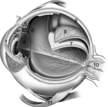
We now know the functions of the various eye components, and most of the steps of visual processing have been investigated (Fig. 1-3). The photoreceptors collect light and feed electrical signals to a network of neurons in the retina that process visual information in complex ways, and that send their output to the brain along the optic nerve. The brain decodes this continuous flow of information resulting in what we call vision.
Figure 1-3. Diagram of the eye. A simplified sagittal section of the human eye, in
which all morphological components are indicated. 1: cornea, 2: iris, 3: pupil, 4: lens, 5: ciliary body, 6: vitreous chamber, 7: retina, 8: choroid, 9: sclera, 10: optic nerve, 11: inferior oblique muscle.
1.1 Morphology of the Retina
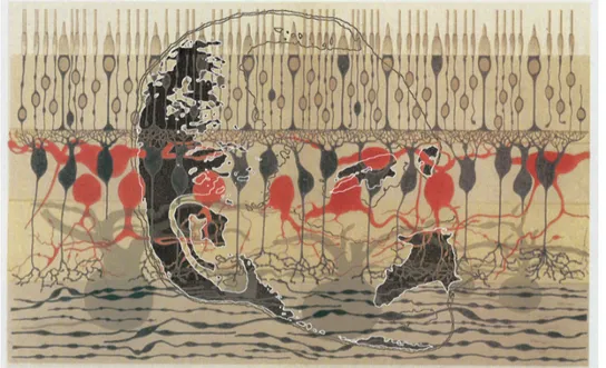
Light, and thus photons, lets us collect visual memories and experiences. This is possible thanks to a complex visual system, which can detect and elaborate an extraordinarily wide range of light input. The neuronal structure designated to detect photons is the retina. The complexity of the retina baffled generations of students of vision, but among these one, in particular, deserves to be mentioned: Charles Darwin. With regards to the idea that the eye had evolved by natural selection, Darwin said: “The eye to this day gives me a cold shudder.”, “Seems, I freely confess, absurd in the highest possible degree.”. Figure 1.1-1 shows the incredible complexity of the retinal network in a vertical section. This is one of the most beautiful early representations of the microscopic structure of retina, by anatomist Ferruccio Tartuferi (in this modern composition the scientist is visible in the background). Tartuferi was a student of Golgi, who had developed the black reaction (reazione nera) in 1873, and published this surprising coloured
Figure 1.1-1. Tartuferi’s retina. The illustration combines one of the early representations of the
microscopic structure of the retina, by anatomist Ferruccio Tartuferi, whose profile can be seen behind. Tartuferi published this coloured illustration of the retina in 1887; his portrait is based upon a photograph from the archives of the University of Pavia, where he worked as a student of Camillo Golgi (Reproduced with permission from http://neuroportraits.eu; ©2012 Nicholas Wade).
illustration of the retina in 1887. Tartuferi was the first to apply the ‘black reaction’, which had initially been demonstrated on the arborizations of cerebellar Purkinje cells, to the retina.
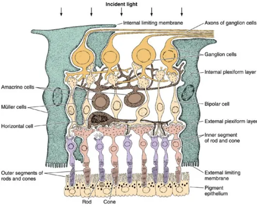
The retina has long been a model system in which to study neuronal morphology and physiology (Werblin and Dowling, 1969), it’s layered structure (Fig. 1.1-2) contains approximately 60 different types of neurons. Synaptic connections are largely formed within two distinct layers, a thin external one and a thicker internal one. In the outer retina, photoreceptors contact horizontal cells and bipolar cells at the outer plexiform layer (OPL); in the inner retina synapses between retinal ganglion cells and their presynaptic neurons, the amacrine and bipolar cells, are localized to the inner plexiform layer (IPL), which is composed of two adjacent sub-laminae.
The outer retina consists of four layers: the retinal pigment epithelium, the photoreceptor outer and inner segments, the outer nuclear layer (ONL) and the OPL. The somas of photoreceptors are located in the ONL. Rods and cones are the main photosensitive cells in the retina (a third one was recently discovered, see below), and their interaction with the pigment epithelium is essential for visual function. Rods and cones have their name according to the shape of their photosensitive portion. Rods outnumber cones by about 20-fold in almost all mammalian species, The peripheral human retina contains about 95% rods and 5% cones, a proportion surprisingly similar to that of the mouse (97% rods and 3% cones) Due to their small size and high density, rods seem to be the second most numerous neuron type of the human nervous system, following the cerebellar granule cells. Photoreceptors occupy much less than 1% of the soma-dendritic volume of a cortical or hippocampal pyramidal cell (Masland, 2012) (Fig. 1.1-3).
Retinal pigment epithelial (RPE) cells, located between the photoreceptors and the choroid, are specialized pigment cells. They are a part of the brain and constitute an epithelial monolayer able to respond to distinct extracellular signals. They provide functions that far exceed those of a light-absorbing screen. The RPE is an important part of the blood-retina barrier and controls the flow of nutrients and metabolites to and from the retina, replenishes 11-cis-retinal by reisomerizing all-trans-retinal generated during photo-conversion, and it is specialized in the phagocytosis of the remnants of photoreceptor outer segments, which are constantly renewed (apical processes from the pigment epithelium
envelop the outer segments of photoreceptors). Furthermore, it prevents the degradation of the visual image by absorbing stray light in melanin granules, and secretes cytokines that locally control the innate and adaptive immune systems. Not surprisingly, RPE cell disease is a major cause of human blindness worldwide (Bharti et al., 2011).
Photoreceptors consist of a cell body, an outer and an inner segment, an axon and a synaptic terminal (Kaneko, 1979). Their distal parts form respectively the outer and inner segment layers (OS/IS). Moving radially inward, at the base of the rod and cone inner segments there is an outer limiting membrane: with a network-like structure it is an alignment of adherent junctions between Müller glia and photoreceptor inner segments (Bunt-Milam et al., 1985). Photoreceptors are specialized to convert light into electrical signals. Two or three types of cone photoreceptors (depending on the species) and a single type of rod photoreceptor
Figure 1.1-2. The three main layers of retinal neurons. The arrows indicate the direction of light.
The flow of visual signals evoked by the incident light proceeds in the opposite direction (Junqueira and Carneiro, 2005).
are present in mammals. However, some lower vertebrate retinas have more cone types.
In good vertical sections of fixed retina, rods and cones are optimally aligned and can be distinguished easily using a light microscope. The cones’ cell bodies are located in the distal part of the ONL in a single row, just below the outer limiting membrane. Their outer segments of cones are shorter than the slim outer segments of rods, and often have a conically-shaped structure (Fig. 1.1-4). Rod and cone outer segments capture light and convert it into membrane potential changes, which are further processed in the proximal parts of the photoreceptor by voltage-gated ion channels (Della Santina et al., 2012) and fed to the inner retina. The outer segments are connected to the inner segments by a narrow stalk.
Rod photoreceptors contain the visual pigment rhodopsin and are sensitive to
blue-green light with a peak sensitivity around a wavelength of 500 nm. With the exception of some diurnal mammals and the foveas of old world primates, the ONL is occupied largely by rod cell bodies. Rods are highly sensitive to light being capable of signaling the absorption of a single photon (Cangiano et al., 2012). This ability makes them crucial for low light/night vision (scotopic conditions).
Cone photoreceptors contain a different set of visual pigments, the cone opsins.
Their peak spectral sensitivity depends on the opsin protein sequence, so that in mammals we find cones maximally sensitive to long wavelengths (L; ‘red’ cones), medium wavelengths (M; ‘green’ cones), or short wavelengths of light (S; ‘blue/ultraviolet’ cones) (Curcio et al., 1987). L and M cones diverged relatively recently in mammalian evolution from a common ancestor, while S-cones have several morphological traits that set them apart, including (1) longer inner segments; (2) smaller and morphologically different pedicles (Ahnelt et al., 1990); (3) a specific retinal distribution (see below). In mouse, which was used as a model system in my thesis, only two cone opsins are expressed, S and M. In the ventral mouse retina, the great majority of cones express both S- and M-opsin, and only a minority of cones are pure S cones (8–20%; Haverkamp et al., 2005). The sparse distribution of pure S cones implies that each one is surrounded by a ring of chromatically different cones. This has been observed also in the peripheral primate retina (in which S cones are sensitive to blue light), where it has been suggested that they may be electrically isolated from
nearby cones to prevent a severe reduction in color discrimination (Hsu et al., 2000, O’Brien et al., 2012). Cones of different spectral sensitivities, and their pathways of connectivity to the brain are, of course, the basis of color perception. Photoreceptors relay their signals to downstream bipolar and horizontal cells at specialized output synapses called spherules (rods) and pedicles (cones). Photoreceptors also communicate with each other, but this aspect has been the target of my doctoral work and will be dealt with separately. The inner retina consists of four layers: the inner nuclear layer (INL), the inner plexiform layer (IPL), the ganglion cell layer, and the nerve fiber layer. The dense neuropil of the IPL is a very fine and complex network in which several pathways connect with each other synaptically and electrically. Only electron microscopy, together with information gained from Golgi-staining, have enabled us to obtain detailed knowledge about this network. The INL contains the cell bodies of four groups of retinal cells: horizontal, bipolar, amacrine, and Müller glial cells.
Figure 1.1-3. A mouse cone photoreceptor (cell inside oval) has been reproduced at its correct
scale on an image showing a macaque cortical pyramidal neuron. Cortical cell modified with permission from Masland (2012), originally from Gilbert (1992).
Horizontal cells process information from the photoreceptors and provide lateral
feedback in the OPL. They are involved in enhancing contrast through lateral inhibition.
Bipolar cells process information from the outer retina and convey it to the
amacrine and ganglion cells, in the inner retina. Early physiological recordings classified four different types of bipolar cells: ON, OFF, sustained, and transient (Kaneko, 1970; Werblin and Dowling, 1969). More recent morphological work, and subsequent physiological evidence has shown that the true number of bipolar cell subtypes is likely to be 12. The IPL is like a tidy stack of sublayers: the bipolar cell axons branch at different levels and form sublayer-specific synapses with postsynaptic neurons (Cajal, 1972).
Amacrine cells of the vertebrate retina are interneurons that create a complex
synaptic network in the IPL, contacting bipolar cells and ganglion cell. They integrate, modulate and shape in the temporal domain the visual message relayed to the ganglion cell (Kolb, 1995). Amacrine cells are so called because most of them are axonless neurons, for this reason it is notoriously hard to recognize the sites of their inputs and outputs (Dowling and Boycott, 1966; Jusuf et al., 2005; Eggers and Lukasiewicz, 2011). The amacrine cells are classified into different types based on morphological characteristics of dendritic tree size, such as small, medium, and large branching, as well as tufted, varicose, linear, beaded, and radiate, and, most importantly, on the stratification of their dendrites in the IPL (Mariani, 1990; Kolb et al., 1992). Amacrine cells are a very diverse group, and can both relay and modulate signals in the inner plexiform layer (Kolb et al., 2002). Some also affect outer retinal neurons by paracrine release of a neuromodulator (e.g. dopaminergic amacrines).
Retinal ganglion cells (RGCs) are the output neurons of the retina and integrate
electrical signals converging from neurons preceding them in the retinal wiring scheme. Their cell bodies are located mainly in the ganglion cell layer, and their dendrites form synapses in the IPL with bipolar and amacrine cells (Wässle, 2004). RGCs are larger on average than most other retinal neurons, and signal by generating spike trains which are sent by their axons onwards to higher visual areas of the brain, through the optic nerve. RGCs integrate visual information over their receptive fields by receiving presynaptic excitatory inputs from bipolar cells, which themselves receive inputs from several photoreceptors, while the inhibitory interactions mediated by horizontal cells and amacrine cells
modulate the structure of the receptive field (Gollisch, 2012). The classification of the different kind of RGCs began with Cajal, in his monumental work on Golgi staining of the vertebrate retina, in which he classified many different varieties of ganglion cell based on form (dendritic morphology), extent (cell body and dendritic tree size), and number of sublayers in which they arborize (stratification levels in the inner plexiform layer). Sun et al. (2002), using the ‘DiOlistic’ method to stain RGCs with lipophilic dye-coated particles delivered with a gene gun (Gan et al., 2000), achieved a complete morphological classification of mouse retinal ganglion cells. They, in accord to a method used for the rat (Huxlin and Goodchild, 1997) classified cells with a large soma and a large dendritic field as RG(A), cells with a small to medium-sized soma and a small to medium-sized dendritic field as RG(B), and cells with a small to medium-sized soma, but a medium-sized to large dendritic field, as RG(C).
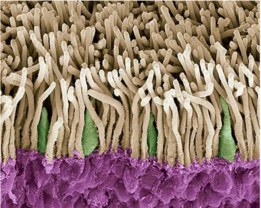
Figure 1.1-4. Colored scanning electron micrograph (SEM) of rods (yellow) and cones (green).
The outer nuclear layer is highlighted in purple. Magnification is x1800 when printed 10 centimetres wide. Credit: STEVE GSCHMEISSNER/SCIENCE PHOTO LIBRARY.
Unequivocally bistratified cells were classified as RG(D) cells. Each group, in turn, was divided into several subtypes. Cells that did not fit into this classification scheme were left unclassified. Currently, we can affirm that RGCs in the mammalian retina comprise at least 10–15 types that are classified according to common structural features (Wässle, 2004). Despite decades of intense study, which criteria one should use to physiologically classify RGCs is still a subject of much discussion and debate (reviewed in: Rowe and Stone, 1977; Rodieck and Brening, 1983; Wässle and Boycott, 1991; Rockhill et al., 2002). Since the time of Cajal it had been believed that one physiological type existed for each morphological type of GC (Boycott and Wässle, 1974; Dacey, 1994; Roska and Werblin, 2001), and numerous studies had shown that retinal ganglion cells exhibit an array of responses to visual stimuli, lending credit to the idea that these cells can be sorted into separate physiological classes (Carcieri et al., 2003). Nonetheless, this great morphological heterogeneity has not been confirmed by physiological findings (Farrow and Masland, 2011). It is well known that RGCs do not respond in the same way to visual stimuli (Hartline, 1938; Kuffler, 1953; Barlow, 1953; Enroth-Cugell and Robson, 1966). For example, when presented with flashing spots in their receptive field centers some respond only to the onset of the spot (ON-center), others only to the offset (OFF-center), and still others to both (ON-OFF-center) (Barlow, 1953; Kuffler, 1953). Similarly, when cells are stimulated with contrast-reversing sine wave gratings, some cells show linearity in their stimulus-to-response transformations, whereas others show striking nonlinearity (Enroth-Cugell and Robson, 1966; Hochstein and Shapley, 1976). However, RGCs shouldn’t necessarily be divided in classes based on their response properties (for review see Rockhill et al., 2002; Stone, 1983; Wässle and Boycott, 1991): the fact that a particular response property varies from cell to cell does not imply that the cells divide into groups with respect to that property. Instead, it has been asserted that RGCs show a functional unimodal distribution, with a single peak and a large standard deviation (Hochstein and Shapley, 1976; Rodieck, 1998; Abbott and Chance, 2002; Mechler and Ringach, 2002). Hence, cells with linear and very nonlinear responses may be endpoints of a continuum that contains cells with varying degrees of nonlinearity in between (Carcieri et al., 2003). A recent important discovery is that of intrinsically photosensitive ganglion cells (ipRGCs), which are a small minority of the total ganglion cell population and express the visual
pigment melanopsin. They have an important role in non image-forming functions as irradiance detectors for behaviors such as the pupillary light reflex and circadian photoentrainment (Lucas, 2013).
Our review of retinal cells ends with Müller glia, which represent the scaffold of the retina and are main glial cells present. They extend processes across the entire retina, with nuclei located in the INL. On vitreal side, the ‘feet’ of Müller cells processes cover most ganglion cell axons and form the inner limiting membrane (ILM) together with a basal lamina. The ILM is the inner surface of the retina bordering the vitreous and thereby forming a diffusion barrier between neural retina and vitreous humor. On the scleral side, Müller glia extend beyond the outer limiting membrane, forming microvilli into the subretinal space, which entail an increase of the surface available for metabolic exchange with the subretinal space. Müller cells don’t carry out only a structural role, but are also involved in the nurture of photoreceptors and represent an important reservoir which buffers changes in the ionic composition of the tight extracellular space during light stimulation. A final unexpected role for Müller glia is that of being conduits of light akin to optic fibers, thus reducing scatter across the retina and contributing to spatial acuity (Franze et al., 2007).
In the center of the retina is the optic disk, a circular to oval white area from which the optic nerve departs. The major blood vessels of the retina also radiate from the optic disc. In the retina of primates, about two and half diameters from the disc, can be seen the oval-shaped and blood vessel-free reddish spot, the fovea, which is at the center of the area known as the macula. The fovea is the area of the finest visual acuity, the result of dense packing of cone photoreceptors. This region is less complicated than the peripheral retina because it has very few rod photoreceptors and rod-specific circuits. There is one inner (invaginating or ON) midget bipolar cell and one outer (flat or OFF) midget bipolar cell for each foveal cone (Kolb, 1970), and one midget ganglion cell for each foveal midget bipolar cell (Boycott and Dowling, 1969; Kolb and DeKorver, 1991; Calkins et al., 1994).
The eye is organized so that the visual image is focused onto the retina with minimal optical distortion. Light traverses the cornea and the lens and, subsequently, crosses the vitreous humor that fills the eye cavity, before reaching the retinal photoreceptors, lying at the back of the eye. To allow light to reach
the photoreceptors with minimal scattering, RGC axons run towards the optic disk unmyelinated.
For cell-to-cell communication retinal neurons use the major neurotransmitter systems, as well as electrical coupling mediated by gap junctions. Glutamate is the primary excitatory neurotransmitter used by neurons in the vertical retinal pathways, including photoreceptors, bipolar, and ganglion cells (Iuvone e al., 2005). The other major excitatory neurotransmitter is acetylcholine (ACh). The inhibitory neurotransmitter GABA appears in many different subtypes of amacrine cells and in one or more subtypes of horizontal cells (Yang, 2004), both cell types being involved in lateral information processing. The inhibitory neurotransmitter glycine is present in most of the small-field types of amacrine cells (Nguyen-Legros et al., 1997).
1.2 Early Retinal Processing of Visual Signals
Visual perception begins with absorption by visual pigments contained in the outer segments of rod and cone photoreceptors. As mentioned above, the outer segment is specialized for phototransduction. The light-absorbing moiety of the visual pigment molecule is attached to a large membrane-spanning protein. Rods and cones have evolved a strategy to increase the surface area of the membrane, and thus increase their light collecting ability, by tightly stacking membranous discs in their outer segments, so that a huge number of these proteins are available for photon capture. In cones the discs are continuous with the plasma membrane, while in rods they pinch off from the plasma membrane becoming intracellular organelles. Like other neurons, photoreceptors do not divide, but their outer segments are constantly renewed, in parallel to the phagocytic activity of the adjacent pigment epithelial cells.
1.2.1 Phototransduction in Rods and Cones
Scotopia, from the Greek words skotos- (darkness) and -opia (sight), is defined as the range of lighting conditions in which only the rods are able to signal,
mediating high-sensitivity but monochromatic vision. Cones mediate coloured vision, but activate at higher light intensities in mesopic and photopic conditions (Fig. 1.2.1-1). Today it has become challenging to avoid light pollution and experience a completely dark natural landscape. In a wide range of dim ambient light levels both rods and cones operate (mesopic conditions): even the light of the full-moon can be sufficient to activate cones somewhat.
Let us delve into the astonishing complexity of phototransduction, step after step (Fig. 1.2.1-2), by describing what happens if one opens the window at twilight, after some hours spent into a completely dark room. Initially the eyes are fully dark adapted and a high concentration of visual pigment, rhodopsin and cone-opsin, is present in the disc membranes of rods and cones.
Both rhodopsin (Rh) and cone-opsins are members of the family of G protein-coupled receptors (GPCRs), which in darkness bind the light absorbing chromophore 11-cis retinal (Palczewski, 2006; Park et al., 2008). The latter is an inverse agonist for the receptor. As long as darkness is maintained, a steady inward current flows through the plasma membrane of the photoreceptor outer segments (the dark current). The dark current is mediated by ion channels gated by cGMP, which has a high concentration in darkness (see below), and are permeable to both Na+ and Ca2+ ions. The dark current enters the outer segment
and keeps the photoreceptor depolarized at –40/–45 mV, a membrane potential at which this inward current is balanced by an outward current in the inner segment
Figure 1.2.1-1. Photopic (left) and scotopic (right) vision in humans simulated from the same
image. The dark central patch in the scotopic image represents the foveal area with its paucity of rods. Photo by S. Asteriti.
mediated by K+ ions. Ionic gradients are maintained by a Na/K ATPase, which
removes Na+ and replenishes K+, and a Na/Ca,K exchanger, which limits the
Ca2+ concentration in darkness at a relatively high level of 500–600 nM. When
photons reach the dark adapted retina, as in our example above, they are absorbed by Rh and cone opsins, which undergo a sequence of conformational changes identifiable biochemically by specific absorption spectra: the early morning twilight is sufficient to stimulate both rods and cones. Since phototransduction has been better studied in rods than in cones, I am going to continue my description referring to Rh, although analogous considerations are valid also for cones.
The active state of Rh (known as Meta-2 and indicated as Rh*), has a half life of a few tens of milliseconds, during which it collides, binds and activates a random number of heterotrimeric G-proteins (transducins). In darkness, inactive transducin is bound to a GDP molecule, but upon collision with Rh* an exchange of GDP for GTP occurs, leading to the dissociation of the heterotrimer in two structural subunits (Tα and Tβγ). Interestingly, rod Transducin α (rTα) and
cone Transducin α (cTα), appear to be functionally interchangeable (Mao et al., 2013).
Figure 1.2.1-2. (A) Schematic anatomy of a representative vertebrate rod. (B) Major proteins and
mechanisms in vertebrate rod phototransduction. Abbreviations: hν, light; Rh*, activated form of the photopigment rhodopsin; GTP, guanosine triphosphate; GDP, guanosine diphosphate; cGMP, guanosine 3ʹ′,5ʹ′-cyclic monophosphate; GMP, guanosine monophosphate; PDE, guanosine nucleotide phosphodiesterase; RGS complex, a group of three proteins including RGS9 which accelerate the hydrolysis of the GTP attached to the alpha subunit of transducin; and Pi, inorganic phosphate. Reproduced with permission from: Fain et al. (2010).
The GTP-bound α-subunit (Tα) is the main effector and diffuses laterally along
the disk membrane until it binds to a γ-subunit of the target enzyme PDE6 (phosphodiesterase 6), which plays a key role in the phototransductive cascade. Two simultaneously-bound Tα-GTP molecules are necessary to elicit maximal
activation of PDE (Liu et al., 2009; Muradov et al., 2010). Activated PDE (PDE*) hydrolyzes cGMP very efficiently, leading to a rapid drop in the local concentration of the cyclic nucleotide. Continuous cGMP synthesis is guaranteed by guanylate cyclase (GC) anchored to the membrane disc. It is the decrease in cGMP concentration occurring shortly after light exposure that leads to closure of the cGMP-gated ion channels. The gain of this last step is enhanced by a cooperativity in the gating action of cGMP at the channels. Light-evoked closure of cGMP-gated channels leads to cell membrane hyperpolarization, since the outward current of the inner segment is now unbalanced by a corresponding inward current in the outer segment. The resulting electrical signal is propagated to the photoreceptor synapse, where it causes a decrease in neurotransmitter release.
In darkness the cytoplasmic free Ca2+ concentration is constant because the rates
of Ca2+ inflow and extrusion are in balance. Since light induces the closure of the
cGMP-gated channels, and these channels are also permeable to this ion (see above), the free [Ca2+] of the outer segment drops inevitably (Sampath et al.,
1999). The activity of the Na/Ca,K exchanger is itself controlled by cytoplasmic free Ca2+, so then the transporter-mediated Ca2+ efflux also decreases following
the channel closure. Under steady illumination the efflux and influx of Ca2+
quickly achieve a new equilibrium at a lower concentration.
Ca2+ is involved in three important feedback mechanisms (Govardovskii et al.,
2000b) essential for the ability of photoreceptors to adapt to increasing light levels. The outer segment [Ca2+], for example, is directly responsible for the
activity of GC in the disk membrane, through an inhibitory effect on the specific GC-activating proteins (GCAPs) (Lolley and Racz, 1982; Koch and Stryer, 1988). The light-dependent change of cytoplasmic Ca2+ controls the function of
several molecules in the transduction pathway: (1) the enzymatic kinetics of GC and GRK (described below); (2) the cGMP-sensitivity of CNG channels, and (3) the transport rate of the Na/Ca,K exchanger. Except for the ion exchanger, Ca2+
control is mediated by the function of soluble, Ca2+-binding proteins that interact
recoverin (Kawamura et al., 1996; Sato and Kawamura, 1997; Tachibanaki et al., 2005). Recoverin is a myristoylated protein, member of the EF-hand superfamily (Flaherty et al., 1993; Gorodovikova et al., 1994; Kawamura et al., 1996; Kawamura and Tachibanaki, 2002; Weiergraber et al., 2003). At high [Ca2+]
recoverin binds to GRK and is physically trapped between Rh* and the kinase, thus preventing GRK from phosphorylating it (Ames et al., 2006). At low [Ca2+]
recoverin does not bind to GRK and the enzyme can act on Rh*.
PDE* hydrolyzes cGMP for as long as it remains active, and its lifetime depends on the dynamics of visual pigment and transducin inactivation, on photoreceptor type and on light-intensity. Photoreceptor-specific opsin-kinases are responsible for visual pigment inactivation. They belong to the large family of GRKs (G-coupled receptor kinases). In photoreceptors two different and specific homologues are expressed: GRK1 and GRK7. GRK7 is expressed essentially in the cone, except in mice and rat cones (Hisatomi et al., 1998; Chen et al., 2001). In the latter animals GRK1 is expressed in all rods and in cones (Weiss et al., 2001). Rod and cone visual pigments have from 6 to 7 consensus phosphorylation sites at the cytoplasmic carboxy terminus, depending on the species. The ability of activated visual pigment to activate transducin falls with increasing phosphorylation of those sites. Finally, the interaction of phosphorylated visual pigment with Arrestin-1 fully inhibits the activation of Transducin (Gurevich et al., 2011). The hydrolysis of the GTP bound to Tα, to
GDP, causes Tα inactivation and the Tα-GDP re-associates with Tβγ. The intrinsic
GTPase activity of Tα-GTP is slow and this rate is accelerated by interaction of
Tα-GTP with the RGS9 multi-protein complex of regulatory proteins (Burns and
Pugh, 2010).
Upon return to darkness PDE* is inactivated and cGMP levels are restored from GTP by membrane-bound guanylate cyclase (GC). In view of what shown up to this point about the Ca2+-mediated feedbacks in the phototransduction cascade,
GC activity is higher in the presence of light than in darkness, because illumination lowers cytoplasmic free [Ca2+].
CNG channels are selectively permeable to cations over anions (Zimmerman and Baylor, 1992; Haynes, 1995). They are permeable to Ca2+ ions, but this ionic
species directly defines a voltage-dependence of the cGMP-gated current, binding itself to specific sites in the open pore from which it can be displaced by
the transmembrane voltage (Colamartino et al., 1991; Zimmerman and Baylor, 1992; Eismann et al., 1994; Ohyama et al., 2002; Picones and Korenbrot, 1995). Note that the relative Ca2+ to Na+ permeability is higher for cone than for rod
CNG channels and because of this, in physiological ionic solutions the Ca2+
current via CNG channels is about twice as large in cones than in rods (Ohyama et al., 2000, 2002).
Currently, it is known that the Na/Ca,K ion exchanger, a member of the superfamily of CaCA (Ca2+/cation antiporter) transport proteins, is the only class
of active ion transport channels present in the outer segment (Cervetto et al., 1989; Lagnado and McNaughton, 1991). As a secondary active transporter, it moves Ca2+ out of the outer segment against its electrochemical gradient,
exploiting the favorable Na+ influx.
The rod and cone CNG channel activity is Ca2+ dependent by the interaction
between the CNG channel protein subunits and a soluble Ca2+ binding modulator
protein. The cGMP concentration that opens a given fraction of the channels is lowered as the Ca2+ concentration decreases (Nakatani et al., 1995; Sagoo and
Lagnado, 1996; Rebrik et al., 2000; Rebrik and Korenbrot, 2004). The channel modulator in rods is calmodulin (Hsu and Molday, 1994; Bauer, 1996), while a novel Ca-dependent modulator of ligand sensitivity has been discovered in cones (Rebrik et al., 2012).
The light responses of rod and cone photoreceptors in the vertebrate retina are quantitatively different. Their respective light sensitivities, as the different kinetics of response to light, match the ecological needs of vertebrates. While cones yield a detectable signal only when several visual pigment molecules per cell are excited (Naarendorp et al., 2010; Korenbrot, 2012), in rods a single light-activated visual pigment molecule is sufficient to generate a signal larger than the intrinsic noise (Baylor et al., 1979). Cones respond to light over a very wide range of ambient light, from twilight up to the maximum irradiance measured on earth’s surface under solar illumination, ~1.6 × 10⁹ cd/m2
(Korenbrot, 2012). On the other hand, rods cover a smaller range of light intensities and a bright steady illumination is able to completely saturate them (Baylor et al., 1984; Schnapf et al., 1990). Human cones are able to preserve around half of their circulating current during steady-state illumination that bleaches 90% of their pigment and, when the darkness is restored, only a few
tens of milliseconds are necessary before the cone circulating current is fully restored. On the contrary, in human rods a background that bleaches a little percentage of the visual pigment is sufficient to totally abolish the circulating current and a full recovery after a large bleach can require in excess of 20 minutes, 50000 times more slowly than in cones (Kenkre et al., 2005). The same occurs in cones of non-mammalian species (Jones et al., 1993). In addition to this sensitivity difference, also the time course of the light response varies between rods and cones, with cones having faster responses than rods. The molecular scheme of the phototransduction pathway is essentially the same in rods and cones, however the enzymes and protein regulators can differ either in their properties or in concentration.
In the phototransductive cascade several substrates of PKA have been identified, such as GC, GCAPs (Wolbring and Schnetkamp, 1996; Peshenko et al., 2004; Osawa et al., 2011), the rhodopsin kinases GRK7 and GRK1 and phosducin (Pagh-Roehl et al., 1995; Willardson et al., 1996). Changes in cAMP content in whole mouse retina (Nir et al., 2002) and in cultured chicken photoreceptors (Chaurasia et al., 2006) were found to occur with a circadian cycle. Therefore, circadian changes in the cAMP content of photoreceptors suggest that cAMP may be directly involved in regulating phototransduction. However, this regulation has not yet been shown in intact photoreceptor cells (Jindrova and Detwiler, 2000).
1.2.2 Rod and Cone Signal Pathways
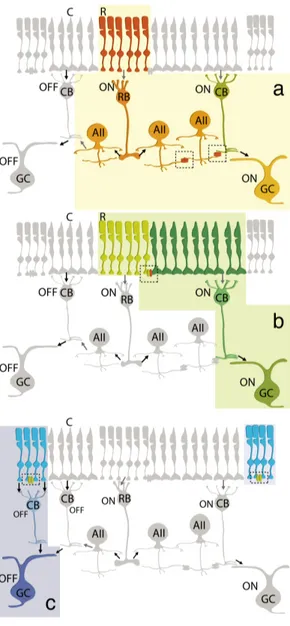
In mammals, rod and cone photoreceptors form chemical synaptic contacts with several bipolar cell types, so that the signals coming from photoreceptors are immediately fed into a number of parallel pathways. The existence of multiple pathways for the transmission of rod signals up to the final output of the retina is supported by studies of anatomy, electrophysiology, and psychophysics (Sharpe and Stockman, 1999; Bloomfield and Dacheux, 2001). The first rod pathway to be identified in the mammalian retina was that in which rod photoreceptors make a sign-inverting synapse onto rod bipolar cells (RBC), which thus depolarize in response to light. Since only one type of dedicated rod bipolar cell exists, rod signals would appear to have only a way to reach ganglion cells: via the RBCs and the postsynaptic amacrine AII cells (Fig. 1.2.2-1, panel a). However, in the
‘90s it was shown in the rabbit that rod signals can bypass RBCs and reach ganglion cells through gap junctions that connect rods to cones (DeVries and Baylor, 1995) (Fig. 1.2.2-1, panel b). Electrical connections between rods and cones had been documented both physiologically and anatomically already in the 70‘s, when ultrastructural analysis of the retina in several vertebrate s p e c i e s s h o w e d t h a t photoreceptors contact each other at specific sites, localized on their synaptic endings, without intervening glia. Raviola and Gilula (1973), applying to the retina the technique of freeze-fracturing, offered a precise structural characterization of these intercellular junctions, which were suggested to be functional by intracellular recordings in turtle (Baylor et al., 1971), and cat retinas (Nelson, 1977). Ultrastructural studies have revealed gap junctions between cones, as well as between cones and rods, in many other mammalian species, including mouse, guinea pig, ground squirrel (Smith et al., 1986; Ahnelt et al., 1990; Tsukamoto et al., 1992, 2001; Kolb et al., 1997; Tsukamoto et al., 2001; Lee et al., 2003; Feigenspan et al., 2004; Li and DeVries, 2004; O'Brien et al., 2004).
Figure 1.2.2-1. The three rod pathways in the
mammalian retina (black arrow: sign-preserving synapse; gray arrow: sign-inverting synapse). Adapted with permission from Bloomfield and Völgyi (2009).
Subsequently, in 1998 Ed Soucy et al. in the mouse demonstrated the presence of a third visual pathway for rod signals. Using a transgenic mouse whose retina lacks cones, they found that rod responses in OFF ganglion cells occurred even when the primary rod pathway was blocked and, since these mice have no cones (and thus no rod–cone gap junctions), this suggested the existence of a third rod pathway, in which rods make a sign-preserving synapse onto OFF bipolar cells, which, in turn, relay to OFF ganglion cells (Fig. 1.2.2-1, panel c) (DeVries et al., 1995). Some studies, subsequently described the presence of chemical synapses between rods and OFF bipolar cells in a number of species, including mammals (Hack, et al. 1999; Li et al., 2004; Völgyi et al., 2004; Pang et al., 2010, 2012). Nevertheless, only one in five rods in the mouse retina seems to be able to make a chemical synapse with an OFF bipolar cell, suggesting that this pathway may play a relatively limited role in scotopic signal transmission (Tsukamoto et al., 2001).
Since, it is known that gap junctions are expressed not only between rods and cones, but also between rods themselves, in mouse, rod–rod coupling could contribute to the third visual pathway by pooling scotopic signals into those sparse rods that do contact OFF-bipolars. The third rod pathway might thus play a role at dusk and dawn, when more photons are available than during starlight (Tsukamoto et al., 2001; Bloomfield and Völgyi, 2009). Indeed, it was shown that it has a lower light sensitivity compared to the other two rod pathways. Interestingly, OFF ganglion cells receiving convergent inputs from all three rod pathways or from the secondary and tertiary pathways together were never observed. It was later demonstrated that the third rod pathway survives in the Cx36-knockout mouse retina (see below for details), suggesting that rod-rod gap junctions do not rely on the Cx36 connexin isoform.
Cone photoreceptors are less sensitive to light than rod photoreceptors and have maximal absorbances at different wavelengths (see above). Cone signals cross the retina and reach ganglion cells via a more direct but diversified route. Cones synapse upon various cone bipolar subtypes rather than on a single type, classified by several parameters. On their scleral pole, characteristic morphological properties are the size of their dendritic field (midget, diffuse, and large-field diffuse) and the type of synaptic contact they form with cone pedicles (i.e. invaginating-ribbon synapses (imb), flat or basal junctions (fmb) or non-ribbon related basal junctions). As we have seen when discussing the primary
rod pathway, rod bipolar cells have a depolarizing response to light (Dacheux and Raviola, 1986), while cone bipolar cells can be either hyperpolarizing (OFF-center) or depolarizing to light (ON-(OFF-center) (Nelson and Kolb, 1983) (Fig. 1.2.2-2).
The ON and OFF bipolars express different glutamate receptor types (Miller and Slaughter, 1986). The ON- or OFF-center phenotype is determined by the nature of the postsynaptic glutamate receptors at the bipolar cell dendritic contacts with the cone. The OFF bipolar receptor is of the AMPA-kainate type, and it is an excitatory ionotropic glutamate receptor (iGluR). In contrast, the ON bipolar cells express mGluR6 metabotropic receptors (Numura et al. 1994; Vardi et al. 1997).
On the vitreal side, electron microscopy and 3D reconstruction of cone bipolar structure in the inner plexiform layer of the cat retina demonstrated that their axons make most of their ribbon output synapses with ganglion cell dendrites (Kolb, 1979). ON and OFF cone bipolars segregate their output synaptic contacts in different sublaminae of the IPL. OFF cone bipolar axons arborize entirely in the outer aspect of the IPL (sublamina a) connecting to OFF-center (center hyperpolarizing) ganglion cells. ON cone bipolars, in contrast, arborize in the inner aspect of the IPL (sublamina b) contacting ON-center (center depolarizing) ganglion cells. Light steps evoke in cone bipolar cells either sustained or transient responses (DeVries, 2000). Thus, a major functional subdivision between cone bipolars is that of ON-sustained, ON-transient,
OFF-sustained, OFF-transient.
One major difference between the circuitry of the cone compared to the primary rod pathway, in mammals, is that cone bipolar cells make direct synapses with ganglion cell dendrites, without the need for intermediate amacrine cell circuitry (Fig. 1.2.2-2). Moreover, fewer cones converge onto cone bipolars than rods onto rod bipolars, and then only a
Fig. 1.2.2-2. ON- and OFF- pathways of cone signals
(black arrow: sign-preserving synapse; gray arrow: sign-inverting synapse). Adapted with permission from Bloomfield and Völgyi (2009).
relatively small number of cone bipolar cells converge onto their ganglion cells. These differences are directly related to the requirements of cone vision for high spatial acuity during daytime, and of rod vision for high photon catch and amplification of scarce light at night. It is interesting to note that, according to visual pigment phylogenetics, cones probably preceded rods, a fact that may explain why the three rod pathways feed into the circuitry of cones (Vinnikov, 1982).
1.3 Gap Junctions and their Role in the Retina
GJs are a ubiquitous feature of the mammalian brain, and while researchers have been gaining an appreciation for their physiological complexity over the last several decades, many of their functional roles within identified neural circuits
Figure 1.3. Simplified schema of gap junctions: hexameric hemichannels cross the plasma
remain elusive. All five primary neuronal cell types in the retina (photoreceptors, horizontal, bipolar, amacrine and ganglion cells) make GJ-mediated synaptic connections, as shown mainly with morphological techniques, providing an approachable model system for exploring the roles that GJs play in neural signal processing. Evidence based on recordings of retinal output signals have provided support for the view that electrical synapses are essential in the processing of visual information.
Gap junctions (GJs) are the morphological substrate of electrical synapses. They are composed of two hemichannels (called connexons), each composed of 6 transmembrane protein subunits, named connexins, that are perpendicularly oriented to the cells’ membranes to form a central pore. Each connexin is in turn formed of 4 transmembrane domains, two extracellular loops, one intracellular loop, carboxyl– and amino–terminals (both in the cytoplasm). Therefore, GJs are composed of two connexons that link across the extracellular space and form a channel that connects the cytoplasms of two neighboring cells. Gap junctional channels can consist of two identical connexons (homotypic channels) or of connexons of different subunit compositions (heterotypic channels). GJs have a dual role, as they both mediate the propagation of electrical signals, as well as enable molecules up to 1000 Da to permeate (Fig. 1.3). The diffusion of small molecules through gap junctional channels has been exploited in the past, and is still used today, to investigate coupling: fluorescent dyes such as Lucifer Yellow or non fluorescent tracers such as neurobiotin have been
used
to collect morphological evidence about coupling between neurons (e.g. Hampson et al., 1994), with the former referred to as dye coupling and the latter as tracer coupling (Hartveit and Veruki, 2012). This dual role adds a layer of complexity beyond that of chemical synapses, and they are now recognized as short and long-term regulatory targets of endogenous systems, in both physiology and disease (e.g. Landisman and Connors, 2005). Connexin isoform 36 (Cx36) is critically involved in neuronal responses to injury (Wang et al., 2012; Belousov and Fontes, 2012). While in the brain the role of GJs in disease is currently a very active field, research in the retina has focused almost exclusively on their role in visual processing. Isoform 36 is widely expressed in the retina (Fig. 1.3.1) and two studies found that this isoform is involved in response to injury also in retinal tissue (Striedinger et al., 2005; Pashcon et al., 2012). These aspects will need to be addressed in greater detail in the future.1.3.1 Connexin 36 is the Major Isoform in the Retina
In the mammalian central nervous system, GJs and hemichannels are variously distributed among cell types. They are regulated at several levels, from transcription to gating. Connexin isoform 36 (Cx36) is the best characterized and predominant connexin in the CNS. Mills and colleagues (2001) performed an extensive investigation of the localization of Cx36 in Rabbit retina. They labeled the processes of AII amacrine cells, a critical interneuron in the rod pathway, and by image analysis found that Cx36 is primarily located at dendritic crossings between overlapping AII amacrine cells. This finding suggested that Cx36 may participate in homotypic gap junctions between pairs of AII amacrine cells. In addition, it was shown that Cx36 colocalized with contacts between AII amacrines and ON cone bipolars, suggesting that also heterotypic GJs are present. Thus, it was clear that Cx36 was an important component of the signaling in the primary rod pathway. Deans et al. (2001) generated mice in which the Cx36 coding sequence was replaced with histological reporters. Analysis of reporter distribution in the retina of heterozygous animals confirmed expression of Cx36 by AII amacrine cells, but also demonstrated expression of Cx36 protein in outer plexiform layer, as well as in two kinds of cone bipolar cells and a small number of cells within the ganglion cell layer. Later, it was shown that also mammalian cone photoreceptors express Cx36 both at cone-cone and rod-cone-cone junctional contacts (Feigenspan et al., 2004; O’Brien et al., 2012).
Deans et al. (2002) produced a direct functional proof that Cx36 is critical for propagation of rod signals across the mammalian retina, by generating a retina-specific Cx36 knockout mouse. Disruption of electrical synapses was tested by intracellular injection of Neurobiotin tracer. Extracellular recording from ON ganglion cells in these animals found that both the primary and secondary rod pathways were absent, implying that AII-AII amacrine and rod-cone gap junctions require Cx36. In vivo electroretinographic (ERG) recordings compared wild type with Cx36 knockout mice (Abd-El-Barr et al., 2009). Larger b-wave amplitudes were observed in the wild type, arguing for the presence of significant levels of rod-cone coupling. These data seem to suggest that junctional coupling mediated by Cx36 at various retinal levels, is important for visual processing.
Importantly, Cx36 shows a marked potential for modulation. An early study on neuroblastoma cell lines (Neuro2A and RT4-AC) and primary cultures of hippocampal neurons, both transfected with a Cx36-enhanced green fluorescent protein (EGFP) expression vector, revealed that junctional currents increased substantially over time during whole cell patch clamp recordings from pairs of coupled cells (Zoidl et al., 2002), a feature that has not been reported for any of the other connexins. Corsso et al. (2012) recently showed that injection of specific peptides corresponding to sites of binding and phosphorylation of CaMKII on Cx36 (Alev et al., 2008) lead to a loss of this “run-up” activity. The same effect was obtained by deletion of corresponding CaMKII binding and phosphorylation sites. According to these results the interaction of Cx36 with
Figure 1.3.1 Sites of gap
junctional contacts between retinal neurons (coloured ovals represent GJ hemi-channels). a) Both hemi-channels of the GJs that couple neighbouring cones (C) express Cx36. b) In rod (R)-cone GJs, only the hemichannel on the cone side contains Cx36; the connexin on the rod side remains unknown. c) The type of connexin in rod–rod GJ is also unknown. d) Horizontal cell (HC) dendrites are extensively coupled. In mammals, axonless horizontal cells express Cx50 whereas axon-bearing horizontal cells express Cx57. e,f) AII amacrine cells form two types of GJ. Those between AIIs seem to be homotypic and comprised of homomeric hemi-channels containing Cx36 e) By contrast, gap junctions between AII amacrine cells and ON cone bipolar cells (CB) can be homotypic or heterotypic, with the AII cell hemi-channels containing Cx36 and the cone bipolar cell hemichannel containing either Cx36 or Cx45. g) Ganglion cells (GCs) are extensively coupled to each other and/or to neighbouring amacrine cells (AC). To date, ganglion cell GJs have been reported to contain Cx36 or Cx45. Figure and legend adapted with permission from Bloomfield and Volgy (2009).
CaMKII may confer a high degree of functional plasticity upon the electrical synapse.
1.3.2 Light Adaptation and Gating of Gap Junctional Coupling
During the day/night cycle, the retina continuously adjusts its signal gain, so as to avoid saturation our visual system and operate over a light intensity range that covers more than 9 orders of magnitude (Rodieck, 1998). In the process, the balance is shifted between optimal contrast detection and acuity in daylight, and high light sensitivity in darkness. Light adaptation mechanisms operate at all levels of retinal processing, from photoreceptors up to ganglion cells, and are known to depend not only on the light stimulus itself, but also on intrinsic circadian rhythms in retinal neurons. Adaptation downstream of phototransduction has been termed ‘network adaptation’, and its main effectors are two key players in retinal signal processing: ion channels and synaptic transmission.
Cell-to-cell electrical coupling, mediated by gap junctions, is an important component of the flow of visual signals through the retina, and is the target of complex regulatory systems in dark/light adaptation (Bloomfield and Völgyi, 2009). It is known that gap junctional coupling in the retina is plastic, similarly to other systems. In particular, a number of studies have shown that coupling strength between retinal neurons changes with illumination. Ambient light regulates the conductance of GJs between horizontal cells and AII amacrine cells (Fig. 1.3.2) (Mangel and Dowling, 1985; DeVries and Schwartz, 1989; Bloomfield et al. 1997; Xin and Bloomfield, 1999; Bloomfield and Völgyi, 2004). More limited evidence points to a light-dependent modulation of coupling between rod and cone photoreceptors (Yang and Wu, 1989; Heikkinen et al., 2011). The general consensus is that in mammalian retinas photopic ambient light promotes a decrease in coupling between neurons, suggesting a rerouting of visual signals away from electrical pathways as we move from night to day. However, this simplistic view is complicated by a number of observations, the primary being those of Bloomfield and Völgyi (2004) in AII amacrine cells, and of Xin and Bloomfield (1999) in horizontal cells. They found that both in darkness (scotopic conditions) and daylight (photopic conditions) these neurons have a low level of mutual coupling, while in dim light (mesopic conditions)
coupling is strong (Fig. 1.3.2). This ‘bell’ shaped regulation of coupling between amacrine cells has been suggested to optimize signal flow in relation to biological noise. Hu et al. (2010) reported an unexpected increase in daylight of the tracer coupling of α-GCs to neighboring ganglion cells and amacrine cells, contrary to the prevailing view of uncoupling by light. This increased coupling seems to be accompanied by a rise in correlated spike activity between neighboring α-GCs, a process that would affect the capacity and efficiency of information flow across the optic nerve.
It must be noted that much of the data on retinal neuron coupling, and its modulation, have been obtained using tracer substances such as neurobiotin. This has provided evidence that histologically–identified GJs represent a viable path for the passage of small molecules between neurons, leaving open the question about their effectiveness as mediators of biologically–relevant electrical signals. As a result, the strength and functional impact of electrical coupling in many retinal neurons remains unclear. This point is particularly relevant for the coupling between mammalian photoreceptors.
Figure 1.3.2. Complex modulation of junctional coupling between AII amacrine cells by ambient
light. Dots below represent the degree of tracer diffusion from a single injection site. Modified with permission from Bloomfield and Völgyi (2009). Photo by P. Prato.
1.3.3 Endogenous Neuromodulators Coordinate Network Adaptation
Network light adaptation is controlled both by mechanisms local to the specific neuron involved, and by global neuromodulatory signals. Two neuromodulators, dopamine (DA) and melatonin, are relatively well established players in coordinating retinal network adaptation, although other transmitters have also been implicated (e.g. NO, GABA, Adenosine). They are synthesized within the retina and released in a paracrine fashion, thus acting on longer time scales (minutes to hours) compared to adaptation in the photoreceptor outer segment, and are controlled both by ambient light levels and intrinsic retinal circadian clocks. Their release by photoreceptors (melatonin) and amacrine cells (DA) (Menger et al., 1998, Doyle et al., 2002), is maintained in antiphase by a mutual inhibitory interaction. DA, in particular, acting through D2-like receptors on the photoreceptor, inhibits melatonin synthesis (Tosini and Dirden, 2000). This results in melatonin being high at night and dopamine high in daytime. DA and melatonin are thought of playing opposing roles in the regulation of retinal light-adaptive physiology: the former functioning as a humoral signal for daylight and promoting network light adaptation, while the latter having dark-adaptive effects (Tosini et al., 2008).
Synthesis and release of DA is under the control of intraretinal clocks, since cyclic oscillations in its levels can be observed also in isolated retinas cultured in constant darkness (Ruan et al., 2008). Daily rhythms are a ubiquitous feature of living systems. They are not just passive consequences of cyclic fluctuations in the environment, but instead originate within the organism. In mammals, including humans, the master pacemaker controlling 24-hour rhythms is localized in the suprachiasmatic nuclei of the hypothalamus, to which the intrinsically photosensitive retinal ganglion cells (ipRGCs) send information about ambient brightness via the direct retinohypothalamic tract. The circadian clock is responsible for the temporal organization of a wide variety of functions, ranging from sleep and food intake, to physiological measures such as body temperature, heart rate and hormone release. The retinal circadian clock was the first extra-SCN circadian oscillator to be discovered in mammals and several studies have now demonstrated that many of the physiological, cellular, and molecular rhythms that are present within the retina are under the control of a retinal circadian clock, or more likely a network of hierarchically organized circadian clocks that are present within this tissue (Tosini et al., 2008).