A
Al
l
ma
m
a
M
Ma
at
te
er
r
S
St
tu
u
di
d
i
or
o
ru
um
m
–
–
U
Un
n
iv
i
ve
er
rs
si
i
tà
t
à
d
d
i
i
B
Bo
ol
l
o
o
gn
g
n
a
a
DOTTORATO DI RICERCA IN
SCIENZE BIOMEDICHE
Ciclo XXIX
Settore Concorsuale di afferenza: 05/H1 Settore Scientifico disciplinare: BIO/16
ADVANCED IN VITRO MODELS TO STUDY THE CROSS- TALK
BETWEEN METASTASES AND BONE MICROENVIRONMENT:
WHICH ROLE FOR OSTEOPOROSIS?
Presentata da: Melania Maglio
Coordinatore Dottorato:
Chiar.mo Prof. Lucio Ildebrando Cocco
Relatore:
Chiar.mo Prof. AlbertoMaria Martelli
Tutor:
Dott.ssa Milena Fini“Research is to see what everybody else has seen,
and to think what nobody else has thought”
Acknowledgments
Special thanks to my tutor Dr. Milena Fini, which gave me the opportunity to follow my research activity at the Rizzoli Orthopaedic Institute, encouraging me with great enthusiasm since the first steps of my PhD course.
Thanks to my supervisor Prof. Alberto Maria Martelli, for the invaluable support in my scientific activity and unfailing care showed me through the years.
Thanks to all the researchers of the Preclinical and Surgical Studies Laboratory and of the BITTA Laboratory of the Rizzoli Orthopaedic Institute.
Contents
CHAPTER 1- INTRODUCTION
1.1 The bone remodeling tale……….…..22
1.2 Skeletal metastases. The great migration to bone……….….27
1.3 From breast to bone: looking for the congenial soil………...……...33
1.4 Osteoporosis……….…..37
1.5 Osteoporosis and bone metastases: the dangerous liason……….….41
AIM OF THE PROJECT
………...45CHAPTER 2 - MATERIALS AND METHODS
2.1 Assessment of rat bone cultures 2.1.1 Calvaria and femoral condyle bone culture- Experimental set up ……….492.1.2 Assessment of bone culture viability………..50
2.1.3 Bone culture microtomography………..50
2.2 Characterization of rat breast cancer cell line (MRMT-1)
2.2.1 MRMT-1 cells proliferation and viability………...52
2.2.2 Wound healing assay………...55
2.2.3 Assessment of activated pathway………53
2.2.3.1 Protein quantification………..53
2.2.3.2 Western Blot………..…..54
2.2.2 MRMT-1 cells sensitivity to doxorubicin ………..58
2.2.2.1 Experimental cultures set up ………..58
2.2.2.2 Evaluation of cells viability……….…58
2.2.3 Statistical analysis………...59
2.3 Co-Culture of MRMT-1 cells and Osteoclasts 2.3.1 Conditioned media preparation………...…60
2.3.2 Isolation of mononuclear cells (PBMCs) and differentiation into osteoclasts (OCs)…60 2.3.3 PBMCs viability………..62
2.3.4 OCs differentiation………..62
2.3.5 OCs synthetic activity……….62
2.4 Assessment of a 3D Model of Rat Bone Metastases In Vitro
2.4.1 Experimental set up……….……64
2.4.2 Statistical analysis……….……..65
2.5 In Vivo Induction of Osteolytic Metastases in osteoporotic rats 2.5.1 Surgical procedure ……….66
2.5.2 Imaging evaluation of lesions development with Positron Emission Tomography (PET)………68
CHAPTER 3- RESULTS
3.1 Assessment of rat bone cultures……….713.2 Characterization of MRMT-1 cells………76
3.3 Co-Culture of MRMT-1 cells and OCs………..82
3.4 Assessment of a 3D Model of Rat Bone Metastases In Vitro………88
3.5 In Vivo Induction of Osteolytic Metastases in osteoporotic rats………92
List of Figures
INTRODUCTION
Figure 1.1: The bone remodeling cycle
Figure 1.2: Histological section of sheep tibia in which the different staining of old and newly fomed bone is appreciable. Fast green staining, 20x magnification.
Figure 1.3: Physiological and pathological stimuli in bone remodeling Figure 1.4: The metastatic journey
Figure 1.5: Vicious cycle of osteolytic bone metastases
Figure 1.6: Images acquired by microCT of proximal epiphisis of tybia from a) healthy and b) osteoporotic rats
MATERIALS AND METHODS
Figure 2.1: Experimental set up of 3D culture of calvaria segments and MRMT-1 cells Figure 2.2: Injection of MRMT-1 cells in rat tibia trough Hamilton syringe
RESULTS
Figure 3.1: Viability trend of calvaria bone cultured up to 14 days evaluated through Alamar Blue assay
Figure 3.2: 3D reconstructions by microCT and histological images of calvaria bone segments after in vitro culture. Hematoxylin/Eosin staining
Figure 3.3: Viability trend of femoral condyle bones cultured up to 14 days evaluated through Alamar Blue assay
Figure 3.4: Fold change in femoral condyles mass during in vitro culture
Figure 3.5: Histological section of femoral condyles after 14 days of culture. Hematoxylin/Eosin staining
Figure 3.6: Microscope images of MRMT-1 cells in culture
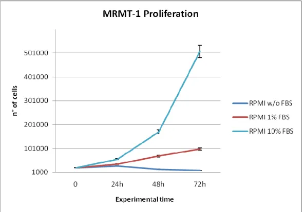
Figure 3.7: MRMT-1 trend proliferation in different culture conditions
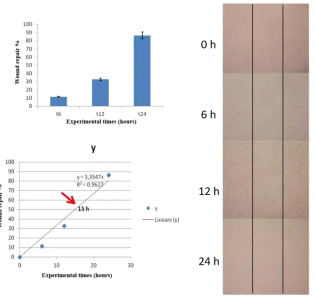
Figure 3.8: Results of wound healing assay: histograms graph of percentage of wound healing recovery, linear regression for assessing timing of 50% of recovery and microscope images of wound healing progression
Figure 3.9: Panel of pathways activated in MRMT-1 cell line Figure 3.10: Doxorubicin chemical structure
Figure 3.11: Effect of different Doxorubicin concentrations on MRMT-1 cell line
Figure 3.13: TRAP staining images of PBMCOVX and PBMCSHAM in CTR-, CTR+, CM-,
CM+ and TW culture conditions at one and two weeks [Salamanna et al, 2016]
Figure. 3.14: Protein content of PBMCOVX and PBMCSHAM in CTR-, CTR+, CM-, CM+ and
TW culture conditions at one and two weeks [Salamanna et al, 2016]
Figure 3.15: Box plot graph of viability values distribution of SHAM, OVX, SHAM+MRMT-1 and OVX+MRMT-1 groups at 7 and 14 days of culture
Figure 3.16: Viability trend of SHAM, OVX, SHAM+MRMT-1 and OVX+MRMT-1 groups at 7 and 14 days of culture
Figure 3.17: Western blot of MRMT-1 protein content after sham and osteoporotic culture Figure 3.18: Histological images of calvaria segments after culture with MRMT-1 cells. Hematoxylin/Eosin staining
List of Abbreviations
BMU- Basic Multicellular Unit OCs- Osteoclasts
OBs- Osteoblasts Ca- Calcium IL- Interleukin
TNF-α- Tumor Necrosis Factor α IGF- Insulin like Growth Factor CSF- Colony Stimulating Factor EGF- Epidermal Growth Factor PDGF- Platelet Derived Growth factor ALP- Alkaline Phosphatase
COLLI- Type I Collagen OC- Osteocalcin
OP- Osteopontin ON- Osteonectin
FGF- Fibroblast Growth Factor
RANKL- Receptor activator of nuclear factor kappa-B ligand OPG- Osteoprotegerin
RANK- Receptor activator of nuclear factor kappa-B EPH- ephrinB2
EphB4- protein ephrinB2 receptor B4 S1P- sphingosine 1- phosphate
VEGF- Vascular Endothelial Growth Factors TGF- - Transforming Growth Factor BMP- Bone Morphogenetic Protein HIF- 1- Hypoxia Induced- Factor
EMT- Epithelial To Mesenchymal Transition
VEGFR- Vascular Endothelial Growth Factors receptor ECM- Extracellular matrix
CTS- Cathepsin
MMP- Metalloproteinase
DKK1- Dickkopf-related protein 1
SDF1/CXCL12- Chemokine Stromal Cell Derived Factor-1 IHH- Indian Hedgehog
PTHrP- Parathyroid hormone-related protein PGE2- Prostaglandin
ER α - Estrogen Receptor α OP- Osteoporosis
BMD- Bone Mineral Density SD- standard deviation Vit.D- Vitamin D
VDR- Vitamin D Receptor gene
CYP2R1- Vitamin D 25-hydroxylase gene
NF- κB -Nuclear Factor kappa-light-chain-enhancer of activated B cells BCAR1- Breast cancer anti-estrogen resistance protein 1
TRAF- TNF receptor-associated factor FSH- Follicle-Stimulating Hormone CTGF- Connective Tissue Growth Factor BMSCs- Bone Marrow Stromal Cells
RPMI- Roswell Park Memorial Institute BSA- Bovine Serum Albumine
DMEM- Dulbecco’s Modified Eagle Medium FBS- Fetal Bovine Serum
°C- Celsius
MicroCT- Microtomography
SDS-PAGE- Polyacrylamide Gel in Sodium Dodecyl Sulphate GLB- Gold Lysis Buffer
AKT- Protein Kinase B
PKR- Protein kinase RNA-activated SHAM- Healthy
OVX- Ovariectomized FCS- Fetal Calf Serum
TRAP- Tartrate-Resistant Acid Phosphatase RGB- Red- Green- Blu
PET- Positron Emission Tomography
18
F-FDG- 18F-Fluorodeoxyglucose
S6RP- Phosphorylated S6 ribosomal protein Bcl2- B-cell lymphoma 2
CHAPTER 1-
INTRODUCTION
1.1 THE BONE REMODELING TALE
Under the deceptive appearance of a static tissue, bone hides a complex remodeling system which is the base of its unique properties. The classical study of bone remodeling arguably takes its first steps from a work of 1969 by Harold M. Frost [Frost et al, 1969], which outlined the main actors and key passages of the process through the use of tetracycline labeling. Particularly remarkable it is the introduction of the “Basic Multicellular Unit” (BMU), intended as the set of cellular components – osteoclasts (OCs), osteoblasts (OBs) and ostecytes- involved in the remodeling process, which is in turn described as a cycle as portrayed below:
Figure 1.1: The bone remodeling cycle
Indeed, it is now well known that bone turnover is a well- balanced fluctuation between degradation of pre- existing tissue by OCs (resorption) and replacement with newly- formed one through OBs activity (formation). [Adachi et al, 2009] (Figure 1.2)
The phase of activation is related to both a systemic and local metabolic activity, usually in response to mechanical loading and microfractures (whose cellular mediators, the osteocytes,
part to the homeostasis of serum calcium (Ca+) and minerals concentration. [Raisz et al,
1999; Henriksen et al, 2009]
Apart from lifestyles, diet, genetic factors and microenvironment, under physiological conditions, there are many local and systemic factors which can control and regulate bone remodeling: classical cytokines and growth factors are Interleukins (IL) like IL-1, 6, 9, 10, Tumour Necrosis Factor α (TNF-α) 1 and 2, Insulin like Growth Factor (IGF) I and II, Colony Stimulating Factor (CSF), Epidermal Growth Factor (EGF), Platelet Derived Growth factor (PDGF). [Hadjidakis et al, 2006]
The mechanism of bone regulation starts also from molecules expressed primarily by the bone cells involved in the process. Alkaline Phosphatase (ALP), Type I Collagen (COLLI), Osteocalcin (OC), Osteopontin (OP) and Ostenectin (ON), key regulators in bone matrix synthesis and mineralization, are expressed by mature OBs whose differentiation is in turn stimulated by Wnt signaling pathway, also involved in OCs maturation. The life cycle of OBs is closed by their transformation in osteocytes, which act on bone formation through the expression of, namely, Dentin Matrix Acidic Phosphoprotein 1 (DMP1), Fibroblast Growth Factor (FGF) 23 and Sclerostin. [Eiksen et al, 2010]
A key role is played by the system of Receptor activator of nuclear factor kappa-B ligand (RANKL) and its decoy receptor Osteoprotegerin (OPG). RANKL (member of TNFs family) is expressed by OBs and stromal cells and induce OCs formation via binding the Receptor activator of nuclear factor kappa-B (RANK), which is in turn expressed by pre- OCs, B and T cells, dendritic cells and fibroblasts. The binding between RANKL/RANK stimulates OCs activation and survival, promoting also their adhesion to the bone and their osteolytic activity. These activities are inhibited by OPG, expressed by OBs and bone marrow cells, via apoptosis of OCs. [Khosla et al, 2001] (Figure 1.3)
Figure 1.2: Histological section of sheep tibia in which the different staining of old and newly fomed bone is appreciable. Fast green staining, 20x magnification.
The coupling activity of OCs and OBs in regulation of bone remodeling is also highlighted by the presence of other cross talks, like the one between the transmembrane protein EphrinB2 (EPH) and its receptor B4 (EphB4), expressed respectively on OBs and OCs. In this pathway it is also involved the Sphingosine 1- Phosphate (S1P), which, when secreted by OCs, activates EphB4 signaling, promoting OBs differentiation and inhibiting OCs activity.
[Ryu et al, 2006].
Among the elevated metabolic activity, another important feature of bone is being a highly vascularized tissue. Vascularization represents a critical step in the phase of bone remodeling, as suggested by its indefeasibility, during the development of human organism, in the correct endochondral ossification, in which blood vessels spread through the cartilaginous matrix, mediated by the Vascular Endothelial Growth Factors (VEGF), promotes and sustains bone formation. VEGF surely plays a pivotal role in angiogenesis and neovascularization; its
activity in the bone tissue is intuitive when considering that many factors involved in bone remodeling, including mechanical stress, are also responsible for the control of the synthesis and activity of VEGF, namely the transcription factor Osterix, growth factors like Transforming Growth Factor (TGF-) and FGF 2, inflammatory cytokines like IL-1, 6 and 8, Prostaglandin E1 and 2, Bone Morphogenetic Protein (BMP), mostly expressed by cell of osteoblastic lineage [Hu et al, 2016].
Although the mechanisms that regulate the role of angiogenesis in bone remodeling have not yet been put fully into focus, it is more and more evident that the link between angiogenesis and osteogenesis lives in the hypoxic environment which characterizes the bone tissue. In fact under hypoxic condition OBs, as well as tumor cells, are stimulated to express the hypoxia induced- factor (HIF-1 alfa), transcription factor which promote VEGF-A mRNA expression, augmenting among angiogenesis also bone formation. This feature appears particularly interesting when considering it is shared with cancer, marking a first point in the affinity between tumor cells and bone. [Schipani et al, 2009]
1.2 SKELETAL METASTASES. THE GREAT MIGRATION TO BONE
The skeleton is the organ more frequently affected by metastases, especially from breast and prostate cancers, whom it is possible to attribute almost the 80% of all bone metastatic diseases; this reflects both the high incidence that the relatively long clinical course of these tumors. Among breast and prostate, even other several common solid tumors, such as tyroide, lung and kidney cancers show tropism for the bone, although with a lower incidence. [Buijs
et al, 2009]
Bone metastases have been commonly classified for a long time as osteolytic or osteoblastic, depending on whether the detected alteration was an excessive loss or apposition of bone. The morbidity and the complications associated with metastatic bone disease, like bone pain, pathologic fractures, spinal cord compression, leukoerythroblastic anemia, bone deformity and hypercalcemia, impair significantly patients’ quality of life. [Roodman et al, 2004] In metastatic patients, long term survival dramatically decreases, and treatments are mostly palliative; among these, surgery still remains the gold standard, often associated with chemotherapy, radiotherapy, therapies with drugs acting on bone remodeling (i.e. biphosphonates), hormonal therapy, thermal ablation and electrochemotherapy [Coleman et
al, 2010]
Although the understanding of the precise mechanisms regulating the predilection of some cancers to bone remains still incomplete, it is now increasingly being recognized that the unique characteristics of the bone microenvironment provide signals that lead to a selective growth advantage for cancer cells and, probably, the resistance to some therapeutic treatments.
However, the study of more effective therapies and the understanding of the underlying mechanisms of tumor escape and resistance to drugs is still an open field. In the last years,
the results highlighting the influence that microenvironment exerts on tumor cells behavior have brought out new scenarios of complexity, from the onset of cancer to metastatic process. It is now clear that the heterogeneity of metastatic patterns is related to the biological cellular and molecular features of both starting cancer cells and metastatized tissues and also the traditional classification of metastases in ostelolytic and osteoblastic seems to be overcome by the evidence of “mixed” metastatic lesions, with hallmarks common to both groups. This reflects the two extremes of a continuous spectrum of changes in bone remodeling during the development of metastasis. [Coleman, 2001]
Several hypotheses have been discussed to speculate the mechanism driving cells from primary tumor to bone, attributing to different factors and conditions some “attractive” properties which might clarify the tropism to the bone.
The multistep process that lead cancer cells towards the road to bone is characterized by the ability of cells to adapt, surviving in many different conditions, and by the affinity that they show for the target organ. (Figure 1.4)
This kind of similarity can be expressed in terms of osteomimetic abilities more than capability to secrete proteins or express genes suitable for bone colonization. The first and more iconic step in this long route is the epithelial to mesenchymal transition (EMT), to which cancer cells undergo in the passage to bloodstream. This transformation, which seemed at first to be the starting point of the metastatic process, actually is more and more recognized as the double face of the same item. In fact, the capability of cancer cells to disseminate and metastatize far away from the primary tumor is marked by the loss of some proteins related to cell-cell junctions and which are typical markers of epithelial cells, namely E-cadherin and cytokeratin. On the other hand, this phenomenon is subverted once reached the target organ, considering that the ability to invade and colonize is mediated by the expression of proteins
characteristics of epithelial cells, and to this end, it occurs the reverted transition named mesenchymal-to-epithelial transition. [Kan et al, 2016]
Figure 1.4:The metastatic journey
Surely, once in the vascular bed, cancer cells can find stimuli driven by factors involved in the various aspects of bone remodeling, namely angiogenesis, minerals release (primary Ca) and high concentrations of growth factors. [Gupta et al, 2006] Some factors are then related to the peculiar characteristics of bone microenvironment, such as low oxygen level and acid pH, which can create a favorable environment for cancer cells colonization and onset of metastases. [Kingsley et al, 2007] The pivotal role of vascular component and blood flow anatomy in this framework is indicated by the preference of cancer cells to metastatize to high-vascularized bone district, such as bone marrow of long bones, pelvis, sternum, ribs and vertebrae.
Studies on animals and on cadavers have shown an involvement of the vertebral venous plexus that could help to explain the tropism of these tumors to the bone. In fact, the venous blood coming from the pelvis and from the breast not only flows into the vena cava, but also directly into the vertebral venous plexus, especially in condition of high intrathoracic or intraddominal pressure. [Kaplan et al, 2006] The tropism for bone containing red marrow seems to have found another explanation in the studies on the relationship between the metastatic cells and the hematopoietic progenitor cells present in the bone marrow. In fact, the combination of chemotactic and adhesion molecules in the endothelium of bone marrow has proved to be particularly favorable to colonization by cancer cells. Recent studies have shown that after about two weeks after implantation of the primary tumor, and before the invasion phase, hematopoietic stem cells expressing the receptor for VEGF (VEGFR-1) are organized to define the contours of what will be the site of metastasis. This structure, called "pre-metastatic niche", works as a physiological niche, in which the VEGFR-1 + cells can maintain the expression of stem cell markers (c-kit, CD34) without differentiating. The signal mediated by VEGF is a critical factor because it stimulates angiogenesis, the growth of the primary tumor and of the metastasis dormant activity. In addition, the VEGFR can induce MET and tumor invasion. [Kaplan et al, 2005]
Finally, it is known that cancer cells can form emboli, which remain blocked in the capillaries beds within the bones. Once the clot or the cancer cells have reached the skeleton, the features of the microenvironment favor its survival. Hypoxia is a hallmark of all solid tumors, whose rapid proliferation prevents adequate angiogenesis and thus a complete perfusion. The hypoxic microenvironment in the bone marrow regulates hematopoiesis. The cells that metastasize to the bone not only are able to survive in low amounts of oxygen, but also are further advantaged, both in terms of proliferation and in terms of resistance to radio / chemotherapy treatments.
The presence of an acid environment also seems to promote the establishment of metastases. Extracellular pH exerts an important effect in bone remodeling, and in this sense, is carefully regulated, leading to an increase in the formation of resorption pit and at the same time to a reduced OBs mineralization and bone formation.
The influence of pH is so pronounced that even a mild but chronic acidosis can strongly stimulates osteoclasts activity up to cause a marked bone loss. [Arnett, 2003] The link between condition of acidosis and tumor invasion seems to live in the alteration of glycolytic metabolism, which characterize many tumors. In fact, even in presence of oxygen, cancer cells exhibit reduced glycolytic pathway, that with lactic acid production and lower pH, give them a more aggressive phenotype and increase their proliferation. Differently, in the same condition of acid- mediated toxicity, normal cells undergo to necrosis and apoptosis, devoiding of the adaptability of cancer cells. At the same time, acid microenvironment leads to extracellular matrix (ECM) degradation and release of Cathepsin B (CTSB), metalloproteinase (MMP) and other proteolytic enzymes with contribute to metastases onset.
[Gatenby et al, 2006].
Once invaded bone, cancer cells start to interact with resident cell populations, as bone cells, hematopoietic stem cells and so on, expressing osteomimetic factors and adhesion molecules so modifying their genotypic and phenotypic profile. These changes might be induced by the interaction with the new microenvironment, whose mineral component makes bone surface five or six times harder than those of soft tissues and consequently more resistant to the action of proteolytic enzymes secreted by tumor cells. [Coleman et al, 2010]
The adhesive interaction with bone matrix and medullar stroma trigger cancer cells to produce angiogenic factors and resorptive molecules that further promote tumor growth. It is more and more evident that the development, more than the onset of bone metastases,
ovariectomy on the establishment of bone metastases in a mouse model didn’t evidenced any difference with sham group in terms of migration of cancer cells to the metastatic site; however, interestingly, cell colonization seemed to be enhanced in osteoporotic environment, with an increase in the expression of genes involved in metastatic spread such as RANKL, Dickkopf-related protein 1 (DKK-1), MMP- 9 and Cathepsin K. Specularly, the same trend can be observed in mice undergone to orchiectomy, with an increased aggressiveness in tumor behavior, secondary to a more intense osteoclastic resorptive activity. [Kan C et al,
1.3 FROM BREAST TO BONE: LOOKING FOR THE “CONGENIAL SOIL”
Breast cancer represents one of the primary tumor that more frequently metastatize to bone, with almost the 70% of patients affected by this type of tumor showing distant skeletal metastatic lesions. For a long time metastases from breast cancer have been classified as osteolytic but now it is increasingly recognized that the old dichotomy between osteolytic and osteoblastic metastases has no more reason to be, as bone metastatic lesions frequently shows mixed signs of unbalanced activities of bone resorption and formation.
The first attempt to find an explanation to the preferential harbor of some site to cancer cells, dates back to nineteenth century, with the study of Ernst Fuchs on the metastatic pattern of uveal melanoma; he first advanced the hypothesis that the metastatic process was not consequence of casualty but of some predispositions of both sites of start and arrival.
Thanks to the inspiration drown by this first suggestion, Stephen Paget started his systematic work that will be resulted in the formulation of the theory known as of “Seed and soil”
[Paget, 1889]. In the 1889, after the autoptic evaluation of 735 cases of breast cancer with
fatal outcome, he hypothesized that the colonization of an organ or a tissue by cancer cells is the result of the productive mutual interaction between tumor cells and the host microenvironment, with the famous claim “When a plant goes to seed, its seeds are carried in all directions; but they can only grow if they fall in congenial soil”.
A great input for breast cancer cells to head to bone is given by the high expression of molecules promoting the invasion and adhesion to the bone tissue, including the receptor tyrosine kinase ErbB2 (Human Epidermal Growth Factor Receptor 2), frequently amplified, which in turn stimulates the expression of CXCR4 (C-X-C Chemokine Receptor Type 4) increasing protein synthesis and inhibiting its degradation. The proliferation and migration of cancer cells is then stimulated by the strengthening of pathways SDF1/CXCL12 (Chemokine
cells. Once in the bone, the SDF1 expressed in specific vascular microdomains can promote transendothelial migration of breast cancer cells expressing CXCR4. The presence of CXCR4 + cells, once in the bone marrow, in synergy with RANKL, can further stimulate cell migration in response to SDF1 and RANK, locally produced. The integrin αvβ3, finally, allows the adhesion of cancer cells to the components of the bone matrix, thus promoting the development of osteolytic metastases [Rose et al, 2006]
A possible relationship between the metastatic tumor cells and stromal cells and OBs present at bone level can be found in the common expression of Cadherin 11, involved in the homophilic cell-cell interactions. It is believed that the preferential migration of breast cancer cells in the bone is due to the interactions mediated by cadherin between the two cell populations. It also seems to be involved in the formation of metastases, enhancing osteoclastogenesis [Tamura et al, 2008]
Finally, a pattern to describe bone remodeling in the presence of cancer cells was introduced in the early '90s and has been called "the vicious cycle of bone metastases" [Chen et al, 2010] It has been suggested that cancer cells metastatize and are then able to live and proliferate preferentially in the bone, thanks to their ability to express factors generally considered related to the bone. It is interesting that most of these osteomimetic factors are modulated by the same transcription factor, Runx2, considered being the key regulator in the commitment and differentiation of OBs. Runx-2 seems to respond to stimulation of the TGF-β activating the expression of Indian Hedgehog (IHH); this pathway, which also involves the cascade of Mitogen-Activate Protein Kinase (MAPK), leads to an increased osteoclastogenesis and osteolysis. [Hansen et al, 2007]
In the vicious cycle of bone metastases, bone destruction increases local levels of Ca, promoting tumor growth and the production of Parathyroid hormone-related protein (PTHrP). Breast cancer cells produce, or induce, IL-1, IL-6, IL-8, IL-11, Prostaglandin (PGE2),
M-CSF, TNF-α, which stimulate OCs formation, activation and migration. In particular, IL-8 may indirectly promote osteoclastogenesis by regulating positively the expression of RANKL on osteoblastic precursors. Conversely, the IL-11 promotes the maturation of OBs from mesenchymal progenitor cells. [Yin et al, 2005] Taken together, these data suggest that
PTHrP is the principal mediator of secondary osteolytic lesions in breast cancer and other solid cancers [Rose et al, 2006] (Figure 1.5)
As inferred from the nature of the osteolytic process, the bone microenvironment contains many proteases and, among these, the MMP family, made up of more than twenty zinc-dependent proteases, can degrade all components of the ECM. Tumor cells, OBs, OCs and endothelial cells produce MMPs [Chen et al, 2010] whose expression is regulated by the levels of TGF-β and TNF-α. MMPs, and in particular the protecapsin D, contribute to the proteolysis of collagen from the surface of osteoid before the attack of osteoclasts, facilitating the metastatic invasion through the removal of this physical barrier. [Gall et al, 2007]
As part of the breast carcinomas an important role is played by the presence of Estrogen Receptor α (ER α), being estrogens mitogenic to the cancer cells. Some studies suggest that TGF-β promotes the transcription mediated by the ER receptors which in turn stimulate the production of PTHrP and should thus have a role in the development and progression of metastatic breast cancer to bone [Chen et al, 2010]
Tumor progression, however, is also critically controlled by angiogenesis. OCs are adjacent to blood vessels growing in the Havers channels; metastases from breast cancer strongly express VEGF, and OCs express both VEGFR1 that VEGFR2. VEGF can promote osteoclastogenesis and the C-terminal region of the PTHrP may induce the expression of VEGF in bone cells. In addition, the VEGF can interact with monocytes to induce the differentiation into OCs [Kaplan et al, 2006].
In parallel to osteolytic lesions, there is a modulation of the function of OBs that induces necrosis and apoptosis. This is mediated by the production, by tumor cells, of IL-1, IL-11, TNF-α and, especially, of Fas ligand (FasL) which, interacting with its receptor Fas, expressed by OBs, activates caspases pathway.
1.4 OSTEOPOROSIS
It dates back to 1820 the appearance of the term “osteoporosis” (OP) to indicate a generic state of pathological alteration of bone. [Schapira D et al, 1992] Since then, many have been the attempts to find an exhaustive definition of OP, but the effort is challenging considering the manifold aspects emerging by the study of etiology and characteristics of this pathology. OP can be defined as a systemic disease affecting bone, clinically identified by a value of Bone Mineral Density (BMD) deviating of at least 2.5 standard deviation (SD) from the mean value of general healthy population (T score <-2.5 SD). Despite this clinical evidence, if it is true that people with such value of BMD are affected by OP, it’s not true the contrary, as not only the course of the condition can remain asymptomatic until the occurrence of the first fracture, but also many patients can suffer from a severe subverted skeletal condition while having a normal BMD index. These observations give reason of the difficulty of setting OP in a well-defined framework. [Salamanna et al, 2015]
The conventional classification of osteoporosis identify mainly two groups:
- primary OP, which assembles OP which occurs after establishment of menopause and the one related to advanced age, affecting the 30% of women;
- secondary OP, which is related to external factors, like unhealthy lifestyles and diet (excessive assumption of alcohol or fat, lack of physical activity, smoking or reduced exposure to sunlight) or to other pathological conditions which can directly or indirectly affect bone, involving between the 30% and 60% of male, and more than 50% of woman.
[Fini et al, 2012]
Among this classical division, it must be considered OP as a more heterogeneous pathology, in whose pathogenesis are also involved genetic factors, which evidence increases the level of complexity, adding to the various variables also the individual predisposition. In recent years
occur in genes involved in the related regulation pathways. Some studies in fact found a correlation between polymorphisms in vitamin D pathway genes, in particular in the gene encoding for Vit.D receptor (VDR) and onset of OP, ascribing to it a connection with lower BMD and consequent development of OP. Polymorphisms of the gene encoding for Vit.D 25-hydroxylase (CYP2R1) are proposed to correlate with alteration in Vit. D metabolism and consequent insufficient levels of Vit.D. This aspect appears particularly intriguing considering that low levels of Vit. D correlate with stimulation of immune system in terms of release of pro inflammatory molecules, TNF-α, resistin, and free fatty acids, and of activation of Toll-like receptors. In the light of the fact that OP can be considered a state of chronic inflammation, alteration of CYP2R1 seems to indirectly promote the osteoporotic condition.
[Bellavia et al, 2016]
OP has nowadays settled as one of the most relevant pathological conditions, being estimated to be cause over the next ten years of at least 3 million of fracture/year and to charge more than $25 billion/year to the health system. The evidence of the extent of the issue has lead to the launch of extensive screening programs to monitor and prevent the morbidity associated and the design of clinical trial to deal with the establishment of the pathology. Considering the relevance that this pathology has assumed in terms of impact on public health and costs for assistance, it could sound strange that the first evidence of the link between menopause and osteoporosis dates back to less than a century. Fuller Albright seems to hold the notable credit for identifying first the possible correlation between the onset of OP and endocrine state [Albright et al, 1940], while a milestone in the classification of various type of OP, different for etiological factors, can be found in the paper by Riggs et al which advanced the so called “Unitary Model for Involutional Osteoporosis”. In this work in particular, starting from the assumption that in the absence of other kinds of pathologies bone remodeling remains quite balanced up to middle age, bone loss consequent to menopause and lowering of
estrogen levels is described as a process articulated in two phases: the first one defined “fast”, characterized by marked bone loss and in particular of trabecular bone, with a ratio between trabecular and cortical bone loss of 3:1. The second phase, which can be considered common to male and female gender, is “slow” and more balanced, with a ratio of 1:1 in terms of loss between the two types of bone. [Riggs et al, 1983] It has been estimated that within the first ten years from the establishment of OP, at least ¼ of the mass of trabecular bone can be lost in woman during the “fast” phase. (Figure 1.6)
Figure 1.6: Images acquired by microCT of proximal epiphisis of ibia from a) healthy and b) osteoporotic rats.
The influence of estrogens in bone development and remodeling is common to lots of species, included rodents and humans, both in female and in male, even though all the aspects of this regulation have not been fully elucidated. Some papers suggest that the primary action of estrogens is not realized in the bone microenvironment but at the level of other targets
feedback regulating follicle-stimulating hormone (FSH) physiological levels, estrogens increase the production of IGF-1 in liver, which has also the effect of enhancing OBs bone deposition. [Imai et al, Mol Endocrinol 2010] Estrogens can also act directly on bone remodeling exerting their actions on bone cells populations, in particular preventing OCs formation. This action is performed interfering with the Nuclear Factor kappa-light-chain-enhancer of activated B cells (NF-κB) /RANKL pathway, via the formation of the complex of Erα with the scaffolding protein Breast cancer anti-estrogen resistance protein 1 (BCAR1), which sequester TNF receptor-associated factor 6 (TRAF6) and prevent monocytes to differentiate into OCs. In addition, estrogens play a role in the activation of bone resorption via inhibition of OBs apoptosis induced by DNA damage and through phosphorylation of Src/Shc/ERK pathway. [Faienza et al, 2013; Imai et al, 2010].
Furthermore, it is more and more evident that the number and the function of ER, expressed by cells of osteogenic lineage and, obviously, estrogen levels in human body, influence bone homeostasis in terms of adaptation to mechanical stress. After all, presence of ERs has been detected during bone callus formation, underlining the role in bone remodeling related to mechanical stress [Zaman et al, 2006].
1.5 OSTEOPOROSIS AND BONE METASTASES: THE DANGEROUS LIASON
One of the most important feature of the bone microenvironment is its sensitivity to the changes exerted by the endocrine system, as bone cells suffer any oscillation in hormones production. In this framework, osteoporotic condition can be considered a possible actor in the onset of metastatic disease, being correlated to the hormonal derangement secondary to the establishment of menopause, with all the possible consequence on bone homeostasis. In fact, in condition of normal production of sexual hormones and in particular in physiological estrogenic condition, the inflammatory state and cytokines production results to be regulated via the inhibition of T-cells. The existence of this control system, aimed to monitor OCs activation and the rate of bone remodeling, is meaningful of the upheaval in bone metabolism in the presence of a state of OP, in which the vertical drop of estrogen levels activates the RANK/RANKL pathway, stimulating bone resorption more than formation, along with lowering OPG levels.
Inflammation in fact is one of the key characteristic of osteoporotic condition, related to the increased bone resorption and consequent osteolysis. [Wu YP et al, 2010] Osteolysis in fact leads to an augmented release of cytokines, like TGF-β, which in turn stimulates the transcription of IL-11 and of genes related to extracellular matrix, like the Connective Tissue Growth Factor (CTGF), which potentiate the metastatic progression. [Kozlow et al, 2005]. In addition, the enhanced bone resorption creates a favorable microenvironment for the metastatic arrangement even thanks to the action of many growth factors, namely TGF-β, IGF, FGF, PDGF, which are released during osteolysis. [Wright et al, 2014]
Among the ways in which the state of chronic inflammation in OP influence the metastatic process, making it easier, a pivotal role is played by the ensuring greater passage through the bloodstream, via the impairment of blood vessels integrity. [Wu YP et al, 2010]
In addition, angiogenesis, which is a well-recognized essential element for establishment of metastases, finds an enhancer both in the action of proteolytic enzymes derived from OCs and in the increased expression of leptin typical in the osteoporotic condition, in which fat mass tends to increase. Leptin, in fact, stimulates VEGF expression and consequently blood vessels growth, and seems to act on proto-oncogene tyrosine-protein kinase Src and integrin α (v) β 5, which in turn act on circulating angiogenic cells. In addition, there are more and more evidence about the influence exerted by leptin on bone metabolism and in particular on bone cell populations. In fact, on the one hand, OBs and chondrocytes are affected by leptin via the expression of the signaling form of its receptor. On the other hand, leptin is involved also in the pathway regulating RANK and RANKL and so influencing OCs differentiation, and also acting positively on the number of bone marrow stromal cells (BMSCs) and peripheral blood mononuclear cells (PBMCs). [Reid, 2008]
The correlation between OP and onset of bone metastases has stimulated a new field in the research of novel reliable markers for early diagnosis of metastatic disease development and for the identification of new therapeutic targets.
Evidences of the fact that alterations in bone status can affect the fate of metastatic cancer cells have emerged in the first instance probably from clinical studies, testing pharmacological treatment for OP and evidencing differences between pre- and post menopausal subjects. The picture that emerges still remains complex, underlining that, if it is true that anti resorptive drugs demonstrated to have an inhibitor effect on the spread of metastatic disease, on the other hand metastases can occur years after the eradication of primary tumor and in older subject with a possible already reverted bone metabolism due to OP.
Ottewel et al, with their pioneeristic work with xenogenic model of osteoporotic mice, demonstrated first that bone microenvironment altered by OP is more receptive for cancer
cells and that the administration of Zoledronc acid, a well-known anti-resorptive agent, is effective in reducing tumor burden but only in osteoporotic animals, suggesting the existence of mechanism related to the metabolic state of bone. [Ottewel et al, 2011]
Starting from these evidence, the deepening of the mechanisms underlying the link between OP and metastatic progression have brought to light an interaction which appears more and more as a mutual rather than one way.
This aspect has been well highlighted in a recent paper by Pagani et al, in which, a co-culture in vitro model between rat breast cancer cells and OBs isolated from healthy and osteoporotic rats was set up to study this reciprocal influence. Results showed in fact, on the one hand, that the presence of cancer cells induced an increasing in the proliferation rate, mineralization and synthesis of ALP, IL-8, RANKL/OPG ratio in osteoporotic OBs and, on the other hand, that the conditioned medium of osteoporotic OBs stimulated positively breast cancer cells migration. [Pagani et al, 2016]
Aim of the project was to evaluate the influence of altered bone remodeling, in particular the condition of OP, on the establishment and development of bone metastases.
To reach the goal, it was proceeded with the following steps:
Development and characterization of in vitro advanced models of bone tissue culture (rat calvaria segments and femoral condyles cultures)
Characterization of rat breast cancer cell line
In vitro co- culture of rat breast cancer cells and OCs isolated from osteoporotic rats Development and set up of an in vitro advanced tridimensional (3D) model co-
culturing rat breast cancer cells with bone segment harvested from health and osteoporotic rats
In vivo set up of a rat model of osteolytic bone metastases in state of OP in order to confirm in vitro assessment
CHAPTER 2-
2.1 ASSESSMENT OF RAT BONE CULTURES
2.1.1 Calvaria and Femoral Condyles Bone Cultures- Experimental set up
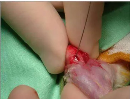
Calvaria bone segments and distal femoral epiphyses were obtained at euthanasia from rats involved in uncorrelated studies, approved by the Rizzoli Orthopedic Institute Ethical Committee, not affecting the health of bone or joints (study code 430/2015-PR “Scaffolds ceramici associati a cellule staminali mesenchimali per l’artrodesi vertebrale: studi in vitro ed in vivo per la chirurgia ‘one step’ in osso sano ed osteoporotico”).
Rats underwent pharmacological euthanasia via injection of 0.3mL/Kg of Tanax- Hoechst. Drugs used for anesthesia and euthanasia were considered not to have effects on bone segments harvested and on the subsequent tests performed. After harvesting, calvaria bone segments were cultured in Roswell Park Memorial Institute (RPMI 1640) medium (Sigma-Aldrich, Saint Louis, Missouri, USA) implemented with Bovine Serum Albumine (BSA) (Sigma-Aldrich, Saint Louis, Missouri, USA) 2 mM glutamine and antibiotics (100 U/ml penicillin, 100 µg/ml streptomycin) (Gibco, INVITROGEN Corporation, Carlsbad, CA). Femoral condyles were cultured in Dulbecco’s Modified Eagle’s Medium (DMEM) High glucose (Sigma, MO, USA) supplemented with 10% fetal bovine serum (FBS, Lonza, Verviers, Belgium), 2 mM glutamine and antibiotics (100 U/ml penicillin, 100 µg/ml streptomycin) (Gibco, INVITROGEN Corporation, Carlsbad, CA).
Both calvaria and femoral condyles were kept in culture at 37°C, 5% CO2 up to 4 weeks, in
vent cap tubes under continue oscillation. Assessment of viability and microtomographical evaluations were performed at time zero; viability was further checked weekly up to 14 days. At the end of the latest experimental time, calvaria segments and femoral condyles were collected and microtomographical and histological evaluations were repeated.
culture period. The samples were weighed in sterility, placed inside a tube of known weight, with an analytical scale and weight was calculated as the difference with the tare of the tube.
2.1.2 Assessment of bone cultures viability
For both bone tissue cultures viability was evaluated through Alamar blue dye test (Serotec, Oxford, UK). Briefly, the reagent is a dye, which incorporates an oxidation-reduction (REDOX) indicator that changes color in response to the chemical reduction of growth medium, resulting from cell growth. The dye was added to each culture (1:10 v/v) for 4 h at 37°C. At the end of experimental time, the supernatants were transferred to 96 well-plates, the absorbance was read spectrophotometrically at 570 and 600 nm wavelengths (for the fully oxidized and reduced forms of reagent) by MicroPlate reader (BioRad, CA, USA). The results, obtained as optical density (OD), were processed following manufacturer’s instruction and expressed as reduction percentage.
2.1.3 Bone cultures microtomography
Both femoral condyles and calvaria segments in culture were scanned at time zero and 14 days with the high-resolution microtomography (microCT) system Skyscan 1172 (Bruker Micro-CT, Belgium). The source voltage was set on 70 kV with a current of 141 μA and an Aluminum filter 0.5 mm thick was interposed between the x-ray source and the sample. Each sample was rotated until 180° with a rotation step of 0.45°. The images obtained from acquisition were later reconstructed by the software NRecon (version 1.6.10) with corrections for alignment, depended on acquisition, beam hardening and ring artifact reduction. Calvaria images resulted jpg images had 2000x2000 pixels with a pixel size of 7.45 μm. Femoral condyles images resulted jpg images had 4000x4000 pixels with a pixel size of 4.96 μm.
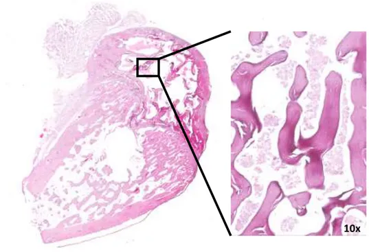
2.1.4 Bone cultures histology
Femoral condyles and calvaria segments were harvested at the end of experimental time and processed for paraffin embedding. More in details, femoral condyles and calvaria segments samples were fixed in 10% neutral buffered formalin (Sigma-Aldrich, Saint Louis, Missouri, USA) for at least 24 hours at room temperature. After rinsing in running tap water, samples were decalcified in 5% solution of formic (ACEF, Fiumicino, Rome, Italy) and nitric acid (Sigma-Aldrich, Saint Louis, Missouri, USA) at 37°C and then extensively rinsed in distilled water. Each condyle was cut along the major axis to help the inclusion, and all samples dehydrated in increasing ethanol solutions (Panreac AppliChem, Barcelona, Spain) for 1 hours each (70%, 95% twice, 100% twice), to remove the aqueous component and facilitate the penetration of the embedding medium, and defatted in xylene (VWR International, Milan, Italy). After overnight infiltration in liquid wax (56°C), samples were finally embedded in paraffin (Sigma-Aldrich, Saint Louis, Missouri, USA). From tissue blocks, thin histological section (5 μm thick) were obtained by a semi-automated microtome (MicromH340E, Germany) and stained with Haematoxylin (Sigma-Aldrich, Saint Louis, Missouri, USA) and Eosin (Bio-Optica, Milan, Italy) (H/E).
Images of obtained sections were acquired with digital scanner at different magnification.
2.1.5 Statistical analysis
Statistical analysis was conducted was performed using the SPSS v.12.1 software (SPSS Inc., IL, USA). Data are reported as mean ± SD at a significance level of p < 0.05. For the evaluation of culture viability and bone mass a two way ANOVA test with Holm Sidak multiple comparison test was performed, in order to assess eventual differences between groups and experimental times.
2.2 CHARACTERIZATION OF RAT BREAST CANCER CELL LINE (MRMT-1)
2.2.1 MRMT-1 cells proliferation and viability
Rat breast carcinoma cells line MRMT-1 were purchased from Cell Resource Center for Biomedical Research Institute of Development, Aging and Cancer (Tohoku University 4-1, Seiryo, Aoba-ku, Sendai, Japan).
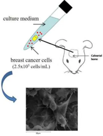
Viability and proliferation of MRMT-1 cells were assessed in different culture conditions, characterized by the addition of growing concentration of FBS to the culture medium. MRMT-1 were seeded in a 24 well plate at a density of 2x10⁵ cells/well and divided in the following groups, representing the different culture conditions:
basal medium (RPMI 1640 medium + 1% penicillin/streptomycin) + 10% FBS
basal medium + 1% di FBS
basal medium only
At the end of the scheduled experimental times of 24, 48 and 72 hours, cells were detached from each well with Trypsin EDTA (Sigma-Aldrich, Saint Louis, Missouri, USA), centrifuged at 1600 rpm, resuspended in an appropriate volume of serum free medium and counted in Burker counting cells chamber using Trypan Blue 1% dye (Sigma-Aldrich, Saint Louis, Missouri, USA), able to color selectively dead cells.
2.2.2 Wound healing assay
The in vitro scratch wound experiments were performed according to previous studies. Briefly, control and treated cells were seeded in 6-well plates (four replicates of each sample) with 3 ml complete medium and allow to reach confluence. A reproducible scratch in the
process of wound closure was monitored at different time points (0, 6, 21, 30, 45 hrs) by photographing the central field of the scratches under an inverted light microscopy (Olympus CKX41, Olympus Corp, Tokyo, Japan) mounted with a digital camera (C-7070 Wide Zoom, Olympus) at 10× magnification. The pictured field was standardized each time against a horizontal line drawn on the base of the plate passing through the center of each well. Morphometric analysis of cell migration was performed by one experienced investigator blinded to the specific experimental conditions using a computerized image analysis system (Qwin, Leica Microsystem Imaging Solution, Ltd). A region of 2.58 x 106 µm2 that included the artificial scratch and the adjacent cell monolayer was selected as the standard region of interest. The wound healing effect was calculated as (1-Ax/A0) %, where A0 and Ax represented the empty scratch area at 0 and x hours, respectively.
2.2.3 Assessment of activated pathway 2.2.3.1 Protein quantification
MRMT-1 cells were collected thorough centrifugation at 1500 rpm for 5 minutes at 4°C. and resuspended in Gold Lysis Buffer (GLB). Lysates were incubated in constant shaking (750 rpm) for at least 1 hour at 4°C and then centrifugated at 13200 rpm at 4°C to remove debris; finally, supernatants with proteic lysate were collected.
Cells (5-10x106) were centrifuged and resuspended in cold ipotonic lysis solution of five times higher volume, kept in ice for 10 minutes and then centrifuged at 1500 rpm for 5 minutes at 4°C. Pelles were resuspended again in cold ipotonic lysis solution of two times higher volume, passed 25-30 times through a syringe with needle of 25 µm and observed at light microscope to assess the lysis.
The lysate was then subjected to a series of centrifugations to separate proteins in the three cells portions, nucleus, heavy membranes and cytosol:
Supernatant containing cytosolic fraction was additioned with PPI inhibitors, Aprotinin, Leupeptin, Na3VO4 and PMSF, while pellet with heavy membrane proteic fraction was lysed in GLB+. All of the fractions were the kept in agitation (750 rpm) for one hour and then the nuclear and heavy membrane fractions were centrifuged at 14000 rcf for 10 minute at 4°C to remove cell particulates.
Quantification of the proteic contents of the samples was evaluated trough the colorimetric Lowry Method (Kit DC Protein Assay, Bio-Rad, Hercules, CA, USA). Briefly, 1 µl of sample was diluted in distilled water (1:20) and additioned with the various reagents of the kit. Samples were incubated at dark and in costant shaking for 15-30 minutes and read spectrophotometrically at 655 nm. For each protein lysate, 50 µg of protein were loaded on the gel. Sample were prepared for loading bringing them all to the same final volume by addition of GLB + and adding to each 5X Protein Sample Buffer. The samples were then boiled for 5 minutes, briefly centrifuged and then subjected to analysis by Western blotting.
2.2.3.2 Western Blotting
The protein lysates were subjected to electrophoresis on polyacrylamide gel in sodium dodecyl sulphate (SDS-PAGE) to obtain the separation of proteins according to their size. The sodium dodecyl sulphate (SDS) (CH3- (CH2) 10-CH2OSO3 -Na +) is an anionic detergent able to strongly bind and denature proteins, with a SDS molecule binding every two amino acid residues. In this way, the original charge of the protein is completely deleted and the polypeptide is covered with negative charges in proportional numbers to amino acids that
constitute it. All gels used were made of acrylamide 10%, except for the one for immunoprecipitation made of 12% and prepared as follow:
Proteins separation was performed in vertical electrophoresis cassettes (Amersham Biosciences UK Limited, Bucks, UK), applying a constant voltage of 100 V and 1X SDS-PAGE Running Buffer made of 1% SDS, 25 mM Tris, 200 mM glycine and distilled water. At the end of electrophoretic run, proteins were transferred through electroblotting technique to a nitrocellulose membrane Hybond-ECL (Amersham), using a Semi-dry Transblotter (Sigma-Aldrich Corporation) and 1X Transblotting Buffer, under the constant amperage of 400 mAmps for almost two hours. The transfer of the proteins to the membrane was proven by Ponceau S staining, which bind proteins nonspecifically.
Membranes were decolored with 1X PBS containing 0.05% Tween-20 (PBS/T) and incubated at 4°C overnight in saturation buffer with 5% non-fat dry milk in PBS/T. Blots were washed three times in PBS/T and incubated overnight at 4°C with the primary antibody diluted in 5% BSA in PBS/T and specifically: α-p-AKT T308 (1:1000), α-p-AKT S473 (1:1000), α-AKT (1:1000), α-p-PKR T451 (1:1000), α-peIF2α S51 (1:1000), α-p-GSK-3α/β S21/9 (1:1000), α-GSK-3β (1:1000) and α-β-actin (Cell Signaling Laboratories, Danvers, MA, USA); α-PKR (D-20) (1:1000) and α-eIF2α (FL-315) (1:1000) (Santa Cruz Biotechnology, La Jolla, CA, USA); α-β-Tubulin I (1:5000) (Sigma-Aldrich Corporation, Saint Louis, MO, USA); α-Lamin B (1:1000) (Oncogene Research).
At the end of incubation, membranes were washed three times with PBS/T and incubated for two hours with secondary antibody conjugated with horseradish peroxidase (HRP) (Cell Signaling Technology, Inc) diluted 1:2000 in 5% of non- fat dry milk in PBS/T. For each primary antibody, one of the two secondary antibody α-rabbit:HRP or α-mouse:HRP was used: α-rabbit:HRP: AKT T308 e S473, α-AKT, PKR T451, α-PKR (D-20),
α-p-eIF2α S51, α-α-p-eIF2α, α-p-GSK-3α/β S21/9, α-GSK-3β and α- β-actina; α-mouse:HRP: α-β-Tubulina I and α-Lamin B.
Blots were washed three times in PBS/T and visualized trough ECL (Enhanced ChemiLuminescence) Western blotting reagent (Amersham, Rockford, IL, USA). It is a method for detection of antigens, directly or indirectly conjugated with an antibody labeled with HRP, which uses the non radioactive light emission. In alkaline conditions, peroxidase catalyzes luminol oxidation which get an excited state; during the decay to the basal condition, luminol releases the excess energy by emitting light that can impress a photographic plate. The ECL Western blotting method uses, also, the presence of chemical enhancers of oxidation (such as phenols), enhancing in this way both the duration and the intensitt of light emission. The bands corresponding to the various proteins of interest were exposed on photographic plates (Kodak) and their intensity was estimated by densitometric analysis using the software Image J. Briefly, plates were scanned and saved as JPEG files 8 -bit greyscale. The percentage of pixels measurable in the images has been set using the adjustment control of the image background and then the number of pixels was estimated in the selected area, corresponding to the band of the protein.
Immunoprecipitation assay exploits the interaction antibody- antigen to precipitate specific antigen present in a solution (for example, cellular lysate). Once added the antibody to the solution, it will recognize and bind its antigen forming soluble antigen-antibody complex. In order to isolate such complexes, an immunoglobulin binding protein, such as protein A or G, was used, associated covalently to an insoluble support, such as agarose, that allows to precipitate by centrifugation, via increasing the molecular weight of the complex. The immunoprecipitation was conducted on CEM cells in logarithmic growth phase. Following extraction of total protein and the determination of protein concentration, two sample volumes corresponding to 500 micrograms of proteins were transferred to 1.5 ml tubes to be
subjected to immunoprecipitation , while 100 micrograms of proteins were transferred to a third tube to be used as a control.
At each of the two samples were added 25 µl of Protein A /G PLUS-Agarose Immunoprecipitation Reagent (Santa Cruz Biotechnology, Inc.) and 200 µL of the GLB + and all was placed in cold room and in rotation for two hours. This phase had the aim of immunoprecipitate cellular proteins capable of forming non-specific bonds with the protein A /G. After incubation, the samples were centrifuged at 13,200 rpm at 4 ◦C for 1 min and the supernatants were transferred to two new tubes to which are added:
• Sample 1: 5 µl antibody α-p-Tyrosine mouse mAb (Cell Signaling Technology) and 25 µl of Protein a / G PLUS-agarose
• Sample 2: 5 µl antibody α-PKR (D-20) (Santa Cruz Biotechnology, Inc.) and 25 µl of Protein a / G PLUS-agarose. Both samples were incubated overnight at 4◦C in rotation. The next day, the antigen-antibody complexes were precipitated by centrifugation at 13200 rpm for 1 minute to 4◦C. The pellets so obtained underwent 4 washes with 500 µL of GLB + after which each pellet was resuspended in 20 µl of GLB + and additioned with 5X protein sample buffer. Samples undergone to immunoprecipitation and control samples were boiled for 5 minutes, centrifuged and analyzed by SDS-PAGE followed by immunoblotting. The blots of the control sample and of the sample with the antibody immunoprecipitated α-p-Tyrosine (samples 1) were incubated with the primary antibody α-PKR (D-20), while for the sample immunoprecipitated with α-PKR (D- 20) α-p-Tyrosine was used as primary antibody. For both the primary antibodies dilutions of 1: 1000 in 5% BSA in PBS / T were used, while the secondary α-rabbit: HRP was diluted 1: 2000 in 5% skimmed milk in PBS / T.
2.2.2 MRMT-1 cells sensitivity to doxorubicin 2.2.2.1 Experimental culture set up
Doxorubicin is a cytotoxic anthracycline antibiotic isolated from cultures of Streptomyces peucetius var. caesius. Doxorubicin binds to nucleic acids, presumably by specific intercalation of the planar anthracycline nucleus with the DNA double helix.
The chemotherapeutic Doxorubicin (Doxorubicin 5 mg powder, Vinci-Biochem, Vinci, Italy) was resuspended in culture medium RPMI 1640 at a concentration of 100 µM. Subsequently serial dilutions of Doxorubicin were prepared to get five drug concentrations (5 µM, 2.5 µM, 1 µM, 0.5 µM, 0.1 µM)
MRMT-1 cells were seeded in a 96 well plate at a density of 2x10⁴ cells/well in standard culture medium. 24 hours after seeding, culture medium was refeeded as described below: - RPMI 1640 medium + 1% penicillin/streptomycin (basal medium) + 10% FBS
- basal medium + 1% FBS - basal medium without FBS
and to each group the different doxorubicin concentrations previously prepared were administered. As controls, for each group cells cultured with the same medium but not exposed to the drug were used. At the end of the expected experimental time, cell viability was assessed trough WST-1 assay.
2.2.2.3 Evaluation of cells viability
Briefly, the WST-1 assay is a colorimetric test to assess cell viability via the mitochondrial respiration. The salt of stable tetrazolium WST-1 is cleaved into soluble formazan by a
bioriduction depends strictly on the glycolitic production of NAD(P)H in viable cells. Therefore, the increase of the formed formazan dye directly correlates with the number of metabolically active cells present in culture.
The cells were washed with PBS 1%, and thereafter WST-1 was added in a concentration of 1:10 to the culture media. The plates were incubated for 4h at 37 ° C, 5% CO2; at the end of
the incubation time, the formed dye is quantified spectrophotometrically at an absorbance of 450 nm.
The IC50, namely the concentration of the drug able to inhibit 50% of cell survival compared
to controls, was determined by comparing cell viability and the logarithmic concentration of doxorubicin used in a dose response semi-log curve.
2.2.3 Statistical analysis
Statistical analysis was conducted on the viability test for Doxorubicin sensitivity to assess differences in the effects of drug concentration. The analysis was performed using the SPSS v.12.1 software (SPSS Inc., IL, USA). Data are reported as mean ± SD at a significance level of p < 0.05. Differences between groups were analyzed using the Kruskall-Wallis test for independent samples.
2.3 CO-CULTURE OF MRMT-1 CELL LINE AND OSTEOCLASTS
Published paper (Salamanna F, Pagani S, Maglio M, Borsari V, Giavaresi G, Martelli AM,
Buontempo F, Fini M. Estrogen-deficient osteoporosis enhances the recruitment and activity of osteoclasts by breast cancer cells. Histol Histopathol. 2016 Jan;31(1):83-93. doi: 10.14670/HH-11-651. Epub 2015 Aug 7.)
2.3.1 Conditioned media preparation
Cells were kept in culture in RPMI 1640 medium (Sigma, MO, USA) supplemented with 10% FBS (FBS, Lonza, Verviers, Belgium), 2 mM glutamine and antibiotics (100 U/ml penicillin, 100 µg/ml streptomycin) (Gibco, INVITROGEN Corporation, Carlsbad, CA) at 37°C 5% CO2. Once at confluence, cell cultures were rinsed and culture medium was
substituted with serum-free RPMI 1640 to obtain conditioned medium (15 ml per T75 flask, ~ 1 x 10⁶ cells/cm2). After twenty-four hours, medium was collected, centrifuged and stored at -80°C.
2.3.2 Isolation of mononuclear cells and differentiation into OCs
PBMCs were isolated from six healthy (SHAM) and six ovariectomized (OVX) Sprague-Dawley adult female rats (Charles River Calco Lecco, Italy) from an uncorrelated study approved by the Rizzoli Orthopedic Institute Ethical Committee to isolate OCs. The establishment of estrogen-deficient osteoporotic condition in OVX animals and the healthy condition in SHAM animals was assessed through quantitative bone ultrasound evaluations, microtomography and histomorphometric analysis of the iliac crest biopsy.
PBMCs were isolated onto Ficoll-Histopaque gradient (Sigma-Aldrich, MO, USA), according to the following protocol. Harvested peripheral blood was diluted 1:1 with PBS and slowly layered on Histopaque 1077 (ratio 2:1). After centrifugation at 700g for 30 min at room temperature, the mononuclear cells fraction was isolated; PBMCs were collected and,