1
Alma Mater Studiorum – Università di Bologna
DOTTORATO DI RICERCA IN
Scienze Chimiche
Ciclo XXVI
Settore Concorsuale di afferenza: 03/C1 Settore Scientifico disciplinare: CHIM/06
Synthesis and surface modification of silver and gold
nanoparticles. Nanomedicine applications against
Glioblastoma Multiforme.
Presentata da: Locatelli Erica
Coordinatore Dottorato
Relatore
Chiar.mo Prof. Aldo Roda Prof. Mauro Comes Franchini
2
Medaglione
Dottorando: Locatelli Erica
Curriculum: Nanoscienze e Nanotecnologie Tutor: Prof. Mauro Comes Franchini
Titolo della Tesi: Synthesis and surface modification of silver and gold nanoparticles.
Nanomedicine applications against Glioblastoma Multiforme.
Relazione:
La Dott.ssa Locatelli Erica durante il triennio del suo dottorato di ricerca si è occupata della sintesi di nanoparticelle metalliche di oro e argento, e del loro successivo intrappolamento in micelle costituite da co-polimeri biocompatibili per applicazioni nel campo del drug delivery ed in particolare alla lotta contro il glioblastoma multiforme, tumore dalla prognosi particolarmente infausta. Nello specifico è stata messa a punto la sintesi di nanosfere di argento e di nanonocilindri di oro in soluzione acquosa e successivamente le due nanostrutture sono state funzionalizzate con leganti organici appositamente sintetizzati per renderle lipofiliche piuttosto che idrofiliche. Infine sono stati sintetizzati copolimeri biocompatibili e biodegradabili a base di acido poli-lattico-co-glicolico (PLGA) e di glicole polyetilenico (PEG). A questo punto si è proceduto allo sviluppo di vari sistemi nanostrutturati complessi costituiti da micelle polimeriche, contenenti al loro interno le nanostrutture di oro e argento, singolarmente o legate tra loro tramite reazione di Click chemistry, oppure le nanostrutture ed eventuali farmaci antitumorali; tali micelle sono inoltre state funzionalizzate in superficie con opportuni agenti di targeting per drug delivery attivo o di imaging. Dopo aver caratterizzato i vari prodotti ottenuti con svariate tecniche analitiche (NMR, AAS, DLS, IR, HPLC, TGA, UV-Vis, etc…) essi sono stati testati da gruppi collaboratori sia tramite tests in vitro ed in
vivo. I risultati sono stati assai incoraggianti in quanto i nanosistemi mostrano una buona
biodistribuzione, capacità di raggiungere il sito tumorale con buoni up-take, possibilità di essere usati quali agenti di imaging diagnostico per una diagnosi precoce del tumore, ed infine una ottima citotossicità contro il glioblastoma multiforme, con in un caso particolare l’ottenimento della riduzione della massa tumorale in vivo in topi del 22% dopo singola somministrazione.
Durante il triennio del dottorato di ricerca la dott.ssa Locatelli Erica è stata coautrice di 11 lavori con peer-review. Ha partecipato a 5 conferenze nell’ambito del progetto europeo
Save-3
Me (CP-IP 263307-2) con 3 presentazioni orali in lingua inglese, ad una scuola di dottorato nazionale (XXXVI Corso Estivo “A. Corbella”, Gargnano, Brescia, 2011) e ad una internazionale (NN13 & ISSON13, Salonicco, Grecia, 2013) con presentazione di posters. Ha inoltre svolto un periodo di ricerca di 4 mesi presso l’istituto di biofarmaceutica di Francoforte (Germania) sotto la supervisione del Professor Jorg Kreuter, durante i quali si è occupata di sintesi di nanoparticelle proteiche a base di siero albumina umana, acquisendo nuove conoscenze nell’ambito della preparazione e caratterizzazione di sistemi biocompatibili per drug delivery.
4
Index
Abstract
List of abbreviations
List of publications
1. Introduction
1.1 Nanomedicine, Nanotechnology and Drug Delivery: an overview. 1.2 Glioblastoma multiforme
1.3 Noble metal nanoparticles and their application in nanomedicine
1.3.1 Properties and advantages of nanoparticles 1.3.2 Gold nanostructures
1.3.3 Silver nanoparticles
1.3.4 Surface modification of metal nanoparticles
1.4Polymeric nanoparticles
1.4.1 Advantages, properties and materials
1.4.2 Polylactic-co-glycolic acid and polyethylene glycol 1.4.3. Preparation methods for polymeric nanoparticles
2. Aim
3. Discussion
3.1 Synthesis of the precursors
3.1.1 Preparation of the organic ligands
3.1.2 Preparation of copolymer for nanocarrier 3.1.3 Synthesis of metal nanoparticles
3.2 GNRs into polymeric nanoparticles: a theranostic tool against Glioblastoma Multiforme
3.2.1 GNRs surface modification
3.2.2 Entrapment of GNRs into polymeric nanoparticles 3.2.3 GNRs-1-PNPs surface conjugation with active agents
5
3.2.4 In vitro and in vivo tests
3.3 AgNPs into polymeric nanoparticles and synergistic effects with antineoplastic drug: towards therapy against Glioblastoma Multiforme.
3.3.1 AgNPs surface modification
3.3.2 Entrapment of AgNPs and/or Alisertib into polymeric nanoparticles 3.3.3 Nanosystems’ surface conjugation with active agents
3.3.4 In vitro and in vivo tests
3.4 Click chemistry on the surface of GNRs and AgNPs
3.4.1 Surface modification of GNRs and AgNPs with ligands 2 and 3 3.4.2 Cycloaddition reaction between the functionalized nanostructures 3.4.3 Entrapment of GNRs-click-AgNPs into polymeric nanocarrier 3.4.4 In vitro evaluation of optoacoustic imaging capability
4. Conclusion
5. Experimental section
5.1 Materials and Methods 5.2 Organic synthesis 5.3. Nanotechnology
6
Abstract
In the last decades noble metal nanoparticles (NPs) arose as one of the most powerful tools for applications in nanomedicine field and cancer treatment. Glioblastoma multiforme (GBM), in particular, is one of the most aggressive malignant brain tumors that nowadays still presents a dramatic scenario concerning median survival. Gold nanorods (GNRs) and silver nanoparticles (AgNPs) could find applications such as diagnostic imaging, hyperthermia and glioblastoma therapy. During these three years, both GNRs and AgNPs were synthesized with the “salt reduction” method and, through a novel double phase transfer process, using specifically designed thiol-based ligands, lipophilic GNRs and AgNPs were obtained and separately entrapped into biocompatible and biodegradable PEG-based polymeric nanoparticles (PNPs) suitable for drug delivery within the body. Moreover, a synergistic effect of AgNPs with the Alisertib drug, were investigated thanks to the simultaneous entrapment of these two moieties into PNPs. In addition, Chlorotoxin (Cltx), a peptide that specifically recognize brain cancer cells, was conjugated onto the external surface of PNPs. The so-obtained novel nanosystems were evaluated for in vitro and in vivo applications against glioblastoma multiforme. In particular, for GNRs-PNPs, their safety, their suitability as optoacoustic contrast agents, their selective laser-induced cells death and finally, a high tumor retention were all demonstrated. Concerning AgNPs-PNPs, promising tumor toxicity and a strong synergistic effect with Alisertib was observed (IC50 10 nM), as well as good in vivo biodistribution, high tumor uptake and significative tumor reduction in tumor bearing mice. Finally, the two nanostructures were linked together, through an organic framework, exploiting the click chemistry azido-alkyne Huisgen cycloaddition, between two ligands previously attached to the NPs surface; this multifunctional complex nanosystem was successfully entrapped into PNPs with nanoparticles’ properties maintenance, obtaining in this way a powerful and promising tool for cancer fight and defeat.
7
List of abbreviations
AAS: atomic absorption spectroscopyAgNPs: silver nanoparticles BBB: blood brain barrier Cy5: cyanine 5
Cltx: chlorotoxin
CTAB: hexadecyltrimethylammonium bromide CuAAC: Copper-catalyzed azide alkyne cycloaddition DLS: Dynamic light scattering
EDC: 1-ethyl-3-(3-dimethylaminopropyl) carbodiimide EPR: Enhanced Permeability and Retention
FDA: Food and Drug Administration GBM: glioblastoma multiforme GNRs: gold nanorods
HPLC: high performance liquid chromatography
ICP-AES: inductively coupled plasma atomic emission spectroscopy IR: infrared
LSPR: localized surface plasmon resonance NMR: nuclear magnetic resonance
NPs: nanoparticles PDI: polidispersity index PEG: poly(ethylene glycol)
PLGA: poly(lactic-co-glycolic) acid PNPs: polymeric nanoparticles PVP: polyvinylpyrrolidone RES: Reticuloendothelial System
RuAAC: Ruthenium-catalyzed azide alkyne cycloaddition sulfo-NHS: N-hydroxysulfosuccinimide
TEM: transmission electron microscopy UV-Vis: ultraviolet-visible
8
List of publications
Papers included and discussed in the present thesis.
1. M. Comes Franchini, J. Ponti, R. Lemor, M. Fournelle, F. Broggi, E. Locatelli:
“Polymeric entrapped thiol-coated gold nanorods: cytotoxicity and suitability as molecular optoacoustic contrast agent”. J. Mater. Chem., 2010, 20, 10908–10914.
Abstract: The behaviour of polymeric entrapped thiol-coated GNRs in culture medium
under biological conditions was analysed. The in vitro cytotoxicity was studied by a Colony Forming Efficiency assay on immortalized mouse fibroblasts (Balb/3T3) obtaining a dose–effect relationship in which a half inhibitory concentration (IC50) was 20.3mM. The suitability of the new nanomaterial as an optoacoustic contrast agent was investigated in phantom studies using a hardware platform suitable for retrieving clinically relevant data. Spherical alginate phantoms containing GNR-2-PNPs at different concentrations were synthesized and the optoacoustic signal amplitudes were measured as a function of concentration. Signals could be obtained with satisfying signal-to-noise ratio (SNR) down to concentrations of 11 mM corresponding to subtoxic concentration in our in vitro model. The nanomaterial proved to be a suitable and promising contrast agent for different optoacoustic imaging modalities including multispectral approaches.
2. E. Locatelli, G. Ori, M. Fournelle, R. Lemor, M. Montorsi, M. Comes Franchini: “Click
Chemistry for the Assembly of Gold Nanorods and Silver Nanoparticles”. Chem. Eur. J.
2011, 17, 9052 – 9056.
Abstract: Click chemistry based on a 1,3-dipolar cycloaddition between lipophilic gold
nanorods (GNRs) containing an acetylene group with spherical silver nanoparticles containing an azide has been accomplished (see scheme). Phantom experiments show that this organic transformation did not affect the suitability of the gold nanorods as contrast agents for optoacoustic imaging.
3. E. Locatelli, F. Broggi, J. Ponti, P. Marmorato, F. Franchini, S. Lena, M. Comes
9
Targeted-PEG-Based Micelles for the Treatment of Glioblastoma”. Adv. Healthcare
Mater. 2012, 1, 342–347.
Abstract: A simple method for the synthesis of lipophilic Ag NPs has been developed. The
coated Ag NPs have been entrapped into a FDA-approved and targetable PEG-based polymeric nanoparticles, and this nanocarrier has been conjugated with the peptide chlorotoxin. Uptake experiments have shown a cell-specific recognition of the Ag-1-PNPs-Cltx on U87MG cell lines in comparison to Balb/3T3. The uptake of Ag into the cells was quantified and an interesting cytotoxic effect (IC50 = 45 μM) has been found on glioblastoma cell lines.
4. E. Locatelli, M. Comes Franchini: “Biodegradable PLGA-b-PEG polymeric
nanoparticles: synthesis, properties, and nanomedical applications as drug delivery system”. J Nanopart Res, 2012, 14, 1316-1333.
Abstract: During the past decades many synthetic polymers have been studied for
nanomedicine applications and in particular as drug delivery systems. For this purpose, polymers must be non-toxic, biodegradable, and biocompatible. Polylactic-co-glycolic acid (PLGA) is one of the most studied polymers due to its complete biodegradability and ability to self-assemble into nanometric micelles that are able to entrap small molecules like drugs and to release them into body in a time dependent manner. Despite fine qualities, using PLGA polymeric nanoparticles for in vivo applications still remains an open challenge due to many factors such as poor stability in water, big diameter (150–200 nm), and the removal of these nanocarriers from the blood stream by the liver and spleen thus reducing the concentration of drugs drastically in tumor tissue. Polyethylene glycol (PEG) is the most used polymers for drug delivery applications and the first PEGylated product is already on the market for over 20 years. This is due to its stealth behavior that inhibits the fast recognition by the immune system (opsonization) and generally leads to a reduced blood clearance of nanocarriers increasing blood circulation time. Furthermore, PEG is hydrophilic and able to stabilize nanoparticles by steric and not ionic effects especially in water. PLGA–PEG block copolymer is an emergent system because it can be easily synthesized and it possesses all good qualities of PLGA and also PEG capability so in the last decade it arose as one of the most promising systems for nanoparticles formation, drug loading, and in vivo drug delivery applications. This review will discuss briefly on PLGA-b-PEG synthesis and physicochemical properties, together with its improved qualities with respect to the single PLGA and PEG polymers. Moreover, we will
10
focus on but in particular will treat nanoparticles formation and uses as new drug delivery system for nanomedical applications.
5. E. Locatelli, W. Bost, M. Fournelle, J. Llop, L. Gil, F. Arena, V. Lorusso, M. Comes
Franchini: “ Targeted polymeric nanoparticles containing gold nanorods: a therapeutic approach against glioblastoma”. J Nanopart Res, 2014, 16, 2304-2013.
Abstract: Chlorotoxin-targeted polymeric nanoparticles containing entrapped gold
nanorods as potential therapeutic agent for glioblastoma multiforme have been developed and evaluated. In first proof of concept experiments, in vitro specific uptake in cancer cells and selective laser-induced cell death have been shown. In vivo studies with optical imaging showed increased retention of targeted NPs in the tumor.
6. E. Locatelli, M. Naddaka, C. Uboldi, G. Loudos, E. Fragogeorgi, V. Molinari, A.
Pucci, T. Tsotakos, D. Psimadas, J. Ponti, M. Comes Franchini: “Targeted delivery of silver nanoparticles and alisertib: in vitro and in vivo synergistic effect against glioblastoma”. Nanomedicine, 2014, In press.
Abstract: Targeted biocompatible nanoplatforms presenting multiple therapeutic functions
have great potential for the treatment of cancer. Materials & methods: Multifunctional nanocomposites formed by polymeric nanoparticles (PNPs) containing two cytotoxic agents – the drug alisertib and silver nanoparticles – were synthesized. These PNPs have been conjugated with a chlorotoxin, an active targeting 36-amino acid-long peptide that specifically binds to MMP-2, a receptor overexpressed by brain cancer cells. Results: The individual and synergistic activity of these two cytotoxic agents against glioblastoma multiforme was tested both in vitro and in vivo. The induced cytotoxicity in a human glioblastoma–astrocytoma epithelial-like cell line (U87MG) was studied in vitro through a trypan blue exclusion test after 48 and 72 h of exposure. Subsequently, the PNPs’ biodistribution in healthy animals and their effect on tumor reduction in tumor-bearing mice were studied using PNPs radiolabeled with 99mTc. Conclusion: Tumor reduction was achieved in vivo when using silver/alisertib@PNPs–chlorotoxin.
11
Papers concerning other research fields
1. E. Locatelli, L. Gil, L. L. Israel, L. Passoni, M. Naddaka, A. Pucci, T. Reese, V.
Gomez-Vallejo, P. Milani, M. Matteoli, J. Llop, J. P. Lellouche, M. Comes Franchini: “Biocompatible nanocomposite for PET/MRI hybrid imaging”. Int. J. Nanomed., 2012, 7, 6021–6033.
Abstract: A novel nanocarrier system was designed and developed with key components
uniquely structured at the nanoscale for early cancer diagnosis and treatment. In order to perform magnetic resonance imaging, hydrophilic superparamagnetic maghemite nanoparticles (NPs) were synthesized and coated with a lipophilic organic ligand. Next, they were entrapped into polymeric NPs made of biodegradable poly(lactic-co-glycolic acid) linked to polyethylene glycol. In addition, resulting NPs have been conjugated on their surface with a 2,2′-(7-(4-((2-aminoethyl)amino)-1-carboxy-4-oxobutyl)-1,4,7-triazonane-1,4-diyl)diacetic acid ligand for subsequent 68Ga incorporation. A cell-based cytotoxicity assay has been employed to verify the in vitro cell viability of human pancreatic cancer cells exposed to this nanosystem. Finally, in vivo positron emission tomography-computerized tomography biodistribution studies in healthy animals were performed.
2. V. Rebuttini, A. Pucci, P. Arosio, X. Bai, E. Locatelli, N. Pinna, A. Lascialfari, M.
Comes Franchini: “Zirconia-doped nanoparticles: organic coating, polymeric entrapment and application as dual-imaging agents”. J. Mater. Chem. B, 2013, 1, 919-923.
Abstract: Zirconia nanoparticles doped with Eu3+, Tb3+ and Gd3+ ions have been synthesized following the benzyl alcohol route. The nanoparticles were coated with N-hydroxydodecanamide and encapsulated in PLGA-b-PEG-COOH nanomicelles. The magnetic and fluorescent properties of these hybrid nanocarriers were investigated, proving them to be potential dual-imaging contrast agents.
3. A. Pucci, E. Locatelli, J. Ponti, C. Uboldi, V. Molinari, M. Comes Franchini: “Click
chemistry on the surface of PLGA-b-PEG polymeric nanoparticles: a novel targetable fluorescent imaging nanocarrier”.
Abstract: In the quest for biocompatible nanocarriers for biomedical applications, a great
deal of effort is put on engineering the nanocomposites surface in order to render them specific to the particular purpose. We developed biocompatible PLGA-b-PEG-based
12
nanoparticles carrying a double functionality (i.e., carboxylic and acetylenic) able to serve as flexible highly selective grafting centers for cancer diagnosis and treatment. As a proof of concept, the nanocarrier was successfully functionalized with a tailored fluorescent molecule by means of click chemistry and with a targeting agent specific for glioblastoma multiforme via amidic bond formation.
4. J. Vandooren, N. Berghmans, C. Dillen, I. Van Aelst, I. Ronsse, L. L. Israel, I.
Rosenberger, J. Kreuter, J. P. Lellouche, S. Michaeli, E. Locatelli, M. Comes Franchini, M. K. Aiertza, L. Sánchez-Abella, I. Loinaz, D. R. Edwards, L. Shenkman, G. Opdenakker: “Intradermal air pouch leukocytosis as an in vivo test for nanoparticles”. Int. J. Nanomed.
2013, 8, 4745–4756.
Abstract: The need for test systems for nanoparticle biocompatibility, toxicity, and
inflam-matory or adaptive immunological responses is paramount. Nanoparticles should be free of microbiological and chemical contaminants, and devoid of toxicity. Nevertheless, in the absence of contamination, these particles may still induce undesired immunological effects in vivo, such as enhanced autoimmunity, hypersensitivity reactions, and fibrosis. Here we show that artificial particles of specific sizes affect immune cell recruitment as tested in a dermal air pouch model in mice. In addition, we demonstrate that the composition of nanoparticles may influence immune cell recruitment in vivo. Aside from biophysical characterizations in terms of hydrodynamic diameter, zeta potential, concentration, and atomic concentration of metals, we show that – after first-line in vitro assays – characterization of cellular and molecular effects by dermal air pouch analysis is straightforward and should be included in the quality control of nanoparticles. We demonstrate this for innate immunological effects such as neutrophil recruitment and the production of immune-modulating matrix metalloproteases such as MMP-9; we propose the use of air pouch leukocytosis analysis as a future standard assay.
5. D. Psimadas, G. Baldi, C. Ravagli, M. Comes Franchini, E. Locatelli, C. Innocenti, C.
Sangregorio, G. Loudos: “Comparison of the magnetic, radiolabeling, hyperthermic and biodistribution properties of hybrid nanoparticles bearing CoFe2O4 and Fe3O4 metal
cores”. Nanotechnology, 2014, 25, 25101-25110.
Abstract: Metal oxide nanoparticles, hybridized with various polymeric chemicals,
represent a novel and breakthrough application in drug delivery, hyperthermia treatment and imaging techniques. Radiolabeling of these nanoformulations can result in new and
13
attractive dual-imaging agents as well as provide accurate in vivo information on their biodistribution profile. In this paper a comparison study has been made between two of the most promising hybrid core–shell nanosystems, bearing either magnetite .Fe3O4/ or cobalt ferrite .CoFe2O4/ cores, regarding their magnetic, radiolabeling, hyperthermic and biodistribution properties. While hyperthermic properties were found to be affected by the metal-core type, the radiolabeling ability and the in vivo fate of the nanoformulations seem to depend critically on the size and the shell composition.
14
1. Introduction
1.1 Nanomedicine, Nanotechnology and Drug Delivery: an overview.
Nanotechnology is a cutting-edge branch of applied science and technology, which deals with the control of matter at dimensions between one and one hundred nanometers and the subsequent realization of devices in such a scale. These dimensions allow matter to have a high ratio of surface area to volume, not present neither in bulk material nor in molecular scale, enabling materials to acquire unique properties (optical, electronic, magnetic, chemical and biologic), which consent nanotechnology to work in a multidisciplinary area of investigation, involving many fields of research, including chemistry, biology, physics, and even medicine.1
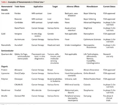
Nanomedicine represents the medical application of the opportunities arising from nanotechnology: indeed by engineering nanomaterials that have specific size, structure, shape, chemical composition and surface chemical characteristic it is possible to obtain new drug delivery systems, diagnostic tools, contrast agents, scaffolds for tissue engineering and a variety of medical devices for the treatment, prevention and diagnosis of numerous diseases.2 Some nanomaterials gained approval by Food and Drug Administration (FDA) and are nowadays commonly used in medicine (Figure 1.1.1).
Figure 1.1.1: some of the nanomaterials undergoing clinical trials or FDA approved. B.Y.S. Kim, N.
15
One of nanomedicine’s major efforts is in the development of new and more efficient cancer treatments: to improve the efficiency of currently available chemo-therapeutic agents as well as to enable new early pre-symptomatic cancer diagnosis, have assumed dramatic importance in the last few decades since still in 2012 one in 4 deaths in Europe and United States is due to cancer.3
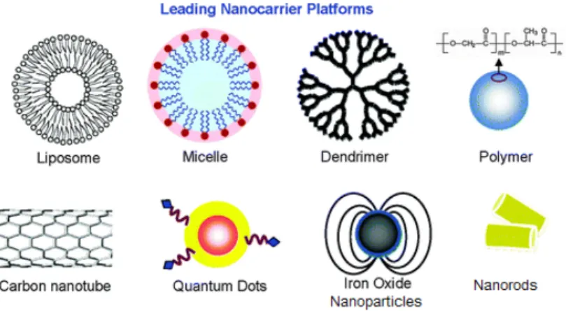
Compared with traditional contrast agents and chemotherapeutic drugs, nanomedicine offers the possibility to incorporate multiple functions in a single system holding great promises of simultaneous monitoring, treatment and targeting of cancer: the term “theranostic” was specifically coined to describe the combination of diagnostic capability and therapeutic efficacy held in a single system (Figure 1.1.2).4
Figure 1.1.2: examples of nanocomposites nowadays developed. Adapted from W. X. Mai.Integr.
Biol., 2013, 5, 19-28.
Moreover, nanomedicine enables the delivery of a drug in a nanosystem within the biological systems and through the biological barriers, which provide advantages due to the high degree of transport and drug protection from immune system so that efficacy can be greatly improved and new therapies became possible. With the term “drug delivery” is generally described the aim of a controlled delivery of drugs to their site of action, without affecting healthy organs, and the release of the same in a time-dependent manner. Drug delivery simultaneously solves common problems ascribed to poor drugs solubility in water and consequently low absorption by the body, short in vivo lifetime due to elevated clearance, leading to various advantages such as an improvement of the therapeutic index, a reduction of the dose of drug to be administered and consequently minimization of the unwanted side effects.5
To specifically target drugs to the desired site of action, especially anti-cancer drugs to tumors, is becoming a challenge that is currently addressed: most of the nowadays-developed
16
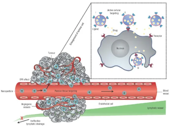
nanosystems for drug delivery take advantages of the particular characteristics of the disease microenvironment and angiogenesis. It is known that tumor or inflamed tissues present an hyper vascularization and leaky connected endothelial cells respect to healthy tissues that allow easy penetration of nanosystems with suitable dimensions; meanwhile they also present a decreased or damaged lymphatic drainage that reduce execration of the nanosystems once entered into the tissues: this set of conditions takes name of Enhanced Permeability and Retention (EPR) effect and the delivery of a drug taking advantages only of that phenomenon is generally recognised as “passive targeting” (Figure 1.1.3). A more efficient drug delivery can be achieved with the so-called “active targeting” that consists in the grafting to the surface of nanosystems of specific ligands able to bond the receptors overexpressed by cancer cells or desired endothelial tissues cells.6
Figure 1.1.3: passive and active targeting of nanosystems. From D. Peer,
Nature Nanotechnology 2007, 2, 751-760.
Proteins, peptides, monoclonal antibodies or small molecules, able to recognize selectively membrane cellular sites, have been widely used in order to enable an efficient active targeting and to obtain a drug release with higher concentration only in the desired site of action. These kinds of ligands must be on the outer shell of a nanosystem since they have to be free for receptors recognition and not hidden into the nanosystem matrix. For surface modification and anchoring of the ligands, covalent bonding of agents is generally preferred to physical
17
adsorption or ionic interaction: its stronger nature prevents leaching from the nanoparticles surface under physiologic conditions avoiding random dispersion within the body.7 Targeting and surface modification of a nanosystem may help not only to reach the desired site of action but also to prolong circulation time and residence time of a drug within the body. This is true especially for injectable particles because, in order to achieve the desired benefits, a drug delivery nanosystem must be present in the bloodstream long enough to reach and recognize its therapeutic site of action. Unfortunately small molecules are rapidly eliminated from the body through the excretory system, especially kidneys, but most of all every foreign object, be it a small molecule or a larger nanosystem, once entered the bloodstream undergo the phenomenon of opsonisation and consequently attack of the immune system and of the Reticuloendothelial System (RES) organs (for instance liver and spleen).
Once entered the bloodstream a foreign organism is immediately surrounded by opsonin proteins so that it becomes subjected to the attack of phagocytic cells.
The phagocytosis brings to the disruption and removal of the undesired materials from the body: in case of nanoparticles, especially polymeric nanoparticles, which cannot be completely destroyed during this process, sequestration from the RES organs frequently occurs. Nanomedicine has long dealt whit this liming factor and even if a completely resolution and prevention of opsonisation is not still achieved, many methods and correlations have been found in order to slow this process. It has been demonstrated that particles’ surface charge and nature is generally related to opsonozation, where hydrophilic as well as neutrally charged particles are less subject to opsonisation than hydrophobic and strongly charged ones. For this reason, one of the most adopted techniques to reduce body clearance is the coating of the nanosystem’s surface with shielding moieties such as long chain hydrophilic polymers, which can help to avoid opsonisation and early elimination.8
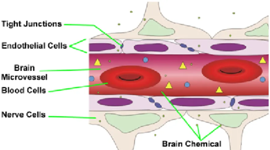
A drug delivery system should not only to have a long blood circulation time to effectively reach the desired site of action but it would also need to permeate through the biological barriers normally protecting tissues and organs. These barriers are numerous and of different nature but the most challenging one to cross remains the Blood Brain Barrier (BBB). The BBB is present all along capillaries in the central nervous system and consists of tight junctions made of adjacent endothelial cells that generally are not found in other body capillaries. Its role consists in the maintaining of an extremely regulated brain microenvironment acting like a physical barrier letting enter into the brain the required nutrients and protecting from virus and bacteria but also from harmful hydrophilic molecules
18
(Figure 1.1.4). The only exchange allowed through the BBB is the one of small hydrophobic molecules such as oxygen, carbon dioxide and hormones.9
Figure 1.1.4: schematic representation of the Blood Brain Barrier. J. Whitebread, The Blood-Brain
Barrier and OCD, Section 23 PSY1001 Fall 2011
Since capillaries in all the body compartments have numerous channels, which allow permeation of molecules up to the size of proteins, capillary permeability is not considered a limiting factor in drug delivery. On the contrary for drug delivery to the brain this factor became of crucial importance and all the predictions for drugs or nanosystems engineering has to be made taking into account BBB low permeability. Nonetheless a number of molecules, such as important nutrient, sugars and amino acids can cross the BBB: this is possible thanks to intramembrane transport proteins that allow a “facilitated diffusion” through the barrier; a specific designed active targeting exploiting these proteins may solve, almost in part, the challenging drug delivery to the brain.10
19
1.2 Glioblastoma multiforme
Cancer remains today one of the leading causes of death in United States and Europe for people from one to 65 years old.11 Medicine has made many strides in cancer treatment and prevention as well as in improvement of quality life for patients with untreatable cancers, but only few sub-types of cancers can be treated in a satisfactory way presenting a good survival rate. Still today, cancer prevention and early detection, instead of therapy, remain the best possibilities for an increment in the survival rate. Cancers that became easily detectable with common and non-expensive techniques (like ultrasound or sample taking) along with cancers that undergo an intensive prevention screening program are today the ones in which the best results were obtained in terms of death reduction and life prolongation. Besides, other types of cancer which present serious difficulties in detection, due to their nature or location in body, remain practically untreatable, and few or nothing at all was possible in order to improve survival rate or patients’ quality life.12
Among these untreatable tumors, brain cancers represent a large portion: they are in fact particularly hard to detect and treat because of their location, clearly not accessible by common diagnostic techniques and surgery. One of the most common and aggressive primary brain tumours is Glioblastoma multiforme (GBM). It is a tumor of the central nervous system, also known as glioma of grade IV, which exhibits one of the lower overall survival rates among all the other cancers. It was estimated that only the 20% of patients survive two years after diagnosis and this number drastically decrease after five years, with less of 5% of patients still alive.13 GBM often affects one of the cerebral hemispheres, but in some cases it can be also found in the cerebellum or sometimes in the spinal cord; furthermore GBM rarely generates metastasis to other parts of the body. GBM tumors can be divided in two macro-categories based on the mode of genesis of the same: these are primary and secondary GBM. The so-called primary GBM is the most frequent case, which prevalently affects elderly patients (more than 60 years old) with a very fast progression, without requiring the presence of a pre-existing lesion. The so-called secondary GBM has a frequency of less than 10% among all the diagnosed tumors, but it affects younger patients (average age at diagnosis of 45 years old); it develops more slowly than the primary one, following the typical progression of a diffuse astrocytoma or anaplastic astrocytoma. In the first case, the median survival is three months after diagnosis while in the second it could increase from six months to one year.14
20
The dramatic scenario of GBM has not improved despite many efforts of traditional medicine and surgery: glioblastoma multiforme shows a large resistance to conventional treatment techniques such as radio- and chemotherapy.15 Nowadays standard therapies consist in surgical removal of the tumor mass when possible, followed by radiation in association with chemotherapy, even though this treatment usually leads only to a small extension of patient’s life, with a median survival of less than one year after diagnosis. Indeed, even in case of successful intervention, with complete removal of tumor mass and no damage at the central nervous system after surgery, glioblastoma multiforme usually recurs in almost all cases in a few months: the average time to recurrence after standard therapy is 7 months. This is why chemotherapy and radiotherapy are always applied after surgery. Recent studies suggest that combination of radio- and chemotherapy, rather than single therapy, is associated to a longer survival time. The aim of radiotherapy is to damage and destroy cancer cells remained in loco after surgery, before their possibility to resume proliferation. Thanks to this technique is possible to attack diseased cells focusing mainly on ionizing radiation in the area of interest, leaving healthy cells almost untouched or exhibited only to a few rays. Dose response relationships demonstrate that median survival rate of 13 weeks is obtained with a total radiation dose of less than 4500 cGy, while with a dose of 6000 cGy is possible to reach a median survival of 42 weeks: of course this dose is usually administered 5 days per week in doses of 1.8-2.0 Gy. Unfortunately, the responsiveness of glioblastoma multiforme to radiotherapy may vary considerably from one patient to another: in most of the cases, a phase of remission is induced by radiotherapy, which can endure for months, with recover of neurological deficits and of patient’s health, but after that, tumor reoccurs with an enhancement in clinical deterioration and in expanded region.16
The use of chemotherapy has increased patients’ possibilities and hope especially in association with the above-described techniques. The chemotherapeutic approach is very common in the treatment of cancer, though due to the poor selectivity of the drugs generally involved, it is impossible to discern diseased tissues from healthy cells, thus causing serious damage to the body and a pool of unwanted side effects, which may sometimes be so debilitating for patients to require the suspension of the therapy.
A novel and promising approach that is arising in these recent years is based on the development of specific therapies, deriving from the studies on the growth mechanism, the genesis of the tumor, the cells proliferation and communication, both at a macroscopic level and a microscopic one. This approach may allow focused therapies, different and specific for every cases, thus increasing the possibility of success by several times.
21
GBM is composed by a heterogeneous mixture of poorly differentiated cancer cells sometimes closely associated to stem cells. The mechanisms that lead to the genesis of GBM were largely investigated and nowadays several possible pathways were recognized as progenitors of the tumor. The most common ones involve the wrong regulation in the receptors of growth factors (EGFR and PTEN protein) or alteration of the reparative p53 protein.17 Generally, in healthy cells a complex mechanism of regulation of cascade-signals takes place: these signals leads to several cells behaviors, such as differentiation, growth, reproduction etc...When one of those regulatory enzymes fails, a series of uncontrolled effects happens, such as an unwanted, extremely fast cells proliferation. For instance, PTEN is a protein encoded by the PTEN gene, which acts as a tumor suppressor by inhibiting the protein kinase and all the following cascade-signals thanks to its dephosphorylation ability; the direct result of this inhibition is a reduced and well-controlled cell proliferation.18 Thus, any alteration of the PTEN protein or of its gene regulation can cause an imbalance in this cascade and produce a fast cell proliferation thus leading to the development of the tumor mass. For this reason PTEN protein, such as several other enzymes or complexes, became the target for the most innovative chemotherapy.
There are many different molecules tested and approved as chemotherapists able to attack one of the metabolic pathways of the tumor growth process or directly their cells DNA, causing cells irreproducibility or cells death, making chemotherapy the most important weapon to defeat cancer. Cancer cells, however, may sometimes develop mechanisms of resistance to the most used drugs, requiring an increase in dosage or administration of different molecules. In the case of glioblastoma chemotherapy has the main purpose of damaging the cells remained in the nervous system after surgery. Unfortunately, the only benefit of this treatment is nowadays the extension of the life of the patient to a few months, since there is still not a definitive cure. Among the drugs used on patients with glioblastoma are doxorubicin and paclitaxel, but recently temozolomide has proved to be particularly efficient when used in combination with radiotherapy, increasing life expectancy of the patient of several months (Figure 1.2.1).19
22
Figure 1.2.1: Temozolomide (left) and its effect on the overall survival when used in combination with radiotherapy (right). From N. Eng. J. Med., 2005, 352, 987-996.
As it is possible to see, a more effective therapy is urgently required in order to really increase patients’ hope and possibilities. Due to this in the last years, new drugs arose to increase the effectiveness of GBM treatment. These new drugs are based on the studies of the mechanism of tumor growth and progression and they are generally high specific against one selected pathways. Among all the other Alisertib (also known as MLN8237) is a small molecule able to inhibit the serine/threonine protein Aurora A kinase (AAK) that are finding application against several types of cancers (Figure 1.2.2). Indeed, AAK localizes itself to the spindle poles and microtubules during cells mitosis, and it is thought to regulate spindle assembly. Its deviant form is expressed in a wide variety of cancers, such as colon cancer, breast cancer and gliomas. The Alisertib activity may result in disruption of the assembly of the mitotic spindle apparatus, with the following disruption of chromosome segregation, and lead to the final inhibition of cell proliferation.
Figure 1.2.2: Alisertib (MNL8237).
Recent studies20 suggest that Alisertib binds and inhibits Aurora A kinase over Aurora B kinase (ABK) with a selectivity of more than 200 times in cells culture, producing a
dose-23
dependent inhibition of proliferation of human tumor cell lines in vitro. This selectivity could also lead to a strong reduction of side effect on healthy cells generally caused by damage in the AKB pathways. On account of this promising activity Alisertib is now in phase II of preclinical trial as novel antineoplastic drugs for gynecological cancers, such as ovarian and fallopian tube tumors.21
In case of gliomas however, the reduced effectiveness of chemotherapy is usually determined by the poor ability of the drug to diffuse into the tumor area. The presence of the blood brain barrier makes difficult or impossible the effective dissemination of the drug into the diseased area. Several drugs including common doxorubicine, have shown good activity against GBM in studies in vitro, but once in vivo they failed because of their low lipophilicity and their high molecular weight, factors that limit the cross of the BBB in an effectual manner, making impossible to reach the tumor site with a sufficient concentration. Unfortunately, traditional techniques used to increase the accumulation of drug in the tumor area, as the disruption of the BBB with drugs, radiation, physical method or direct intracranial injection are highly invasive and cannot be exploited for prolonged treatments.
The use of nanomedicine drug delivery systems could leads to a sensible overcoming of these problems, especially by exploiting a drug delivery targeted towards the crossing of the BBB. Several studies have been carried out with the purpose of identifying possible receptors or enzymes that may help both in BBB crossing and/or in the tumor uptake thanks to various mechanisms of internalization.
One of the most promising agents is the Chlorotoxin, a 36 amino acids protein that was originally isolated from the venom of the Giant Israeli scorpion Leiurusquinquestriatus. Its amino acids sequence is MCMPCFTTDHQMARKCDDCCGGKGRGKCYGPQCLCR and nowadays it could be easily purchased by several companies on the market. Chlorotoxin presents two distinct binding domains designated α and β, each capable of binding tumor cells. As a matter of fact, this protein became known and was found particularly interesting thanks to its ability to bind selectively the trans-membrane matrix metalloproteinase two (MMP-2).22 The MMP-2 is an enzyme presents in the membrane of several cells but it results particularly overexpressed in many cancer cell lines and it seems to act as promoter for tumor invasion, metastasis and angiogenesis. More specifically, even if MMP-2 can be overexpressed in several solid tumors (such as lung, prostate and melanoma cancer) as well as non-solid tumors (leukemia, myeloma, lymphoma etc…)23 it is particularly found on glioma and astrocytoma cancer, including glioblastoma multiforme. Therefore, chlorotoxin interacts predominantly with isoforms of MMP-2, present in organelles involved in signal transduction,
24
cell invasion, adhesion, and motility, which are particularly up-regulated in cancer cells of neuroectodermal origin, but they are not present, or are only present at low level, in normal and healthy neurons or glia cells. This is the reason why chlorotoxin has become the favored targeting agent for glioma tumours and other primary human brain tumours.24 The proposed interaction mechanism of chlorotoxin with the MMP-2 enzyme seems to involve also the chloride trans-membrane transport channel, with a reduction in the permeability of the membrane for the Cl- ions and a consequent higher permeability to foreign agents: this is justified by the driving force to preserve unchanged the volume of cells. In conclusion the chlorotoxin could be internalized by glioma cells and, if conjugated to the surface of a nanosystem, it may enhance the natural, but low, tumor uptake provided by the EPR effect, increasing in this way also the cross of the BBB and providing in the end an optimal platform for the development of focused drug delivery systems.
25
1.3. Noble metal nanoparticles and their application in nanomedicine
1.3.1 Properties and advantages of nanoparticles.
At the present moment, there is not an overall internationally accepted definition of a nanoparticle. Some scientists generally considered a nanoparticle or a cluster as the aggregation of atoms in a number ranging between 3 and 107, while other defined nanoparticles as “objects with at list one of the three external dimensions in the nanoscale”.25 Surely in nanoparticles, due to the reduced dimensions, novel properties that are neither those of molecules nor those of bulk material arise and allow filling the gap between molecular chemistry and material science. These properties could be optical, electrical, electronic, magnetic, and biological so that they can be exploited in all the compartments of science. Among all, in the last decades, metal nanoparticles have been the subject of increasing interest attributable to their unique optical properties, especially the great capacity for absorption and scattering of light. This ability is due to the resonant collective oscillation of the free electrons on the metal surface of the nanostructure in the presence of luminous radiation, a phenomenon called localized surface plasmon resonance (LSPR), which is common to all the nanoparticles but is particularly pronounced in those of transition metals.26 For metal nanoparticles the surface plasmon band is observed like a strong absorption of light radiation and is particularly noticeable for noble metal Ag and Au.
Both for gold and silver nanoparticles, the frequency of LSPR is located in the visible range, which is the reason why colloidal solutions of these elements are strongly coloured (Figure
1.3.1.1) and have attracted interests since historical times as pigments for glass.
26
The phenomenon of the LSPR can be described in details thank to the theory of Gustav Mie (1908), who solved the Maxwell equations giving a quantitative explanation of the resonance. Generally, simplified equations deriving from Mie theory are used to explain the importance of many parameters that strongly affects LSPR intensity and wavelength.27 By way of illustration, for many metals in the bulk state free electron behaviour is predominant and the wavelength of the plasmon absorption peak depends on the equation:
λ2= λ
p2 (ε∞ + 2εm)
where
λp2 = (2πc)2/ωp2
is the metal's bulk plasma wavelength, ε∞
is the high frequency dielectric constant due to interband and core electrons’ transitions, εm is the dielectric constant of the surrounding
medium and the resonance frequency (ωp) is given by:28
ωp= (N*e2/ε0*me)1/2
where N is the concentration of free electrons in the metal, e is the charge of the electron, m is the mass of the electron and ε0 is the vacuum permittivity.
Looking at the equations it becomes clear that the LSPR is strongly affected by several factors such as size and shape of the nanoparticles and most of all by the nature of the surrounding environment due to a direct dependence on the dielectric constant of the medium (εm) in
which the nanoparticles are dispersed; every modification in the interface with the surrounding environment of these particles leads to significant shifts to the LSPR wavelength and intensity.
The tunability of LSPR by changing size, shape or medium refractive index of nanoparticles has been exploited for several applications in order to develop optical, nanomedicine and biosensor devices.
Moreover, owing to the plasmon resonance, metal nanoparticles could either radiate light through scattering phenomena (Mie scattering), or absorb light and rapidly convert it to heat: the first process has found great applications in optical and imaging field, while the second dissipation has enabled possibilities in several new areas, such as hyperthermia and cancer ablation.29
27 1.3.2 Gold nanostructures
Unlike other metal nanostructures, gold nanospheres have been known for centuries for their intense red-pink coloration and have been used in the creation of artefacts and for glass painting. The most famous artefact containing gold nanoparticles is the Lycurgus Cup (Figure
1.3.2.1), a Roman glass cage cup dating from the 4th century showing a red colour when lit from behind and a green one when lit from the front: it contains gold and silver nanoparticles of 70 nm diameter, probably formed during the glass manufacturing and deriving from waste of the previous works. The nature of the phenomenon was not known, but gold nanoparticles continued to be used during the following centuries as pigment and they were known as “Purple of Cassius”, who used tin(II) chloride to reduced gold salts. It was only with the work of Michael Faraday in 1857 that the genesis of the colour becomes clear and was attributed to the small dimensions of gold nanoparticles.30 After that several preparation’s methods arose, the easier one remains the Turkevich method (1951).31
Figure 1.3.2.1: Lycurgus Cup
The typical red-pink colouration that made gold nanoparticles famous is attributable to the LSPR maximum absorption band of about 520 nm. This happens for gold nanospheres of around 10-20 nm diameters while an increase of size brings to a redshift of the absorption band’s maximum: the colour could so varies from red to blue.
Apart from colour and painting purpose, more recently gold nanoparticles were firstly used as novel catalyst in organic and inorganic reactions, for instance the hydrogenation of ethylene or decomposition of hydrogen peroxide. Effects of size or combination of various other metals with gold nanoparticles were investigated and they still nowadays are under examination for the development of always-new efficient catalysts. In recent decades, they have also found applications in several other fields, from optical to nanomedicine.32
28
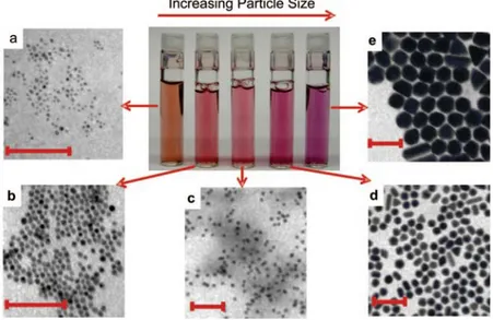
The extraordinary success that gold nanoparticles obtain for medical purpose is almost in part related to the fact that gold has always been considered a highly biocompatible material: it presents a very low toxicity even at high concentrations, no damage to organs occurs after prolonged exposition to gold nanoparticles, it is generally taken up by various organs especially by liver and spleen and could be excreted via the hepatobiliary system.33 Even if additional studies have to be conducted prior to claim the non-toxicity of gold nanoparticles, this fact together with its unique properties has attracted great interest. As already said, the possibility to get more and different properties arises not only from changing size but also by varying shape; this is why during the last decades controllable synthesis of gold nanospheres as well as anisotropic structures have been developed and investigated especially for medical purpose (Figure 1.3.2.2).34
Figure 1.3.2.2: effect of varying size of gold nanoparticles. TEM images (scale bars 100 nm) and picture of different solutions of gold nanoparticles whit increasing size (from a to e). From C.J.
Murphy, Acc. Chem. Res., 2008, 41, 1721-1730.
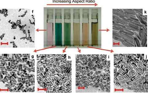
Particularly the nanostructures of gold in the form of rods, called gold nanorods (GNRs), have appeared fascinating. Indeed, they show two distinct plasmon resonance bands: the first, which falls around 520 nm as well as the nanospheres’ one, is due to oscillations of electrons on the transversal (short) axis of the rod, and it is called transversal plasmon resonance (TPR), while the second, which falls instead to longer wavelengths, around 700 nm, is caused by oscillations of electrons along the longitudinal (long) axis and it is called longitudinal plasmon resonance (LPR). This second band is particularly interesting for nanomedicine applications owing to the fact that is much more intense than the other and can be finely
29
“moved” toward higher wavelengths (smaller frequencies) up to 1200 nm by increasing the aspect ratio (ratio between length and width) (Figure 1.3.2.3).
Figure 1.3.2.3: effect of varying the aspect ratio of gold nanorods. TEM images (scale bars 100 nm) and picture of different solutions of GNRs whit increasing aspect ratio (from f to j). From C.J.
Murphy, Acc. Chem. Res., 2008, 41, 1721-1730.
It is known that in the range 800-1200 nm is placed the so-called Near Infrared Window (NIR): this represents for physicians a particularly attractive range of wavelengths due to the high transmittance (low absorbance) of water, deoxygenated haemoglobin and oxygenated haemoglobin in this, which allows the use of laser without interfering with or burning healthy tissues and organs (Figure 1.3.2.4).35
Figure 1.3.2.4: absorption spectra of GNRs (left) and absorbance of water, deoxygenated haemoglobin (Hb) and oxygenated haemoglobin (HbO2) (right).
30
Due to these characteristics GNRs are finding applications in nanomedicine as real theranostic agents. Undeniably, they represent an innovative tool for diagnostic techniques that require the presence of a contrast agent. Several imaging modes can exploit GNRs, such as dark-field microscopy, optoacoustic and photo-optoacoustic imaging, two-photon enhanced luminescence, X-ray tomography, etc. GNRs present considerable advantages in comparison to other common contrast agents as molecular dyes, fluorophores or quantum dots in terms of prolonged stability, reduced photo bleaching under common imaging conditions, drastically reduced toxicity, enhanced absorption cross section and high scattering signal.36 Moreover GNRs can be used as therapeutic tool due to the high capacity in absorbing radiation and convert it into heat. In GNRs more than 96% of the absorbed radiation is converted into heat by non radiative processes: this high optothermal conversion efficiency occurs as a consequence of the higher absorption cross section of GNRs than other nanostructures. GNRs rapidly absorb light but the relaxation process is slower and the result is energy release in form of heat. The generated local hyperthermia originates an increase in the temperature of the surrounding media of several degrees.37 A so strong localized increment in temperature can be exploited to selectively destruction of cancer cells or diseased tissues as a powerful alternative to medical surgery or invasive therapies, making GNRs a real and appealing theranostic agent.
1.3.3 Silver nanoparticles
The history of silver’s use has probably been long as well as the gold one. The people of the ancient Greeks, and Romans after them, already knew the bactericidal properties of silver and they exploited them for food and water disinfection, putting silver coins inside the amphorae to keep beverage and supplies fresh.
In centuries, products containing silver particles in the nanoscale dimensions have been developed and commercialized as pigments (see the Lycurgus cup, Figure 1.2.2.1), wound treatments and biocides. More recently, and especially in the last 100 years, silver nanoparticles (AgNPs) have also found applications in photographs, as conductive composites, new catalysts and from 1980 as enhancer of organic molecules’ signals in Raman spectroscopy. In 1920 FDA approved the first silver solution for antibacterial purpose. Despite the fact that an extraordinary amount of researches has been conducted for the development of silver composites it was only in the last century that became clear that silver
31
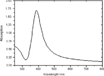
nanoparticles and not simply silver ions were present in most of those products. In 1889, M. C. Lea38 firstly reported the synthesis of a silver colloid stabilized with citrate and having a diameter between 7 and 9 nm, while in 1902 the stabilization of AgNPs by using proteins was reported and the so-obtained composite was firstly used in application for medical purpose.39 Together with gold, also AgNPs possess unique optical properties due to the strong LSPR. For particles of around 10-20 nm the band falls in the visible range at 410 nm, giving the typical yellowish colour to the solutions (Figure 1.3.3.1).
Figure 1.3.3.1: UV-Vis spectrum of silver nanoparticles in aqueous medium.
These special properties made AgNPs suitable for the development of sensors, optical and electronic devices so in the last decades a wide variety of papers and patents regarding their optimal synthesis were spread in the scientific community, making silver colloidal chemistry the most studied one. Silver is much more reactive than gold and the preparation of stabilized nanoparticles is more challenging than the previous one. Indeed AgNPs, as gold nanoparticles, are usually synthesised by reduction of silver salts with or in the presence of stabilizing agents such as citrate or polymers (polyvinyl alcohol or polyvinylpyrrolidone), which avoid a fast re-oxidation of the particles’ surface and consequent aggregation in solution.40
Knowing the potential bactericidal activity of silver ions, AgNPs have attracted great interest for manufacturing of medical and highly specialized devices: textile industry inserts AgNPs into clothes and shoes to prevent sweat odour generation, toothbrushes with AgNPs on their surface were placed on the market to ensure high health and prevent colonies formation, etc. Silver ions are known for they ability to kill bacteria through different pathways: thiol groups present in amino acids and many other thiolated compounds represent an important target for
32
silver attributable the strong interaction between these two atoms. This leads to an increase of concentration of cations in the bacteria cells compartment with the consequence of a strong release of K+ ions, which irreversibly alter the electrolyte transport. Moreover Ag+ ions could enter in the active site of enzymes where sulphate or thiol groups are present blocking some essential metabolic process and leading to cell death. Silver ions also interact with metabolism with the result of an increased reactive oxygen species (ROS) production: this leads to the creation of imperfection in the double layers of membrane cells and brings to the collapse or the release of cytoplasm.
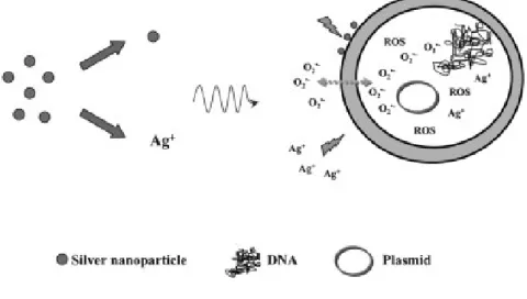
The activity of AgNPs as antibacterial agent is still under investigation, but the release of silver ions from the surface imperfections of the nanoparticles seems to be the driving force of the phenomenon. Certainly silver ions concentration is much lower in cells exposed to AgNPs than silver salts, because nanoparticles ions’ release is slower and time dependent, while silver ions deriving from salts are immediately available (Figure 1.3.3.2).41
Figure 1.3.3.2: induced cell apoptosis by silver nanoparticles and silver ions.
E. T. Hwang, Small, 2008, 4, 746-750.
1.3.4 Surface modification of metal nanoparticles.
As already seen, the synthesis of metal nanoparticles generally occurs in water medium with the assistance of various kinds of surfactants, which stabilized the growing nanoparticles against aggregation phenomena.42 These surfactants remain adsorbed or deposited onto the
33
nanoparticles surface once the process is finished, avoiding post-synthesis collapse of the created nanoparticles.
However, the possibility of transferring the nanoparticles already formed from the aqueous phase to an organic one offers several advantages: in fact, to be able to convey a therapeutic agent and/or a diagnostic tool within a cell it is necessary to entrap them in polymeric micelles that will perform the real drug delivery. This is actually quite complex, since it requires several conditions to be successful, for instance the obtainment of nanoparticles soluble in organic solvent.
The transfer of nanoparticles into an organic phase requires the development of specific ligands: these must replace the surfactants and simultaneously continue to prevent the aggregation phenomena, but at the same time, they must also ensure the solubility in common solvents.
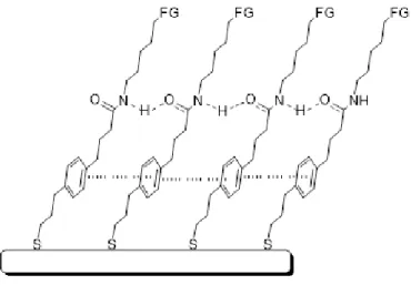
A suitable method to coat nanostructures is the so-called self-assembled monolayer (SAM): it consists of an ordered monolayer, generally with an inclination of 30° respect to the surface, which is formed spontaneously by immersing a solid substrate in a solution containing particular molecules, with "amphifunctional" characteristics. Such molecules must present a "head" with a strong affinity to the metal surface, a central body usually constituted by a long chain and finally a "tail", or a functional group, which will determine the properties both of the monolayer formed and of the entire system (Figure 1.3.4.1).
Figure 1.3.4.1: general representation of a SAM onto a metal structure.
For what concerns the functional group that must bind to the metal surface, several studies have been performed in order to evaluate their affinity with different kinds of metal. It is known as various organic molecules or functional groups are able of chemo-adsorb on
34
surfaces of inorganic solids. Typical examples of these molecules are amides, siloxanes, ethers, acetylacetonates, carboxylic acids, phosphonic acids, and hydroxamic acids:43 all these molecules are able to provide excellent interactions with metal surfaces; in the specific case of gold, however, none of these presents an affinity as high as thiols due to the particularly strength of the interaction sulfur-gold;44 for this reason in recent years researchers have indeed followed this trend and gold nanostructures have been coated with various organic ligands containing thiols but also disulfides functional groups. On the contrary, in the literature there are still a few examples regarding the functionalization of silver nanostructures using thiols groups, and in the majority of the studies conducted on these nanoparticles a coating with other functional groups, such as amines, has been preferred, although even this noble metal presents strong affinity with ligands containing sulfur.45
As regards the chain which constitutes the "body" of the ligand, previous research has demonstrated the importance of the presence of amide junctions and aromatic components, the first capable of forming hydrogen bridges between the various chains, the second able to give π-stacking interactions that ensure greater stability to the structure.46
Finally, the terminal functional group of the ligand must confer the desired properties to the entire system. By introducing appropriate functional groups at the end of the ligands it is possible to obtain stability in different solvents or for instance make them react with common reactions of organic chemistry once they are already firmly anchored to the nanoparticles’ surface.
By exploiting these potentialities, many opportunities, still poorly investigated, become accessible, such as the possibility to attach biomolecules directly onto the nanoparticles surface, thus creating hybrid systems applicable in nanomedicine, or in addition, the assembly of two different nanostructured systems by surface chemical reaction, in order to obtain a single complex system where the properties of the nanostructures remain intact thanks to the link of separation formed by the ligands themselves. Indeed, in the literature there are several examples of the creation of complex nanostructured systems obtained by growing a nanoparticle on the surface of another already existing, but at the same time examples of conjunction of two of them by simply organic reactions are still few or missing.47
Among these simple organic reactions, the famous click chemistry must be mentioned. The click chemistry’s concept was firstly introduced by Barry Sharpless in 2001 as an innovative synthetic strategy for the preparation of substances in a quickly and reliably manner.48 The reactions that meet the cardinal principles of click chemistry are those that occur under mild reaction conditions, that do not involve harmful solvents, or that are solvent-free and produce
35
high yield, harmless by-products, high stereospecificity, high atom economy, easily isolable products. The reaction should possess a large thermodynamic driving force (greater than 84 KJ/mol) in order to obtain a single reaction product, condition made possible thanks to reagents strongly activated known as spring-loaded reagents. It is clear that it is nearly impossible for a reaction to meet all the above criteria, but some of them are very close to the goal such as nucleophilic substitutions of epoxides and aziridines, additions to activated carbon-carbon double bonds, the Diels-Alder cycloaddition and 1,3-dipolar cycloadditions.49 The 1,3-dipolar cycloaddition reactions are an important class of reactions for the synthesis of polyfunctionalised heterocycles in a regio- and stereoselective manner.
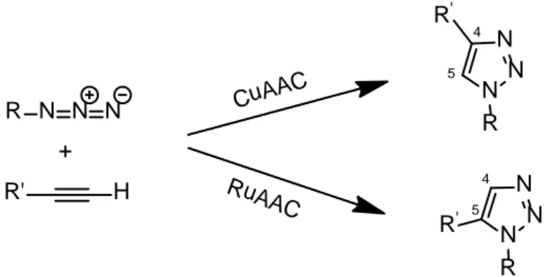
The most famous example is the Huisgen’s cycloaddition, whose copper catalyzed version is often simply known as the click reaction.50 It provides for the coupling between an azide and an acetylene functional group forming a triazole, which can be variously substituted; however both azides and alkynes are kinetically stable and for that reason the reaction requires high temperatures and long times to be completed, or specific catalysts.
Copper (I) is the most used catalyst, insomuch that the reaction in this case takes the name of Copper-catalyzedazide alkyne cycloaddition (CuAAC); more specifically, copper catalyst not only greatly accelerates the reaction bringing it to completeness in a few hours, but at the same time leads to the regioselective generation of only the 1,4-regioisomer and not both the 1,4- and 1,5- as is the case of the non-catalyzed reaction. In recent years further possible catalysts have been investigated: among these ruthenium gave excellent results, in the so called "Ruthenium-catalyzed azide alkyne cycloaddition" (RuAAC),51 which is capable, as copper is, to accelerate the reaction and to make it regiospecific, but generating only the opposite regioisomer, namely the 1,5 -substituted triazole. (Figure 1.3.4.2)
R N N N + R' H CuAAC RuAAC N N N R R' N N N R R' 4 5 4 5
Figure 1.3.4.2: schematic representation of the Huisgen copper- and ruthenium-catalized azide alkyne cycloaddition.
36
Moreover, this reaction presents other substantial advantages since it can be conducted in common solvents such as tetrahydrofuran (THF), N,N-dimethylformamide (DMF), dimethylsulfoxide (DMSO), but also in water; it also occurs at ambient temperature, does not fear the presence of air and essentially generates no by-products, therefore it can be considered ideal for the reaction of complex and labile systems, such the nanostructured ones.
37
1.4. Polymeric nanoparticles
1.4.1 Advantages, properties and materials
As discussed in chapter one, the transport and delivery processes of nanoparticles through the body are of critical importance to obtain satisfactory results in nanomedicine. These processes are strongly affected by the physic-chemical characteristics of the nanoparticles, such as size, shape, surface charge, as well as their chemical properties. For instance, it has been demonstrated that nanoparticles with a mean diameter smaller than 60–70 nm are excreted fast via urinary system, while larger nanoparticles, with diameter larger than 250 nm can be easily sequestrated by liver or spleen: this shows how important the control of nanoparticles size is for successful applications in vivo.52 In light of these considerations, it is clear how much importance plays the choice of materials for the creation of nanoparticles and nanocarriers especially. Nanoparticles for drug delivery applications are generally prepared from a wide variety of materials,53 including metals, lipids and polymers.54
Metal nanoparticles, as previously shown in chapter two, surely offer the most promising opportunities in terms of special properties, therapeutic efficacy, imaging possibilities etc., but they also present several limitations for medical application when used alone, especially due to their fast elimination from body and/or degradation in physiological conditions.
In addition to this, polymeric nanoparticles (PNPs) have spread for applications in drug delivery and nanomedicine because they proved to be easily synthesizable with tunable characteristics and able to entrap and transport common poor water-soluble drugs greatly improving their solubility, bioavailability, retention time and release profile.55 By using polymeric nanoparticles, several disadvantages belonging to traditional therapies can be overcome. These generally concern a not efficient drug absorption in body, a too fast elimination through kidneys or liver and the spread of the drug in various tissues or non-target organs; the major consequences are the reduction of the therapeutic index of the drug, more doses necessary to obtain efficacy and several unwanted side effects. PNPs carrying drug or other moieties can penetrate deep into tissue through capillaries and reach merely the target tissue, where they can be taken up by the cells, limiting simultaneously fast clearance and side effects.56 Having seen all the benefits and advantages in the use of PNPs, many efforts have been made in the last decades to fulfil several requirements necessary for the application of these systems in vivo: scientists investigated numerous physical and biochemical properties,