Alma Mater Studiorum – Università di Bologna
DOTTORATO DI RICERCA IN
BIOLOGIA CELLULARE E MOLECOLARE
Ciclo XXVI
Settore Scientifico Disciplinare: 05/E2
Settore Concorsuale di afferenza: BIO/11
TITOLO TESI
Polymerizing activity and regulation of group B
Streptococcus pilus 2a sortase C1
Presentata da:
Francesca Zerbini
Coordinatore Dottorato
Relatore
Chiar.mo Prof. Chiar.mo Prof.
Vincenzo Scarlato
Vincenzo Scarlato
Co-relatore
Dott.ssa
Roberta Cozzi
2
Oggetto del mio progetto di dottorato, presentato in questo lavoro di tesi, è stato lo studio del meccanismo di assemblaggio del pilo 2a di Streptococcus agalactiae
(Streptococco di gruppo B, GBS), focalizzandomi soprattutto sull‟attività e la
regolazione della sortasi C1.
Il lavoro svolto durante lo svolgimento di questo progetto di dottorato è stato oggetto delle seguenti pubblicazioni:
- Cozzi R*, Zerbini F*, Assfalg M, D'Onofrio M, Biagini M, Martinelli M, Nuccitelli A, Norais N, Telford JL, Maione D, Rinaudo CD. Group B Streptococcus pilus sortase regulation: a single mutation in the lid region induces pilin protein polymerization in vitro. FASEB J. 2013 Aug;27(8):3144-54. Epub 2013 Apr 30.
* These authors contributed equally to this paper
- Cozzi R, Nuccitelli A, D'Onofrio M, Necchi F, Rosini R, Zerbini F, Biagini M, Norais N, Beier C, Telford JL, Grandi G, Assfalg M, Zacharias M, Maione D, Rinaudo CD. New insights into the role of the glutamic acid of the E-box motif in group B Streptococcus pilus 2a assembly. FASEB J. 2012 May;26(5): 2008-18.
3
Table of Contents
Abstract ... 6
Chapter 1. Introduction ... 7
1.1Structure and assembly of Gram-positive bacterial pili ... 7
1.2.1 Streptococcus agalactiae (Group B streptococcus, GBS) ... 15
1.2.2 Identification of novel genomic islands coding for pilus-like structures in Streptococcus agalactiae ... 17
1.3 Sortase enzyme in Gram-positive bacteria ... 23
1.4 The sortase A class ... 28
1.5 Protein engineering using sortase enzymes... 33
1.5.1 Engineering of bacterial surfaces ... 33
1.5.2 C-Terminal and N-Terminal ... 34
1.5.3 Other sortases application as protein engineered ... 37
1.6 Sortases that assemble pili: class C enzymes ... 38
1.7 Class C sortases in GBS ... 40
1.7.1 Structural organization and biochemical characterization of PI-1 and PI-2a sortase C enzymes ... 41
Aim of the thesis ... 52
Chapter 2. Results ... 53
2.1 Recombinant S. agalactiae SrtC1 of PI-2a production... 53
2.2 Recombinant S. agalactiae and S.pneumoniae backbone proteins production ... 55
2.3 Wild-type SrtC1 is not able to induce recombinant BP polymerization in vitro ... 58
2.4 BP-2a high molecular weight structures can be assembled in vitro by recombinant SrtC1 lid mutant ... 61
4
2.5 Lysine 189 in the putative pilin motif and the IPQTG sorting signal of BP-2a
are essential for pilus formation in vivo ... 63
2.6 The IPQTG sorting signal is essential for the transpeptidation reaction mediated in vitro by the SrtC1Y86A mutant ... 65
2.7 The SrtC1Y86A active mutant is able to polymerize in vitro backbone proteins of other GBS pili and/or pathogens ... 67
2.8 The GFP protein containing a C-term LPXTG-motif is polymerized in vitro by SrtC1Y86A ... 69
2.9 Biochemical characterization of SrtC1-2a wild type and active mutant reveals that the lid is involved in protein stability ... 70
2.10 Lid anchoring to the active site leads to an overall protection of SrtC1 from proteolysis ... 74
2.11 SrtC1 enzyme deleted of the entire N-terminal region is active in polymerizing BP in vitro ... 78
Chapter 3. Discussion ... 80
Chapter 4. Experimental procedures ... 86
4.1 Materials and reagents... 86
4.2 Bionformatics ... 86
4.3 Bacterial Strains, Media and Growth Conditions ... 86
4.4 PI-2a SrtC1 recombinant cloning and expression ... 87
4.5 Recombinant backbone proteins cloning and expression ... 88
4.6 In vitro pilus polymerization ... 90
4.7 Differential scanning fluorimetry (DSF) ... 90
4.8 Antisera ... 91
4.9 Bacterial strains and growth conditions ... 91
4.10 Construction of complementation vectors ... 91
4.11 Western Blot Analysis ... 92
4.12 Limited proteolysis assay ... 93
5
4.14 Analytic size-exclusion chromatography ... 93 4.15 NMR spectroscopy ... 94 Bibliography ... 96
6
Abstract
Group B Streptococcus [GBS; Streptococcus agalactiae] is the leading cause of life-threatening diseases in newborn and is also becoming a common cause of invasive diseases in non-pregnant, elderly and immune-compromised adults. Pili, long filamentous fibers protruding from the bacterial surface, have been discovered in GBS, as important virulence factors and vaccine candidates. Gram-positive bacteria build pili on their cell surface via a class C sortase-catalyzed transpeptidation mechanism from pilin protein substrates. Despite the availability of several crystal structures, pilus-related C sortases remain poorly characterized to date and their mechanisms of transpeptidation and regulation need to be further investigated. The available three-dimensional structures of these enzymes reveal a typical sortase fold except for the presence of a unique feature represented by an N-terminal highly flexible loop, known as the “lid”. This region interacts with the residues composing the catalytic triad and covers the active site, thus maintaining the enzyme in an auto-inhibited state and preventing the accessibility to the substrate. It is believed that enzyme activation may occur only after lid displacement from the catalytic domain. In this work we provide the first direct evidence of the regulatory role of the lid, demonstrating that it is possible to obtain in vitro an efficient polymerization of pilin subunits using an active C sortase lid mutant carrying a single residue mutation in the lid region. Moreover, biochemical analyses of this recombinant mutant reveal that the lid confers thermodynamic and proteolytic stability to the enzyme. A further characterization of this sortase active mutant showed promiscuity in the substrate recognition, as it is able to polymerize different LPXTG-proteins in vitro.
7
Chapter 1. Introduction
1.1 Structure and assembly of Gram-positive bacterial pili
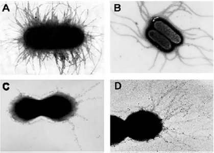
Pili, or fimbriae, are protein polymers that form long, filamentous structures that extend from bacterial cells, mediating adhesion to host cells, colonization, biofilm formation and sometimes motility (Proft and Baker 2009). Pili of pathogenic organisms are also highly immunogenic, making them attractive for vaccine development. The best-known and characterized pili are those of Gram-negative bacteria: the Type I and Type P pili of Escherichia coli, and the Type IV pili of Neisseria species (Waksman and Hultgren 2009), which form rod-like bundles of non-covalently assembled subunits. In contrast, the pili on Gram-positive bacteria are fundamentally different. They are long (2–5 µm) but extremely thin (about 3 nm), assembled by enzymes called sortases, and they are rare examples of covalent polymers (Fig.1). Despite many years of study of Gram-positive bacterial pili, they remained largely unnoticed until very recently (Kang and Baker). Their characterization followed the discovery of sortases and the availability of genome sequences (Kang and Baker). The assay generally used to determine the expression of pilus structures is to subject the total bacterial cell lysate to boiling in SDS followed by SDS-PAGE. A protein that is part of a pilus will appear as a high molecular weight (HMW) ladder in immunoblot. Another method used to detect pili is visualization by negative staining, or, more specifically, by immunogold electron microscopy (IEM), which can reveal the localization of a protein within the pilus structure. Gram-positive pili are composed of multiple copies of a single pilin shaft, other than additional proteins
8
associated with the shaft, but not required for the integrity or synthesis of the pilus (Ton-That and Schneewind 2003).
Early data from studies of oral Gram-positive pathogens indicated that such structures are involved in adhesion and attachment to host cell, in the interaction with components of the extracellular matrix (ECM), and in biofilm formation (Konto-Ghiorghi, Mairey et al. 2009). Additionally, a recent study provided evidence for an active role of S. agalactiae pilus proteins in the newly discovered paracellular translocation through the epithelial barrier, during host colonization (Soriani, Santi et al. 2006). Gram-positive pili could be considered important virulence factors for several diseases (Nallapareddy, Singh et al. 2006), in particular infections of the urinary, genital and gastrointestinal tracts. Furthermore, in pathogenic Streptococcus species pili are reported to be also promising vaccine candidates (Maione, Margarit et al. 2005).
Figure 1. Different examples of pilus-like structures in negative and Gram-positive bacteria. Electron micrographs of fimbriae in Gram-negative organisms : E. coli
9 Gram-positive bacteria: fibrils in Streptococcus salivarius (C) and pili in Streptococcus agalactiae (D) stained by immunogold labeling (Telford, Barocchi et al. 2006).
Thon-That and Schneewind, working on Corynebacterium diphteriae as a model, have provided the first insights into the assembly mechanism of Gram-positive pili (Ton-That and Schneewind 2003). The three pilus proteins together with genes coding for sortases, that are required for pilus assembly, are encoded in a small gene cluster within pathogenicity islands which are known as Pilus Islands (PIs). The genes are transcribed in the same direction, indicating that they are part of an operon. The three pilus components are characterized by the presence of an N-terminal signal peptide together with a C-terminal cell-wall sorting signal (CWSS), that is found in many surface proteins and is required for the attachment to the peptidoglycan of the cell wall. The CWSS comprises the amino acid sequence “LPXTG” (where X denotes any amino acid) or a variation of this motif (such as VV/PXTG in the case of the main pilin subunit of Group A streptococcus, Cpa), followed by a hydrophobic membrane-spanning domain and a positively charged tail. This motif is targeted by sortase enzymes, which are membrane-bound transpeptidases catalysing the covalent linkage of LPXTG motif proteins to the peptidoglycan. During pilus formation, specific pilus-related sortases catalyse the covalent attachment of the pilin subunits to each other and to the peptidoglycan cell wall (Telford, Barocchi et al. 2006). Immunogold electron microscopy (IEM) using antisera specific for the three pilus components revealed that pilus shaft is a polymer of one pilin called backbone protein (BP), and the other two components are ancillary proteins (AP). Backbone protein specific
10
antisera stain the whole length of the pilus structure (Telford, Barocchi et al. 2006).
The first insights into the assembly mechanism of Gram-positive pili were provided by a study performed on Corynebacterium diphteriae (Ton-That, Marraffini et al. 2004).
Initially, the three pilus components containing an LPXTG motif are secreted in a Sec-dependent way (Telford, Barocchi et al. 2006). Each component remains anchored to the cell membrane, owing to the presence of the C-terminal transmembrane domain.
The second step involves a sortase-dependent reaction in which the membrane-anchored proteins are cleaved at the LPXTG motif, between the threonine (T) and glycine (G) residue. This reaction leads to the formation of acyl-enzyme intermediates in which a covalent thioester bond is formed between the thiol group of the cysteine residue located in the catalytic pocket of the sortase and the carboxyl group of the threonine residue in the LPXTG motif of the pilin protein (Telford, Barocchi et al. 2006). Because sortases are membrane-associated enzymes, the acyl-enzyme derivatives that are formed are retained on the external side of the membrane (Fig. 2).
The following steps of the assembly process involve the oligomerization of the pilus protein subunits and the anchoring of the oligomerized structure to the cell wall.
These steps require the nucleophilic attack of the thioester bond in the acyl-enzyme intermediate. During pilus polymerization the nucleophile is provided by the ε-amino group of a specific lysine (K) residue within the “pilin motif”,
11
WXXXVXVYPKN (where X denotes any amino acid), which has been found in most of the pilin subunits that have been characterized (Ton-That and Schneewind 2003). The nucleophilic attack results in cleavage of the thioester bond and concomitant formation of an amide bond between the carbonyl-group carbon of the threonine residue of the pilin subunit (present in the catalytic pocket of the sortase) and the lysine side-chain (ε-amino group) of the pilin motif of the neighboring pilin subunit. This leads to the formation of a membrane-associated covalently linked dimer with a pilin motif that can interact with other sortase- associated pilin subunits, forming an elongated pilus fiber. Ton-That and co-workers have shown that replacing the lysine residue in the pilin motif with an alanine residue abolishes the polymerization process, highlighting the importance of this conserved sequence in pilus formation (Telford, Barocchi et al. 2006). According to this model, pilus growth occurs by subunit addition at the base of the pilus (Fig. 2), and the length of the pilus depends on the relative abundance of the pilus subunits that are coupled to the membrane-associated sortases (Telford, Barocchi et al. 2006). Finally, the association of the membrane-proximal pilus subunit with the cell wall occurs when the thioester bond between the subunit and the sortase is subject to nucleophilic attack by the amino group in the cross-bridge of the peptidoglycan precursor lipid II (Ton-That and Schneewind 2004), and this leads to the formation of an amide bond between the basal subunit and the bacterial cell wall.
12
Figure 2. General model for pilus assembly in Gram-positive bacteria (Telford, Barocchi et al. 2006). (A) In the first step, proteins that contain the amino-acid motif
LPXTG are targeted to the cell membrane by Sec-dependent secretion (not shown). This is followed by a sortase-mediated reaction (indicated by the arrows) in which the LPXTG motif is cleaved between the threonine (T) and glycine (G) residues. (B) The reaction leads to the formation of an acyl-enzyme intermediate in which a covalent thioester bond is formed between the thiol group of a cysteine residue in the sortase and the carboxyl group of the pilin threonine residue. (C) Oligomerization occurs after the nucleophilic attack provided by the e-amino group of the lysine residue in the pilin motif on the cysteine residue of the sortase. (D)The thioester bond between the pilin subunit and the sortase is targeted by the amino group of the pentapeptide of lipid II, the precursor of peptidoglycan. (E) This leads to the formation of an elongated pilus covalently linked to the cell wall peptidoglycan. NAG, N-acetyl glucosamine; NAM, N-acetyl muramic acid (Telford, Barocchi et al. 2006).
13
It has been suggested that another conserved aminoacidic sequence in the backbone subunit, called the “E-box” (consensus YxLxETxAPxGY), due to a highly conserved glutamic acid residue, plays a role in pilus polymerization (Telford, Barocchi et al. 2006).
Despite low sequence similarities, the pilin subunits of gram-positive bacteria show very similar tridimensional structure comprising immunoglobulin G (IgG)-like domains of shared evolutionary origin. Each pilin subunit is stabilized by intramolecular isopeptide bonds, and all contain sequence elements and/or residues that are essential for pilus assembly, and which are conserved among pilin subunits in different bacteria (Rosini, Rinaudo et al. 2006). Such motives include the above mentioned pilin motif, the cell-wall sorting signal (CWSS) containing the sortase recognition site LPxTG motif, and the E-box motif as assigned for the first time in the major pilin subunit SpaA of Corynebacterium diphtheriae (Ton-That, Marraffini et al. 2004) and subsequently in other bacterial pilins (Mandlik, Swierczynski et al. 2008). The E-box contains a conserved glutamic acid residue, which in C. diphtheria SpaA (Glu-446) has been demonstrated to be essential for the incorporation of the minor pilins SpaB and SpaC (Ton-That, Marraffini et al. 2004). Intriguingly, in SpaA, this glutamate is the catalytic residue that mediates the formation of the Lys-363–Asn-462 intramolecular isopeptide bond (Kang, Paterson et al. 2009), similar to the role assigned to Glu-258 in GAS Spy0128, in which this residue was shown to be essential for the corresponding intramolecular reaction to occur (Kang, Coulibaly et al. 2007). Moreover, several X-ray crystal structures of backbone pilins have shown that the E-box domain is involved in the formation of such isopeptide
14
bonds and that these linkages confer higher stability to the monomeric subunit (Hendrickx, Budzik et al.; Kang and Baker; Kang, Coulibaly et al. 2007). Recently, the X-ray crystal structure of the shaft-forming backbone protein of S. agalactiae pilus 2a (BP-2a) was solved (Nuccitelli, Cozzi et al.). The 3-D structure revealed an IgG-like fold domains organization, comprising 4 structural units, designated D1–D4. The domains D2, D3, and D4 are each stabilized by an intramolecular Lys-Asn isopeptide bond, located in a largely hydrophobic pocket, comprising several aromatic residues, including a bond-catalyzing aspartyl or glutamyl residue (Fig.3) (Nuccitelli, Cozzi et al.). However, the role of intramolecular isopeptide bonds and of the E-box motif in pilus assembly still needs to be clarified (Cozzi, Nuccitelli et al. 2012).
Figure 3. Structural analysis of BP-2a-515. (A) Ribbon representation of the crystal
structure of BP-2a-515 (residues 190–640), illustrating the N and C termini, domains D2, D3, and D4, two potassium ions (blue spheres), and the three intramolecular isopeptide bonds (spheres). (B) Superimposition of BP-2a-515 (purple) with RrgB from
15 Streptococcus pneumoniae (blue), highlighting the structural similarity between the two proteins. (C) Structural details of the D2, D3, and D4 domains in the regions involved in isopeptide bond formation. All images were generated using Pymol Version 1.1r1 (www.pymol.org) (Nuccitelli, Cozzi et al. 2011).
In conclusion, pilus assembly in Gram-positive bacteria seems to occur by a universal mechanism of ordered cross-linking of precursor proteins, whose multiple conserved features are recognized by designated sortase enzymes (Ton-That and Schneewind 2003; Ton-(Ton-That, Marraffini et al. 2004).
1.2 Pili in Group B Streptococcus
1.2.1 Streptococcus agalactiae (Group B streptococcus, GBS)
Streptococcus agalactiae (commonly referred to as Group B Streptococcus or GBS) is an encapsulated Gram-positive coccus, catalase negative and facultatively anaerobic. It generally grows in pairs or in long chains of spherical bacteria, less than 2 m in size (Fig.4A). It displays beta-hemolysis when cultured on blood agar plates and produces zones of hemolysis that are only slightly larger than the colonies themselves (Fig.4B) (Gibbs, Schrag et al. 2004). GBS strains are classified into nine serotypes according to immunogenic characteristics of the capsule polysaccharides (Ia, Ib, II, III, IV, V, VI, VII, VIII and IX). Approximately, 10% of serotypes are non-typeable (Kogan, Uhrin et al. 1996).
16
Figure 4. Streptococcus agalactiae. (A) Scanning Electron Microscopy (SEM) of
Streptococcus agalactiae. (B) Colonies of Streptococcus agalactiae on a blood agar plate. Note the zone of clear haemolysis.
Consistent with other streptococcal species (Mitchell 2003), Streptococcus agalactiae is present on the mucosal surfaces of animals and humans. In fact, GBS can usually colonize as a normal commensal the intestinal and vaginal tract but also the pharyngeal mucosa of human adults (Baker 1997) and 20–40% of healthy women carry GBS (Baker 1997; Hansen, Uldbjerg et al. 2004; Yamamoto, Pargade et al. 2006).
Invasive group B streptococcal disease emerged in the 1970s as a leading cause of neonatal morbidity and mortality in the United States (McCracken 1973), and represents the most common etiological agent of invasive bacterial infections (pneumonia, septicaemia and meningitis) in human neonates (Nizet, Gibson et al. 1996; Davies, Adair et al. 2001; Gibbs, Schrag et al. 2004). Most infections and colonization of newborns are due to aspiration of contaminated amniotic and vaginal fluid before or during delivery (Doran and Nizet 2004).
Streptococcus agalactiae is also associated to a number of postpartum sequelae, such as urinary tract infections, amnionitis, endometritis, as well as to wound
17
infection and mortality or morbidity in immunocompromised adults (Schuchat 1998).
Among them, pili have been recently implicated in mediating attachment to human epithelial cells (Dramsi, Caliot et al. 2006), and in the binding and invasion of brain microvascular endothelial cells (Maisey, Hensler et al. 2007).
1.2.2 Identification of novel genomic islands coding for pilus-like structures
in Streptococcus agalactiae
A Reverse Vaccinology approach (De Groot and Rappuoli 2004) has been used to identify protective antigens for inclusion in a vaccine against GBS. Five proteins were found to elicit protection against GBS in a mouse maternal immunization assay (Maione, Margarit et al. 2005). Furthermore, analysis of the eight sequenced genomes of GBS has shown that four of these five protective antigens, GBS80 (TIGR annotation SAG0645), GBS104 (SAG0649), GBS67 (SAG1408) and GBS59 (SAG1407), are located in tandem in two different genomic islands that belongs to the “dispensable genome” of GBS (Tettelin, Masignani et al. 2005) (Lauer, Rinaudo et al. 2005; Rosini, Rinaudo et al. 2006). The genes coding for GBS80 and GBS104 are localized in a genomic island, named Pilus Island 1 (PI-1), containing genes coding for three LPXTG proteins and two sortases with similar organization to the genes coding for pilus-like structures in C. diptheriae (Fig.5A) (Ton-That and Schneewind 2003).
The genes are transcribed in the same direction, indicating that they are part of an operon. GBS80, GBS104, and GBS52 (SAG0646), represent the three LPXTG motif containing proteins of this island. The other two genes (SAG0647 an
18
SAG0648) code for sortase enzymes, which are known to catalyse the covalent linkage of LPXTG motif proteins to the peptidoglycan (Fig.5A).
Figure 5. Schematic representation of GBS pilus-island regions. (A. pilus island 1; B.
pilus island 2) Genes coding for LPXTG-containing proteins are represented with orange arrows, whereas transcriptional regulators are in green and conserved flanking genes are in grey. At least two sortases are present in each PI (black arrows), while a signal peptidase is present in PI-2b (yellow arrow). In PI-1, transposable elements are also present (blue arrows), as well as interrupted or frame-shifted genes (white arrows). The insertion site for the 51 kb prophage in PI-1 of strains A909 and CJB111 is shown. For PI-1 and PI-2a, gene numbers are relative to the database annotation for strain 2603 V/R, while for PI-2b, gene numbers are relative to COH1 strain. DR: direct repeat (Rosini, Rinaudo et al. 2006).
19
PI-1 consists of an approximately 16 kbp-long DNA region flanked by 11 bp of direct repeats, and it has been found in ≈ 70% of the GBS strains that have been analysed (Tettelin, Masignani et al. 2005; Margarit, Rinaudo et al. 2009). Two conserved genes (sag0633 and sag0652), that are present in all GBS strains that have been analysed, flank this DNA region. In strains that lack the region, the flanking genes are contiguous. In addition to the pilus genes, the genomic island contains a gene that encodes an AraC-type transcriptional regulator, as well as a gene (spy0123) that encodes a heat-shock protein (Hsp33) and remnants of transposase-like genes. Two strains, A909 and CJB111, contain an insertion of a 51.2 kb-long prophage at one end of the 16 kbp-long island (Fig. 5A) (Rosini, Rinaudo et al. 2006). The overall organization of this genomic region suggests that the complete island may have been acquired by horizontal DNA transfer. The other two protective antigens, GBS67 and GBS59, are located in a second island with a similar organization to Pilus Island-1 and for this reason named Pilus Island 2 (PI-2) (Fig. 5B). As PI-1, the second pilus locus is located in a variable region of the genome and contains genes coding for three LPXTG proteins (GBS67, GBS59, and GBS150) and two sortases (Fig. 5B).
There are two variants of this region (PI-2a and PI-2b), which differ in an 11-kb segment of DNA that is flanked by identical conserved genes (sag1403 and sag1410). The two variants encode for distinct pili that have only limited amino-acid sequence similarity. PI-2a contains, in addition to pilus genes, a gene that encodes for a RogB-type transcriptional regulator. PI-2b lacks the transcriptional regulator but contains a gene that encodes for a protein similar to the LepA-type signal peptidase of Gram-negative bacteria (Fig.5B).
20
In summary, there are three genomic islands in GBS that are found at two different genomic locations. The three islands are similar in organization but poorly conserved among different isolates. All strains analyzed carried at least 1 of the islands, and 94% expressed pili on their surface (Margarit, Rinaudo et al. 2009). PCR and FACS analysis on a wide panel of GBS clinical isolates revealed that pilus 2a is the most represented and surface exposed among the three pilus types (Margarit, Rinaudo et al. 2009).
Immunoblot analysis, using sera raised against the three LPXTG proteins present in each island, showed that all proteins were part of high molecular weight (HMW) covalently-linked polymers (Fig.6A, C and E). Immunogold electron microscopy (IEM), using antibodies raised against GBS80 (for PI-1), GBS59 (for PI-2a) and GBS1518 (for PI-2b) showed that these polymers constitute pilus-like structures extending beyond the bacterial surface (Lauer, Rinaudo et al. 2005) (Rosini, Rinaudo et al. 2006) (Fig. 6B, D and F).
Each PI of GBS contains two genes encoding SrtC transpeptidases. Generation of deletion mutants showed that both enzymes are capable of polymerizing the backbone pilus subunit, but each preferentially incorporates one of the two ancillary proteins (Rosini, Rinaudo et al. 2006).
21
Figure 6. Novel genomic islands code for pilus-like structures. (A) Immunoblots of
total protein extracts from JM9130013 strain probed with antisera specific for PI-1 proteins GBS80 (α-80), GBS104 (α-104) and GBS52 (α-52). (B) Immunogold labeling and transmission electron microscopy of GBS80 in strain JM9130013, showing long pilus-like structures.(C) Immunoblots of total protein extracts from 515 strain probed with antisera specific for PI-2a proteins GBS59 59), GBS67 67) and GBS150 (α-150). Asterisks (*) indicate the monomeric form of GBS59, GBS67 and GBS150. (D) Immunogold electron microscopy of 515 strain incubated with sera raised against GBS59 protein and labeled with secondary antibodies conjugated with 10nm gold particles. (E) Immunoblots of total protein extracts from JM9130013 strain probed with antisera specific for PI-2b proteins SAN1518 1518), SAN1519 1519) and SAN1516 (α-1516). (F) Immunogold electron microscopy of JM9130013 wt strain incubated with sera
22 raised against GBS1518 protein and labeled with secondary antibodies conjugated with 10nm gold particles (Rosini, Rinaudo et al. 2006).
There is growing evidence that, in addition to the SrtC transpeptidases, the housekeeping SrtA may play a role in GBS pilus assembly. Indeed, a study based on the generation of a knock-out strain for srtA gene revealed that the enzyme is not involved in pilus polymerization, but it is essential for the permanent anchoring of GBS pilus 2a to the cell wall (Nobbs, Rosini et al. 2008). Moreover, a detailed analysis of PI-2a identified the ancillary protein GBS150 as the substrate for SrtA.
23
1.3 Sortase enzyme in Gram-positive bacteria
In Gram-positive bacteria, a class of surface proteins are covalently anchored on the cell wall by a transpeptidase, which has been called sortase (Srt) (Paterson and Mitchell 2004) (Ton-That, Marraffini et al. 2004) (Clancy, Melvin et al.). Sortases are positioned at the cytoplasmic membrane via a membrane anchor located either at the N- or C-terminus, contain the active site, LxTC motif (Marraffini, Dedent et al. 2006), of which cystein is essential for the sortase activity (Ton-That, Liu et al. 1999) and recognize their substrate proteins via a common C-terminal pentapeptide sequence, which acts as a cell wall sorting signal.
So far, more than 700 putative sortase substrates encoded by more than 50 different prokaryotic genomes have been identified (Nguyen, Phan et al.).
These enzymes have also been developed into powerful molecular biology reagents to site-specifically attach proteins to a variety of biomolecules (Tsukiji and Nagamune 2009) (Popp and Ploegh). Although they are not essential for bacterial viability when cells are grown in rich media, sortases can be important virulence factors as they display surface proteins that mediate bacterial adhesion to host tissues, host cell entry, evasion and suppression of the immune response and acquisition of essential nutrients. The sorting reaction catalyzed by the sortase A protein from Staphylococcus aureus (Sa-SrtA) is the best understood and begins when a full-length precursor protein containing an amino terminal leader peptide is exported from the cytoplasm through the secretory pathway (Fig.7). The C-terminal CWSS is then processed by Sa-SrtA. The CWSS consists of a
24
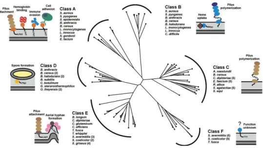
LPXTG motif, followed by a segment of hydrophobic amino acids, and a tail composed primarily of positively charged residues. The C-terminal charged tail presumably retards export, positioning the protein for processing by the extracellular membrane associated Sa-SrtA enzyme. A highly conserved active site cysteine residue in Sa-SrtA then nucleophilically attacks the backbone carbonyl carbon of the threonine residue in the LPXTG motif, breaking the threonine and glycine peptide bond and creating a sortase-protein complex in which the components are linked via a thioacyl bond. The protein is then transferred by Sa-SrtA to the cell wall precursor lipid II, when the amino group in this molecule nucleophilically attacks the thioacyl linkage to create an isopeptide linked protein-lipid II product. Transglycosylation and transpeptidation reactions that synthesize the cell wall then incorporate this product into the peptidoglycan, where it is covalently linked to the cross-bridge peptide. Other sortases catalyse a similar transpeptidation reaction, but join remarkably different LPXTG motifs and amino groups. Since the discovery of Sa-SrtA a little more than decade ago by Schneewind and colleagues (Mazmanian, Liu et al. 1999), over 800 genes encoding related proteins have been identified in ~260 distinct bacterial species (Finn, Mistry et al.). The vast majority of sortases is found in Gram-positive bacteria that contain a conventional cell wall (they are absent in Mollicutes) (Pallen, Lam et al. 2001). Most bacterial species contain multiple sortase enzymes that have been named in an ad hoc manner (e.g. SrtA, SrtB, SrtC, etc.). To provide a framework in which to discuss their functions, the sortases from Gram-positive bacteria were grouped into families based upon their primary sequences (Fig.8). Approximately 60% of all sortase proteins can be partitioned into six
25
distinct families of enzymes that share related amino acid sequences, these include class A to F enzymes (Comfort and Clubb 2004) (Dramsi, Caliot et al. 2006). Experimental and bioinformatics analyses indicate members of each group recognize distinct CWSSs in which the LPXTG sequence is varied (hereafter called sorting signal motifs). Class A enzymes are present in Firmicutes and have been studied extensively. They appear to perform a housekeeping role in the cell as members of this group are capable of anchoring a large number of functionally distinct proteins to the cell wall. Class B enzymes are also present in Firmicutes and can have distinct functions. Some members of this group attach haem-receptors to the peptidoglycan, while others assemble pili. Most surface proteins attached by class A enzymes contain a canonical LPXTG motif within their CWSS and have diverse functions that can promote bacterial adhesion, nutrient acquisition, host cell invasion, and immune evasion. Class A enzymes have attracted significant interest as potential drug targets because a number of clinically important pathogens use these sortases to display virulence factors and they are attenuated in their virulence if their srtA gene is eliminated (S. aureus, L. monocytogenes, Streptococcus pyogenes and Streptococcus pneumoniae among others) (Naik, Suree et al. 2006) (Maresso, Chapa et al. 2006).
26
Figure 7. Mechanisms of sortase mediated attachment of surface proteins and pilus assembly at the bacterial cell wall.
(A) The S. aureus housekeeping sortase A anchors surface proteins to the peptidoglycan.
The precursor protein containing an amino terminal leader peptide is secreted across the membrane through the Sec pathway. The exported protein (light blue) is processed by the sortase enzyme (dark blue, labelled „A‟), which recognizes the LPXTG sequence and cleaves the surface proteins between the threonine and glycine residues of the motif. The enzyme then recognizes the pentaglycine cross-bridge peptide of lipid II as the second substrate. Subsequent formation of a peptide bond between the carbonyl of the threonine and the free amino group of the cross-bridge peptide results in covalent attachment of the protein to lipid II. The surface protein is then fully incorporated into the cross-linked peptidoglycan via the transglycosylation and transpeptidation reactions during the bacterial cell wall synthesis. The sphere coloured light blue represents the folded form of the cell surface displayed protein. (B) Pilin-specific and housekeeping sortases assemble the SpaA pilus in C. diphtheria. The formation of complexes between the pilus-specific sortase C (light green) and the tip protein SpaC (light orange) initiates pilus assembly. The class C enzyme also recognizes the main pilin subunit SpaA (orange) forming SrtC– SpaA complexes. Nucleophilic attack by the free amino group originating from a lysine residue present in SpaA results in dissolution of the sortase–SpaC intermediate and the formation of a sortase–SpaA–SpaC complex. Repetition of this transpeptidation reaction results in pilus elongation. The class C sortase also incorporates the minor pilin SpaB (red) into the growing shaft by an analogous mechanism. Termination of pilus biogenesis is presumably initiated when the pilin polymer is transferred to the class E typehousekeeping sortase (dark blue), which subsequently catalyses the nucleophilic attack by the amino group within lipid II. In the final assembly step the lipid II linked pilus is incorporated in the murein sacculus via normal cell wall biosynthesis (Spirig, Weiner et al.).
27
Class C enzymes are broadly distributed in Gram-positive bacteria and function as pilin polymerases that construct pili. Class D enzymes predominate in Bacilli and in Bacillus anthracis; this type of enzyme anchors proteins to the cell wall that facilitate sporulation. Actinobacteria contain class E and F enzymes whose functions are largely unknown. In Corynebacterium diphtheriae a class E enzyme appears to perform a housekeeping function similar to class A enzymes (Ton-That and Schneewind 2003), while class F enzymes have yet to be studied. Sortases are also present in a few Gram-negative and archaebacterial species, but the functions of these enzymes are unknown (Pallen, Lam et al. 2001; Pallen, Chaudhuri et al. 2003; Comfort and Clubb 2004).
Figure 8. Phylogenic tree showing the relationships among the six classes of sortases from Gram-positive bacteria. A multiple sequence alignment based on pairwise
constraints of a selected set of 73 sortase proteins was generated using the program COBALT and a phylogenetic tree constructed using the neighbour joining method (Papadopoulos and Agarwala, 2007). The analysed sortases can be partitioned into six distinct subfamilies based on their primary sequences. It should be noted that the class D and E enzymes described here are collectively referred to as a class D enzymes by Bierne and colleagues (Dramsi et al., 2005). Class D and E enzymes have also previously been
28 referred to as subfamily-4 and -5 enzymes (Comfort and Clubb, 2004). The bacterial species associated with the enzyme classes A–F are listed and schematic representations of the main biological function of their corresponding sortase substrates are illustrated (Spirig T. et al, Molecular Microbiology, 2011).
1.4 The sortase A class
Members of this subfamily play a pivotal role in the cell, anchoring a large number of diverse proteins to the cell wall. The majority of surface proteins (a total of 511) are predicted to be anchored by SrtA-type sortases, which are distributed in a wide range of Gram-positive bacterial genera (Bacillus, Enterococcus, Lactobacillus, Lactococcus, Listeria, Staphylococcus, and Streptococcus). The prototype SrtA from S. aureus is included in this subfamily. Bacteria always encode only a single SrtA-type homolog, which on average is predicted to anchor a large number of proteins (≈ 12 substrates). The genes of the target proteins are never proximal to the gene encoding SrtA-type enzyme. The analysis of their predicted substrates suggests that members of this subfamily target the sequence LPXTG, in which X is often a lysine, a glutamate, an asparagine, a glutamine or an alanine (Fig. 9). A Pfam (Protein Family database) analysis of the predicted substrates indicates that they are functionally diverse (Bateman, Birney et al. 2000).
29
Figure 9. Sorting signals categorized by subfamily type. The figure shows the
position-specific frequency of amino acids within the sorting signals of different types of sortases. The one-letter symbol for the amino acid residue is given for each position in the six-residue motif. The font size of each letter is proportional to the frequency with which an amino acid occurs. If an amino acid appears in fewer than 8% of the substrates, then the letter does not appear in the figure. When one type of amino acid is completely conserved at a particular position of the sorting signal motif or when one type of amino acid occurs in more than 92% of the CWS-containing proteins, then only one letter is present in a position. When no amino acid type is predominant in a given position of the motif, then the amino acid types found in the motif are given in brackets (Comfort and Clubb 2004).
Sortase A harbors an N-terminal hydrophobic segment that functions as a signal peptide for secretion and as a stop transfer signal for membrane anchoring (Fig. 10). The enzymes belonging to this subfamily adopts a type II membrane topology, with the N-terminus inside the cytoplasm and the C-terminal enzymatic portion located across the plasma membrane.
SrtA-type homologs have a low percentage of aa sequence identity (about 30% of S. aureus SrtA with those of other Gram-positive). This may suggest that sortases have coevolved with their substrates. Importantly, Gram-positive bacteria display significant differences at the third position of the stem peptide of a peptidoglycan subunit that can be substituted by variable side chains. Similarly, it has become
30
increasingly apparent that variation within the CWSS motif exists (Comfort and Clubb 2004). The amino acid composition and length of the transmembrane part or the charged tail constituting the CWSS vary between different Gram-positive bacteria. These observations suggest a coevolution of substrate(s)–enzyme pairs.
Figure 10. Four structural classes of sortases in Gram-positive bacteria. All sortases possess
at their N-terminus the signal peptide and three conserved domains D1, D2 and D3. The two key amino acids forming the catalytic site are found in domains D2 (His120) and D3 (Cys184) of all sortases (numbering is according to the canonical Staphylococcus aureus SrtA sequence). Each class of sortases also possesses a specific pattern of conserved amino acids (Dramsi et al., 2005). The sortase B class (SrtB) possesses three additional amino acid segments (B1, B2, B3), which are not found in SrtA and the TLXTC motif, in which X is often a serine residue. The sortase C class (SrtC) possesses a typical C-terminal hydrophobic domain (TM) and a conserved proline residue located after the catalytic site TLXTC (Dramsi, Magnet et al. 2008).
The structure of S. aureus sortase A (206 amino acids) has been studied by nuclear magnetic resonance (NMR) and X-ray analysis.
A truncated version that lacked the first 59 amino acids retained the ability to cleave the LPXTG peptide and the transpeptidase activity in vitro (Ton-That, Liu et al. 1999). This enzyme adopts a unique eight-stranded β-barrel fold, which contains several short helices and loops (Fig. 11) (Ilangovan, Iwahara et al. 2001; Ilangovan, Ton-That et al. 2001; Zong, Bice et al. 2004).
31
The active site was found within an elongated hydrophobic groove formed by the β4, β7 and β8 strands. Two conserved residues, His120 and Arg197, are positioned in close proximity to the active site sulphydryl of Cys184 (Ilangovan, Ton-That et al. 2001; Zong, Bice et al. 2004).
Mutagenesis studies revealed that both His120 and Arg197 are involved in catalysis. Replacement of Cys184 by Ala completely abolished sortase activity both in vitro and in vivo and replacement of His120 or Arg197 by Ala drastically reduced the enzymatic activity (Ton-That, Mazmanian et al. 2002; Marraffini, Ton-That et al. 2004; Frankel, Tong et al. 2007). Cocrystals of sortase and LPETG peptide revealed that Cys184 and Arg197 reside between the side chains of the scissile T-G peptide bond (Zong, Bice et al. 2004).
Arg197 presumably stabilizes the binding of the substrate in the active site by donating hydrogen bonds from its guanidine group to the backbone carbonyl oxygens of the leucine and proline residues of the sorting signal (Suree, Liew et al. 2009).
Meanwhile, the histidine residue in a charged state may act as a general acid to protonate the leaving amide group of the scissile bond, facilitating collapse of the tetrahedral intermediate.
In the NMR structure, the β3/β4 and β6/β7 loops contain a set of acidic residues involved in calcium binding. Addition of Ca2+ in the reaction stimulates sortase activity eightfold, probably by a mechanism that may facilitate substrate binding (Naik, Suree et al. 2006). It has been shown that calcium ions are involved in structural rearrangements of a disordered loop (β6/β7 loop) covering the active site.
32
Regarding the LPXTG-binding site, mutagenesis and NMR studies revealed the importance of β6/β7 loop in determining substrate selectivity. Particularly, Val168 and Leu169 are important for binding the Leu-Pro region of the LPXTG peptide (Bentley, Lamb et al. 2008). Recently NMR structure of the covalent SrtA-substrate complex identified the LPXTG binding site in a more large groove, formed by β4 and β7 strands, together with β7/β8, β3/β4 and β2/H1 loops, other than the β6/β7 loop already identified (Suree, Liew et al. 2009).
33
Figure 11. NMR solution structure of the S. aureus SrtAΔN59-LPAT* complex.
Ribbon drawing of the structure of the SrtAΔN59-LPAT* complex. The covalently bound
peptide is shown in a red ball-and-stick representation with its amino acids labeled. A yellow sphere represents the calcium ion. The core of SrtAΔN59 is an 8-strand β-barrel. β4,
β7 and β8 form a concave β-sheet, surrounded by some loop regions. The three important catalytic residues Cys184, His120 and Arg197 are located in the middle of the β-sheet (cys184 is labeled). (Suree, Liew et al. 2009).
1.5 Protein engineering using sortase enzymes 1.5.1 Engineering of bacterial surfaces
The sortase-mediated system of anchoring proteins to the cell wall of Gram-positive bacteria was first exploited to decorate these microbes with heterologous proteins. Such experiments require the creation of a genetic fusion of the heterologous protein to the sorting motif. The heterologous protein is then
34
expressed and directed to the surface though the normal cell-wall sorting pathway. In this manner, the enzyme alkaline phosphatase has been anchored to the cell wall of Staphylococcus aureus (Schneewind, Model et al. 1992), the E7 protein of Human papilloma virus 16 (HPV16) has been displayed on Streptococcus gordonii, a commensal microbe in the oral cavity (Pozzi, Contorni et al. 1992), and a-amalyase has been affixed to the peptidoglycan of Bacillus subtilis, helped by coexpression of the sortase gene from Listeria monocytogenes (Nguyen and Schumann 2006). The peptidoglycan cell wall can even be decorated with non-natural entities (fluorescein, biotin, azide) by incubating dividing S. aureus cultures with chemical probes appended to the N terminus of an LPXTG peptide (Nelson, Chamessian et al.). The incorporation of what are in essence N-terminal labeling probes occurs through use of the endogenous sortase enzyme and anchors the exogenously provided probes onto available pentaglycine side chains of the cell wall (Popp and Ploegh).
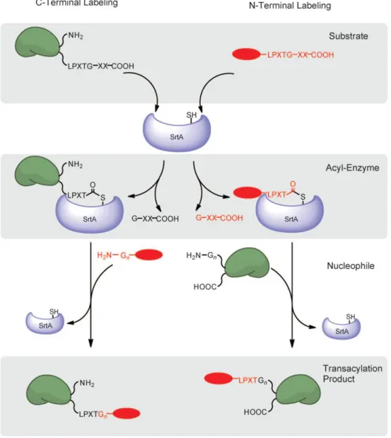
1.5.2 C-Terminal and N-Terminal
The ability of sortase to recognize the sorting motif when transplanted onto recombinantly expressed proteins allows the site-specific incorporation of moieties and functional groups that cannot be encoded genetically (Fig.12). This method requires only that the LPXTG motif be solvent exposed and usually results in high yields of the desired transpeptidation product. Indeed, many substrate proteins have now been labeled with probes bearing a wide range of functionalities, including biotin, fluorophores, cross-linkers, and multifunctional
35
probes (Popp, Antos et al. 2009). The labeling of recombinant proteins by sortase A requires no sophisticated synthetic chemistry; most of the probes are readily accessible by standard peptide synthesis, using off-the shelf reagents. The production and folding of recombinant substrate proteins is not usually compromised by the presence of the small C-term LPXTG tag. Since all transformations are carried out using sortase under physiological buffer conditions (pH, ionic strength, ionic requirements) on substrates whose proper folding and activity status can be ascertained prior to starting the reaction, loss of biological activity is rarely, if ever, observed for the final product. The ability to engage in a sortase-catalyzed transacylation appears to be determined solely by the accessibility and flexibility of the sorting motif. The utility of the sortase labeling method stems from the fact that the enzyme tolerates substrates unrelated in structure and sequence immediately upstream from the cleavage site. This property is not unexpected, given the role of sortase in anchoring a broad range of protein substrates to the cell wall (Popp and Ploegh).
Protein labeling at the N terminus can be accomplished simply by moving the placement of the sortase recognition element from the protein to the short peptide probe and by inclusion of a suitable number of glycine residues at the N terminus of the target protein (Fig.12). Both methyl ester mimetics of the sortase motif (Antos, Chew et al. 2009) as well as the complete LPXTG sortase recognition motif can be used as scaffolds for such probes (Yamamoto and Nagamune 2009). Conceptually, this labeling technique is analogous to the C-terminal labeling, except the acyl-enzyme intermediate is generated between sortase and the peptide probe, and the protein to be labeled bears several glycine residues at the N
36
terminus, the NH2 group of which serves as the nucleophile. This strategy was
used to install fluorescent probes at the N terminus of membrane proteins in living mammalian cells after a clever initial unmasking step by sortase itself to expose the nucleophilic glycine (Yamamoto and Nagamune 2009).
Figure 12. Site-specific and N-terminal labeling scheme using sortase A.
C-Terminal labeling (left) and N-terminal labeling (right) proceed through a substrate-recognition step (top), followed by generation of a thioacyl intermediate (middle) and resolution of the acylated enzyme by an exogenously added nucleophile (bottom) (Popp and Ploegh).
37
1.5.3 Other sortases application as protein engineered
Sortase methods allow the production of homogeneous recombinant protein preparations that are modified with nongenetically templated post-translational modifications. Glycoproteins, normally elaborated by a complex set of enzymatic events in the secretory pathway, can thus be constructed. LPXTG-tagged proteins and peptides can be modified with 6-aminohexose-based sugar nucleophiles, including aminoglycoside antibiotics and their analogues (Samantaray, Marathe et al. 2008). Glycosylphosphatidylinositol (GPI) anchors, normally attached at the C terminus of proteins, can be phenocopied by ligation of LPXTG peptides to synthetic glycine nucleophiles, which in turn are linked to the phosphoethanolamine moiety on a GPI derivative (Guo, Wang et al. 2009). Lipidation of proteins is yet another important post-translational modification that has been poorly studied because of the lack of tools available to obtain homogeneous preparations of lipoproteins. Sortase has been used to fill this void (Antos, Miller et al. 2008). A glycine-based scaffold was modified with a panel of linear alkyl chains (C12–C24) as well as with cholesterol or adamantane, and then used to modify a suitably LPETG-tagged version of eGFP. These eGFP lipoproteins associated with the plasma membranes of living cells in a chain-length-dependent fashion (the optimum being a C22 chain), from where they gained access to the endosomal compartment.
Moreover, covalent immobilization of proteins onto solid supports has been accomplished by sortase. A major advantage of the method is that the specificity of the enzyme enables proteins to be immobilized uniformly and in a defined
38
orientation on the solid surface for subsequent exposure to the analyte of interest (Popp and Ploegh).
1.6 Sortases that assemble pili: class C enzymes
Gram-positive bacteria use class C enzymes to build pili that promote microbial adhesion and biofilm formation. First, one or more class C enzymes form the long thin shaft of the pilus by linking together pilin subunits via isopeptide bonds. The base of the pilus is then anchored to the cell wall by a housekeeping sortase or, in some cases, the class C enzyme itself (Spirig, Weiner et al.).
Recently, an extensive characterization of pilus-associated sortases from Streptococcus pneumoniae pilus 1 (SrtC-1, SrtC-2, and SrtC-3) was performed, and the X-ray structures of all 3 SrtC enzymes have been solved (Neiers, Madhurantakam et al. 2009) (Manzano, Izore et al. 2009). The overall fold of all three enzymes is very similar to other known sortases, corresponding to a β-barrel structure, composed of eight anti-parallel β-strands linked by multiple helices. The catalytic triad (constituted of His131, Cys193, Arg202 in SrtC1; His159, Cys221, Arg230 in SrtC2; His144, Cys206, Arg215 in SrtC3) within the substrate binding region is encapsulated by the lid, which maintain the active site in a closed conformation in the absence of substrate. The lid anchoring within the active site is through multiple interactions with key catalytic residues (Manzano, Contreras-Martel et al. 2008; Neiers, Madhurantakam et al. 2009). Structural comparison of the three pilus-associated sortases revealed some slight differences in terms of flexibility, positioning and number of residues of the lid and B-factor
39
values of the N-terminal helices. An additional helix in the C-terminal region is only present in SrtC-3. Some structural differences suggested a molecular explanation for the functional differences observed among these sortases, in terms of substrate specificity and incorporation of the ancillary pilins into pili.
Manzano et al. have also showed that site-specific mutations of the anchor residues in the lid region did not affect backbone protein recognition or the formation of the acyl-intermediate; however, the stability and the efficiency of the enzyme were negatively affected (Manzano, Izore et al. 2009). While the catalytic triad of Cys, His, and Arg side chains within the active site cleft is absolutely conserved among different classes of sortases (Zong, Bice et al. 2004) (Zong, Mazmanian et al. 2004), including SrtA from Staphylococcus aureus, the region corresponding to the lid is thus far found only in X-ray diffraction solved crystal structures of pilus-related C sortases in Gram-positive bacteria (Manzano, Contreras-Martel et al. 2008) (Weiner, Robson et al.).
The crystal structures of several other pilin-related class C sortases, including AcSrtC-1 from Actinomyces oris (Persson 2011), SrtC1 from S. suis (Lu, Qi et al. 2011) and GBS (Cozzi, Malito et al. 2011; Khare, Fu et al. 2011; Khare, Krishnan et al. 2011), have been reported. These structures all reveal a core 8-stranded β-barrel, with the catalytic triad (His, Cys, Arg) situated in the active site at the end of a groove along one side of the β-barrel. The GBS and S. suis SrtC1 structures were determined with the active-site in the „open‟ conformation, while the other structures showed the active site occluded by a loop region, termed the lid. The lid in SrtC1 from GBS PI-2a (SrtC1-2a) and Actinomyces oris SrtC2 is dispensable for sortase activity in vivo (Wu, Mishra et al.; Cozzi, Malito et al. 2011).
40
1.7 Class C sortases in GBS
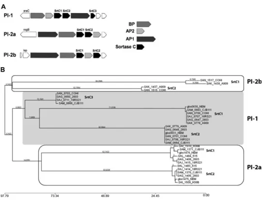
Sequence comparison by multiple alignment and phylogenetic analysis permitted the identification of 3 major clusters, corresponding to class C sortases of PI-1, PI-2a, and PI-2b, with amino acid identities ranging from 15 to 60% (Fig. 13). While both variants of PI-2 (PI-2a and PI-2b) contain 2 sortase genes (SrtC1 and SrtC2), PI-1 carries a third gene (SrtC3) predicted to code for a C sortase not directly involved in pilus polymerization (Rinaudo, Rosini et al.; Buccato, Maione et al. 2006; Rosini, Rinaudo et al. 2006). Moreover, the crystal structure of the sortase SrtC1 of PI-2a (SAL_1484) was solved at high resolution (Cozzi, Malito et al. 2011).
Figure 13. Class C sortases in PIs of GBS. (A) Schematic representation of GBS PIs. (B) Phylogenetic tree inferred from the alignment by the neighbor-joining distance-based
method of C sortases from the available genomes of GBS. Single sortases are indicated by TIGR annotation. The 3 major clusters, highlighted in the boxes, include C sortases of each PI (Cozzi, Malito et al. 2011).
41
1.7.1 Structural organization and biochemical characterization of PI-1 and PI-2a sortase C enzymes
The ectodomain of sortase SrtC1 of PI-2a (residues 43–254) was crystallized and the structure was solved by molecular replacement, using as the initial search model the coordinates of sortase C1 of S. pneumoniae (PDB 2W1J).
The overall folding of GBS SrtC1 is highly similar to the folding of previously determined pilus-associated sortases. A β-barrel made of 9 antiparallel β-strands forms the core of the enzyme; a so-called roof made of 3 α-helices positioned above the β-barrel and a loop (known as the “mobile lid”) that covers the active site (Fig.14A). This last one is positioned on one inner side of the β-barrel core and is made of the catalytic triad His157-Cys219-Arg228. The lid of SrtC1 harbors 3 residues, Asp84, Pro85, and Tyr86 ,which make interactions with residues of the active site and surroundings. While Asp84 and Pro85 are highly conserved, an aromatic residue (Tyr86 in SAL_1484) generally occupies the third position. The carboxylate group of Asp84 forms a salt bridge with the side chain of the conserved catalytic residue Arg228 (Fig. 14B) and with a water molecule (W76). The ring of Tyr86 is positioned in a pocket lined by highly conserved hydrophobic residues (Leu131, Leu138, Val153, Leu217) on one side and by the catalytic residue His157 on the other side. The residue Pro85 points toward the same hydrophobic pocket. The aromatic benzene ring of Tyr86 is close enough to the catalytic Cys219 side chain to make an aromatic-sulfur interaction (Cozzi, Malito et al. 2011). As shown previously, this sulfur-aromatic interaction is conserved in other sortases (SrtB and SrtD), and this finding suggests that this serves as a general mechanism of anchoring the lid within the active site (Viguera
42
and Serrano 1995) (Fig.14B). This sulfur-aromatic interaction has been postulated to strengthen the anchoring of the lid within the active site (Neiers, Madhurantakam et al. 2009). In addition, the hydroxyl group of Tyr86 makes H-bond interactions with the hydroxyl side chain of the highly conserved Thr155 and with the backbone amino group of the conserved Ala156 (Fig.14B). The aromatic ring of Tyr86 is also positioned in a hydrophobic environment where it potentially can be involved in CH-π weak polar interactions. This network of interactions between catalytic residues and those located on the lid (Asp84 and Try86) is postulated to regulate the movement of the lid and therefore the access of LPXTG substrates to the active site (Manzano, Izore et al. 2009; Cozzi, Malito et al. 2011).
The active site of GBS SrtC1 is made of the highly conserved catalytic triad His157, Cys219, Arg228 (Fig.14) and through site-directed mutagenesis and in vivo complementation studies, it has been demonstrated that each residue in the catalytic triad is essential for pilus polymerization; these data confirm the relationship between GBS C sortases and other members of sortase family. However previously reported NMR data on SrtA of S. aureus describe large chemical shift changes in the amide nitrogen and proton atoms of residues localized in specific loops on calcium ion addition, leading to the prediction that those residues form a structurally ordered calcium-binding site (Ilangovan, Ton-That et al. 2001; Cozzi, Malito et al. 2011). The absence of significant NMR chemical shift change on addition of EDTA or CaCl2 to GBS SrtC1 indicates that,
while calcium binding is required for the activity of the housekeeping SrtA in S. aureus, GBS SrtC1 does not bind any calcium ion (Cozzi, Malito et al. 2011).
43
The crystal structure of GBS SrtC1 showed that catalytic residues are not accessible to pilus substrates (Fig.14), as they are locked by the lid. Moreover, the mutations of key residues Asp84 and Tyr86 in the lid region or the deletion of the entire lid region had no effect on pilus protein polymerization (Fig.15A) (Cozzi, Malito et al. 2011).
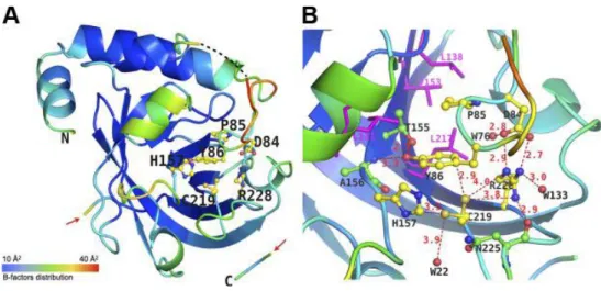
Figure 14. Overall folding of SAL_1484 and active site organization. A) Overall
folding and B factors of SAL_1484. SAL_1484 is represented as a cartoon, colored according to B-factor distribution, from low (blue) to high (red). Residues forming the mobile lid and the active site are shown as balls and sticks and are labeled. N and C termini are labeled. Carbon, oxygen, an nitrogen atoms are depicted in yellow, red, and blue, respectively. Position of residues 92–93 of the mobile lid, missing from the model because of poor electron density, is indicated by black dashes. Red arrows indicate the gap in the C-terminal region, fragment of residues 240–249. B) Active site of SAL_1484. Residues forming the mobile lid (Asp84, Tyr86) and the active site (His157, Cys219, Arg228) are shown as balls and sticks, with carbon, oxygen, and nitrogen atoms in yellow, red, and blue, respectively. Conserved surrounding and interacting residues (Thr155, Ala156, Asn225) are shown as balls and sticks, with carbon, oxygen, and nitrogen atoms in green, red, and blue, respectively. Conserved hydrophobic residues are shown as magenta sticks and labeled in magenta. Distances between atoms are labeled and shown as red dashes. Water molecules are shown as red spheres. Background cartoon representation of SAL_1484 is colored according to B factors as in panel A(Cozzi, Malito et al. 2011).
44
Figure 15. Lid region is not essential for pilus protein polymerization. Immunoblots
of total protein extracts from 515 mutant strain of both sortase C genes _(SrtC1_SrtC2) complemented by plasmids expressing SrtC1 wild-type (SrtC1WT) or SrtC1 carrying the
mutation D84A (SrtC1D84A) or Y86A (SrtC1Y86A) or the deletion of the entire lid region
(SrtC1Δlid). Nitrocellulose membranes were probed with antisera specific for the BP (A) and the ancillary proteins, AP1 (B) and AP2 (C) (Cozzi, Malito et al. 2011).
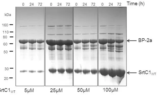
To better investigate the role of this region in catalysis, in vitro measurements of the kinetic properties of recombinant SrtC1Y86A and SrtC1ΔLID in comparison with
the SrtC1WT were performed. Accordingly to the in vivo data, the lid mutants can
efficiently cleave the substrate peptide, and the rate of peptide cleavage by lid mutant variants was even higher than that obtained with the wild-type (Fig.16) (Cozzi, Malito et al. 2011).
45 Figure 16. FRET assay with wild-type SrtC1 and lid mutants. (A) Progress curves of the cleavage reaction of PI-2a BP fluorescent peptide catalyzed by recombinant SrtC1 wild-type (SrtC1WT), SrtC1 carrying the mutation Y84A (SrtC1Y86A) and the deletion of
the entire lid region (SrtC1Δlid). Reactions containing 25µM of enzyme and from 2 to 256
µM of fluorescent peptide were performed at 37°C in 20 mM Tris (pH 7.5), 75 mM NaCl, and 1 mM DTT. (B) Rate [relative fluorescence units (RFU)/min] vs. concentration of substrate (Cozzi, Malito et al. 2011).
These results fit with the role of the lid suggested by the crystal structure, in which the lid covers the active site and sterically blocks the access of substrate. Structural and biochemical data suggest that the lid maintains the enzyme in an inactive closed conformation and that, for the enzyme activation, the lid needs to move. Therefore the deletion of the lid region does not abrogate pilus protein polymerization because its role is not catalytic; rather, it is a catalytic cleft-blocking loop, and only its movement can activate the enzyme in vivo (Cozzi, Malito et al. 2011).
The main questions remain to understand how this movement can be regulated by the interaction with the pilus proteins and to identify which are the residues involved in stabilizing the active open lid conformation of the enzyme. Based on
46
these analyses, the SrtC enzymes can be considered as having two functional domains: (i) an N-terminal regulatory region that contains the flexible inhibitory, pseudo-substrate lid, involved in enzyme regulation and probably specificity; and (ii) an enzymatic region, the b-barrel core that contains the catalytic triad (Cozzi, Prigozhin et al. 2012).
Moreover, the predicted C- and N-terminal TM domains of GBS SrtC1 are absolutely required for sortase biological function (Cozzi, Malito et al. 2011). The importance of TM domains for the enzyme activity has been recently reported for the pilus-associated sortase of Corynebacterium diphtheriae. Ton-That and co-workers (Guttilla, Gaspar et al. 2009) showed that the predicted C-terminal TM domain of pilus-associated sortase SrtA is essential for efficient pilus polymerization in C. diphtheriae. In addition, the evidence that the substitution in GBS SrtC1 of the C-terminal TM region with the corresponding hydrophobic helix of the backbone subunit of pilus type 2a could not restore enzyme activity strengthens the view that this region could play a key role in enzyme function. Finally, also in an in vitro FRET assay using fluorescent peptides mimicking the natural LPXTG substrates, the activity of SrtC enzymes was detected only when the enzyme contains the C-terminal TM region. These data suggest that the N-terminal hydrophobic helix could have a role in protein anchoring to the membrane, while the C-terminal TM region could be also involved in enzyme activity (Cozzi, Malito et al. 2011).
The crystal structures of GBS PI-1SrtC2 and SrtC1 were determined (Fig.17) . In both enzymes, the catalytic residues are not accessible to pilin substrates, suggesting that the enzymes cannot bind substrates in this conformation. Also
47
these sortase C enzymes contain an additional N-terminal extension of approximately 50 residues, composed of one or two α-helices and a lid that blocks the access of substrates to the active site. Ligand-free SrtC structures are more similar to the peptide-bound SrtA structure than to apo-SrtA. The structural similarity between the LPXTG peptide in the active site of SrtA suggests that the conserved residues in the lid that interact with the active site of GBS sortase act as a pseudo-substrate (Cozzi, Prigozhin et al. 2012). This observation further supports the already proposed regulatory role played by the lid in restricting the access of the pilin substrates to the catalytic cleft (Manzano, Contreras-Martel et al. 2008; Neiers, Madhurantakam et al. 2009; Cozzi, Malito et al. 2011). Moreover, structural analysis combined with in vitro experiments performed with fluorogenic peptides and with N-terminal deletion mutants of SrtC1 and SrtC2 show that the entire N-terminus, and not just the lid, as shown for GBS PI-2a SrtC1 (Cozzi, Malito et al. 2011), is disposable for catalysis (Cozzi, Prigozhin et al. 2012). Thus, the minimum active sortase region is the β-sheet core seen in the S. aureus SrtA structure and common to all sortase family members. The N-terminal extension is a unique feature of class C sortases and appears to function as a regulatory motif. Both class A and class C sortases cleave LPXTG-like motifs, but only sortase C can polymerize the pilus proteins to form high molecular weight structures. Hence, the different function of SrtC compared to SrtA, in terms of regulation, specificity or localization, may be due to the presence in this specific class of enzymes of a highly specialized N-terminal segment (Cozzi, Prigozhin et al. 2012).