FACOLTÀ DI INGEGNERIA DEI SISTEMI
DIPARTIMENTO DI ELETTRONICA, INFORMAZIONE E BIOINGEGNERIA DOTTORATO DI RICERCA IN BIOINGEGNERIA
C
HARACTERIZATION OF
A
UTONOMIC AND
C
ARDIORESPIRATORY
C
ONTROL IN
N
EWBORN
P
OPULATIONS AT
R
ISK FOR
S
UDDEN
I
NFANT
D
EATH
S
YNDROME
Doctoral Dissertation of:
Maristella Lucchini
Advisor:
Prof. Maria Gabriella Signorini
Co-Advisor:
Prof. William P. Fifer
Tutor:
Prof. Pietro Cerveri
Supervisor of the PhD Program:
Prof. Andrea Aliverti
XXX edition 2014-2018
i
A
BSTRACT
Sudden Infant Death Syndrome (SIDS) is defined as a sudden and unexplained death of an infant younger than 1 year of age. Although infrequent, SIDS is still the most common cause of infant death between one month and one year of age in developed countries. Thanks to epidemiological, animal and pathophysiological studies, possible risk factors and mechanisms leading to this death are more understood, nonetheless number of deaths has reached a plateau in the last decade and no reliable quantitative tool to assess risk exists.
The purpose of this Ph.D. thesis is to employ novel signal processing methodologies in order to accurately estimate the effect of several risk factors on the autonomic and cardiorespiratory control in newborn infants. In particular, the effects of sleep states, sleep position, prematurity and exposure to alcohol and smoking during pregnancy are investigated.
The original contribution of this thesis centers on the utilization of noninvasive methodologies, to analyze physiological signals routinely acquired in hospital and home settings. Moreover, the approach to signal analysis incorporates complex system generation and interaction, with a systemic view of the cardiorespiratory physiology. Different modes of interactions were explored, ranging from amplitude to phase modulation.
Parameters proposed proved to be capable of characterizing multiple autonomic profiles, providing information in line with pathophysiological findings. Results highlighted sleep position effects on autonomic control, with less parasympathetic activity in prone position, especially at 2 months of age. Prematurity greatly influenced the cardiorespiratory coupling, both in terms of strength and directionality, with weaker respiratory drive associated with prematurity. Lastly, neonates exposed to smoking and alcohol during pregnancy showed a blunted autonomic response when exposed to the autonomic challenge of head up tilt.
ii In conclusion, the methods and results of this Ph.D thesis show the utility and power of complex and multivariate signal processing techniques in the investigation of an enigmatic syndrome such as SIDS, where multiple organs and their control systems are involved.
iii
SUMMARY
Introduction
Sudden Infant Death Syndrome (SIDS) is defined as a sudden and unexplained death of an infant younger than 1 year of age. It occurs in an infant considered healthy and it remains unexplained even after autopsy, a careful examination of the death scene and a review of the clinical history [1]. Although infrequent, SIDS is the most common cause of infant death between one month and one year of age in developed countries, with around 2000 death a year only in the United States [2].
Thanks to the information from epidemiological, animal and pathophysiological studies, we have now knowledge of risk factors and have formulated hypotheses regarding the underlying mechanisms. Currently, the most supported theory is the triple risk model proposed by Filiano and Kinney in 1994 [3]. The model views SIDS as the result of the simultaneous occurrence of three factors: an underlying vulnerability (i.e. prematurity, genetic abnormalities, etc.), a critical developmental period in homeostatic control (i.e. the first year of life), and exposure to exogenous stressor(s) (e.g. newborn placed in a prone sleep position, overheating, inadequate oxygen supply etc.).
One crucial aspect in this and many other models proposed is the involvement of autonomic control circuits and their role in cardiorespiratory regulation. Brainstem alterations and altered serotonin receptors were found in SIDS cases autopsies [4], [5], and altered heart rate (HR), breathing and arousing patterns were identified in vulnerable populations and conditions, such as prematurity and prone sleeping [6], [7].
Nonetheless, currently there is no marker for risk stratification nor measures to quantify the impact of the identified risk factors on autonomic development. This is even more relevant given that in the past
iv 10 years death rates have reached a plateau, with no major further decline since the successful Back to Sleep campaign [8].
Thus, the principal purpose of this Ph.D. thesis is to provide quantitative and noninvasive tools to evaluate the impact of several risk factors on the functionality of cardiorespiratory and autonomic control. The ultimate objective is to provide noninvasive and early markers to assess risk.
The tools employed are time series methods and analysis of neonatal HR and breathing, obtained from noninvasive collection in hospital. Heart rate variability (HRV) was a primary direction given the plethora of publications linking risk with HRV measures and sympatho-vagal autonomic activity. Given the relevant role of the cardiorespiratory interaction in maintaining homeostasis and responding to variable physiological demands, in our analysis we added breathing and its interaction with HRV to provide a more complete approach.
The research was conducted under the guidance of several collaborators from two institutions, the Politecnico di Milano and the Columbia University Medical Center. Datasets for the analyses were used in collaboration with Drs. Rakesh Sahni and Nina Burtchen, and the PASS Research Network. The design and realization of the analytic and statistical part were developed under the supervision of Professors William P. Fifer, Michael M. Myers and Maria G. Signorini.
Advanced methods of analysis.
The autonomic nervous system (ANS) is a part of the peripheral nervous system that acts to control physiology of many bodily functions. It is divided in two branches, sympathetic and parasympathetic, acting synergistically for the regulation of physiology and behavior crucial for survival. Primary autonomic functions include the regulation of HR, blood pressure, rate of respiration, body temperature, sweating, gastrointestinal motility and secretion. One of the most interesting function relevant to this thesis is the control over the heart muscle: sympathetic fibers increase HR, atrioventricular conduction, and contractility of cardiac muscle, while they dilate the coronary arteries. In contrast, parasympathetic nerves slow down the HR and reduce heart contractility, favoring conservation of energy. These effects are integrated with several other factors, such as the spontaneous contractility of the heart itself, the peripheral resistance of the vascular tree, the effects of circulating hormones and the metabolic supply to the heart.
v This complex interplay generates a range of heart rate variability (HRV) patterns, which reflects ANS mediated change in the time intervals between adjacent heartbeats. For this reason, HRV has been for long time considered a powerful tool, providing a window to observe non-invasively the interaction between the sympathetic and parasympathetic nervous systems and their capability to respond properly to internal and external challenges [9]. HRV is also influenced by respiration; the neuronal control of breathing and HR are closely linked, functionally as well as anatomically. The close interaction between cardiac and respiratory control is critical for survival. This synergy is crucial for the homeostatic regulation of blood gases, and essential for regulating central nervous functions, such as arousal [10], [11].
Thus, breathing and HR are the output of a complex network of controlling mechanisms, which constantly adapt to the ever-changing need of the organism. Since the 1960s, many efforts have been spent to describe the systems behind these signals, starting with time domain and following with frequency domain approaches [12]. Nonetheless, the complex origin of these signals often makes traditional linear signal processing approaches unsuitable or only partially capable of characterizing the systems generating the data. More parameters coming from nonlinear chaos theory and the information theory can provide alternative approaches to extract meaningful information and describe the behavior of the systems generating the data under analysis [13]. Furthermore, advanced bivariate techniques have been proposed in order to highlight different types of interactions among systems and unveil their relationships.
Previous studies had addressed the questions of autonomic and cardiorespiratory regulation in the context of SIDS, but mostly employing traditional spectral techniques to asses cardiac and respiratory functioning and their interaction [14]–[17]. One of the objectives of this thesis is to implement complex signal processing methods to characterize newborn HRV and cardiorespiratory activity. For this reason, we selected entropies and phase rectified signal averaging (PRSA) techniques, given previous positive applications in adults, newborns and fetuses.
In 1993, Pincus was the first to introduce a practical measure of complexity, namely Approximate Entropy (ApEn), showing that infants who had an apparent life threatening event presented greater ApEn instability across quiet sleep than any normal infant exhibited, with incidents of extremely low values [18]. Following this work, the group of Moorman applied a modified version of ApEn, called Sample Entropy, to detect sepsis in newborns [19]. These entropy measures aim to quantify the
vi regularity, defined as the presence of repetitive patterns in a time series within a certain tolerance r and at different lags [19], [20]. They provide entropy estimation indices even with relatively short segments and without making any assumptions about the underlying structure of the system. A challenging problem for entropy estimation is the choice of the tolerance r, to assess if two patterns could be defined similar. In 2006, Lake proposed a new parameter called quadratic sample entropy (QSE) [21]. It removes the dependency on r, which can be optimally varied for each data record. One way to optimally vary r, is that of setting a minimum number of matches (MCM), while minimizing r, in order to have a stable and consistent entropy estimate. We also discuss ways to optimally select the MCM.
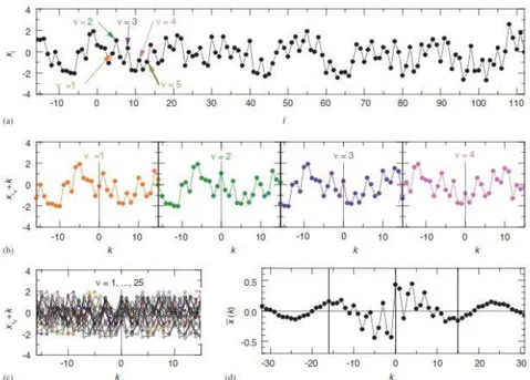
Another approach for nonlinear characterization of HRV is PRSA, which offers the possibility to analyze separately HR accelerations and decelerations and select parameters to specify the frequencies of interest. This affords the opportunity to investigate rapid parasympathetic influences as well as slower sympathetic ANS mechanisms. This technique requires relatively long recordings since it is based on averaging many segments in order to discard irrelevant data. It was applied successfully in the identification of intra uterine growth restricted fetuses and detection of cardiovascular risk in adults [22], [23].
Our second objective was to characterize and quantify the mutual influence of cardiovascular and respiratory rhythms. Many contributions were found in the literature, ranging from cross-spectral analysis to nonlinear methods, e.g., mutual information or time delay stability [24], [25]. These include linear and nonlinear relations between HR and respiration signal. Nonetheless, a limitation of all these techniques was that they did not measure the directionality of the relationships, and thus, they could only partially reveal the underlying interacting mechanisms responsible for the changes in complexity, especially when knowledge of the underlying physiology was limited.
Transfer Entropy (TE) was developed to address precisely this issue. Its focus is on tracking the information flow between two systems. Specifically, TE can enhance the quantification of the directional coupling between respiration and HR in order to incorporate both sympathetic and parasympathetic regulatory influences. The main advantages of this method are that it captures both linear and nonlinear contributions to information flow, and it can differentiate the directionality of transfer. Applying TE on shifted signals we can also evaluate how long the effect of a system on the other lasts and thus inquire their relationships at different time scales. This is relevant since we know
vii that different time scales might reflect the effect of different autonomic branches on the cardiac regulation.
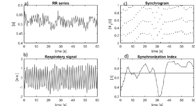
Another approach is that of describing the cardiorespiratory coupling as a dynamic synchronization process, meaning an interaction between two subsystems which can be modeled as two weakly self-sustained chaotic oscillators [26]–[28]. The basic idea is that given two weakly coupled systems, the amplitude of their oscillations may remain uncorrelated whereas their phases do mutually perturb each other. With this assumption, it becomes possible to investigate cardiorespiratory synchronization by means of a phase analysis of RR series and respiratory signal with a method called Phase Locking, rather than applying a classical amplitude analysis. The behavior of the cardiorespiratory system can be seen as synergetic, i.e. a multi-stable system switching between several phase configurations, with a preference for a specific set of phase relations, which can be seen as attracting frequency ratios [29]. In this context, Rosenblum et al. also proposed a method which exploits the notion of phase synchronization of irregular oscillators in order to reveal whether the interaction is bi- or unidirectional and to quantify the degree of asymmetry in the systems’ coupling [30].
Interestingly, different modes of interaction are not exclusive, rather they may simultaneously coexist, representing different aspects of neural regulation and acting on different time scales [31], [32]. The performance of these novel techniques were compared with that of traditional approaches, proposed by the Task Force of 1995 [33]. Specific guidelines for newborns are lacking, and their different degree of maturation has a big impact on many variables. As a matter of fact, newborns’ HR is double adults’ HR. For this reason, we utilized three minute segments, which contain roughly the same number of beats as the five minutes, usually used for adults. Additionally, we tested the performance of fetal parameters, given the developmental overlap in timing of nervous system development, especially for premature populations. Parameters estimated for the time domain include mean RR, Standard Deviation of Normal to Normal RR intervals (SDNN) and Root Mean Square of the Successive Differences (RMSSD), Long-Term Irregularity (LTI) and Short-Term Variability (STV). Spectral and cross-spectral analyses were also performed, extracting areas under the curve (signal variance) for Low and High frequency bands.
These techniques were employed to quantify how SIDS risk factors may alter cardiorespiratory interaction and autonomic activity. In particular, we investigated the influence of sleep state, sleep
viii position, prematurity and the combined effect of prenatal smoking and alcohol exposure on baseline physiology and a physiological challenge, i.e. the head up tilt. These conditions were chosen to cover both intrinsic and extrinsic risk factors, as proposed by the triple risk model, as well as their interaction.
HRV and Cardiorespiratory Analysis Results
As a preliminary step, we decided to address the question of how sleep states affect cardiorespiratory measures, in particular the novel measures introduced in this study, in order to be able to account for this influence in larger databases and following studies. From the literature we know that HR and its long term variability, measured by parameters such SDNN, are generally higher in active sleep (AS), while short term variability, for instance measured by RMSSD, is higher in quiet sleep (QS). These findings have been attributed to the increased vagal activity in QS and increased body movement and sympathetic activity in AS. With respect to the quantification of cardiorespiratory interaction as a function of sleep state, QS generally presents higher values of coherence at high frequencies (HF), indicating well established linear coupling in that frequency range.
We tested the effect of sleep state on complex measures of cardiorespiratory coupling in 151 full term newborns studied at birth and at one month of age [34], [35]. Entropy values (SampEn and QSE) were higher in QS. TE values showed that in QS coupling is stronger both with HR driving breathing and vice versa. Moreover, only in QS information flow from in the direction of respiration to HR was dominant. TE implemented on shifted signals showed how different directionalities of interaction act on different time scales, probably driven by the different branches of the ANS.
Our results indicate that, while QS generally presents lower global HRV, higher complexity values were observed probably due to increased interactions among physiological systems, as higher TE values seem to indicate. Moreover, a difference in directionality balance based on sleep state was observed, which could be driven by differences in the average breathing frequency. That is when breathing frequency is higher there is less opportunity for the respiration to dynamically modulate HR. Higher breathing rates occur more often in AS, and this could account for an absence of a dominant directionality. With respect to the cardiorespiratory synchronization, our findings showed a significantly higher value both in percentage of time spent in phase-coupling and in length of the coupled epochs in QS, both at the newborn and one-month stage.
ix When looking at the parameters behavior as a function of age, time domain parameters showed an increase in HR and a decrease in variability. In TE the major differences occurred in QS, with an increase in information flow in both directions with age. Moreover, the ratio of the dominant synchronization shifted from a majority of 3:1 (3 beats in 1 breath) in newborns, to 4:1 (4 beats in 1 breath) in one-month infants. This is of particular interest, since the peak of SIDS incidence is between 2-4 one-month of age.
Following the characterization of autonomic and cardiorespiratory activity by state, we addressed the effects of risk factors in three populations.
First, we investigated the effect of position during sleep in a group of 35 premature infants, with data acquired prior to discharge from the hospital (gestational age (GA) at birth 28.7 ±2 weeks and post menstrual age at time of study 37.7 ±2.4 weeks). Twenty four of these infants were studied again two months after discharge (GA at birth 27.9 ±1.6 weeks and post menstrual age at time of study 49.2±3.2 weeks) [36]. All infants underwent the same study protocol, which included baseline sleep recordings in both prone and a baseline in supine positions. ECG was acquired at 500 Hz and RR series were extracted.
Analysis by sleep position in the first study showed clear differences both in the long-term variability parameters (LTI and PRSA) and in the short-term variability parameters (SampEn and QSE), with supine position being characterized by higher variability but lower complexity. At 2 months of age, sleep position influenced mostly short-term variability parameters, specifically RMSSD and STV, and entropies, all parameters generally associated with parasympathetic regulation. With increasing post-natal age, infant parasympathetic control should dominate HR autonomic regulation: the fact that at 2 months of age measures of vagal activity were found to be diminished in prone position might suggest a suppressive effect of this position, changing the sympatho-vagal balance.
Next, we addressed the effect of prematurity in a dataset of 329 subjects, from 35 to 40 weeks GA [37], [38]. Infants were tested 12 to-84 hours after delivery, with a 10-minute baseline recording of ECG and respiration, at 500 Hz and 200 Hz respectively. Following the guidelines of the American College of Obstetricians and Gynecologists, infants were divided into three groups: newborns whose GA was (1) 35-36 weeks, labelled Late Preterm (LPT); (2) GA 37-38 weeks, Early Term (ET) and (3) GA 39-40 weeks, Full Term (FT). Results showed increasing mean RR intervals, short-term HRV (RMSSD), HR complexity
x (QSE) and linear cardiorespiratory coupling (spectral HF content) as a function of GA, indicating a significant increasing cardiorespiratory coupling and autonomic control as a function of GA at birth. Measures of cardiorespiratory coupling and directionality indicated no relationship between time spent in phase-synchronized state and GA group, but GA at birth influenced significantly directionality of interaction. In QS all the three GA groups showed the dominant influence of breathing on HR, and this relationship grew with GA. In AS a balanced relationship was present in LPT and it moved toward a dominant relationship from breathing to HR in FT.
Mrowka et al. had hypothesized that directionality could depend on respiratory frequency. In particular, respiration rate would act as a low pass filter, i.e. below a set respiratory frequency (proposed to 0.6 Hz) directionality would mainly be from respiration to HR, whereas above it the interaction would become bidirectional. We tested this hypothesis in our dataset and found the occurrence of this bimodal influence: concurrently with breathing frequency <0.6 Hz, an increased polarization toward values of directionality from breathing to HR occurred, especially in ET and FT. Nonetheless, the mean breathing frequency did not change significantly in the GA window analyzed (35-40 weeks); thus, the significant change in directionality with GA at birth could not be explained solely by breathing frequency. Our explanation for this phenomenon is that the threshold for the low pass filter effect is still adapting between 35-40 weeks GA. At a younger GA, for instance LPT, the value for the cutoff frequency might be lower when compared to more mature conditions, such as FT. Given that this cutoff frequency is potentially related to vagal nerve regulation, these findings would be consistent with previous studies showing immature vagal function in premature infants. Further confirmation of this hypothesis comes from the fact that we found directionality to be negatively correlated with measures of parasympathetic activity.
Lastly, the effects of prenatal alcohol and smoking exposure on tilt response in newborn infants were investigated in a population of 28 subjects (GA 39 ±1 weeks) , enrolled in the Safe Passage study [39]. Data was acquired within 48-96 hours of birth, with ECG and breathing for 10-minutes baseline period and a 45° head-up tilt, while the infant was sleeping prone. The tilt session was divided in: 30-sec just prior tilt (B); 15-seconds block right after the infant reached head-up position (I); three per 30-seconds blocks (T1, T2, T3) before returning to flat position. A modification of the Timeline Follow-Back Interview was employed to guide mothers’ self-reported estimation of their tobacco and alcohol consumption during pregnancy [39]. Based on the assessment, subjects were divided in a control group (no prenatal
xi exposure to alcohol or tobacco smoke), an exposed group (heavy prenatal exposure to both substances during the first, second or third trimester of pregnancy).
Graded head-up tilt has been widely used as a test to observe and quantify autonomic control. The tilt should elicit a shift of the sympatho-vagal balance toward a sympathetic activation and parasympathetic withdrawal. In this protocol, breathing signals were of very low quality due to movement during tilt, so the analysis focused on HR parameters. It was decided to estimate vagal withdrawal with RMSSD, HF power, QSE and RSA amplitude, all parameters which had been linked with vagal activity by previous literature [40], [41]. This expected behavior was displayed by the control group, with decreases of all the parameters selected after tilt, in particular at T2. The exposed group instead showed a blunted response, with no significant differences across the blocks.
In literature, prenatal exposure to smoke has been reported to effectively impair receptors in the medullary 5-HT system, as well as alter in cardiorespiratory control mechanisms and induce abnormalities in the pathogenesis of the parasympathetic systems [42]. It is possible to speculate that the illustrated difference in vagal activity comparing the control and the exposed groups may effectively arise as a consequence of prenatal exposure and may contribute to the decreased infant’s ANS capability to maintain homeostatic control when exposed to a direct physiological challenge. From a methodological perspective, an important finding was that we showed that the introduction of QSE calculated with the Minimum Count of Matches (MCM) approach led to a significant improvement in entropy estimation because QSE was less dependent on the length of the segment analyzed and this technique allowed to adjust the tolerance r in order to obtain more stable and consistent results. Moreover, QSE was less influenced by artifacts than other traditional measures, and thus could be applied with minimal preprocessing, with great advantage in real time applications. Secondly, we showed the usefulness of estimating PRSA curves by incorporating adjustments to the parameter T and, thus, vary the upper frequency limit of the detectable periodicities. TE implemented on shifted signals was useful to assess systems memory when interacting with each other.
Lastly, we compared measures of vagal activity on short segments and showed that even when parameters are highly correlated they are not interchangeable due to different statistical characteristics.
xii
Conclusions
The main purpose of this Ph.D. thesis was to characterize autonomic and cardiorespiratory regulation in newborn populations at risk for SIDS, who are particularly vulnerable to failure of the autonomic and cardiorespiratory control systems. We showed how complex parameters and techniques for bivariate analysis of breathing and HR provide additional information with respect to traditional techniques in the description of how physiological systems dynamically interact to maintain an optimal health status. Sleeping position, prematurity and exposure to smoke and alcohol during pregnancy all impaired physiological control, generally leading to an altered sympatho-vagal balance and diminished cardiorespiratory interaction.
Results show how traditional indices routinely employed in clinical studies often only scratch the surface of a more complex picture. The proposed novel techniques are advantageous for addressing specific time scales and different modes of interaction for data collected under standard clinical conditions with artifacts and noise. Moreover, these techniques were capable of characterizing systems interactions. This supports our aim to utilize nonlinear advanced parameters to obtain reliable physiological and clinical indices. This approach could lead to a quantitative autonomic profile to assess vulnerability in populations at risk for SIDS. This could grant the possibility of the definition of an “elastic” triple risk model, where the contributions of the various risk factors could be weighted and updated based on infants physical and environmental conditions, hopefully improving predictability and generating novel monitoring solutions.
xiii
T
ABLE OF
C
ONTENTS
ABSTRACT... i
SUMMARY ... iii
1. SUDDEN INFANT DEATH SYNDROME: ... 5
1.1. Introduction to Sudden Infant Death Syndrome ... 5
1.2. SIDS Risk Factors ... 6
Socio-Demographic ... 6
Pregnancy Related ... 6
Maternal Substance Abuse ... 7
Infant Sleep Practices and Environment ... 8
Genetic ... 9
1.3. Hypothesized Mechanisms Underlying SIDS ... 9
Respiratory Function ... 9
Cardiovascular Function ... 10
Nervous System Abnormalities ... 10
Immune responses and infectious agents ... 11
1.4. Risk Factors Interactions and Triple Risk Model ... 12
1.5. Sleep, Autonomic Nervous System and Cardiorespiratory Control ... 13
1.5.1. Sleep Development... 13
1.5.2. Autonomic Nervous System ... 14
1.5.3. Heart Rate Variability and Cardiorespiratory Interaction ... 15
1.5.4. Effects of sleep states on autonomic cardiorespiratory control and arousal ... 18
2. HEART RATE VARIABILITY AND CARDIORESPIRATORY ANALYSIS ... 19
2.1. Introduction... 19
xiv
2.1.1. Traditional HRV Analytic Approaches ... 20
2.1.2. Nonlinear HRV Analytic Approaches ... 21
2.3. Bivariate Analysis ... 25
2.3.1. Traditional Cardiorespiratory Analytic Approaches ... 25
2.3.2. Novel Approaches... 27
3. HEART RATE ANALYSIS RESULTS ... 37
3.1. Introduction... 37
3.2. Sleep States Effects on Cardiorespiratory Regulation ... 38
3.2.1. Experimental Protocol and Dataset ... 39
3.3. Effect of External Stressor: Supine vs Prone Position ... 47
3.3.1. Experimental protocol and data set ... 47
3.3.2. Results ... 48
3.4. Effect of Intrinsic Vulnerability: Prematurity ... 51
3.4.1. Experimental Protocol and Dataset ... 51
3.4.2. Results and Discussions. ... 52
3.5. Combination of Intrinsic Vulnerability and External Stressor: Alcohol and Smoking Exposure during Pregnancy and Head-up Tilt... 60
3.5.1. Experimental protocol and data set ... 60
3.5.2. Results ... 62
4. DISCUSSION AND CONCLUSIONS ... 67
Clinical impact ... 74
Ongoing developments ... 76
Application to PASS dataset ... 76
Extension of the Network Physiology framework ... 77
Point process application ... 77
1
L
IST OF
T
ABLES
Table 1-1: Summary of risk factors for SIDS. ... 6 Table 3-1: Parameter values for newborn and 1-month-old infants in active and quiet sleep. ... 40 Table 3-2:Parameters’ results for infants in the first and follow up study for position comparison ... 49 Table 3-3: Descriptive statistics of time and frequency domain parameters by gestational age in quiet sleep and active sleep ... 53 Table 3-4: Descriptive statistics of the transfer entropy, of the square root of the percentage of time in synchronized state and the average synchronization duration and number of subjects by gestational age in quiet and active sleep... 53 Table 3-5: 2-way Anova p-values of the square root of the percentage of time in synchronized state and the average synchronization duration by gestational age and sleep state ... 53 Table 3-6: Descriptive statistics for directionality index and frequency of respiration by gestational age and sleep state and number of subjects ... 55 Table 3-7: Multivariate model analysis of directionality index and frequency of respiration by gestational age and sleep state along with their interaction effect and post hoc analysis ... 55 Table 3-8: Mean ± std of parameter for each tilt block and within-subjects results of repeated measures ANOVA for CG and EG. ... 63 Table 3-9: Pearson correlation coefficient for RMSSD, RSA,HF and QSE ... 64 Table 3-10: Skewness and Kurtosis for the parameters proposed and for their log transformation when
the original parameters did not pass the requirement for normality distribution 64 Table 4-1: Summary of the methods and the findings of all the studies performed for the PhD thesis 73
3
L
IST OF
F
IGURES
Figure 1-1:Age distribution ny month of age from 15 global data sets comprising 19,949 SIDS cases .... 7
Figure 1-2: US SIDS death count during transition period following the introduction of supine vs prone sleep recommendations ... 7
Figure 1-3: Hypothesis of re-breathing mechanism leading to SIDS ... 10
Figure 1-4:The triple-risk model for SIDS ... 12
Figure 1-5: General plan of the distribution of autonomic nerves and ganglia... 15
Figure 1-6: Example of RSA and phase locking.. ... 17
Figure 2-1: A graphic representation of the process of match finding to calculate SampEn and ApEn . 22 Figure 2-2: Illustration of the PRSA technique ... 24
Figure 2-3: Steps involved in coherence analysis with surrogate data adapting threshold... ... 26
Figure 2-4: Example of coherence analysis with surrogate data adapting threshold. ... 26
Figure 2-5: Transfer entropy calculation steps ... 29
Figure 2-6: TE calculation at different lags ... 30
Figure 2-7: a): The RR series b): Respiratory signal c): The synchrogram d) synchronization index (λ). . 33
Figure 2-8: Presentation of phase locking procedure in a block scheme. ... 34
Figure 3-1: Heatmaps of coherence values for 10 minutes recordings of 2 subjects. ... 39
Figure 3-2: Boxplot of Transfer entropy values at lag=0. ... 41
Figure 3-3: In the upper panel, TE RR-> RESP is portrayed at different lags, while on the lower panel, TE RESP-> RR is shown. ... 42
Figure 3-4: Total percentage of time spent in coupling and length of the coupled epochs in active and quiet sleep ... 44
Figure 3-5: Bargraph of the percentage of synchronization on different n:m ratios for newborns and one month infants ... 44
4 Figure 3-6: PRSA curves of respiratory phase signal are shown in blue, BPRSA curve in green. The upper panel refers to increment APs, the lower panel to decrement APs. ... 45 Figure 3-7: Scatterplot of percentage of synchronization and mean IBI in quiet and active sleep, showing
a positive correlation between the two variables ... 46 Figure 3-8: On the left a representation of p-values for dx and dy in AS as a function of the parameter T,
showing dy reaching significance only after T>33. On the right a comparision of 2 PRSA curves obtained with T=51 and L=150 with one subject in prone and in supine position ... 50 Figure 3-9: On the left a graph showing the area under the Roc curve for the QSE parameter as a function
of M, the minimum count of matches. On the right SampEn and QSE for a subject in supine and prone position as a function of N, the length of the segment analyzed... 50 Figure 3-10: a): Boxplots show breathing frequency by gestational age and sleep state b): Boxplots show directionality index by GA and sleep state. ... 54 Figure 3-11: In each panel, histograms of directionality index distribution are plotted, on the left for breathing frequencies < 0.6 Hz, and on the right for breathing frequencies ≥ 0.6 Hz in order for LPT, ET and FT ... 56 Figure 3-12: On the left, the scatterplot of directionality index and RMSSD and on the right the scatterplot of the directionality index and QSE. In blue are reported values for LPT, in red for ET and in green for FT ... 57 Figure 3-13: a) RR interval series for a three 3-minute segment. b) ECG tracing corresponding to RR series in a) from 466 to 485 s showing transient bradycardia. c) RMSSD values for the three segments in red when no preprocessing is applied on the RR interval series, and in black after the RR artifact removal is applied. d) QSE m=1 values with the same logic as panel c). In panel c) and d), averages and standard deviations for the FT group are shown in blue. ... 58 Figure 3-14:Values of RMSSD and QSE of the three populations before and after RR intervals series preprocessing are plotted against one another. ... 58 Figure 3-15: Correlation matrices for all the indices calculated in this section, on the left in active sleep and on the right in quiet ... 59 Figure 3-16: Schematic of cardiorespiratory assessments... 61 Figure 3-17: Left and right panel show the mean values of the four extracted parameters and their trends when plotted based upon the tilt blocks, with the control group on the left and exposed group on the right. ... 62
5
1. S
UDDEN
I
NFANT
D
EATH
S
YNDROME
:
R
ISK
F
ACTORS AND
P
OSSIBLE
M
ODELS OF
P
HYSIOLOGICAL
D
ETERMINANTS
1.1.
Introduction to Sudden Infant Death Syndrome
Sudden Infant Death Syndrome (SIDS) is defined as a sudden and unexplained death of an infant younger than 1 year of age. It occurs in an infant considered healthy and it remains unexplained even after an autopsy, a careful examination of the death scene and a review of the clinical history [1]. Although infrequent, SIDS is still the most common cause of infant death between one month and one year of age in developed countries, with around 2000 death a year in the United States [2].
Despite years of research, SIDS remains an enigma. Over the past few decades, numerous, world-wide, epidemiological studies have identified factors that appear to contribute to SIDS deaths, a summary of which will be presented in first part of this chapter. A review of the main risk factors interaction and proposed models will be then described, with particular attention to the most well know triple risk model. The last section is focused on the development of sleep, autonomic control and cardiorespiratory interaction in newborns. These elements are all believed to play a crucial role in the cascade of events leading to SIDS.
6
1.2.
SIDS Risk Factors
Multiple epidemiological studies have determined that several factors, both modifiable and not, have significant associations with SIDS, both using case-control and cohort designs. A summary of these factors is presented in Table 1-1, and a detailed description follows.
Socio-Demographic
o SIDS affects infants from all social backgrounds, nevertheless lower socioeconomic status, younger maternal age, lower maternal education level and single marital status are consistently associated with a higher risk of SIDS. For instance, in the United States, SIDS rates are highest for non-Hispanic black and American Indian mothers—2.1 and 1.9 times those for non-Hispanic white mothers, respectively [43]. Following educational campaigns in the last few decades we have witnessed a decline in SIDS across all social and racial groups, but recent trends indicate that there are now even greater social and racial disparities [44], [45].
o SIDS can occur at any time during the first year of age, but approximately 90% of cases happen in the first six months of life, with increased incidence between 2 and 4 months of age, as portrayed in Figure 1-1 [46].
o Boys are 30%–50% more likely than girls to be affected, and this ratio was not influenced by the reduction of SIDS incidence in the last decades, as shown in Figure 1-2 [1], [47].
Pregnancy Related
o Low birth weight, preterm birth, intrauterine growth retardation and shorter intervals between pregnancies affect SIDS incidence [48]–[50].
Table 1-1: Summary of risk factors for SIDS, categorized in two groups. On the left are listed maternal and antenatal risk factors, with infant risk factors listed on the right
Maternal and antenatal risk factors Infant risk factors Smoking, alcohol, illegal drug use
Inadequate antenatal care Low socioeconomic status Low maternal age
Single marital status Low education level Fetal growth retardation
Male sex
Age (peak 2-4 months)
Race/ ethnic background (e.g. black, Native Indians, etc.)
Prematurity/low birth weight Prone/side sleeping
Overheating Soft bedding Bed sharing
7 o Studies have shown that mothers of SIDS infants generally receive less prenatal care and initiate
care later in pregnancy than do mothers of living control infants [47].
Maternal Substance Abuse
o There is a major association between intrauterine and postnatal exposure to cigarette smoking and risk of SIDS. The main limitations are related to the accuracy of self-reported cigarette smoking data and the difficulty in disentangling the independent effects of pre vs postnatal exposure to
Figure 1-1: Age distribution by month of age from 15 global data sets comprising 19,949 SIDS cases [223]
Figure 1-2: US SIDS death count during transition period following the introduction of supine vs prone sleep recommendations, showing that the male fraction remains constant at about 0.61 [222]
8 environmental tobacco smoke because parental smoking behaviors during and after pregnancy are highly correlated [51], [52].
o The evidence linking prenatal illegal drug use and SIDS is conflicting. Overall, studies do link maternal prenatal drug use, especially opiates, with an increased risk of SIDS ranging from 2- to 15-fold [53], [54].
o Studies regarding the association between SIDS and maternal alcohol use prenatally or postnatally are still inconclusive. In one study of Northern Plains American Indians, periconceptual maternal alcohol consumption was associated with a 6-fold increased risk of SIDS, and binge drinking during the first trimester of pregnancy was associated with an 8-fold increase [55], [56]. It remains to be established whether these effects are primarily attributed to true biological effects, or to sociodemographic and lifestyle factors that co-occur with pregnancy drinking, or a combination and possibly synergistic effect.
Infant Sleep Practices and Environment
o The prone sleep position was noted in multiple case-control studies to be associated with SIDS, as early as 1965 in the United Kingdom. Following numerous public health campaigns in Europe, Australia and New Zealand in the 1980s a decline in SIDS rate occurred. In 1992 the American Academy of Pediatrics (AAP) recommended that healthy term infants be placed to sleep in the non-prone position until 6 months of age. Since the 1992 AAP recommendation and the following 1994 Back to Sleep campaign, the US percentage of SIDS has diminished more than 40% [57]. o Soft bedding such as comforters, has been associated with a 2–3-fold increased risk of SIDS.
Combinations of sleep related risk factors result in even higher risk; for example, prone sleeping in soft bedding has been associated with a 20-fold increased risk of SIDS [58], [59].
o Overheating has been associated with increased risk of SIDS based on indicators such as increased room temperature, high body temperature, sweating, and excessive clothing or bedding [60]. o Several studies have suggested bed sharing as a risk factor for SIDS. Earlier case–control studies in
England and New Zealand showed a 5–9-fold increased risk associated with bed sharing only among infants of mothers who smoked. More recent studies showed that bed sharing was associated with increased risk of SIDS even when mothers did not smoke or if they breastfed, particularly among younger infants [61], [62].
9
Genetic
o In postmortem analysis of brainstems of infants who died of SIDS, serotonin receptor abnormalities were found throughout the ventral medulla [5]. Several studies have identified polymorphisms in the promoter region of serotonin transporter (5-HTT) protein gene in infants who have died of SIDS [63].
Brainstem findings include persistent increases in dendritic spines (indicating neuronal maturational delay) and delayed maturation of synapses in medullary respiratory centers, decreased tyrosine hydroxylase immunoreactivity in catecholaminergic neurons [64].
o Long QT syndrome is associated with sodium- and potassium-channel polymorphisms. Overall, it is estimated that 5%–10% of SIDS cases are associated with a defective cardiac ion channel and hence an increased potential for a lethal arrhythmia [65], [66].
o Genetic studies have identified mutations in SIDS infants pertinent to early embryologic development of the autonomic nervous system [67].
1.3.
Hypothesized Mechanisms Underlying SIDS
Given that the definition of SIDS is reliant on the elimination of other causes of death, it is not surprising that there are no conclusive findings on etiology. This has led to a vast number of theories on the mechanisms responsible for SIDS. The population of infants who die of SIDS is likely to represent a mixed population with various etiologies and disease entities contributing to one common endpoint (i.e. death) rather than all deaths being attributed to one single cause [68]. In the following paragraph, the main putative pathways will be described.
Respiratory Function
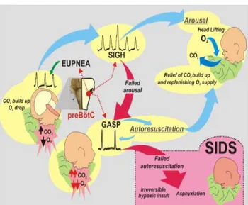
Respiratory failure has been proposed to contribute to SIDS, given that sleep increases the possibility of airway obstruction and apparent life-threatening events such as prolonged apneas. Furthermore, some studies have shown that SIDS infants present defects in respiratory control resulting in altered respiratory function, prolonged periods of “breath holding”, a failure of autoresuscitation, and defective arousal mechanisms, for example, in response to altered oxygen or carbon dioxide levels [69]. Figure 1-3 illustrates a hypothesized series of events involving respiratory failure leading to SIDS. Nonetheless, studies investigating alterations in breathing patterns or respiratory rates are inconclusive [70]–[73]. Other hypotheses involve peripheral sensory chemoreceptors, including the
10 carotid body, or dysfunction or immaturity in centrally located brainstem networks controlling upper airway functions.
Cardiovascular Function
Extended evidence in SIDS infants including altered heart rate (HR) and heart rate variability (HRV), defects in centrally mediated cardiac control (primarily brainstem centers), autonomic imbalances, prolongation of the QT interval and severe bradycardia has led to the conclusion that arrhythmia and cardiovascular changes are involved in the death in SIDS infants [70], [74], [75]. Nonetheless, inconsistencies in findings make it difficult to confirm a cardiac cause for SIDS.
Nervous System Abnormalities
Nervous system dysfunction has been proposed as a major factor leading to SIDS, potentially due to abnormal development or to maturational delay in particular brainstem regions, given their direct influence over homeostatic processes including cardiorespiratory control, sleep regulation, and arousal. Moreover, the peak period for SIDS, i.e. 2 to 4 months of age, is a crucial time for changes in neural control. Nonetheless, until now the literature has been contradictory as the ability to apply
Figure 1-3: During prone sleeping, re-breathing exhaled air can increase CO2 and decrease O2 levels. The
described mechanism should initiate the arousal response, beginning with sigh generation during continued eupneic breathing. If the arousal is successful, the infant will lift the head and reposition, relieving the build -up of CO2. If arousal fails, a more severe hypoxic state is reached and e-upneic breathing will transition to gasping, thanks to the network reconfiguration of the pre-Bötzinger complex (preBötC). Brainstem abnormalities, however, may alter preBötC network and impair sigh and gasp generation which may increase SIDS vulnerability [69].
11 histological and molecular techniques to examine post-mortem specimens are limited due to rapid deterioration of tissues after death.
Studies over the last 30 years show that the brainstems of infants who died from SIDS exhibit abnormalities in many major neurotransmitters and receptor systems including: catecholamines, neuropeptides, acetylcholinergic, indoleamines (predominantly involving serotonin and its receptors), aminoacids, brain derived neurotrophic growth factors, and some cytokines. A pattern is emerging for particular brainstem and hypothalamus nuclei being consistently affected, including the dorsal motor nucleus of the vagus, nucleus of the solitary tract, arcuate nucleus and raphe [76]. These changes have been attributed to ischemic damage, but many pathological findings overlap with observations from controls [77], [78]. Furthermore, while all these abnormalities have the potential to alter brain function, it should be noted that most of these studies report findings only in a subset of SIDS infants [79]–[81]. Moreover, the reported differences in neurotransmitter or receptor expression between SIDS cases and control cases can further be influenced by factors such as maternal cigarette smoking during pregnancy, highlighting the importance of accounting for these factors when trying to interpret neurochemical changes [4], [82].
Changes in the peripheral nervous system may impact on SIDS as well. Studies have reported histological changes in the carotid bodies in SIDS cases, which may suggest exposure to sustained hypoxemia and have the potential to impact the ability of chemoreceptors to respond adequately to changes in oxygen levels [83]. However, conflicting outcomes regarding carotid body size, histological changes, and the number of neurosecretory granules and transmitter levels have been reported make interpretation of the results challenging.
Lastly, since peripheral and central networks are highly integrated, it is likely that a change in one system may subsequently affect the other, and thus that both processes could contribute to nervous system dysfunction in SIDS.
Immune responses and infectious agents
Presence of a mild cold or upper respiratory infection [84] close to the time of death and findings of markers of infection and inflammation in many SIDS infants have led to the hypothesis that they might be immunologically underdeveloped and that stressors on the immune system may contribute to death [85]. Increased levels of immunoglobulins have been reported in SIDS victims [86], and reports of the presence of viruses (e.g. rhinovirus, cytomegalovirus) and of bacteria in the pathology of SIDS
12 have strengthened the argument for immune-mediated responses in the pathology of SIDS. However, many of these are also present in control cases, suggesting that their presence may be more co-incidental than causative. Thus, the presence of one, or a combination of, infectious agents is likely to increase the vulnerability rather than being responsible for death in SIDS infants.
1.4.
Risk Factors Interactions and Triple Risk Model
It has become evident that the mechanisms of death in infants classified as SIDS involve a complex interplay of individual vulnerabilities with developmental stages and environmental factors, rather than a convenient and simplistic “single cause”. In 1970, Bergman was the first to hypothesize that SIDS did not depend on any single characteristic but on an interaction of risk factors with variable probabilities [87]. Not long after, Wedgwood expanded this concept and grouped these risk factors into the first “triple risk hypothesis” consisting of general, developmental and physiological factors [88]. These factors needed to overlap and their synergy to exceed the threshold for survival for an infant to succumb to death. This concept has evolved in time, with many contributions suggesting different definitions and varied emphasis on the various risk factors [89], [90]. These theories culminated in the currently most accepted model in the field, the triple risk model presented by Filiano and Kinney in 1994 [3]. They posit that SIDS results from the simultaneous occurrence of: (1) an underlying vulnerability (i.e. prematurity, genetic abnormalities, etc.) (2) a critical developmental period in homeostatic control (i.e. the first year of life), and (3) exposure to exogenous stressor(s) (i.e.
Figure 1-4: The triple risk model for SIDS, presenting the three elements that must be present: 1) baby’s vulnerability, 2) critical developmental period, 3) exposure to one or more outside stressor
13 being placed in a prone position for sleep, overheating etc.). Figure 1-4 provides a schematic representation of this model.
Even if the factors involved may be multiple, the triple risk model does not exclude the possibility that most SIDS deaths might occur with a single common pathway upon which multiple stressors impinge to produce sudden death during the critical period.
1.5.
Sleep, Autonomic Nervous System and Cardiorespiratory Control
Even if consensus on the mechanisms leading to SIDS has yet to be reached, a failure of autonomic control of the cardiorespiratory system and an impaired arousal from sleep have been identified as crucial players in the cascade leading to SIDS, and both these factors are heavily influenced by sleep patterns. For this reason, the next section will present an overview on the development of sleep patterns and arousals in newborns, and a summary of the autonomic nervous system structure and functions, with a focus on the regulation and interaction of HRV and breathing
1.5.1. Sleep Development
The maturation of sleep is one of the most important physiological processes occurring during the first year of life and is particularly rapid during the first six months after birth. A human infant shows prolonged and characteristic epochs of stable behavior, that have been called 'behavioral states'. Many physiological variables are inter-related and mutually influencing during the state cycles and change their properties at the transitions [91].
Sleep states architecture in infants is quite different from those in adults: in infants, sleep states are classified as active sleep (AS) and quiet sleep (QS), which can be seen as precursors of adult rapid eye movement sleep (REM sleep) and non-rapid eye movement sleep (NREM sleep), respectively. During QS, high voltage low amplitude electroencephalograph activity is observable, with absence of eye movements and regular HR and respiration. On the other hand, AS is characterized by low amplitude high frequency electroencephalograph activity, eye movements, and irregular HR and respiration. Additionally, a third state, indeterminate sleep (I), can be defined when criteria for AS and QS are not met fully [92].
Interestingly, cyclical sleep patterns can be observed already in the human fetus from 28 weeks of gestation. By this time, AS can be easily identified, while QS becomes clearly identifiable only around the last month of gestation. The percentage of time spent in QS increases with gestational age and by two months of age infants spends equal amounts of time in both states. From term to six months of
14 age, the proportion of AS decreases. While the specific functions of the different sleep stages are not yet well understood, many believe that deep sleep is essential for physical rest, while REM sleep is important for memory consolidation [93]–[95]. The major change in sleep-wake pattern occurs between six weeks and three months post-term age. During the first six months after term, consolidation and entrainment of sleep at night develops and sleep periods lengthen.
1.5.2. Autonomic Nervous System
The autonomic nervous system (ANS) is a component of the peripheral nervous system and comprises a set of nerves and nerve cells that innervates blood vessels and the airways, heart, intestines and urogenital organs. These nerves regulate and coordinate bodily functions based on secretory activity of glands, on contraction and relaxation of smooth muscle and cardiac muscle, and on sensations arising from deep viscera.
The ANS consists of a central and a peripheral component: it is controlled by centers located in the spinal cord, brain stem and hypothalamus. Nerves sprout from this central part, reaching autonomic ganglia from which other nerves connect with the peripheral tissues that are the target of this system, such as smooth muscles, cardiac muscle and secretory cells.
The autonomic nerves arrange in an intricate network with many branching and merging points. Along these nerves there are prominent swellings, the autonomic ganglia, which consist of large aggregates of ganglion neurons and efferent nerve fibers. Within a ganglion, the incoming fibers end many of terminal boutons forming synapses on ganglion neurons. New fibers emerge from these neurons, directed to a target in the peripheral organs, whereas other incoming fibers pass through one ganglion to terminate in another ganglion. In addition, there are autonomic afferent (sensory) fibers sprouting from neurons in the cranial and spinal sensory ganglia and are distributed to the peripheral organs. A schematic of the ANS structure is shown in Figure 1-5 [96].
The peripheral ANS is made of two branches, the sympathetic and parasympathetic. The sympathetic system typically releases norepinephrine in response to intrinsic and/or extrinsic stressors via afferent autonomic pathways, which transmit responses to visceral organs via efferent autonomy pathways. Conversely, the parasympathetic system typically releases acetylcholine to restore the body to restful conditions after stressful events [97].
15 Autonomic activity increases with gestational age in the fetus during pregnancy and postnatally in the infant. Sympathetic development occurs early on, while the parasympathetic activity matures mostly during the last trimester and the first months of life [98].
1.5.3. Heart Rate Variability and Cardiorespiratory Interaction
Core functions of ANS are the regulation of HR, blood pressure, breathing rate, thermoregulation, sweating, gastrointestinal motility and secretion, as well as other visceral activities that maintain homeostasis. One of the most interesting functions and a primary focus of this thesis is ANS control over the heart muscle: sympathetic fibers increase HR, atrioventricular conduction, and contractility of cardiac muscle, while they dilate the coronary arteries. In contrast, parasympathetic nerves slow down the HR and reduce heart contractility, favoring conservation of energy. These effects are integrated with several other factors, such as the spontaneous contractility of the heart itself, the peripheral resistance of the vascular tree, the effects of circulating hormones and the metabolic supply to the heart.
Figure 1-5: General plan of the distribution of autonomic nerves and ganglia, as they are spread between the spinal cord (left) and the peripheral organs. The central component is represented by some nuclei in the brainstem (top left) and a long column of neurons in the spinal cord .
16 This complex interplay generates the so-called heart rate variability (HRV), the change in the time intervals between adjacent heartbeats. For this reason, HRV has been for long time considered a powerful tool, providing a window to observe non-invasively the interaction between the sympathetic and parasympathetic nervous systems and their capability to respond properly to internal and external challenges [9].
HR is also influenced by respiration; the neuronal control of breathing and HR are closely linked, functionally as well as anatomically. Several modes of interaction exist between these two subsystems: perhaps the most well-known example is respiratory sinus arrhythmia (RSA) that was firstly observed as early as 1733 [99]. It consists of a HR increase during inspiration and decrease during expiration [100], [101]. This phenomenon involves both central and reflex interactions, with the regulation of cardiac vagal outflow involving the same neuronal processes that generate the respiratory rhythm and reside within the brainstem, and a mechanical interaction of the two systems in the thoracic cavity [100], [102]. RSA amplitude is generally categorized as a linear interaction, but the cardiovascular and respiratory systems also show complex interplay with both linear and nonlinear interactions.
Other modes of interactions are related to the signal phase rather than the amplitude, such as the phase locking of HR and respiratory rate [27], [29], [103], [104]. Two interacting oscillators are n:m phase locked if marked events of one oscillator occur at fixed phases of the other oscillator. This phase synchronization condition implies also the frequency synchronization between the two rhythms, i.e. n periods of the first rhythm have exactly the same duration as m periods of the second one. Thus, period of cardiovascular phase locking can be described as short intermittent periods during which the phases of the R peaks and respiration maintain a stable relation with different integer ratios, known as phase locking ratios. The physiological mechanisms behind the phase synchronization are still not fully understood. Recent results have led to an hypothesized link to central nervous coupling factors; these physiological circuits might coordinate cardiovascular and respiratory rhythms in the brainstem through the control of phase synchronization between nerve discharges in order to improve energy efficiency [105], [106].
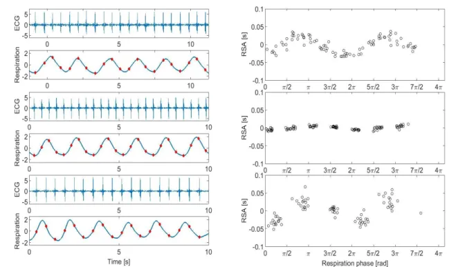
These different modes of interactions are not exclusive, rather they may simultaneously coexist, representing different aspects of neural regulation and acting on different time scales [31], [32]. Figure 1-6 shows three examples, one where RSA is present, another where phase locking occurs and one where they both occur simultaneously.
17 RSA has been observed in full-term infants indicating the presence of cardiorespiratory coupling even at this early life stage [107]. This coupling increases in strength and consistency with GA at birth, reflecting the transition from sympathetic to parasympathetic dominance during the postnatal period with an increasing influence of breathing modulating HR [108]. Overall, immaturity in linear cardiorespiratory coupling is observed in preterm infants, mostly in the form of lower short-term HRV, an indirect measure of RSA [109].
The close interaction between cardiac and respiratory control is critical for survival. This synergy is crucial for the homeostatic regulation of blood gases, and essential for regulating central nervous functions, such as arousal [10], [11]. Arousal from sleep involves both physiological and behavioral responses and has long been considered a vital survival response for restoring homeostasis in reaction to various life-threatening situations, such as prolonged hypoxia or hypotension. There are two distinct
Figure 1-6: Example of presence of RSA and phase locking. On the left there are three 10 seconds segments of ECG and breathing, and on the right the corresponding values of RSA and distribution of R peaks occurrence with respect to respiratory phase. In the first row there is an example of RSA without phase locking. In the second row an example of phase locking (4:1) without RSA. In the third row an example of phase locking (3:1) concurrent with RSA.
18 arousal types defined in infants, subcortical activation and full cortical arousal, which reflect the hierarchical activation from the brainstem (including heart rate, blood pressure, and ventilation changes) to the cortex.
1.5.4. Effects of sleep states on autonomic cardiorespiratory control and arousal
There are marked differences in cardiorespiratory control between AS and QS. Assessment of cardiovascular control is commonly made by studies of HR and HRV and blood pressure and its variability. In infants, HR and overall HRV are higher in AS compared to QS, as a result of the predominance of sympathetic activity in AS. Sleep state also affects blood pressure, with higher values in AS and increased variability. Respiratory control is also affected by sleep state [94]. In infants, respiratory rate decreases from wake to sleep, and breathing is more variable in AS, with short apneas occurring more frequently in this state than in QS [93]. Voluntary control of ventilation is abolished at sleep onset and upper airway resistance is increased due to muscle hypotonia. It is nowadays recognized that the two sleep states have different effects on the control of the respiratory rhythm generator, respiratory muscles and lung volumes. During QS, when lying supine, due to tonic activity in the diaphragm and intercostal muscles, abdominal and thoracic respiratory movements are largely synchronous with each other. In contrast, during AS, there is a reduction in the tonic activity of the diaphragm and intercostal muscles that produces a paradoxical inward rib cage motion during inspiration while the abdominal wall moves outwards. In addition, the horizontal configuration of the diaphragm in neonates causes the lower ribcage to move inward during inspiration, thereby reducing diaphragmatic efficacy. Oxygen saturation levels are consistently lower and more variable in AS than QS due to the paradoxical inward rib cage motion and increased oxygen consumption in AS. Although end-tidal carbon dioxide levels do not change from one to six months of age in the term infant, they are consistently lower in AS than QS. The rate and depth of breathing during sleep oscillate in cycles of 7–13s in both AS and QS; minute ventilation is higher in AS than QS due to increases in rate while depth remains relatively constant [94].
Infant arousal responses are affected by postnatal age and these maturational effects are also sleep-state-dependent. Previous studies have demonstrated that in response to respiratory, tactile and auditory stimulation, total arousability is reduced with increasing age during QS, whilst remaining unchanged in AS [110], [111]. Nonetheless, there are conflicting findings, depending on what type of stimulus is used and what measure is employed to identify arousal.
19
2. H
EART
R
ATE
V
ARIABILITY AND
C
ARDIORESPIRATORY
A
NALYSIS
2.1.
Introduction
In the previous chapter the definition of SIDS and the hypothesized mechanisms leading to it have been highlighted. Furthermore, the crucial role of autonomic activity and cardiorespiratory control was discussed.
The first part of this chapter will present methods proposed for heart rate variability analysis, as a marker of autonomic nervous system activity. No clinical standards are in place for the newborn population, thus adaptation of guidelines for adults and fetuses was necessary. We also evaluated nonlinear parameters which are not yet used in clinical work but can increase our understanding of the complex behavior of autonomic regulation.
In the second part of the chapter, methods that address cardiorespiratory interaction will be presented. Few studies in the past have been performed in populations at risk for SIDS (near-miss SIDS, SIDS siblings, premature infants etc.) regarding their respiratory behavior, for example investigating periodic breathing, apneic events etc. Even fewer studies have addressed the coupling between breathing and HRV dynamics. Given the hypothesized SIDS mechanisms presented in the previous chapter, the contribution of our work is extremely novel and relevant.
Furthermore, this comprehensive view of the cardiac and respiratory systems follows a more physiological perspective, since systems are not analyzed separately but are looked as a complex network, encompassing control cycles using either positive or negative feedback mechanisms and showing complex behavior. Our approach finds its root in a new field of study, called Network
20 Physiology, which views the human organism as an integrated network of interconnected and interacting organ systems, each representing a separate regulatory network. The behavior of one physiological system may affect the dynamics of all other systems in the network of physiologic networks. Due to this intertwining, failure of one system can trigger a cascade of failures throughout the entire network. Network Physiology offers a framework to address the question of how different physiological systems interact and behave together. It stresses the advantages of this holistic approach over the reductionist methods, which analyze every system separately. Recent publications have highlighted the new insights afforded by this novel approach [112]. Looking at different physiological systems as dynamically interacting could shine light on the process of horizontal integration at the level of organ to organ interaction required to maintain an optimal health status.
2.2.
Monovariate analysis
Newborn HRV analysis encompasses techniques from various domains. There are still no standard norms developed for this particular population, thus parameters from adult and fetal HR analysis are usually adapted. The Task Force of 1996 suggests the evaluation of parameters on five-minute windows for adults’ HR [33]. However, since neonatal HR is higher, a similar number of beats occurs in about three minutes allowing a shorter reference length.
2.1.1. Traditional HRV Analytic Approaches
Time domain
For the time domain analysis, measures adapted from adult studies’ have a long history: SDNN, which is the standard deviation of normal to normal intervals (NN), defined as the RR distances excluding anomalous beats (e.g. ectopic beats), and root mean of successive differences, RMSSD. SDNN estimates overall HRV, RMSSD estimates short-term components of HRV [33]. In addition to these traditional parameters, standard measures from fetal HR analyses were computed: short term variability (STV), interval index (II), differential index (DI) and long term irregularity (LTI) [113], [114]. All these measures are calculated in seconds. Generally long-term variability parameters evaluate a combination of sympathetic and parasympathetic nervous systems. In contrast, measures related to the beat to beat variability are influenced largely by the parasympathetic nervous system, tied to rapid ANS reactivity.

![Figure 1-2: US SIDS death count during transition period following the introduction of supine vs prone sleep recommendations, showing that the male fraction remains constant at about 0.61 [222]](https://thumb-eu.123doks.com/thumbv2/123dokorg/7521559.106095/23.918.262.736.141.370/figure-transition-following-introduction-recommendations-showing-fraction-constant.webp)