A
A
l
l
m
m
a
a
M
M
a
a
t
t
e
e
r
r
S
S
t
t
u
u
d
d
i
i
o
o
r
r
u
u
m
m
–
–
U
U
n
n
i
i
v
v
e
e
r
r
s
s
i
i
t
t
à
à
d
d
i
i
B
B
o
o
l
l
o
o
g
g
n
n
a
a
DOTTORATO
DI
RICERCA
IN
Scienze Farmaceutiche
Ciclo XXIV
Settore Concorsuale di afferenza: CHIM/06
S
YNTHESIS
A
ND
S
UPRAMOLECULAR
A
RCHITECTURES
O
F
L
IPOPHILIC
D
ERIVATIVES
O
F
G
UANINE
.
Presentata da:
R
OSARIAC
ARMELAP
ERONECoordinatore Dottorato
Relatore
Prof.Maurizio Recanatini
Prof.Gian Piero Spada
L’auto-assemblaggio è descritto come il processo mediante il quale un sistema
disordinato di componenti pre-esistenti forma una struttura supramolecolare, in
conseguenza di specifiche interazioni deboli tra i componenti del sistema stesso.
Studi precedenti, hanno evidenziato la capacità di auto-assemblaggio di derivati
lipofili delle guanosine; tali composti sono capaci di estrarre degli ioni dalla fase
acquosa e trasportarli in fase organica mediante la formazione di specifiche
strutture supramolecolari. In presenza di cationi le guanosine lipofile formano
aggregati colonnari mentre in loro assenza generano delle strutture nastriformi. Lo
scopo principale della seguente tesi è stato la sintesi di derivati lipofili della
guanina, in particolare di derivati della guanina alchilati in posizione N9 e di un
derivato
della
guanosina
funzionalizzato
con
un
sostituente
perclorotrifenilmetilico(PTM). Lo scopo principale era osservare in che modo
l’assenza dello zucchero, in un caso, e la presenza di un sostituente chirale con un
elevato ingombro sterico (GaceHPTM) nell’altro, potessero influenzare il
comportamento supramolecolare delle molecole sintetizzate. Utilizzando le
guanine invece delle guanosine, non solo vengono conservati tutti i gruppi donatori
e accettori di legami a idrogeno richiesti per l’aggregazione supramolecolare, ma
viene ridotto l’ingombro sterico che in alcuni casi ostacola la formazione di
aggregati (guanine con sostituenti in N9 diversi dallo zucchero possiediono una
maggiore libertà conformazionale anche in presenza di un gruppo ingombrato in
posizione C8 della guanosina). L’auto-assemblaggio di derivati lipofili della
guanina è stato studiato in soluzione mediante spettroscopia NMR e dicroismo
circolare mentre su superficie lo studio è stato effettuato mediante STM (scanning
tunneling microscopy). Allo stesso modo dei derivati lipofili della guanosina, i
seguenti derivati formano strutture nastriformi e colonnari variando le condizioni
sperimentali.
Abstract
Self-assembly relies on the association of pre-programmed building blocks through
non-covalent interactions to give complex supramolecular architectures. Previous
studies provided evidence for the unique self-assembly properties of
semi-synthetic lipophilic guanosine derivatives which can sequestrate ions from an
aqueous phase, carry them into an organic phase where they promote the
generation of well-defined supramolecular assemblies. In the presence of cations
lipophilic guanosines form columnar aggregates while in their absence they
generate supramolecular ribbons. The aim of this thesis has been the synthesis of
guanine derivatives, in particular N9-alkylated guanines and a guanosine
functionalized as a perchlorotriphenylmetil moiety (GaceHPTM) in order to
observe their supramolecular behaviour in the absence of sugar (ribose or
deoxyribose) and in the presence of a bulky and chiral substituent respectively. By
using guanine instead of guanosine, while maintaining all the hydrogen bond
acceptor and donor groups required for supramolecular aggregation, the steric
hindrance to supramolecular aggregation is notably reduced because (i.e. guanines
with groups in N9 different from sugar are expected to have a greatest
conformational freedom even in presence of bulky groups in C8). Supramolecular
self-assembly of these derivatives has been accomplished in solutions by NMR and
CD spectroscopy and on surface by STM technique. In analogy with other
guanosine derivatives, also N9-substituted guanines and GAceHPTM form either
ribbon-like aggregates or cation-templated G-quartet based columnar structures.
Chapter 1. Introduction
1.1 Concepts
2
1.2 Supramolecular chemistry
2
1.3 Self-assembly in supramolecular system
4
1.4 Guanine and G-quartet
5
1.5 DNA G-quadruplexes and their functions
7
1.5.1 Telomers and telomerases
9
1.5.2 Transcription
11
1.5.3 G-quadruplexes ligands
12
1.5.4 Methods for studying G-quadruplexes
14
1.6. Conclusions
19
References
20
Chapter 2. Supramolecular organization of guanosine derivative
2.1 Concepts
24
2.2 G-quartet based assemblies
24
2.3 Linear ribbon of guanosine
29
2.4 Switching between supramolecular assemblies of Lipo-G.
33
2.4.1 Addition/removal of cations
34
2.4.2 Variation of solvent polarity
36
2.4.3 UV-VIS irradiation
37
2.5 Conclusions
39
Chapter 3. N
9-alkyl guanines
3.1 Concepts
44
3.2 Sinthetic approaches
45
3.3 Supramolecular studies in solution
49
3.4 N
9-alkylated guanines: STM study
55
3.4.1 Introduction
55
3.4.2 Results and discussions
59
3.4.3 Conclusions
64
3.5 Reversible assembly/ Reassembly process
65
References
69
Chapter 4. Synthesis of a guanosine functionalized as PTM derivatives
4.1 Concepts
73
4.2 Overview on PTM derivatives
75
4.2.1 Synthesis of PTM radicals
77
4.2.2 PTM properties
78
4.3 HPTM-GACE: synthetic approaches
83
4.4 HPTM-GACE: studies in solution
89
References
98
Chapter 5. Experimental part
102
As the wind of time blows into the sails of space, the unfolding of the universe nurtures the evolution of matter under the pressure of information. From divided to condensed and on to organized, living, and thinking matter, the path is toward an increase in complexity through self-organization. (J.M.Lehn)
Introduction
1
2
1.1
Concepts
Molecular chemistry has created a wide range of ever more sophisticated molecules and materials. Supramolecular chemistry aims at developing highly complex chemical systems from molecular components interacting via non-covalent intermolecular forces. Through the appropriate manipulation of these interactions, it became progressively the chemistry of molecular information at molecular level.[1]
1.2
Supramolecular Chemistry
Supramolecular chemistry is often defined as being “chemistry beyond the molecule”, which is rather vague expression. Therefore, in order to get across the basic concepts of
supramolecules and supramolecular chemistry, it could be worth using an analogy of
daily life. Many sports involve teams of players, one of the main objectives in such sports is to organize the team such that the performance of team is significantly greater than the sum of the performances of each team –member. This concept of a good team being greater than the sum of its part can also be applied to a supermolecule. Indeed according to Dr. Lehn , a supermolecule is an organized, complex entity that is created from the association of two or more chemical species held together by intermolecular forces. Supramolecular structures are the result of not only additive but also cooperative interactions, including elettrostatic interactions, hydrogen bonding, interactions ,dispersion and hydrophobic interactions, and their properties are different than the sum of the properties of each individual component.
Supramolecular chemistry can be classified into three categories:
chemistry associated with a molecule recognizing a partner molecule
chemistry of molecules built to specific shape
chemistry of molecular assembly from numerous molecule.
This classification is deeply related to the size of target molecular system. Molecular recognition chemistry generally deals with the smallest supramolecular systems and encompasses interaction between just a few molecules. In contrast, the chemistry of molecular assemblies can include molecular systems made from countless number of
Introduction
3 molecules.[2]Molecular recognition can be regarded in many ways as the most fundamental kind of supramolecular chemistry. Its importance came to light in the middle of nineteenth century before the concept of supramolecules was established. During microscopic observation Pasteur noticed that crystals of tartaric acid occurred in two types, that were mirror images of each other, and found that mold and yeast recognize and utilize only one of this types. In 1894 Emil Fischer proposed the lock and key mechanism, this concept proposed that the mechanism by which an enzyme recognizes and interacts with a substrate can be likened to a lock and key system. In 1967 Cram established a new field of chemistry ,host-guest chemistry, where the host component is defined as an organic molecule or ion whose binding sites converge in the complex and the guest component is any molecule or ion whose binding sites diverge in the complex.[3] Only in 1978 J.M. Lehn attempted to organize these novels chemistries and proposed the term supramolecular chemistry.
Such supramolecules have geometrically specific shapes for example rotaxane which contains molecules that are threaded by linear molecules or catenane which contains entangled molecular rings. These molecules can be obtained introducing a strategy based on supramolecular chemistry. Controlled molecular association results in the spontaneous formation of supramolecules with specific shape and characteristcs. This process is called
self- assembly or self-organization. This process can be distinguished in two types: strict
or looser. The first type involves associations formed through hydrogen bonds, in the other type the main binding forces come from hydrophobic interactions in aqueous media.
4
1.3
Self-assembly in supramolecular system
Self –assembly may be defined as the process by which a supramolecular species forms spontaneously from its components. Generally we can make two distinctions between static self assembly (SSA) and dynamic self-assembly (DySA) by considering the thermodynamic description of the resulting assemblies (fig.1 2)[4].
SSA refers to a stable equilibrium structures by a maximum(local or global) in the system’s entropy and no systematic energy flows, examples are organic and inorganic crystals, block copolymer assemblies and supramolecular system.
Figure 1.2. Schematic representations of (a) static and dynamic assembly (b). (a) In static
self-assembly, components form an ordered, thermodynamically isolated aggregate whose structure does not change in time. (b) Dynamic self-assembly involves a disordered collection of components in an ordered structure through input of energy from an external source and dissipates this energy on the environment.
Biological system aside, self-assembly is also a common place throughout chemistry: the growth of crystals, the formation of liquid crystals, the spontaneous generation of synthetic lipid bilayers, the synthesis of metal co-ordination complexes and the alignment of molecules on surfaces are but a few of the many manifestation of self-assembly in chemical systems. Whereas self-assembly may be taken as a simple collection and aggregation of components into a confined entity, we shall considered self-organization as the spontaneous but information-directed generation of organized functional structures in equilibrium conditions.[1] These structures are held together by a variety of
Introduction
5 weak non covalent interactions as: hydrogen bonding, stacking, dipolar interactions, van der Waals forces and hydrophobic interactions. A distinctive feature of using this weak forces in molecular assemblies is that such interactions are normally readily reversible, so that the final product is in thermodynamic equilibrium with its components. This leads to an additional proprerty of most supramolecular systems: they have an built-in capacity of error-correction not available to covalent systems. Among non-covalent interactions the use of hydrogen bonding and interactions have tended to receive most attention in the design of supramolecular system but van der Waals considerations are often also of crucial importance. A self –organization process may be considered to involve three main stages :
Molecular recognition for the selective binding of the basic components.
Growth through sequential binding of multiple components in the correct relative disposition.
Termination of the process, requiring a built-in feature, a stop signal, that specifies the end point.
Suitable encoding by manipulation of structural subunits and processing through interactional algorithms should give access to a variety of systems.
1.4
Guanine and G-quartets
Nucleobases have been proposed as basic units of supramolecular motifs due to the potential non-covalent interactions.[5] They are well known for their ability to form complementary H-bonds with their base pairs. The double helix of DNA is due to the canonical Watson and Crick base pairing (fig. 1.3), adenine forms a pair with tymine and guanosine with cytosine.
6 Figure 1.3
However alternative hydrogen bonding patterns, such as the Hoogsten base pairing can occur giving rise to complex and functional tertiary structures. This one implies the N7 position (as a hydrogen bond acceptor) and C6 amino group (as a donor)of the purine base, which bind the Watson-Crick (N3–N4) edge of the pyrimidine base.
Figure 1.4: Hoogsten base pair adenine- tymine
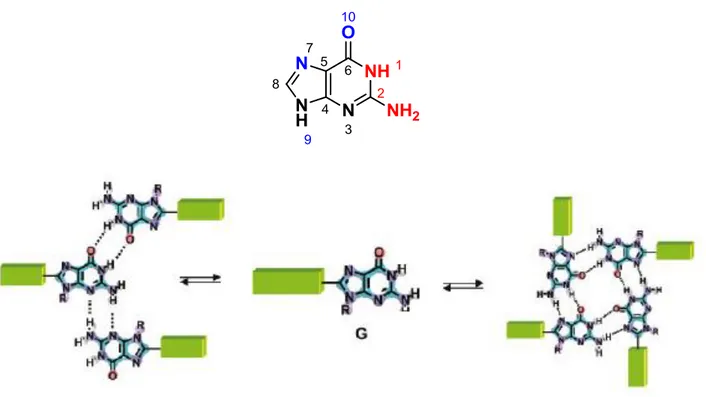
Among the other nucleobases, guanine is the most versatile indeed as it contains both a Watson-Crick edge and Hoogsteen edge.[6] Moreover, the Watson-Crick edge has two hydrogen bond donors (N1H, N2H) that can hydrogen bond with the two hydrogen bond acceptors (O6, N3 and N7) on the Hoogsteen edge. This leads guanine to form different supramolecular structures. Among these the most known is the so-called G-quartet which is a macrocyclic array of four guanines, hydrogen bonded through their self-complementary Watson-Crick (N1H and N2H) and Hoogsten (O6and N7) edges.[7] In addition, guanine can form two ribbon-like structures, characterized by different hydrogen-bonded patterns.
Introduction
7
Fi
2
1.5
DNA G-quadruplexs and their functions
The G-quartet was identified by Davies and coworkers in 1962 as the structural unit behind hydrogels formed by 5’-guanosine monophosphate(5’-GMP).[9] But, only several years later did the possible biological relevance of DNA structures based on this moiety begin to be addressed.[10] G-quadruplexes, have a core that is made up of guanine bases only, with four guanines arranged in a rotationally symmetric manner, and held together hydrogen bonds between N1–O6 and N2–N7 around the edges of the resulting square.[11] These planar structures are called G-quartets, and are stabilized by monovalent cations, in particular K+ and to a lesser extent NH4+ and Na+, which interact with the lone pairs on the O6 atoms surrounding the central core. They can form spontaneously at sufficiently high concentrations of guanine. These G-quartets have large -surfaces, and hence tend to stack on each other. In particular,oligonucleotides with contiguous runs of guanine, such as d(TGGGT) can form stacked structures with the G-quartets linked by the sugar– phosphate backbone. These are called G-quadruplexes and can form from DNA or RNA strands.
Figure 1.5 Guanine and supramolecular structure Schematic representation of the equilibrium between a
8 Figure. 1.6 Schematic rapresentation of G-quadruplexes structure. Four G-quartet are held togheter
from-stacking and sugar backbones
These strands have a directionality described as from 5’-end to 3’-end and they can be parallel or antiparallel. At a molecular level, the different directionality of the strands relates to the conformational state of the glycosidic bond between the guanine base and the sugar. This may be either syn or anti, when all bases are in anti conformation the four strands are parallel, when the bases are in the syn all strands are antiparallel. This then affects the orientation of the backbone relative to the G-quartets, and hence results in grooves of different sizes.[12] G-quadruplexes may be comprised of four separate strands, as in figure 1.7, forming tetramolecular G-quadruplexes, which are always found in the all-anti parallel form. Alternatively, they may be formed from two strands, each with two sets of contiguous guanines, or just from one strand, folding back on itself to form an intramolecular structure. In either of these cases, there will be loops that serve to connect the strands of the structure together.
Depending on which strands are connected, these loops may cross diagonally across the top of the structure, joining diagonally opposed antiparallel strands; go across a side, linking adjacent antiparallel strands; or may loop around the side of the structure linking parallel strands and forming a double-strand reversal loop.
Introduction
9 Figure 1.7 Different stoichiometries and folding patterns of G-quadruplexes. (A) Tetramolecular
structure with all strands parallel; (B) bimolecular antiparallel structure with adjacent parallels trands; (C) unimolecular antiparallel structure with alternating parallel strands
1.5.1 Telomeres and telomerases
It is important for eukaryotic cells, which have linear chromosomes, to be able to distinguish between chromosome ends and unexpected breaks in the DNA. [12] In order to facilitate this discrimination, they have repeated sequences at the ends, called telomeres[13] Human cells’ telomeres represent the chromosomal ends (preventing them from fusion events), ranging in length from 3000 to 15000 bases, composed of tandem repeats of the 5´-GGTTAG-3´ sequence with a 3’ overhang of the G-strand,which plays an important structural and functional role.[14] Telomere length decreases with each cell division event, while reversion of this degradation by a specialized enzyme called telomerase increases cellular replicative capacity, leading to uncontrolled proliferation: in the majority of tumour cells (85–90%) this enzyme is over-expressed. Therefore there is a great deal of interest in developing approaches to reduce the activity of telomerase for therapeutic purposes.[15-16].
The human telomeric sequence, d(GGGTTA)n folds spontaneously into an intramolecular
G-quadruplex form, with the GGG runs forming the G-quartet core, and TTA forming the loops of the structure[14]. This structure is stable under physiological conditions, with a thermal melting temperature of around 65 °C. All telomeric sequences studied to date can also form G-quadruplex structures with comparable thermal stability. It has therefore been proposed that the physiologically relevant structure of the telomeric overhang has a series of G-quadruplexes, much like beads on a string. This telomeric repeat sequence has become an important target for drug development,[15-16] as it has been shown that by binding to and stabilizing telomeric G-quadruplexes, it is possible to block telomerase
10 from acting and extending the telomeres, hence preventing the immortalisation of cancerous cells.
Figure 1.8 (a) Side view of the antiparallel human telomeric G-quadruplex structure solved by Wang and
Patel using NMR spectroscopy, from PDB entry 143D. (b) Detailed view of the central G-quartet from PDB entry 143D. (c) Side view of the parallel human telomeric G-quadruplex structure solved by Parkinson, Lee and Neidle using X-ray crystallography, from PDB entry 1KF1. (d) Top view of the parallel structure from PDB entry 1KF1. In all cases, guanines are shown as cylinders, other bases as balls and sticks. Potassium ions are shown in magenta.
The structures formed by the telomeric repeat have been the subject of considerable study, and various structures have been solved for the telomeric repeat, using slightly different sequences and conditions. More recent studies have shown that the telomeric sequence can form a wide variety of different structures, which all seem to exist in equilibrium with each other.[17]
Introduction
11
1.5.2
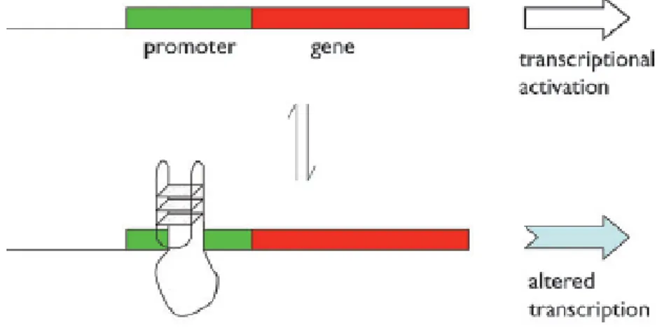
Transcription regulation
One method that is used in some cases to regulate gene transcription, is based on the presence of G-quadruplexes located in the promoter region of a gene, broadly speaking the kilobase upstream of the transcription start site (TSS).[16-18].This model was originally demonstrated by Hurley and co-workers for the oncogene c-myc,[19] an important transcription factor involved in regulating around 15% of all human genes. As a result of this, overexpression of c-myc has been implicated in a wide range of cancers including colorectal cancer. This one controls the vast majority of the transcription of the gene, and studies in vitro of the sequence d(GGGGAGGGTGGGGAGGGTGGGGAAGG) show that it is capable of forming a family of polymorphic G-quadruplexes, using various combinations of the guanine runs underlined.[20] It has further been shown that the G-quadruplex ligand TMPyP4binds to this element leading to downregulation of c-myc expression.[21] This clear proof of principle led to the proposal that this may be a general mechanism for gene regulation. The simplest form of the model (fig. 1.9) proposes that there is an equilibrium between two forms of the DNA.
Figure 1.9 The formation of a G-quadruplex in a promoter can affect the level and nature of transcription
from that gene. At the simplest level, it may act as a steric block to the transcription machinery.
On one side of the equilibrium is double helix DNA, and transcription occurs as normally; on the other side, one strand is separated, and has folded up into a quadruplex. This structure then acts as a steric block to transcription. Addition of a quadruplex ligand, whether a small molecule or a protein, will energetically favour the
G-12 quadruplex form, and hence move the equilibrium towards that side and reduce the transcriptional activity.
1.5.3 G
-quadruplexes ligands
[12-13]A range of G-quadruplex ligands have been shown to bind quadruplexes in vitro.[22.]
Some very interesting examples of compounds capable of stabilizing G-quadruplexes show a binding mode based on a loop and groove interaction, however the major part of ligands show the same chemical chrateristics such as: an aromatic core which favours stacking interactions with G-tetrads and basic side-chains (positively charged under
physiological conditions) which interact with the quadruple helix groove.[23] These
molecules recognize quadruplex DNA, adopting a terminal stacking mode as in figure 1.10: some of them have also been shown to induce telomere shortening or telomere
instability, triggering apoptosis and/or senescence programs in various cell lines.[24-25]
It is worth noting that, as a result of their direct interaction with telomeres, G-quadruplex ligands have shown more rapid and specific effects than those that would be expected for
simple telomerase inhibitors. Using the above principles, it is relatively easy to design
compounds that will bind G-quadruplexes, although not necessarily with high affinities. Nonetheless, some good G-quadruplex binders have been developed, such as those depicted in figure 1.11. These include the cationic porphyrin 5,10,15,20-tetra(N-methyl-4-pyridyl) porphin, TMPyP4 (although widely used, this has only limited selectivity for
Figure 1.10 Representation of a complex between a coronene derivative (CORON3)[26] as a ball and-stick model with a grey surface and a monomeric G-quadruplex (black surface).
Introduction
13 G-quadruplexes over duplexes) and a variety of acridine and acridone compounds, such as the 3,6,9-substituted acridine BRACO-19.
In order for ligand binding to be therapeutically effective, it is not enough for the ligand to bind to G-quadruplexes, or even to be very highly selective for them over duplex DNA. It must also be able to bind selectively to one G-quadruplex over another. This is a big challenge, since there are relatively few recognition points to discriminate different G-quadruplex structures. Various methods have been used to try to combine targeting of the loops and grooves of each structure with targeting of the G-quartet core, but to date this has only provided limited success.[27]
14
1.5.4 Methods for studying G-quadruplexes
There are a number of different experimental techniques used to study G-quadruplex formation, each examining different aspects of the structures, and hence reporting on different aspects of their formation. The majority of these techniques are principally descriptive, and complete structure determination requires the use of either NMR structure determination or X-ray crystallography. NMR spectroscopy requires much less sample preparation than crystallography, but does require very pure and high-concentrated samples.
At the simplest level, it is possible to gain much information even from a 1-D 1H NMR spectrum, as there are a relatively small number of protons in nucleic acids and the guanine NH1 imino protons have a characteristic shift when hydrogen bonded.
In addition, they exchange relatively slowly with the deuterated solvent when compared to non-hydrogen-bonded protons. This may therefore be used to show G-quadruplex formation. In order to provide more detailed analysis, multi-dimensional techniques are needed, which allow the complete assignment of resonances to the sequence being studied.[12, 28].
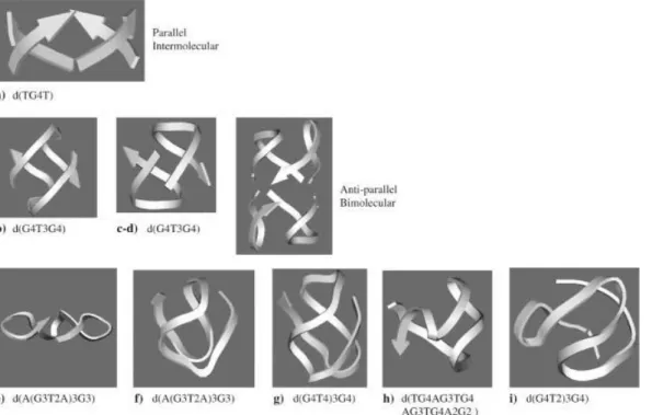
Figure 1.12 Typical folding topologies of G4-DNA forming sequences: a parallel intermolecular structure
(a), bimolecular antiparallel structural motifs (b–d), intramolecular parallel (e) and antiparallel (f–i) monomolecular structures. Arrows represent backbones running from 5’to 3’end.
Introduction
15 Circular dichroism (CD) is another common technique used to study G4-DNA.: it has been used to study 3D-structures, ligand binding and effect of cations. Although the topology of the folding of G4-DNA strands is very complex , only two basic types of CD spectra, which have been associated with the relative orientation of the strands parallel and antiparallel, are investigated (fig. 1.12).
The spectra of parallel quadruplexes, in which four strands with all glycosidic bonds in
anti have a dominant positive band at 260 nm, and a negative band at 240 nm while the
spectra of antiparallel quadruplexes ( where guanines alternate syn and anti glycosidic conformations along each strands) have a negative band at 260 nm and a positive band at 290 nm[29-30] Although this empirical relationship for many case is the interpretation for most CD spectra of G4-DNA, [11,29.31,32] it cannot be considered of general validity (fig. 1-13).
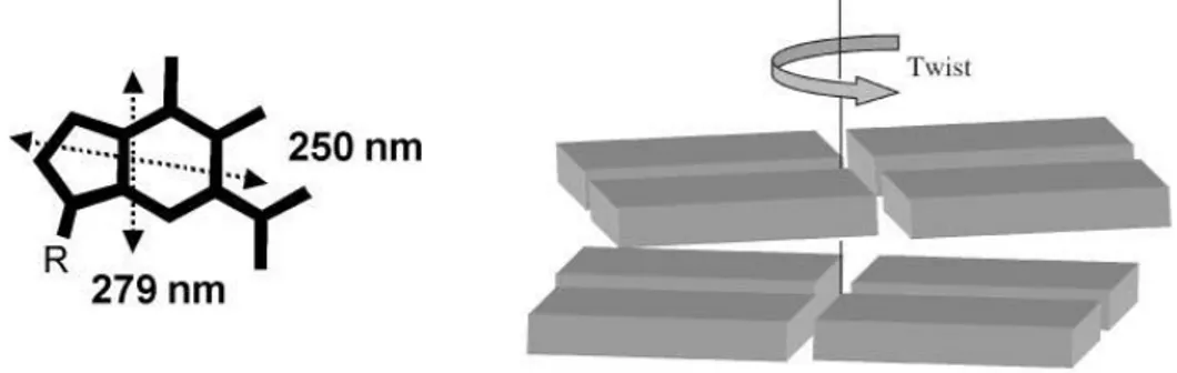
However starting from the chromophore it could be possible to explain how the different folding patterns origin different CD. In the case of G4-DNA the chromophore is represented by guanine, this one has two absorption band in 240-290 nm region connected to two transitions at 279nm and 248 nm.These transitions are short and long axis polarized respectively . In G4-DNA , G-quartets are stacked one on the top of Figure 1.13 CD spectra of guanine quadruplexes. Left side: the parallel stranded quadruplex [d(G4)]4
16 the other and they are rotated one with respect to the adjacent one:this rotation causes a chiral exciton coupling between transition dipole moments located in near-neighbour guanines.
The first non empirical interpretation of CD of G4 DNA has been reported by Spada et al. by modeling the spectrum of polyguanilic acid , which shows the tipical spectrum of “parallel G4-DNA and it has been reproduced by an exciton calculation considering only near-neighbour interactions between the guanine transitions of two stacked G-quartets.
[33-34].The two faces of G-quartet are diasterotopic so when the G-tetrads are piled, each
quartet can stack onto the adjacent one through the same (head-to-head or tail-to tail) or the opposite (head to tail) face leading to a heteropolar or homopolar stacking.
Figure 1.14Orientation of the two most relevant electric transition moments (dotted double-head arrows) of the guanine chromophore (left) and a sketch of the chiral arrangement of two adjacent G-quartets (each parallelepiped represents a guanine base).
Figura 1.15 Top view of the heteropolar and homopolar stacking of two G-quartets: the “head” and the
“tail” sides of the G-quartets are represented in red and green, respectively (the double-head arrows represent the transition moments corresponding to the absorption band at ca. 250 nm).
Introduction
17 Considering the case of paralle G4-DNA, the disposition of two adjacent G-quartets in a H-to-T orientation is that reported in the figure 1.15 where the electric moments of a couple of near neighbour guanines have been proposed. Applying the simplified model of the exciton coupling it emerges that this chiral arrangement is expected to exhibit a positive exciton centerd at 250 nm (see figure1.16). When the glycosidic bonds of the guanines alternate in syn and anti conformations along each strand (antiparallel strands) the G-quartet polarity also alternates, while quadruplexes with parallel strands and all
anti glycosidic bonds have a non-alternating G-quartet polarity. CD spectra in these two
cases are expected to be different. Indeed in the heteropolar H-to-H stacking of two quartets, the relative orientation of the closest dipole moments is different from the case described above. Using the qualitative approach to exciton coupling , the chiral arrangement is expected to give a negative couplet centered at 250 nm ( fig. 1.16)
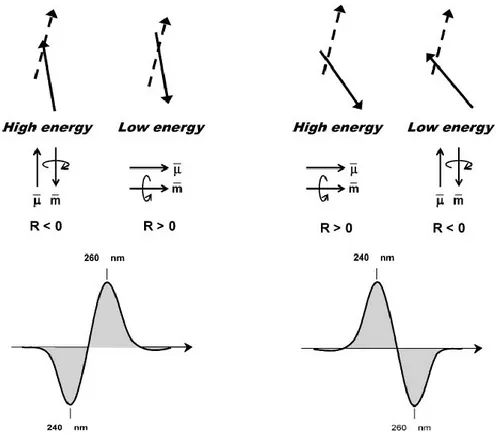
Figure 1.16 A simplified model for the origin of the positive (left side) and negative (right side) exciton
couplets for the head-to-tail (H-to-T) and head-to-head (H-to-H) G-quartet stacking, respectively. Top: the arrangement of two 250 nm electric transition moments (full line: front vector; dashed line: rear vector) located in two closest guanines. Middle: themagnetic (m) and electric (l)moments generated by the coupling of the two guanine chromophore (more in details, in the high energy coupling of the left-side panel, the two electric transition moments–top–sum to a total electric vector pointing upward– middle–and generate a charge rotation with a resulting magnetic moments pointing downward, that is antiparallel). Bottom: the predicted CD spectra.
18 CD spectral calculations with the dipole approximation of the two G-quartet stacked with the same or the opposite polarity has been performed by Gray at al.(fig. 1.17)[34].and their results confirm the late computations by Spada et al.[33].on the homopolar stacked system and show how heteropolar stacking explains the emergence of a positive CD signals at 290 nm. The kind of CD spectrum is actually not directly related to the relative strands orientation: the stacking orientation of G tetrads obviously depends on the folding of the strands, however no direct relationship can be established between the two topological features.
Figure 1.17 Superimposed calculated CD spectra of two G-quartets stacked in the to-T (solid line) or
H-to-H (dashed line) orientation as shown in Fig. 1.15. The relative orientation of the G-quartets for the calculation were extracted from the solution structure of d(G3T4G3) that present mixed polarities of stacked G-quartets.
Introduction
19
1.6
Conclusions
G-quadruplex structures are interesting on many levels. Structurally, they display a fascinating array of polymorphic structures, and we are unable as yet to predict their structure or stability theoretically. They seem to play a number of important biological roles, including regulating the critical processes of transcription and translation, and there is pharmaceutical interest in being able to manipulate these processes to develop novel therapeutics. They can also be used for a range of innovative nanotechnological applications—including all the ones no-one has yet envisaged. It is a rapidly growing field, with promise for chemists, biologists, physicists and computer scientists.
20
Bibliography
1 J. M. Lehn, Proc. Natl. Acad .Sci USA, 2002, 99, 4763-4768
2 K. Ariga, T. Kunitake, Supramolecular chemistry: fundamentals and applications, 2006
3 Cram, D. J, Angew. Chem., Int. Ed. Engl. 1986, 25, 1039–1134.
4 N. Krasnogor, S. Gustafon, D. A. Pelta, J. L. Verdegay Systems-self-assembly:
multidisciplinary snapshots
5 S. Sivakova, S. J. Rowan, Chem. Soc. Rev. 2005, 34, 9.
6 W Saenger, Principles of Nucleic Acid Structure, Springer-Verlag: New York,
1984.
7 G.P.Spada et al., Chem. Eur. J. , 2009, 15, 7792-77806
8 D.Gonzales Rodriguez and A.P.H.J. Schenning, Chem. Mater. 2011, 23, 310-325 9 M. Gellert, M. N. Lipsett, D. R. Davies, Proc. Natl. Acad. Sci.USA 1962, 48,
2013–2018
10 D. Rhodes, R. Giraldo, Curr. Opin. Struct. Biol., 1995, 5, 311–322
11 S. Burge, G. N. Parkinson, P. Hazel, A. K. Todd and S. Neidle, Nucleic Acids Res.,
2006, 34, 5402
12 J. L. Huppert, Chem. Soc. Rev., 2008, 37, 1375–1384
13 M. Franceschin, Eur. J. Org. Chem. 2009, 2225–2238
14 S. Neidle, G. N. Parkinson, Curr. Opin. Struct. Biol. 2003, 13,275–283
15 J. L. Mergny, J. F. Riou, P. Mailliet, M. P. Teulade-Fichou, E.Gilson, Nucleic Acids Res. 2002, 30, 839–865
16 D. J. Patel, A. T. Phan and V. Kuryavyi, Nucleic Acids Res., 2007,35, 7429
17 J. Dai, C. Punchihewa, A. Ambrus, D. Chen, R. A. Jones and D. Yang, Nucleic Acids Res., 2007, 35, 2440.
18 . S. Dexheimer, M. Fry and L. H. Hurley, DNA quadruplexes and gene regulation,
in Quadruplex Nucleic Acids, ed. S Neidle and S Balasubramanian, Royal Society
of Chemistry Cambridge, UK, 2006.
19 A. Siddiqui-Jain, C. L. Grand, D. J. Bearss and L. H. Hurley, Proc. Natl. Acad.
Introduction
21 20 J. Seenisamy, E. M. Rezler, T. J. Powell, D. Tye, V. Gokhale,C. S. Joshi, A.
Siddiqui-Jain and L. H. Hurley, J. Am. Chem. Soc., 2004, 126, 8702.
21 . C. L. Grand, H. Han, R. M. Muñoz, S. Weitman, D. D. Von Hoff, L. H. Hurley and D. J. Bearss, Mol. Cancer Ther., 2002, 1, 565.
22 C. M. Incles, C. M. Schultes, S. Neidle, Curr. Opin. Investig.Drugs 2003, 4, 675–
685
23 a) S. M. Haider, G. N. Parkinson, S. Neidle, J. Mol. Biol. 2003,326, 117–125; b)
S. Neidle, R. J. Harrison, A. P. Reszka, M. A. Read, Pharmacol. Ther. 2000, 85, 133–139.
24 J. F. Riou, L. Guittat, P. Mailliet, A. Laoui, E. Renou, O. Petitgenet, F.
Mégnin-Chanet, C. Hélène, J. L. Mergny, Proc. Natl. Acad. Sci. USA 2002, 99, 2672–2677
25 E. Salvati, C.Leonetti, A. Rizzo, M. Scarsella, M. Mottolese, R. Galati, I. Sperduti,
M. F. Stevens, M. D’Incalci, M. Blasco, G. Chiorino, S. Bauwens, B. Horard, E. Gilson, A. Stoppacciaro, G. Zupi, A. Biroccio, J. Clin. Invest. 2007, 117, 3236– 3247.
26 M. Franceschin, A. Alvino, V. Casagrande, C. Mauriello, E.Pascucci, M. Savino, G. Ortaggi, A. Bianco, Bioorg. Med.Chem., 2007, 15, 1848–1858.
27 P. S. Shirude, E. R. Gillies, S. Ladame, F. Godde, K. Shin-Ya, I.Huc, S. Balasubramanian, J. Am. Chem. Soc., 2007, 129, 11890–11891.
28 Stefano Masiero, Roberta Trotta, Silvia Pieraccini, Stefano De Tito, Rosaria Perone, Antonio Randazzo * and Gian Piero Spada *, Org. Biomol. Chem., 2010,
8, 2683–2692
29 P. Balagurumoorthy, S. K. Brahmachari, D. Mohanty, M. Bansal and V. Sasisekharan, Nucleic Acids Res., 1992, 20, 4061.
30 D. M. Gray and F. J. Bollum, Biopolymers, 1974, 13, 2087; R. Jin, B. L. Gaffney, C. Wang, R. A. Jones and K. J. Breslauer, Proc. Natl. Acad.Sci. U.S.A., 1992, 89, 8832.
31 M. Lu, Q. Guo and N. R. Kallenbach, Biochemistry, 1993, 32, 598.
32 F.W. Smith and J. Feigon, Nature, 1992, 356, 164; C. Kang, X. Zhang, R. Ratliff, R. Moyzis and A. Rich, Nature, 1992, 356, 126; J. R. Williamson, Annu. Rev.
22 33 G. Gottarelli, P. Palmieri and G. P. Spada, Gazz. Chim. It., 1990, 120,101.
34 D. M. Gray, J.-D. Wen, C. W. Gray, R. Repges, C. Repges, G. Raabe and J. Fleischhauer, Chirality, 2008, 20, 431
Supramolecular organization of guanosine derivatives
23
2 Supramolecular organization of
24
2.1
Concepts
Although the large variety of supramolecular networks originated by guanosine derivatives has been investigated for a couple of decades, only in recent years research groups focused their attention on their application in supramolecular chemistry. Guanine moiety is a versatile hydrogen bonding building block. In particular, lipophilic guanosines or guanines can undergo different self-assembly pathways, originating different nanoarchitectures depending on environmental conditions, the two typical assemblies being ribbons and cyclic G-quartet systems. Furthemore the easy functionalisation of guanosine in the sugar hydroxyl groups or in the aromatic base makes it promising building block for the fabrications of complex architectures with functional units located in preprogrammed positions.[1]
2.2
G-quartet based assemblies
Lipophilic guanosines (LipoG) can undergo in the presence of cation different self-assembly pathways depending on experimental conditions. In 1995.[2] our group showed that LipoG, in particular deoxyguanosine derivative dG(C10)2 1, extracted K+ picrate
from water into chlorinated solvent (CDCl3) giving an octamer.The presence of cation was essential, for the formation of this lipophilic octamer. At the same time Davis at al.[3]. demonstrated analogous behaviour for isoguanosine derivative.
Supramolecular organization of guanosine derivatives
25 Figure 2.18: the cation-templated self-assembly of Lipo-G 1 from the unassembled molecule to an
octameric species and finally to a pseudopolymeric aggregate (the spheres represent the cation).
For different Lipo-Gs,[4] different, yet G-quartet based, different supramolecular assemblies have been characterized, depending on chemical modification of the guanosine and on the relative guanosine to metal ion ratio. In the last years, NMR spectroscopy has become a valuable tool for the characterization of these supramolecular systems. It provides informations on the structural size of complexes, the effect of the solvent, the role of the cation and anion. After reporting that 1 behaves as an ionophore and a solution of it in chloroform is able to extract potassium picrate (KPic) from water (or crystal state),[5] two different supramolecular assemblies were proposed and later on solved (figure 2.19): depending on the relative amount of KPic (1:8 or 1:4 K+/G ratio) used in the extraction either a C4-symmetric octamer or a pseudopolymeric-assembly can be obtained. In the 1:8 ratio case, the H NMR spectra,[1] essentially temperature independent over more than 100°C, show two sets of signals in a 1:1 ratio corresponding each to nucleosides with different glycosydic conformation (syn-like and anti-like). It should be noted that there is another stereochemical consequence to the cation-templated self-assembly of guanosine derivatives.
As described in figure 1.54 the two faces of the G-quartets are diasterotopic and can be labelled head and tail (fig. 2.19).
pseudo-polymer Octamer
26 Figure 2.19. a) A G-quartet showing its head diastereotopic face. Schematic drawing of b) a C4-symmetric and c) a D4-C4-symmetric octamer. In the C4- C4-symmetric octamer (obtained, e. g., from 2), an syn G-quartet (S, black disk) with its tail-side (lower face) stacks on the head-side (upper face) of an all-anti (A, white disk) quartet. In the D4-symmetric octamer (obtained, e.g., from 3), two all-syn G-quartet stack facing their tail sides. Clockwise and counter-clockwise arrows refer to the head and tail faces, respectively.d) Cartoon of the assembly of the polymeric aggregate obtained from dG 1. White, grey and black disks refer to all-anti (A), all-syn1 (S1) and all-syn2 (S2) quartets, respectively; clockwise and counter-clockwise arrows refer to the quartet head and tail faces, respectively.
In principle, the two quartets in the octamer can be arranged in three different orientations: head-to-tail (C4 symmetry, fig. 2.19 b), head-to-head and tail-to-tail (D4 symmetry; in figure 2.19 c the tail-to-tail arrangement is shown). Therefore, considering these two stereochemical aspects (syn/anti glycosydic conformation and relative G-quartets orientation) several diastereoisomers are possible for the octamer.
Remarkably, the NMR data indicated that this octamer was a single diastereomer of C4 symmetry. In one G-quartet, all monomers had a syn conformation, while the other tetramer had an “all-anti” conformation. NOE interactions indicated a relative orientation with the head-side of the “all-anti” quartet facing the tail-side of the “all-syn” G-quartet (Figure2.19 b). While derivative 1 forms the K+-templated C4-symmetric octamer structure or pseudo-polymeric assembly in solution, other lipo-G derivatives (especially those with ribose, in place of deoxyribose, including 2’,3’di-O-isopropylidene-guanosine derivative 2 can give a different stereoregular octamer with a D4 symmetry.[6-7]
While in the case of 1 two sets of signals are observed in the 1H NMR spectrum (the two G-quartets are diastereotopic), in the latter case only a single set of signals is observed for
Supramolecular organization of guanosine derivatives
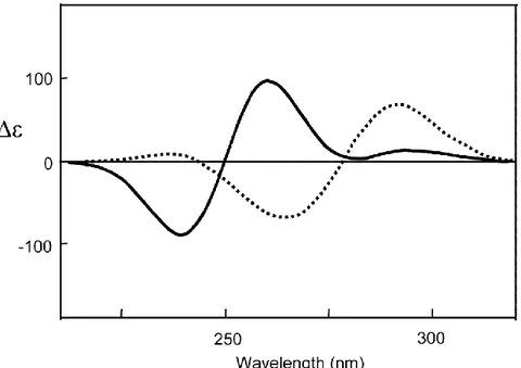
27 the two homotopic G-quartets. Furthermore circular dichroism is diagnostic of the stacking polarity of two contiguous G-quartets.[8].
The tetramers do not stack in register, but are rotated with respect to each other to give, in the 230–300 nm region characteristic of the * transitions of guanine chromophore, a double-signed exciton- like CD signal. This couplet, the sign of which allows the assignment of the stacking helicity (handedness), exhibits opposite signed bands at about 260 and 240 nm for the head-to-tail (C4-symmetric) stacking in compound 1, while both bands are blue-shifted by 20–30 nm in the case of D4-symmetric stacking (compound 2).
Figure 2.20 Comparison between CD spectra of C4- (solid line) and D4-symmetric(dashed line) octamers
G8·M+ obtained from 1 and 2, respectively (Data from references [2-9]).
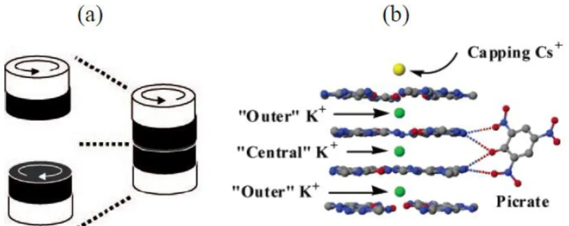
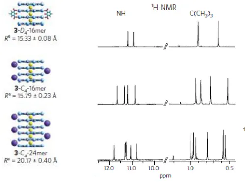
Another different metal ion templated supramolecular structure was observed for the compound 3. In the presence of KPic.[6] NMR experiments (including DOSY NMR data)[10] indicated that a D4-symmetric hexadecamer composed of four stacked G-quartets is the major species in solution (figure 2.21 ). H NMR spectra show two sets of signals,
28 present in a 1:1 ratio, assigned to the distinct “outer” and “inner”quartets. X-ray crystallography confirmed that derivative 3 forms an ordered hexadecamer.[11] This assembly, with empirical formula G16·3K+/Cs+·4 Pic-, is stabilised by four co-axial cations and by four picrate anions. It can be described as a pair of head-to-tail C4-symmetric octamers G8·M+, with each octamer using its eight carbonyl oxygen atoms to coordinate a K+ ion, while a third K+ ion holds the two G8·M+ octamers together in a head-to-head orientation: this leads to a D4-symmetric assembly. Finally, a Cs+ ion loosely bound, in solution caps the structure. In addition to stabilization by cations, four picrate anions form hydrogen bonds to N2 amino groups that extend from the two “inner” G-quartets. The lipophilic G-quadruplex looks like a cation channel with an anionic belt wrapped around its middle. Similar solid-state structures for G16·M22+·4 Pic-4 were obtained with the divalent cations Ba2+ and Sr2+.[12]
Figura 2.21 a) Schematic cartoon of the quartet assemblies in the D4-symmetric hexadecamer (black and
white refer to inner and outer quartets, respectively; clockwise and counter-clockwise arrows refer to the head and tail faces, respectively); b) a schematic showing the nucleobase–picrate hydrogen bonds in the hexadecamer G16·3K
+
/Cs+·4Pic-. ( Figure 2.21b adapted from reference [11] )
Recent studies have already underlined the subtle role of specific anions in this self-assembly process. For instance, in the presence of dinitrophenolate (DNP) salts, two octamers are linked to produce a D4-symmetric hexadecamer, owing to the formation of bifurcated hydrogen bonds between the DNP anion and the amino protons of the inner G- quartets.[13-11] Other studies provide a more general qualitative analysis of the role of the anion, or, more concretely, of the Coulombic interactions between the dissociated ion pairs, in the thermodynamics of G-quadruplex self-assembly. Because the cation is complexed inside the G-quartet cavity, the energy of such interactions can be modulated within a certain range by tuning the stability of the dissociated anion in solution as a
Supramolecular organization of guanosine derivatives
29 function of three factors: solvent polarity, the nature of the anion and the cation–anion distance (modified by steric effects around the complexes). They demonstrate that the role of the ion-pair separation energy, although it can usually be neglected in aqueous solutions, is important in G-quadruplex self-assembly and becomes a critical factor in less polar solvents.[14] This Coulombic contribution alone obviously does not explain the rich polymorphism observed in G-quadruplex self-assembly, but it has been underlined how it can be adjusted within a certain range to regulate, in a precise manner, the growth of G-quadruplex stacks, to form quantitatively assemblies of 8, 12, 16 or 24 guanosine molecules. The last of these species, in particular,comprising 5 cations, 5 anions, and 24 hydrogen-bonded molecules self-assembled in a single object, represents a record in the supramolecular synthesis of discrete, well-defined nanostructures.
2.3
Linear ribbons from guanosine
In the absence of metal ions, 1 has been shown to self-assemble into ribbon-like structures in organic solvents.[15] The same AADD homocoupling at the basis of the G-quartet formation may indeed drive to the formation of a different supramolecular motif
Figure 2.22 Model of the complex formed, experimental hydrodynamic radii Re, amide and t-butyl regions
of the 1H-NMR spectra (0.02 M; 298 K) of 3 in THF-d8 (+0.25 equivalents KMeDNP); THF-d8 (+0.25 equivalents KPF6); and acetone-d6 (+0.25 equivalents KPF6)
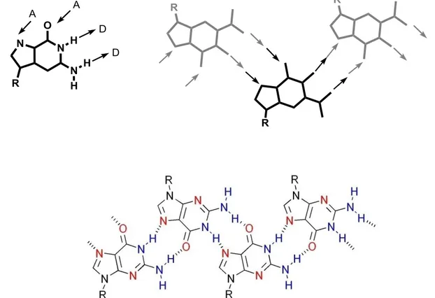
30 (fig. 2.23): in fact, when a couple of guanines exposes to the observer their opposite sides, an infinite H-bonded motif is obtained (“ribbon A”). Furthermore, a different homocoupling (ADDA) in which different H-bonding sites of the guanine are involved, leads to the formation of a further kind of H-bonded ribbon (“ribbon B”). The transformation over time from “ribbon A” (non centrosymmetric) to “ribbon B” (centrosymmetric) has been observed in CDCl3 by solution-state NMR.[16]
Figura 2.23 The non-centrosymmetric ribbon supramolecular structure by guanine, characterized by
N2–H…O6, as composed by AADD homocoupling of guanines (“ribbon A”).
R R R N N N N N H H O H R
A
A
D
D
1 2 3 6 7 8 9 R R R N N N N N H H O H RA
A
D
D
1 2 3 6 7 8 9 1 2 3 6 7 8 9Supramolecular organization of guanosine derivatives
31 These long anisometric supramolecular ribbons may form in organic solvents liquid-crystalline phases. [17] For example derivative 1 in hexadecane above a critical concentrations, gives a viscous birefringent liquid-crystalline phase. A texture of this phase is reported in figure 2.25.
While it has not been possible to obtain a diffraction structure for the longer-chain derivative 1, an X-ray single-crystal diffraction structure for the three-carbon atom tail derivative dG(C3)2 reveals the “ribbon A” type self-assembly [18]
R R R
D
A
A
D
N
N
N
N
H
H
O
H
R
1 2 3 6 7 8 9 R R RD
A
A
D
N
N
N
N
H
H
O
H
R
1 2 3 6 7 8 9 1 2 3 6 7 8 9Figura 2.24 The centrosymmetric ribbon supramolecular structures by guanine, characterized by N1–H
O6, as composed by ADDA homocoupling of guanines (“ribbon B”).
32 Figure 2.26 Model for gel like phases in hydrocarbon solvent (a) and in chloroform (b) of dG(C3)2derivative
Similar ribbon-like self-assembly is observed in crystal structures presented by Araki and co-workers for a guanosine derivative with three Me2t-BuSi- substituents[19] and deoxyguanosine derivatives with two Ph2t-BuSi- or two i-Pri3Si substituents[20], while longer-chain alkylsilyl derivatives have been shown to self assemble into supramolecular films [21] or supramolecular vesicles composed of two-dimensional hydrogen-bonded sheets.[22] Guanosine ribbon-like and G-quartet self assemblies have been observed on surfaces by SFM and STM.[16],[23],[24],[25] Nikan and Sherman have shown that guanosine-linked cavitands also exhibit guanosine quartets in the absence of metal ions.[26] LipoG 1 has been used to fill the gap between nanocontacts obtained by electron beam lithography, so as to produce devices with interesting electrical properties, namely, photoconductive devices [27], and, when the gap between the contacts is smaller than 100 nm, they act as rectifiers. [28],[29]
Derivative 1 has also been used for biophotonic applications [30] and to produce a molecular electronic device with rectifying properties when conjugated to a wide band gap GaN semiconductor.[31]
Supramolecular organization of guanosine derivatives
33
2.4
Switching between supramolecular assemblies of Lipo-Gs
The control of molecular assembly into well-defined structures on the nanoscale is a key step to improve the performances of materials [32]-[33] to be used, for example, as components in electronic nanodevices, such as solar cells, light-emitting diodes (LEDs), and field effect transistors (FETs). This control has enormous potential for materials science due to the possibility of bridging the gap between the molecular scale and the macroscopic one in terms of structural order, when precise control of such self-assembly processes is achieved. Among weak interactions, π-stacking has been the first to be employed to drive the self-assembly of conjugated (macro)-molecular systems into well-defined nanoscale assemblies that feature a high degree of order at the supramolecular
level.[34]-[35] Further control of nanoarchitectures might be possible by incorporating more
specific noncovalent interaction sites in the building blocks.[36]-[37] Among the various non-covalent interactions, multiple hydrogen bonds have been widely adopted because of their directionality and selectivity. [38] Many examples of bottom-up nanostructurization of π-conjugated oligomers assisted by multiple hydrogen-bonding interactions have been reported.[36]
Guanine moiety is a versatile hydrogen bonding building block. In particular, lipophilic guanosines can undergo different self-assembly pathways originating diverse nanoarchitectures, and two typical assemblies are the ribbons and the cyclic-quartet system previously described. The equilibrium between the different nanoarchitectures can be controlled and accordingly some physical properties, possibly relevant for molecular electronics, organic photovoltaics, photonics and spintronics, can be tuned. Three types of stimuli will be considered:
chemical stimulus (namely, addition or removal of cations) variation of solvent polarity
34
2.4.1 Addition/removal of cations
Considering that the supramolecular motifs obtained in the presence or in the absence of cations are different, an obvious chemical stimulus is represented by the addition and removal of potassium ions to/from a solution of a LipoG, e.g. 1.
We could control the addition/removal of K+ ions by means of cryptand [2.2.2].[39] In fact
this cryptand has a high affinity for K+ and allows its removal from the system. However,
the ability of [2.2.2] to capture the K+ ion is pH-dependent and in its protonated form this
macrocycle is no longer active as cryptand. This fact can be exploited to switch reversibly from one supramolecular motif to the other by successive addition of acid and
base.[38].More in detail, the addition of potassium picrate to a chloroform solution of 1
transforms the supramolecular ribbon into the octameric complex based on the G-quartet motif. Upon subsequent addition to the quadruplex solution of the [2.2.2] cryptand, potassium is captured by the cryptand (hence the cryptate is formed) and the system reverts to the original G-ribbon. At this point upon addition of an acid (namely, triflic
acid), K+ is released from the cryptate and the G-quartet based system is regenerated.
Finally, adding thereafter a base (namely triethylamine) the protonated cryptand
deprotonates, the free cryptand recaptures K+ and the G-ribbon is formed again. The
acid/base addition steps can be repeated several times. 1H-NMR and Circular Dichroism
(CD) can be both exploited to monitor the ribbon-quadruplex interconversion. Without entering into details, NMR spectra of octamer and ribbon are definitely different: for example, the former has a double set of signals (a few selected signals are marked with triangles in fig. 2.29) arising from its C4-symmetry, while the latter shows a single set (marked with a star in fig.2.29).
Supramolecular organization of guanosine derivatives
35 This same chemical-stimulus-controlled self-assembly has been exploited with guanosine derivatives armed with one or two persistent paramagnetic units, 4-carbonyl-2,2,6,6-tetramethylpiperidine-1-oxyl (TEMPO).[41],[42].As shown in the ESR spectra, in the absence of metal cations the spectrum of LipoG 4 (fig. 2.30, trace a) is characterised by three equally spaced lines (with a broadening between them indicating that intramolecular spin exchange is occurring). In sharp contrast, the ESR spectrum recorded after solid-liquid extraction of potassium picrate shows mainly one very broad signal whose integrated intensity corresponds to the initial amount of radicals (fig 2.30, trace b). The broadening of the signal is independent of concentration and temperature, and thus inter-assembly interactions and motional broadening can be discounted. This spectrum is reminiscent of those obtained from very concentrated nitroxide solutions (>0.05 M).[43].Since the spectrum was obtained at 0.5 mM concentration, the signal broadening is ascribed to the proximity of spin centers of LipoG 4 within the framework of the octamer.
Figure 2.29 1H-NMR (left) and Circular Dichroism (right) spectral variation upon reversible quartet-to-ribbon transformation upon acid/base addition.
36 The octameric assembly allows the confinement of 16 paramagnetic units in a small volume giving rise to a drastic change of magnetic properties. Since the relative geometry of the radical units is the outcome of K+-directed self-assembly, the spin-spin interaction is suppressed by removing the alkaline ion by means of the cryptand/acid-base system described above.
2.4.2 Variation of solvent polarity
In favourable conditions also a variation of solvent properties may control the type of supramolecular organisation of LipoGs.[44].
Figure 2.30 The EPR spectrum of LipoG 4 before and after K+-directed formation of an octameric G-quartet based species. Stars mark the signals due to intermolecular spin-spin exchange. Molecular model of the assembled species..
Figura 2.31 CD spectra of 5-KPic in CHCl3 (blue line) and in CH3CN/CHCl3 9/1 (red line). The idealised models represent the supramolecular structures in the two solvent conditions (in red the guanine, in light blue the sugar and in yellow the terthienyl moiety)
Supramolecular organization of guanosine derivatives
37 An interesting case is represented by LipoG 5, armed with a terthiophene unit, that can form in THF either a ribbon-like motif or a G-quartet based columnar structure in the absence or presence of alkali metal ions, respectively[45], thus allowing the control of the inter-oligothiophene interactions. Interestingly, LipoG 5 undergoes a pronounced variation of its supramolecular organisation by changing the polarity of the solvent.[46] In chloroform the guanosine derivative, templated by alkali metal ions, assembles via H-bonding in G-quartet based D4-symmetric octamers; the polar guanine bases are located
into the inner part of the assembly and act as a scaffold for the terthienyl pendants. On the other hand, in the more polar (and H-bond competing) acetonitrile (CH3CN) different
aggregates are observed, where the terthiophene chains are - stacked in a helicoidal (left-handed) arrangement in the central core and the guanine bases, free from hydrogen bonding, are located at the periphery and exposed to the solvent. The system can be switched from one state (guanine-directed) to the other (thiophene-directed) by subsequent addition of chloroform and acetonitrile. The solvent-induced switching can be easily followed by Circular Dichroism spectroscopy (fig. 2.31): the CD exciton-couplet in the guanine chromophore absorption region observed in chloroform disappears after addition of acetonitrile, indicating the disassembly of the G-quartet based octameric structure, while an intense quasi-conservative exciton splitting in the 300-450 nm spectral region becomes predominant in the CD spectrum. This latter strong bisignate optical activity can be ascribed to the helical packing of conjugated terthiophene moieties stabilised by - interactions. NMR spectra and photophysical investigations confirm the structures of the guanine-directed and thiophene-directed assemblies in chloroform and acetonitrile, respectively.
2.4.3
UV-
VIS IRRADIATIONIn the following it was described the photocontrolled self-assembly of a modified guanosine nucleobase. We investigated the behaviour of LipoG E-6,[47] whose photoresponsive structure was inspired by previous work of Ogasawara and coworkers on oligonucleotides containing a modified guanosine.[48],[49]
Supramolecular organization of guanosine derivatives
38 Figure 2.32Top: Cartoon of the photo-triggering of self-assembly of 6. Bottom: CD spectra of a solution of E-6/KI (dashed line), of 6/KI at the Z-PSS (dotted line) and 6 at the Z-PSS (solid line) in acetonitrile.
When a weighted amount of KI is added to an acetonitrile solution of E-6, 1H-NMR and CD spectra are indicative of the formation of stacked G-quartets templated by the cation. In particular, an octameric species composed of two stacked G-quartets arranged in a D4-symmetry is formed. Although no detailed information on the electronic transitions are available so far for 8-styrylguanine chromophore, the CD spectral changes observed upon addition of potassium ion closely resemble those reported for analogous modified lipophilic guanosines. [1],[50] The strong increase of the CD signal associated with the formation of the E-6/K+ aggregate can analogously be attributed to interchromophore couplings taking place in the stacked complex (fig. 2.32, dashed line in the CD spectra). When samples of the E-6/K+ octameric complex are irradiated at 365 nm, photoconversion to the Z isomer takes place and the Z-PSS is reached. The photoisomerization has a dramatic effect on the assembled species. The CD spectrum of the solution of 6/KI recorded at the Z-PSS shows very weak signals (fig. 2.32, dotted line in the CD spectra): this spectrum is practically
E-5
E
Z
E-5
E-5
E
Z
E-5
Z
E-5
E-5
Z
E 6E-5
E
Z
E-5
E-5
E
Z
Supramolecular organization of guanosine derivatives
39 superimposable to the CD spectrum of Z-6 prior to KI addition (fig. 2.32, solid line in the CD spectra) and it is similar to that of uncomplexed E-6. The disappearance of the strong CD bands at 255 and 350 nm is an evidence of the complex decomposition: stacked G-quartets no longer exist in solution. The absence of G-G-quartets in the case of Z-6 is likely due to the fact that in the Z form the phenyl group of the styryl unit is twisted with respect to the G-quartet plane. The consequent steric hindrance could force quartets away from van der Waals contact or it could produce a conformational change around the glycosidic bond, which, in turn, would hamper the stacking. Additionally, in the Z form the N7 is probably shielded by the styryl unit and is no longer available for H-bonding. The Z form can be converted back to the E form either photochemically, by irradiating at 254 nm, or thermally. Retroisomerization to the E isomer determines, at the supramolecular level, the recreation of the octameric complex: the CD spectrum of the solution at this point perfectly overlaps to the starting (E-6)K+ trace. Thus, the G-quartet based complex can be cyclically assembled and disassembled by light.
2.5
Conclusions
The combination of a variety of effects, such as - stacking, van der Waals interactions, H-bonding, amphiphilicity, may allow fine tuning of the self-aggregating behaviour. These findings extend the comprehension of the experimental tools available for controlling the supramolecular organization of multifunctional derivatives. Any stimulus acting on one (or more) of the above mentioned effects, will, in principle, affect the self-aggregation behaviour and hence the properties of the system.
![Figure 2.20 Comparison between CD spectra of C4- (solid line) and D4-symmetric(dashed line) octamers G8·M+ obtained from 1 and 2, respectively (Data from references [2-9])](https://thumb-eu.123doks.com/thumbv2/123dokorg/8195245.127746/33.892.179.745.381.937/figure-comparison-spectra-symmetric-octamers-obtained-respectively-references.webp)