timed stimulation with interferon (IFN)-γ (d 0), anti-CD3 moAb OKT3 (d 1) and interleukin (IL)-2 (from d 1 to the end) (8–10). The MHC-independent tumor-killing ability of CIK cells favors their possible clinical translation, as, in theory, they could be applied to all patients regardless their human leuko-cyte antigen haplotype. CIK cells have a T-NK mixed phenotype with variable rates of CD3+CD56+ cells, considered mainly responsible for the antitumor activity (1,11,12). CIK cells express some activating receptors shared with natural killer (NK) cells such as NKG2D, DNAX accessory molecule-1 (DNAM-1) and low levels of NKp30, while they do not express NKp44 and NKp46, in-hibitory killer immunoglobulin- like mixed T-NK phenotype and endowed
with wide MHC-unrestricted antitumor activity against both solid and hemato-logic malignancies (1–7). CIK cells can be easily expanded ex vivo up to clinical relevant rates from circulating periph-eral blood mononuclear cells (PBMCs), according to a standard protocol involving inTRODUCTiOn
Adoptive immunotherapy with cyto-kine-induced cells holds promise as a new therapeutic approach in the setting of metastatic solid tumors refractory to standard treatments. Cytokine-induced killer (CIK) cells are heterogeneous ex vivo expanded T lymphocytes with
Cytokine-induced Killer Cells
Giulia Mesiano,
1,*Roberta Zini,
3,*Giulia Montagner,
4Nicoletta Bianchi,
4Rossella Manfredini,
3Antonella
Chillemi,
5Massimo Aglietta,
1,2Giovanni Grignani,
1,2Ilaria Lampronti,
4Erika Fiorino,
2Fabio Malavasi,
5Dario Sangiolo,
1,2Roberto Gambari,
4and Davide Ferrari
4,61Division of Medical Oncology, Experimental Cell Therapy, Candiolo Cancer Institute, FPO-IRCCS, Candiolo, Torino, Italy, 2Department of Oncology, University of Torino, Candiolo, Torino, Italy, 3Centre for Regenerative Medicine “Stefano Ferrari,” Department of Life Sciences, University of Modena and Reggio Emilia, Modena, Italy, 4Department of Life Science and Biotechnology, Sections of Microbiology and Applied Pathology; Biochemistry and Molecular Biology, University of Ferrara, Ferrara, Italy, and 5Laboratory of Immunogenetics and CeRMS, Department of Medical Sciences, University of Torino, Torino, Italy
Adoptive immunotherapy with cytokine induced killer (CIK) cells has shown antitumor activity against several kinds of cancer in preclinical models and clinical trials. CIK cells are a subset of ex vivo expanded T lymphocytes with T-NK phenotype and MHC- unrestricted antitumor activity. The literature provides scant information on cytokines, chemokines and growth factors secreted by CIK cells. Therefore, we investigated the secretory profile of CIK cells generated from tumor patients. The secretome analysis was performed at specific time points (d 1, d 14 and d 21) of CIK cell expansion. Mature CIK cells (d 21) produce a great variety of interleukins and secreted proteins that can be divided into three groups based on their secretion quantity: high (interleukin [IL]-13, regulated on activation normal T cell expressed and secreted [RANTES] chemokine, MIP-1α and 1β), medium (IL-1Ra, IL-5, IL-8, IL-10, IL-17, IP-10, INF-γ, vascular endothelial growth factor [VEGF] and granulocyte-macrophage colony- stimulating factor [GM-CSF]) and low (IL-1β, IL-4, IL-6, IL-7, IL-9, IL-12, IL-15, eotaxin, platelet-derived growth factor-bb, basic fibroblast growth factor, G-CSF and monocyte chemoattractant protein [MCP]-1). Moreover, comparing peripheral blood mononuclear cells (PBMCs) (d 1) and mature CIK cells (d 14 and 21) secretomes, we observed that IL-5, IL-10, IL-13, GM-CSF and VEGF were greatly upreg-ulated, while IL-1β, IL-6, IL-8, IL-15, IL-17, eotaxin, MCP-1 and RANTES were downregulated. We also performed a gene expression profile analysis of patient-derived CIK cells, showing that mRNA for the different cytokines and secreted proteins was modulated during PBMC-to-CIK differentiation. We highlight previously unknown secretory properties and provide, for the first time, a com-prehensive molecular characterization of CIK cells. Our findings provide a rationale to explore the functional implications and possible therapeutic modulation of CIK secretome.
Online address: http://www.molmed.org doi: 10.2119/molmed.2017.00084
*Authors equally contributed.
Address correspondence to Davide Ferrari, Department of Life Science and Biotechnology, Sections of Microbiology and Applied Pathology; Biochemistry and Molecular Biology, University of Ferrara, Ferrara, Italy. Phone: + 39-0532-455406; E-mail: [email protected]. Submitted May 15, 2017; Accepted for Publication August 1, 2017;
receptors NKG2A and CD94 (13). The antitumor activity of CIK cells is mainly due to the NKG2D receptor in-tensely expressed on the membrane of CIK cells. The main ligands recognized by NKG2D are MHC class I–related mol-ecules A and B (MIC A/B) and members of the unique long 16-binding proteins, stress-inducible proteins expressed by tumor cells of various origin (3,4,14–18). Recent clinical trials support their initial activity and excellent safety profile in chal-lenging settings such as lung, renal, liver, breast and gastrointestinal cancers (19).
It is known that CIK cells have a pre-dominant Th1 phenotype, with reported secretion of IFN-γ and tumor necrosis factor (TNF)-α (20,21), which are in-volved in regulating innate and adaptive immunities. The other positive regula-tory cytokines that are secreted by CIK cells are IL-2 and IL-4 (20,21).
Comprehensive information on the secretory activity of CIK cells is limited and needs to be more deeply explored to improve our knowledge of CIK cell biology and possible clinical applications. Investigation of CIK cell secretome can provide novel insights into its physiolog-ical mechanisms as well as a better un-derstanding of immunological processes in this context. CIK cell performance is positively or negatively modulated by both cell-to-cell interactions and soluble factors secreted by CIK cells themselves or other cells. T regulatory lymphocytes (Tregs) have been shown to impair CIK cell activity. It has been demonstrated that depletion of Tregs before starting the ex vivo culture improved CIK cell prolifera-tion and tumor-killing activity (22). These effects were at least in part attributed to TGF-beta1 and glucocorticoid-induced tumor necrosis factor receptor (22). We hypothesize that other plasma membrane molecules or soluble factors have a role in modulation of CIK cell performance. It has been reported that IL-10 suppresses CIK cell activity and the co-culture of CIK cells with DC can reverse its effect (23).
In this study we explored the com-prehensive secretory activity of patient- derived CIK cells, at both the protein and
mRNA level. Furthermore, we conducted a dynamic analysis to highlight possible variations of different factors (cytokines, chemokines and growth factors) during the ex vivo expansion of CIK cells. MaTERiaLs anD METHODs
Ex Vivo Expansion and Phenotype Characterization of CiK Cells
CIK cells were expanded from periph-eral blood collected from five patients with histologically confirmed gastrointes-tinal stromal tumors (GISTs) at the Candiolo Cancer Institute, Fondazione del Piemonte per L’Oncologia–IRCCS. All individuals provided informed con-sent for blood donation according to a protocol approved by the internal review board and ethics committee. As control for the cytokines secretion analysis, we also generated CIK cells from five healthy donors. Cryopreserved PBMCs were seeded at a concentration of 2 × 10⁶ cells/ mL according to the standard protocol (8,9), including 21 d of culture in RPMI-1640 medium (Gibco BRL Life Technol-ogies Italia, Monza, Italy) supplemented with 10% fetal bovine serum (Sigma Al-drich, Milan, Italy), 100 U/mL penicillin and 100 U/mL streptomycin (Gibco BRL Life Technologies Italia) at 37°C and 5% CO2, with the timed addition of IFN-γ (1000 U/mL on d 0), Ab anti-CD3 OKT3 (50 ng/mL on d 1) and IL-2 (300 U/mL on d 1 up to the end, refreshing the me-dium every 2–3 d) (all factors are from Miltenyi Biotec, Calderara di Reno, Bolo-gna, Italy). At d 0, a small amount of each PBMC culture (7 × 10⁶) was seeded at the same concentration and culture con-ditions (2 × 10⁶ cells/mL in RPMI-1640 medium with 10% fetal bovine serum, 100 U/mL penicillin and streptomycin at 37°C and 5% CO2) but without the addi-tion of INF-γ. Aliquots of culture medium (4.5 mL) and 3 × 10⁶ of PBMCs were collected at d 1 for quantification of cyto-kines and RNA extraction, respectively. Culture medium was centrifuged to pre-cipitate floating cells and stored at –30°C, while PBMCs were collected in Invitro-gen™ TRIzol™ (Thermo Fisher Scientific,
Milan, Italy) and stored at –80°C. The same procedure was repeated for each CIK cell culture at d 14 and 21.
The final expansion rate calculated on both bulk CIK cells and the CD3+CD56+ subset was determined. Phenotype of CIK cells was weekly analyzed starting from d 0 by standard flow cytometric assay. The following monoclonal anti-bodies (mAbs) were used: CD3-FITC, CD4–PE, CD56-APC, CD8–PE and CD314-APC (anti-NKG2D) (all mAbs were from Miltenyi Biotec). The Miltenyi Biotec Treg Detection kit (CD4/CD25/ Foxp3) (APC) was used to detect T reg-ulatory cells within CIK cultures at d 14 and 21. Labeled cells were read on FACS CyAn ADP (Beckman Coulter, Cassina De’ Pecchi, Milan, Italy) and analyzed using Summit software.
Quantification of Cytokines and CiK Cell secretome
Cytokines released in culture medium by PBMCs (d 1, absence of INF-γ) and CIK cells (d 14 and 21) were measured by Bio-Plex cytokine assay (Bio-Rad Laboratories, Hercules, CA, USA) as described elsewhere (24). The Bio-Plex cytokine assay is designed for multi-plexed quantitative measurement of dif-ferent cytokines in a single well using as little as 50 μL of supernatants collected at three time points. To perform the ex-periments, we used premixed multiplex beads of the Bio-Plex human cytokine Human 27-Plex Panel (Bio-Rad Labo-ratories), which included 27 secreted proteins: cytokines, chemokines and growth factors (IL-1β, IL-1Ra, IL-2, IL-4, IL-5, IL-6, IL-7, IL-8, IL-9, IL-10, IL-12 p70, IL-13, IL-15, IL-17, fibroblast growth factor [FGF]-basic, eotaxin, granulocyte colony-stimulating factor [G-CSF], gran-ulocyte-macrophage colony-stimulating factor [GM-CSF], IFN-γ, IP-10, mono-cyte chemoattractant protein [MCP]-1 [MCAF], macrophage inflammatory pro-tein [MIP]-1α, MIP-1β, platelet-derived growth factor [PDGF]-bb, regulated on activation normal T cell expressed and secreted [RANTES] chemokine, tumor necrosis factor [TNF]-α and vascular
significant when *p < 0.05 and highly significant when **p < 0.01.
All supplementary materials are available online at www.molmed.org.
REsULTs
Ex Vivo Expansion and Phenotype of CiK Cells
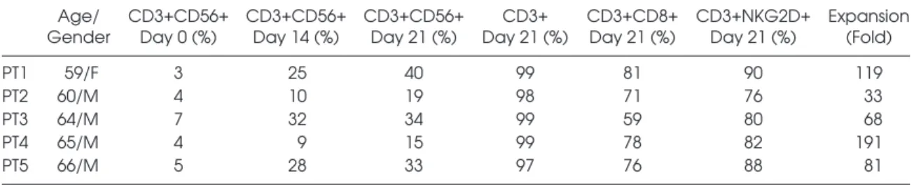
CIK cells were successfully generated from five GIST patients starting from cryopreserved PBMCs within 3 wks of culture according to a standard protocol that included timed addition of IFN-γ, Ab anti-CD3 OKT3 and IL-2 (8,9). Four out five patients had a diagnosis of advanced-stage GIST, treated with mo-lecular targeted therapy (imatinib n = 3, sunitinib n = 1). One patient (PT 3) had localized-stage GIST, treated with surgical resection followed by adjuvant therapy with imatinib. The median expansion of bulk patient-derived CIK cells, calculated on the total CD3+ fraction, was 69-fold (range 12–191). The median expression of the CD3+CD56+ cell fraction was 34% (15–50). The presence of pure NK (CD3– CD56+) cells was negligible, median 0.93% (0.23–3.8) at the end of the expansion. The subset of mature patient-derived CIK cells co-expressing CD3 and CD56 molecules (CD3+CD56+) was present with a median of 34% (range 15–50), whereas 74% (45–90) of total bulk CIK cells were CD8+. The me-dian membrane expression of the NKG2D receptor on expanded patient-derived CIK cells was 84% (61%–96%). We confirmed the absence of T regulatory cells within CIK cultures at either d 14 or d 21 (n = 2: PTs 2 and 4). A summary of patient char-acteristics and relative CIK cell expansion and phenotypes are reported in Table 1. Bioanalyzer. NanoDrop ND-1000
spec-trophotometer was used to evaluate the RNA sample concentration, and 260/280 nm and 260/230 nm ratios were consid-ered to verify the purity of total RNA. For microarray analysis, we selected the three RNA sample pairs with the best RNA integrity number. Briefly, cDNA synthesis and biotin-labeled target syn-thesis were performed using the Gene Atlas 3′ IVT Plus Reagent Kit according to the standard protocol supplied by Affymetrix. The HG-U219 Array Strip (Affymetrix, Santa Clara, CA, USA) hy-bridization, staining and scanning were performed using the GeneAtlas platform.
Gene expression profile data were an-alyzed with the Partek GS 6.6 software package and normalized using the robust multiarray average procedure (25). Differ-entially expressed genes (DEGs) were then selected using a supervised approach with the analysis of variance module included in the Partek GS package. In particular, we selected all the probe sets with a fold change contrast ≥2 in the pairwise compar-ison of CIK cells with PBMCs, and a false discovery rate (q-value) <0.05. Ingenuity Pathway Analysis (IPA) (www.ingenuity. com) was used to study cellular processes and pathways modulated in CIK cells.
All microarray data were submitted to the Gene Expression Omnibus repository (www.ncbi.nlm.nih.gov/geo/query/ acc.cgi?acc = GSE97581).
statistics
Results are expressed as mean ± stan-dard deviation. Comparisons between sam-ples of d 14 and d 1 and between d 21 and d 1 were made by using nonparametric unpaired t test (Mann-Whitney). Differ-ences between groups were considered endothelial growth factor [VEGF]). IL-2
detection was included to have an inter-nal positive control. Briefly, 50 μL of cyto-kine standards or samples (supernatants from seeded cells) were incubated with 50 μL of anti-cytokine conjugated beads in 96-well plates for 30 min at room tem-perature (RT) with shaking. Plates were then washed three times with 100 μL of Bio-Plex wash buffer using the Bio-Plex Pro Wash Station (Bio-Rad Laboratories), then 25 μL of diluted detection antibody was added and plates were incubated for 30 min at RT with shaking. After three washes, 50 μL of streptavidin- phycoerythrin was added, and the plates were incubated for 10 min at RT with shaking. Finally, plates were washed three times, beads were suspended in Bio-Plex assay buffer and samples were analyzed on Bio-Rad 96-plate reader using the Bio-Plex suspension array system and Bio-Plex manager software (Bio-Rad Labo-ratories) (24). Secretome of patient- derived CIK cells was compared with those of healthy donors to evaluate possible differ-ences in the secretory pathways.
Rna Extraction and secretome Gene Expression Profile
Total cellular RNA was isolated from 3 million PBMCs (d 1, absence of INF-γ) and CIK cells (d 14) obtained from 5 GIST patients, using Invitrogen TRIzol (Thermo Fisher Scientific). We performed a microarray analysis of CIK cells at d 14 instead of d 21 (mature CIK cells) to study gene expression changes during CIK cell maturation. Disposable RNA chips (Agilent RNA 6000 Nano Lab-Chip kit; Agilent Technologies) were used to determine the purity/integrity of RNA samples with an Agilent 2100
Table 1. Characteristics of patients and CIK cells. Age/
Gender CD3+CD56+ Day 0 (%) CD3+CD56+ Day 14 (%) CD3+CD56+ Day 21 (%) Day 21 (%)CD3+ CD3+CD8+ Day 21 (%) CD3+NKG2D+ Day 21 (%) Expansion (Fold)
PT1 59/F 3 25 40 99 81 90 119
PT2 60/M 4 10 19 98 71 76 33
PT3 64/M 7 32 34 99 59 80 68
PT4 65/M 4 9 15 99 78 82 191
secretome of CiK Cells
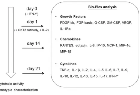
Secretome analysis was performed at specific time points during the three-week ex vivo expansion: at d 1 on PBMC supernatants collected after 24 h of cul-ture in the absence of any cytokines; at d 14 on CIK cell cultures; and at d 21 on supernatants of mature CIK cells (see also Materials and Methods). Figure 1 describes the experimental design and shows the complete list of growth factors, chemokines and cytokines an-alyzed. IL-2 detection was included to have an internal positive control (data not shown).
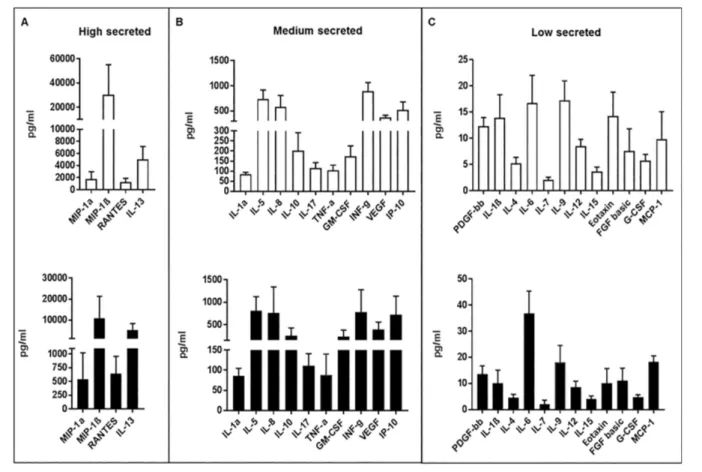
Mature CIK cells produced multiple soluble factors, including cytokines, chemokines and growth factors. The secreted proteins were schematically di-vided based on their concentration into three groups: high, medium and low (Figure 2). We found that IL-13 (5054 ± 2117 pg/mL), RANTES (1244 ± 642 pg/ mL), MIP-1α (1755 ± 1228 pg/mL) and MIP-1β (30570 ± 24613 pg/mL) were secreted at high levels (Figure 2A). The
medium secreted factors were IL-1Ra (85 ± 9 pg/mL), IL-5 (741 ± 174 pg/mL), IL-8 (579 ± 228 pg/mL), IL-10 (202 ± 89 pg/mL), IL-17 (115.5 ± 27 pg/mL), IP-10 (518.5 ± 162 pg/mL), INF–γ (893.5 ± 172 pg/mL), VEGF (372 ± 45 pg/mL) and GMCSF (174 ± 51.5 pg/mL) (Figure 2B). Finally, IL-1β (14 ± 4 pg/mL), IL-4 (5 ± 1 pg/mL), IL-6 (17 ± 5 pg/mL), IL-7 (2 ± 0.5 pg/mL), IL-9 (17 ± 4 pg/mL), IL-12 (8.5 ± 1 pg/mL), IL-15 (4 ± 1 pg/mL), eotaxin (14 ± 5 pg/mL), PDGF-bb (12 ± 2 pg/mL), FGF basic (8 ± 4 pg/mL), G-CSF (6 ± 1 pg/mL) and MCP-1 (10 ± 5 pg/mL) secreted at low levels (Figure 2C). The secretion pattern ob-tained from mature patient-derived CIK cells (d 21) trended similarly to that ob-served from control CIK cells generated from healthy donors.
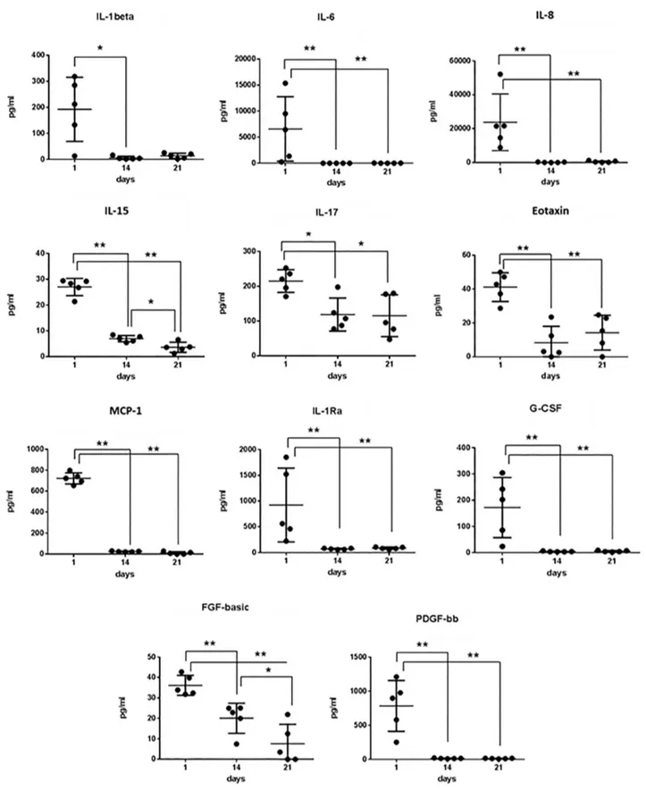
To explore the dynamic of CIK cell secretome, the main classes of secreted proteins, i.e., growth factors, cytokines and chemokines, were analyzed at dif-ferent time points. We compared their release during the ex vivo expansion at
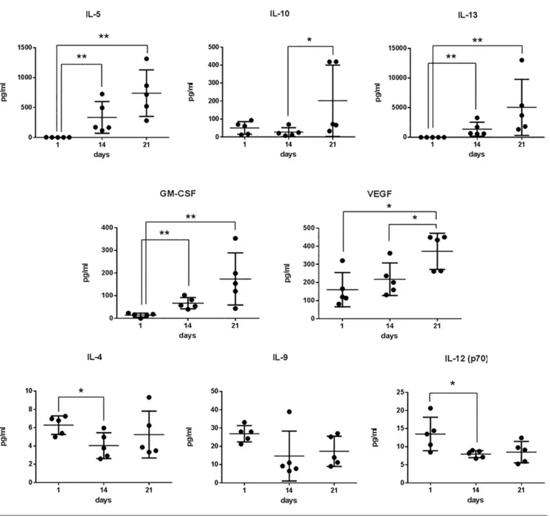
d 1 (as unstimulated PBMCs), d 14 and d 21 (mature CIK cells). We observed that PDGF-bb, IL-1Ra, G-CSF, IL-1β, IL-6, IL-8 and MCP-1 were released on d 1, but their levels progressively and intensely de-creased by d 14 (Figure 3A). A similar di-minishing trend, although less pronounced, was observed for FGF-basic, IL-15, IL-17, eotaxin and RANTES (Figure 3A). On the contrary, the concentrations of GM-CSF, VEGF, IL-5, IL10 and IL-13 progressively increased during the ex vivo expansion of CIK cells (Figure 3B). The concentration levels of IL-4, IL-9 and IL-12 did not sig-nificantly vary over time (Figure 3C). secretome Gene Expression Profile of CiK Cells
We investigated the molecular net-works activated during the ex vivo ex-pansion of CIK cells. We performed gene expression profiling of RNA obtained from CIK cells collected at specific time points: at d 1 (basal PBMCs) and at d 14 (CIK cells) (see Materials and Methods). PTs 2, 3 and 4 were selected for microar-ray analysis based on the best RNA in-tegrity number.
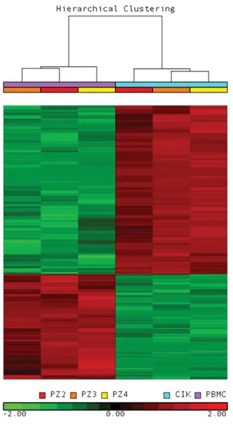
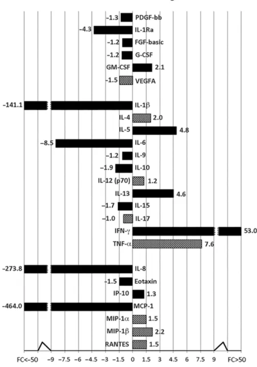
The principal component analysis (PCA) (Supplementary Figure S1) shows that the CIK cell samples clustered together and were clearly separated from PBMCs. Next, using an analysis of variance–based supervised approach to compare CIK cells with PBMCs, we identified 3799 DEGs. As shown in Figure 4, hierarchical clustering analysis using the list of DEGs correctly grouped the different cell phenotypes. First, we checked the mRNA expression levels of 27 secreted proteins analyzed by Bio-Plex assay, and good correspondence between transcriptomic and proteomic profiles was detected. In particular, we confirmed upregulation of GM-CSF, IL-5, IL-10, IL-13 and IFN-g in mature CIK cells. Moreover, consistent with secre-tome data, IL-1Ra, IL-1b, IL-6, IL-15, IL-8, eotaxin (CCL11) and MCP-1 were down-regulated in mature CIK cells compared with PBMCs. On the contrary, PDGF-bb, FGF-basic, G-CSF, IL-9 and IP-10 were not significantly modulated (Figure 5).
Figure 1. Bio-Plex analysis and experimental design. Secretome analysis was performed on supernatants collected at d 1 (no cytokines), d 14 and d 21 of ex vivo expansion of CIK cells. The right panel shows the complete list of growth factors, chemokines and cytokines analyzed by Bio-Plex. IFN-γ (1000 U/mL) was added at d 0, while OKT3 antibody (50 ng/mL) and IL-2 (300 U/mL) were added at d 1 and up to the end, refreshing the medium every 2–3 d.
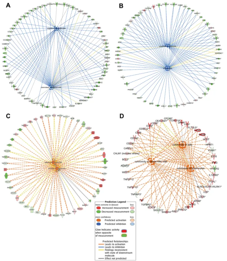
expansion of CIK cell precursors induced by in vitro treatment of PBMCs. In par-ticular, Figure 6C shows the expression of genes that play a pivotal role in B cell apoptosis. Moreover, IPA analysis predicted as activated categories “cyto-toxicity of lymphocytes,” “cyto“cyto-toxicity of cells” and “cytotoxicity of natural killer cells” (Supplementary Table S3). In particular, genes involved in cyto-toxic mechanism of CIK cells like NCR3/ NKp30, KLRK1/NKG2D, TRAIL/TNFSF10, CD226/DNAM1, CD244, CD69, CD96, GZMA, GZMB and PRF1 are differently expressed (Figure 6D).
DisCUssiOn
Immune cells play a fundamen-tal role against cancer; however, in many cases tumor cells become able to circumvent the activity of innate and adoptive immune response (26). NKG2D, NCR3/NKp30 and TRAIL/
TNFSF10.
Next, to identify differentially ex-pressed pathways during CIK cell maturation, we performed a functional analysis by using IPA software (Decem-ber 2016 release). Among inactivated functions in CIK cells, we found “chemo-taxis of myeloid cells,” “phagocytosis,” “migration of granulocytes” and “en-gulfment of leukocytes” (Supplementary Table S3).
Figure 6 shows the modulated expres-sion of genes related to the chemotaxis and phagocytosis processes (panels A and B, respectively). Of note, functional gene categories “proliferation of cells,” “cell death of lymphocytes,” “cell death of mononuclear leukocytes,” “apopto-sis of B lymphocytes” and “quantity of CD4+ T-lymphocytes” were activated, in agreement with the selection and Of note, among DEGs we identified
other secreted molecules that were up-regulated in mature patient-derived CIK cells, which could contribute to their tumor-killing activity: GZMA, GZMB, GZMK, PRF1, IL-32 and LTA (Supplementary Table S1). Furthermore, as shown in Supplementary Table S2, to better characterize the CIK cell phe-notype, we studied the surface antigen expression. As expected, we found downregulation of several myeloid dif-ferentiation markers (CD14, CD9, CD93, CSF2RA, CSF2RB, EPB41L3, receptors for Fc fragments of immunoglobulins, ITGAX, MCEMP1 and TREM1) and B cell antigens (CD19, CD24, CD79A, CXCR5 and MS4A1). Moreover, microar-ray data confirmed upregulation in CIK cells of well-known surface antigens like CD3D, CD3G, IL-2Ra, IL-2RG, CD226/ DNAM1, ITGAL/LFA-1, KLRK1/
Figure 2. Secretome of mature CIK cells. Secretion of 27 cytokines and cell soluble factors were measured by Bio-Plex cytokine assay on CIK cell supernatants at d 21 of culture. Histograms (white for patients, black for healthy donors) represent mean values ± standard devi-ation (pg/mL) of secreted proteins.
Figure 3. Secretion kinetic of patient-derived CIK cells. Cell culture supernatants were collected at the indicated time points and ana-lyzed by Bio-Plex assay. Differences between groups were considered significant when *p < 0.05 and highly significant when **p < 0.01.
obvious impact of the study is that it provides a rationale to explore the func-tional implications and possible thera-peutic modulation of CIK secretome.
To unravel CIK secretory properties, we undertook a thorough investigation, starting from the first stages of differ-entiation and ending at d 21, which is the time point of infusion of CIK cells in patients with cancer. We found that While CIK cells are usually generated
by a standard protocol, many efforts are ongoing to enhance their efficacy by exploring new or additional cytokine in-tegrations (33–36).
Identification of cytokines secreted by CIK cells during their differentiation sheds light on mediators that could pos-itively or negatively modulate tumor growth by acting at different levels. An Cancer immunotherapy holds great
po-tential against various forms of cancer (27,28), exploiting either antibody or cell-mediated responses. Among adop-tive immunotherapy approaches, CIK cells represent an intriguing therapeutic option, since they are endowed with intense MHC-independent antitumor activity against both hematopoietic and solid tumors (9,29–32).
IL-5, IL-10, IL-13, GM-CSF and VEGF production greatly increased during dif-ferentiation, whereas IL-4, IL-9 and IL-12 cytokine secretion levels changed mini-mally. On the contrary, IL-1β, IL-6, IL-8, IL-15, IL-17, eotaxin, MCP-1, IL-1Ra, G-CSF, FGF-basic and PDGF-bb were downregulated over the same time.
GM-CSF mediates a wide variety of effects on cells of the immune system (37). Although GM-CSF is among the growth factors known to promote the expansion of immunosuppressive my-eloid-derived suppressor cells (MDSCs), thus promoting tumor growth and met-astatic dissemination (38), many reports have also shown relevant positive effects of GM-CSF for immunotherapy (39,40). These include T cell activation, matura-tion of dendritic cells and stimulamatura-tion of humoral and cell-mediated responses (41). Data obtained in mice, using herpes simplex virus armed with GM-CSF, have shown its efficacy against pancreatic can-cer (42). Preclinical and clinical studies have shown GM-CSF antitumor activity against melanoma, either alone or in association with antimelanoma vaccines (43,44). GM-CSF represents a success-ful choice in association with anti-GD2 monoclonal antibodies, also for patients with high-risk neuroblastoma (39).
An interesting observation from CIK cell secretome relates to the very low production of IL-6. Indeed, IL-6 is gain-ing attention for its role in tumor biol-ogy. It has recently been found in tissue samples of human prostate obtained by needle biopsy (45), and an integrated gene and miRNA expression analysis of prostate cancer–associated fibroblasts supports a prominent role for IL-6 in fibroblast activation (46). Moreover, IL- 6–mediated signaling in hepatocellular carcinoma has been considered crucial for blocking initiation and malignant growth of this neoplastic disease by the anticancer agent icaritin (47). A protec-tive role in hepatocellular carcinoma has been shown for chemerin, also known as retinoic acid receptor responder protein 2, which inhibits IL-6 and GM-CSF ex-pression and MDSC accumulation (48).
Figure 4. Gene expression profile was analyzed in PBMCs (d 1) and CIK cells (d 14) obtained from three GIST patients. The heat map was computed on the gene list in-cluding 3799 DEGs using the clustering algorithm included in the Partek GS package by means of Euclidean distance and average linkage. Gene coloring is based on normal-ized signals, as shown at the bottom of the figure; green indicates reduced expression, red increased expression. CIK cell and PBMC groups clustered separately in the dendro-gram as shown at the top of the heat map.
suppress activated T cells and promote tumor growth (42).
Our analysis shows that human CIK cells secrete another crucial cytokine that has both positive and negative effects IL-6 is also among those cytokines
re-cently identified as tumor-derived fac-tors inducing CD38 expression in ex vivo MDSCs. Interestingly, highly expressing CD38 MDSCs have an elevated ability to
depending on tissue context and condi-tions. IL-10 exerts positive homeostatic ef-fects by downmodulating global immune response, thus preventing tissue damage and chronic inflammation; however, many reports have shown that IL-10 impairs cy-totoxic responses of immune cells against tumors (49). Accordingly, elevated IL-10 concentration in serum and cerebrospinal fluid has been linked to poor prognosis in different tumors (50–53), and inhibition of IL-10–mediated signaling increases T cell infiltration and responses against mouse tumors (54). However, recent findings demonstrated that IL-10 in combination with oncolytic virotherapy can enhance pancreatic cancer rejection (55).
Another cytokine playing a role in tumor biology is IL-13. Besides CIK cells, IL-13 is secreted by a variety of cell types, including T helper type 2 lympho-cytes, mast cells, basophils, eosinophils, dendritic cells and CD8+ T lymphocytes (56). It is released upon stimulation by proteases or allergens, thus inducing eosinophilic inflammation and immuno-globulin E class switching in B cells (57). In monocytes and macrophages, IL-13 inhibits the production of prostaglandins, reactive oxygen, nitrogen intermediates and proinflammatory cytokines, among them IL-1, IL-6, IL-8, TNF-α and IL-12 (58). It has been shown that IL-13 exerts multiple effects on tumor cells. Hence, it favors growth of cutaneous T cell lym-phoma and its concentration increase correlates with the number of MDSCs in pancreatic, esophageal and gastric cancer. Accordingly, targeting of the IL13Ralpha2 subunit of IL-13R suppresses breast cancer lung metastasis in mice (59,60). Our study shows that IL-13 is highly produced by CIK cells, therefore it would be worth-while to study in depth the repercussions of CIK-secreted IL-13 on in vitro and in vivo tumor growth. Chemokines play mul-tiple roles in cancer biology and recruit-ment of cancer responsive immune cells. We also showed that CIK cells produce VEGF during differentiation, giving rise to questions on potential side effects (ie, favor tumor survival and dissemination). The role of VEGF in cancer angiogenic
Figure 5. Gene expression profile of CIK cells and correspondence with secretome. mRNA expression was analyzed in PBMCs (d 1) and CIK cells (d 14). Protein levels of secreted proteins previously analyzed by the Bio-Plex platform were compared with the corre-sponding mRNA expression profile. The black blocks display the mRNA expression data that confirm the secretome analysis results. Instead, the chess pattern displays mRNA expression data that are inconsistent with secretome analysis.
Figure 6. IPA functional analysis of gene expression data from CIK cells discovered different inhibited/activated processes based on the expression pattern of regulatory molecules: (A) chemotaxis, (B) phagocytosis, (C) apoptosis of B lymphocyte, (D) cytotoxicity. The orange lines show that the activation effect of protein was positively confirmed by the IPA Knowledge database. The blue lines show that the inhibitory effect of protein was positively confirmed by the IPA Knowledge database. The gray lines indicate that the proteins lacked literature support to predict the activation effect. The yellow lines indicate that our results and the literature in IPA are not consistent. Solid lines: direct interactions; dashed lines: indirect interactions.
UL16 and stimulate NK cytotoxicity through the NKG2D receptor. Immunity. 14:123–33. 18. Groh V, et al. (1999) Broad tumor-associated
expression and recognition by tumor-derived gamma delta T cells of MICA and MICB. Proc. Natl. Acad. Sci. USA. 96:6879–84.
19. Jäkel CE, Schmidt-Wolf IG. (2014) An update on new adoptive immunotherapy strategies for solid tumors with cytokine-induced killer cells. Expert Opin. Biol. Ther. 14:905–16.
20. Linn YC, Hui KM. (2010) Cytokine-induced NK-like T cells: from bench to bedside. J. Biomed. Biotechnol. 2010:435745.
21. Zhu BQ, Ju SW, Shu YQ. (2009) CD137 enhances cytotoxicity of CD3(+)CD56(+) cells and their capacities to induce CD4(+) Th1 responses. Biomed. Pharmacother. 63:509–16.
22. Li H, et al. (2007) CD4 +CD25 + regulatory T cells decreased the antitumor activity of cytokine-in-duced killer (CIK) cells of lung cancer patients. J. Clin. Immunol. 27:317–26.
23. Hombach AA, Rappl G, Abken H. (2013) Arm-ing cytokine-induced killer cells with chimeric antigen receptors: CD28 outperforms combined CD28-OX40 “super-stimulation.” Mol. Ther. 21:2268–77.
24. Penolazzi L, et al. (2008) Evaluation of chemokine and cytokine profiles in osteoblast progenitors from umbilical cord blood stem cells by BIO-PLEX technology. Cell Biol. Int. 32:320–25. 25. Irizarry RA, et al. (2003) Exploration,
normaliza-tion, and summaries of high density oligonucle-otide array probe level data. Biostatistics. 4:249–64. 26. Vesely MD, Kershaw MH, Schreiber RD, Smyth
MJ. (2011) Natural innate and adaptive immu-nity to cancer. Annu. Rev. Immunol. 29:235–71. 27. Grierson P, Lim KH, Amin M. (2017)
Immuno-therapy in gastrointestinal cancers. J. Gastrointest. Oncol. 8:474–84.
28. Kumai T, Fan A, Harabuchi Y, Celis E. (2017) Can-cer immunotherapy: moving forward with pep-tide T cell vaccines. Curr. Opin. Immunol. 47:57–63. 29. Mesiano G, et al. (2012) Cytokine-induced killer
(CIK) cells as feasible and effective adoptive im-munotherapy for the treatment of solid tumors. Expert Opin. Biol. Ther. 12:673–84.
30. Introna M, Golay J, Rambaldi A. (2013) Cytokine induced killer (CIK) cells for the treatment of hae-matological neoplasms. Immunol. Lett. 155:27–30. 31. Wong E, Ritchie DS, Davis JE. (2016) CIK immu-notherapy in refractory hematologic malignan-cies. Leuk. Res. 49:60–61.
32. Li R, et al. (2012) Autologous cytokine-induced killer cell immunotherapy in lung cancer: a phase II clinical study. Cancer Immunol. Immunother. 61:2125–33.
33. Jiang J, Wu C, Lu B. (2013) Cytokine-induced killer cells promote antitumor immunity. J. Transl. Med. 11:83.
34. Rettinger E, et al. (2012) The cytotoxic potential of interleukin-15-stimulated cytokine-induced killer cells against leukemia cells. Cytotherapy. 14:91–103.
3. Baker J, Verneris MR, Ito M, Shizuru JA, Negrin RS. (2001) Expansion of cytolytic CD8(+) natural killer T cells with limited capacity for graft-versus-host disease induction due to interferon gamma production. Blood. 97:2923–31. 4. Verneris MR, Karami M, Baker J, Jayaswal A,
Negrin RS. (2004) Role of NKG2D signaling in the cytotoxicity of activated and expanded CD8+ T cells. Blood. 103:3065–72.
5. Olioso P, et al. (2009) Immunotherapy with cyto-kine induced killer cells in solid and hematopoi-etic tumours: a pilot clinical trial. Hematol. Oncol. 27(3):130–39.
6. Schmidt-Wolf IG, et al. (1999) Phase I clinical study applying autologous immunological effector cells transfected with the interleukin-2 gene in patients with metastatic renal cancer, colorectal cancer and lymphoma. Br. J. Cancer. 81:1009–16.
7. Schmidt-Wolf IG, Negrin RS, Kiem HP, Blume KG, Weissman IL. (1991) Use of a SCID mouse/ human lymphoma model to evaluate cyto-kine-induced killer cells with potent antitumor cell activity. J. Exp. Med. 174:139–49.
8. Gammaitoni L, et al. (2013) Effective activity of cytokine induced killer cells against autologous metastatic melanoma including cells with stem-ness features. Clin. Cancer Res. 19(16):4347–58. 9. Sangiolo D, et al. (2014) Cytokine-induced killer
cells eradicate bone and soft-tissue sarcomas. Cancer Res. 74:119–29.
10. Schmeel LC, Schmeel FC, Coch C, Schmidt-Wolf IG. (2014) Cytokine-induced killer (CIK) cells in cancer immunotherapy: report of the interna-tional registry on CIK cells (IRCC). J. Cancer Res. Clin Oncol. 141(5):839–49.
11. Franceschetti M, et al. (2009) Cytokine-induced killer cells are terminally differentiated activated CD8 cytotoxic T-EMRA lymphocytes. Exp. Hematol. 37:616–28.
12. Sangiolo D, et al. (2008) Alloreactivity and an-ti-tumor activity segregate within two distinct subsets of cytokine-induced killer (CIK) cells: implications for their infusion across major HLA barriers. Int. Immunol. 20:841–48.
13. Pievani A, et al. (2011) Dual-functional capability of CD3+CD56+ CIK cells, a T-cell subset that acquires NK function and retains TCR-mediated specific cytotoxicity. Blood. 118:3301–10. 14. Verneris MR, Baker J, Edinger M, Negrin RS.
(2002) Studies of ex vivo activated and expanded CD8+ NK-T cells in humans and mice. J. Clin. Immunol. 22:131–36.
15. Jamieson AM, et al. (2002) The role of the NKG2D immunoreceptor in immune cell activa-tion and natural killing. Immunity. 17:19–29. 16. Diefenbach A, Jamieson AM, Liu SD, Shastri
N, Raulet DH. (2000) Ligands for the murine NKG2D receptor: expression by tumor cells and activation of NK cells and macrophages. Nat. Immunol. 1:119–26.
17. Cosman D, et al. (2001) ULBPs, novel MHC class I-related molecules, bind to CMV glycoprotein
activity has been deeply investigated, showing that it represents a powerful way for tumors to induce formation of new vessels by both autocrine and paracrine stimulation, thus favoring tumor meta-static dissemination (61). Although the effects of anti-VEGF–based therapy can be transitory (62), VEGF still represents a tar-get for pharmacologically controlling and inhibiting neoangiogenesis and metastatic diffusion (63,64), therefore the previously unknown capacity of CIK cells to secrete this pro-angiogenic factor is relevant (65). COnCLUsiOn
In this report we analyzed the secre-tory panel of human CIK cells laying a foundation for future studies on modu-lation of CIK secretome. This will hope-fully represent a new way to increase their performance against neoplastic cells and/or contribute to the establishment of a micro-environment of unfavorable conditions for cancer growth.
aCKnOWLEDGMEnTs
This work was supported in part by FPRC ONLUS 5 × 1000, Ministero della Salute 2012; Associazione Italiana per la Ricerca sul Cancro (AIRC), project no. 10005 Special Program Molecular Clini-cal Oncology 5 × 1000 to AIRC-Gruppo Italiano Malattie Mieloproliferative, AIRC project no. 15337. DS is the recip-ient of a grant from Ricerca Finalizza-ta-Giovani Ricercatori Ministero della Salute (GR-2011-02349197).
DisCLOsURE
The authors have no competing inter-ests as defined by Molecular Medicine or other interests that might be perceived to influence the results and discussion reported in this paper.
REFEREnCEs
1. Lu PH, Negrin RS. (1994) A novel population of expanded human CD3+CD56+ cells derived from T cells with potent in vivo antitumor activ-ity in mice with severe combined immunodefi-ciency. J. Immunol. 153:1687–96.
2. Schmidt-Wolf IG, et al. (1994) Propagation of large numbers of T cells with natural killer cell markers. Br. J. Haematol. 87:453–58.
induce TGF-β1 expression and IL-10 expression and secretion in the cervical cancer cells, result-ing in protection from cytotoxic T cell activity. Cytokine. 76:382–90.
50. Zhao S, Wu D, Wu P, Wang Z, Huang J. (2015) Serum IL-10 predicts worse outcome in cancer patients: a meta-analysis. PLoS One. 10: e0139598. 51. Wang H, et al. (2015) Increased serum levels of
interleukin-10 predict poor prognosis in extran-odal natural killer/T-cell lymphoma patients receiving asparaginase-based chemotherapy. Onco. Targets Ther. 8:2589–99.
52. Sasayama T, et al. (2016) Tumor-associated macrophages associate with cerebrospinal fluid interleukin-10 and survival in primary central nervous system lymphoma (PCNSL). Brain Pathol. 26:479–87.
53. Dwivedi S, et al. (2015) Functional genetic vari-ability at promoters of pro-(IL-18) and anti-(IL-10) inflammatory affects their mRNA expression and survival in prostate carcinoma patients: five year follow-up study. Prostate. 75:1737–46.
54. Chen S, et al. (2016) Blocking IL-10 signalling at the time of immunization renders the tumour more accessible to T cell infiltration in mice. Cell Immunol. 300:9–17.
55. Chard LS, Lemoine NR, Wang Y. (2015) New role of interleukin-10 in enhancing the antitumor efficacy of oncolytic vaccinia virus for treatment of pancreatic cancer. Oncoimmunology. 4:e1038689. 56. Wills-Karp M, et al. (1998) Interleukin-13: central mediator of allergic asthma. Science. 282:2258–61. 57. Guo L, et al. (2015) Innate immunological function
of TH2 cells in vivo. Nat. Immunol. 16:1051–59. 58. Minty A, et al. (1993) Interleukin-13 is a new
human lymphokine regulating inflammatory and immune responses. Nature. 362:248–50.
59. Wasik MA. (2015) IL-13 as a novel growth factor in CTCL. Blood. 125:2737–38.
60. Gabitass RF, Annels NE, Stocken DD, Pandha HA, Middleton GW. (2011) Elevated myeloid-de-rived suppressor cells in pancreatic, esophageal and gastric cancer are an independent prognostic factor and are associated with significant eleva-tion of the Th2 cytokine interleukin-13. Cancer Immunol. Immunother. 60:1419–30.
61. Quail DF, Joyce JA. (2013) Microenvironmental regulation of tumor progression and metastasis. Nat. Med. 19:1423–37.
62. Shinkaruk S, Bayle M, Laïn G, Déléris G. (2003) Vascular endothelial cell growth factor (VEGF), an emerging target for cancer chemotherapy. Curr. Med. Chem. Anticancer Agents. 3:95–117. 63. Bergers G, Hanahan D. (2008) Modes of
re-sistance to anti-angiogenic therapy. Nat. Rev. Cancer. 8:592–603.
64. Heath VL, Bicknell R. (2009) Anticancer strat-egies involving the vasculature. Nat. Rev. Clin. Oncol. 6:395–404.
65. Gensicka M, Głowacka A, Dzierzbicka K, Cholewinski G. (2015) Inhibitors of angiogene-sis in cancer therapy: syntheangiogene-sis and biological activity. Curr. Med. Chem. 22:3830–47. 35. Tao Q, et al. (2013) IL-15 improves the
cyto-toxicity of cytokine-induced killer cells against leukemia cells by upregulating CD3+CD56+ cells and downregulating regulatory T cells as well as IL-35. J. Immunother. 36:462–67.
36. Iudicone P, et al. (2016) Interleukin-15 enhances cytokine induced killer (CIK) cytotoxic potential against epithelial cancer cell lines via an innate pathway. Hum. Immunol. 77:1239–47.
37. Becher B, Tugues S, Greter M. (2016) GM-CSF: From growth factor to central mediator of tissue inflammation. Immunity. 45:963–73.
38. Chiodoni C, Sangaletti S, Colombo MP. (2017) Matricellular proteins tune myeloid-derived sup-pressor cell recruitment and function in breast cancer. J. Leukoc. Biol. 102(2):287–92.
39. Cheung IY, Hsu K, Cheung NK. (2012) Activa-tion of peripheral-blood granulocytes is strongly correlated with patient outcome after immu-notherapy with anti-GD2 monoclonal antibody and granulocyte-macrophage colony-stimulating factor. J. Clin. Oncol. 30:426–32.
40. Wei XX, et al. (2016) Systemic GM-CSF recruits effector T cells into the tumor microenvironment in localized prostate cancer. Cancer Immunol. Res. 4:948–58.
41. Sakamoto C, et al. (2017) Therapeutic vaccination based on side population cells transduced by the granulocyte-macrophage colony-stimulating factor gene elicits potent antitumor immunity. Cancer Gene Ther. 24(4)165–74.
42. Liu H, et al. (2013) Preclinical evaluation of her-pes simplex virus armed with granulocyte-mac-rophage colony-stimulating factor in pancreatic carcinoma. World J. Gastroenterol. 19:5138–43. 43. Grotz TE, Kottschade L, Pavey ES, Markovic SN,
Jakub JW. (2014) Adjuvant GM-CSF improves survival in high-risk stage IIIc melanoma: a sin-gle-center study. Am. J. Clin. Oncol. 37:467–72. 44. Kaufman HL, Ruby CE, Hughes T, Slingluff CL.
(2014) Current status of granulocyte-macrophage colony-stimulating factor in the immunotherapy of melanoma. J. Immunother. Cancer. 2:11. 45. Miličević N, Mrčela M, Galić J, Marjanović K.
(2015) Expression of proinflammatory cytokine interleukin-6 in tissue samples of human prostate obtained by needle biopsy. Pathol. Res. Pract. 211:865–70.
46. Doldi V, et al. (2015) Integrated gene and miRNA expression analysis of prostate cancer associ-ated fibroblasts supports a prominent role for interleukin-6 in fibroblast activation. Oncotarget. 6:31441–60.
47. Zhao H, et al. (2015) A novel anti-cancer agent Icaritin suppresses hepatocellular carcinoma ini-tiation and malignant growth through the IL-6/ Jak2/Stat3 pathway. Oncotarget. 6:31927–43. 48. Lin Y, et al. (2017) Chemerin has a protective role
in hepatocellular carcinoma by inhibiting the expression of IL-6 and GM-CSF and MDSC accu-mulation. Oncogene. 36(25):3599–3608.
49. García-Rocha R, et al. (2015) Mesenchymal stro-mal cells derived from cervical cancer tumors
Cite this article as: Mesiano G, et al. (2017) Analytic and dynamic secretory profile of patient-derived cytokine-induced killer cells. Mol. Med. 23: 235–46.