U
N I V E R S I T Y OF
C
A T A N I A
I
NTERNATIONAL
P
H
D
C
OURSE
–
XXIX
C
YCLE
“B
ASIC AND
A
PPLIED
B
IOMEDICAL
S
CIENCES
”
Coordinator: Prof. Ferdinando Nicoletti
Dr. Giuseppe Lanza
Doctoral dissertation
I
DENTIFICATION OF EARLY NEUROPHYSIOLOGICAL MARKERS
RELATED TO COGNITIVE
-
BEHAVIOURAL DISORDERS AND
PROGRESSION OF VASCULAR COGNITIVE IMPAIRMENT
:
A
T
RANSCRANIAL
M
AGNETIC
S
TIMULATION STUDY
Tutor: Prof. Giovanni Pennisi
SUMMARY
Page
Abstract 3
Abstract (Italian) 4
List of abbreviations 5
1. From Vascular Cognitive Impairment to Vascular Dementia: a predictable continuum? 6
1.1 Definition and terminology 6
1.2 Subtypes 7
1.3 Pathogenesis and pathophysiology 8
1.4 Diagnosis 12
1.5 Disease progression and management 19
2. “Vascular Depression”, the hypothesis linking vascular disease and late-life depression 22
2.1 Definition 22
2.2 The role of white matter lesions: “MRI-defined Vascular Depression” 22
2.3 Neuropathological evidences 23
2.4 “Depression-executive dysfunction syndrome” and other cognitive deficits 24
2.5 Course of Vascular Depression 25
2.6 Vascular risk genes in late-life depression 27
2.7 Mechanistic hypotheses 28
3. Dementia through the looking glass of Transcranial Magnetic Stimulation 33
3.1 Introduction 33
3.2 Cortical excitability and plasticity in Vascular Cognitive Impairment 34
3.3 TMS: basic principles and derived parameters 36
4. TMS study of depression in the course of Vascular Cognitive Impairment-no Dementia 41
4.1 Aim and hypothesis 41
4.2 Materials and methods 41
4.3 Results 48
4.4 Discussion 54
5. Conclusions and future outlooks 57
ABSTRACT
Transcranial magnetic stimulation (TMS) highlighted functional changes in dementia, whereas there are few data in patients with Vascular Cognitive Impairment–No Dementia (VCI-ND) at risk for cognitive worsening. Similarly, little is known about the neurophysiological impact of Vascular Depression (VD) on deterioration of cognitive functions. We performed a longitudinal TMS study to test whether the presence of depression might affect not only cognition but also the functioning of specific cortical circuits in patients with subcortical vascular damage. In this study, 16 VCI-ND and 11 VD patients, age-matched with 15 healthy controls, underwent a baseline evaluation including clinical-cognitive, neuroimaging and TMS assessment. After approximately two years of follow-up, all participants were prospectively re-evaluated. At baseline, a significant more pronounced intracortical facilitation (ICF) at paired-pulse TMS was found in VCI-ND patients only. Re-evaluation revealed an increase of the global excitability at single-pulse TMS in both VCI-ND and VD. At follow-up, the ICF of VCI-VCI-ND become similar to the other groups. Only VD patients showed cognitive deterioration. In conclusion, in VCI-ND specific measures of cortical excitability, namely the high level of ICF found at baseline, suggests an enhanced glutamatergic neurotransmission that might contribute to the preservation of cognitive functioning; conversely, a lack of this hyperfacilitation in VD might be associated with clinical progression. The hyperexcitability to single-pulse TMS observed at follow-up in both group of patients also suggests functional changes in glutamatergic neurotransmission. This suggests that the mechanisms enhancing the risk of dementia in VD might be related either to subcortical changes produced by vascular lesions or to the lack of compensatory functional cortical changes.
Key words: cognitive decline; cerebrovascular disease; late-life depression; non-invasive barin
ABSTRACT (
ITALIAN
)
La Stimolazione Magnetica Transcranica (TMS) ha messo in luce specifiche alterazioni funzionali in pazienti con demenza. Tuttavia, i dati in soggetti con deterioramento cognitivo vascolare a rischio per demenza (VCI-ND) sono limitati, così come poco è noto sull’impatto neurofisiologico della depressione vascolare (VD) nel deterioramento delle funzioni cognitive nell’anziano. In questo studio, abbiamo valutato prospetticamente le modificazioni elettrocorticali alla TMS in soggetti con VCI-ND con e senza depressione al fine di verificare se la presenza di sintomatologia depressiva nel contesto della cerebrovasculopatia cronica sottocorticale possa influire negativamente non solo sulle capacità cognitive ma anche sul funzionamento di specifici circuiti cortico-sottocorticali. Sedici soggetti con VCI-ND ed 11 con VD, paragonati a 15 anziani sani di pari età, sono stati sottoposti ad una valutazione di base comprendente l’esame clinico-cognitivo, neuroradiologico e TMS. All’ingresso, i pazienti con VCI-ND esibivano un aumento della facilitazione intracorticale (ICF) alla TMS a doppio stimolo rispetto agli altri 2 gruppi. Dopo circa 2 anni, tutti i pazienti (con e senza depressione) mostravano un aumento globale
dell’eccitabilità corticale alla TMS a singolo stimolo; inoltre, l’ICF dei VCI-ND risultava ora sovrapponibile agli altri due gruppi. Solo i pazienti depressi si erano deteriorati cognitivamente. In conclusione, specifiche misure di eccitabilità corticale, quale l’iperfacilitazione di base dei pazienti con VCI-ND, suggeriscono un’aumentata neurotrasmissione glutammatergica che potrebbe
contribuire a preservare lo status cognitivo di coloro senza depressione; al contrario, la carenza di facilitazione dei soggetti depressi potrebbe accompagnarsi alla loro progressione clinica.
L’ipereccitabilità osservata al follow-up potrebbe anch’essa essere espressione di modificazioni funzionali dell’attività glutammatergica cerebrale, sebbene senza correlazione con la presenza di depressione. I meccanismi neurofisiologici che espongono maggiormente al rischio di demenza i soggetti depressi potrebbero dunque risiedere sia in alterazioni sottocorticali dovuti alla malattia cerebrovascolare sia al deficit di risposte plastico-compensatorie a livello corticale.
List of abbreviations
A ratio = CMAP/MEP amplitude ratio ACC = anterior cingulate cortex ACE = angiotensin converting enzyme AD = Alzheimer’s disease
ADL = Activity of Daily Living AGTR = angiotensin receptor ApoE = Apolipoprotein E AS = Apathy Scale
BDNF = Brain-derived Neurotrophic Factor CBF = cerebral blood flow
CMCT = central motor conduction time
CMCT-F = CMCT estimated by using the F wave CoG = center of gravity
CSF = cerebro-spinal fluid
CSP = contralateral cortical silent period DLPFC = dorsolateral prefrontal cortex
DSM-IV-TR = Diagnostic and Statistical Manual for Mental Disorders-Forth Edition-Text Revised DTI = Diffusion tensor imaging
EEG = electroencephalography EMG = electromyographic FA = fractional anisotropy
FAB = Frontal Assessment Battery FDI = first dorsal interosseous
FLAIR = Fluid-attenuated Inversion Recovery GABA = gamma-aminobutyric acid
HDRS = 17-item Hamilton Depression Rating Scale IADL = Instrumental Activity of Daily Living ICF = intracortical facilitation
IFCN = International Federation of Clinical Neurophysiology
ISI = interstimulus interval
LAI = long-latency afferent inhibition LLD = late-life depression
LTD = long-term depression LTP = long-term potentiation M1 = primary motor cortex MCI = Mild Cognitive Impairment MEP = motor evoked potential MID = multi-infarct dementia MMP = Matrix metalloproteinases MMSE = Mini Mental State Examination MRI = Magnetic Resonance Imaging NMDA = N-methyl-D-aspartate PAS = paired-associative stimulation PSD = post-stroke dementia
RAS = renin-angiotensin system rMT = resting motor threshold
rTMS = repetitive Transcranial Magnetic Stimulation SAI = short-latency afferent inhibition
SICI = short-latency intracortical inhibition SIVD = subcortical ischemic vascular disease SNRI = Serotonin Noradrenaline Reuptake Inhibitors SSRI = Selective Serotonin Re-uptake Inhibitors Stroop E = Stroop Color-Word test – number of errors Stroop T = Stroop Color-Word test – total time TMS = Transcranial Magnetic Stimulation VaD = Vascular Dementia
VCI = Vascular Cognitive Impairment
VCI-ND = Vascular cognitive impairment – No Dementia
VD = Vascular Depression WML = white matter lesion
CHAPTER 1
From Vascular Cognitive Impairment to Vascular Dementia:
a predictable continuum?
1.1 Definition and terminology
Vascular cognitive impairment (VCI) is a heterogeneous group of cognitive disorders that share a presumed vascular origin.1 VCI is of interest to both clinicians and researchers because it is a common condition, costly, and possibly preventable.2 As kwnon, about a third of cases of dementia show substantial vascular pathology on autopsy,3,4 and depending on our understanding of cerebrovascular mechanisms, VCI can be deemed to be the most common form of cognitive impairment,5 affecting about 5% of people over the age of 65.6 However, unlike neurodegenerative cognitive disorders, such as Alzheimer’s disease (AD), VCI can be potentially prevented and the course of cognitive decline significantly improved.2
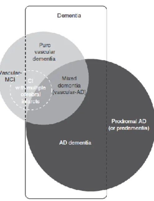
Historically, the idea of VCI grew from dissatisfaction with the term “multi-infarct dementia” (MID),7 which replaced the term “senile dementia” due to hardening of the arteries.8 MID is reported to account for 15% of dementias (figure 1),9,10 and even today, it is sometimes incorrectly used interchangeably with the broader concept of Vascular Dementia (VaD). As shown in Figure 1, VCI should be considered as an “umbrella term”, that includes VCI–No Dementia (VCI-ND), VaD, and cognitive impairment of mixed origin (usually AD + VaD).1 Within the VCI construct, the parallel condition to Mild Cognitive Impairment (MCI) is VCI–ND, and indeed the term “vascular MCI” is sometimes used for VCI–ND.11 The MCI concept distinguishes amnestic from non-amnestic MCI, as well as single-domain from multi-domain impairment.12 Non-amnestic MCI can, in some cases, be vascular in origin, and other subtypes of MCI can progress to VCI.13,14
Figure 1 Links between the main entities associated with Vascular Cognitive Impairment and Alzheimer’s disease.
CI = cognitive impairment; MCI = mild cognitive impairment; AD = Alzheimer’s disease.15
1.2 Subtypes
Because it is a large and heterogeneous group of disorders, VCI has been subgrouped for clinical and research uses,1 being the subtypes characterised by different risk factors, mechanisms, pathology, clinical features, neuroimaging findings, and response to treatment.16
The VaD subtype includes disorders that are in the original VaD construct, such as post-stroke dementia (PSD), MID, subcortical ischemic VaD, and hemorrhagic dementia.2 VCI–ND describes those individuals whose symptoms are not associated with substantial
functional impairment, including a high proportion with subcortical ischemia with cognitive impairment of presumed vascular cause. Patients with VCI–ND have a high risk of progression into dementia, mixed primary neurodegenerative dementia with VaD, or VaD
per se,17,18 particularly if they have recurrent strokes.19
Mixed dementia describes the presentation of individuals with clinical, and commonly neuropathological features of AD and VaD. The way in which AD relates to VCI is incompletely understood but their coexistence is well recognised20 (Figure 2).
Figure 2. Potential mechanisms between vascular risk factors, cerebrovascular dieases and Alzheimer’s pathology leading to Vascular Cognitive Impairment.
1.3 Pathogenesis and pathophysiology
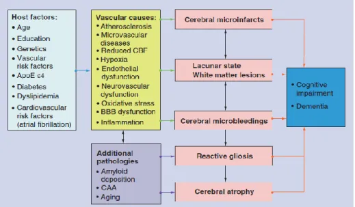
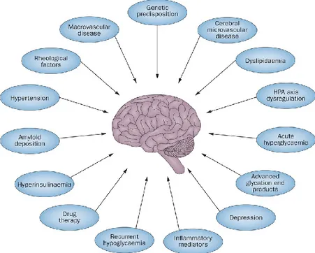
The pathogenesis and pathophysiology of VCI continue to be investigated. A mechanistic approach separates VCI due to large vessel disease from that associated with small vessel disease, including subcortical ischaemic vascular disease, and non-infarct ischemic changes2 (Figure 3).
Figure 3. Schematic interplay of pathogenic factors causing Vascular Cognitive Impairment and dementia.
1.3.1 Large vessel disease
The clinical archetype of large vessel disease is PSD, defined as a substantial cognitive impairment that follows stroke (usually within 3 months).2 The prevalence of dementia after stroke ranges from 14% to 32%, and incident PSD from 20% at 3 months to 33% at 5 years.21,22 Risk factors for PSD include age and low education; of note, the number of vascular risk factors might be more important for predicting cognitive impairment than any individual factor.23 PSD is more common in people who have had strokes and pre-existing neurodegenerative dementia.24,25
PSD can follow a single strategic infarct in the thalamus, angular gyrus, caudate, globus pallidus, basal forebrain, or hippocampus. Such strokes commonly have characteristic cognitive features; for instance, case reports indicate that angular gyrus infarction is associated with an acute onset of fluent dysphasia, visuospatial disorientation, agraphia, and memory loss that can be mistaken for AD.26 Dementia can also result from the cumulative effects of several cortical infarcts of varying size and number, which is the basis of MID, described by Hachinski.7 MID can also result from thromboembolic disease or, less commonly, cerebral vasculitis.2
PSD is seldom found in isolation. In older patients, small vessel disease (overt or covert) is ubiquitous and can accelerate the clinical progression of AD.27 Small vessel disease includes leukoaraiosis, subcortical infarcts, and incomplete infarction associated with cognitive impairment, and might be the most common cause of VCI.28,29 When both large and small vessel disease are present, vascular pathology can act in association with neurodegenerative changes.2
1.3.2 Small vessel disease
Small vessel disease is commonly discussed in relation to the white matter changes seen with neuroimaging, and the term “leukoaraiosis” is often used to describe them. The increased sensitivity of Magnetic Resonance Imaging (MRI) has led to reduced specificity and predictive validity of leukoaraiosis, which can now be detected in more than 90% of older patients.30 White matter lesions (WMLs) are not specific to infarcts; frank infarction might be rare in leukoaraiosis
compared with deep WMLs, and the causal pathway between leukoaraiosis and vascular changes is not well understood.31 The cognitive domains affected by leukoaraiosis are not clearly established, but the association with slow AD-like cognitive and functional decline is robust31,32 (Figure 4). In general, patients with confluent lesions have a worse prognosis than those with punctuate lesions, but decline in cognition and function are more consistently related to measures of atrophy.23
In contrast to cortical infarcts, subcortical vascular injury due to small vessel infarct or ischemia occurs within the cerebral white matter, basal ganglia, and brainstem. Lacunae are typically seen in the corona radiata, internal capsule, centrum semiovale, thalamus, basal ganglia, and pons.29 Infarcts less than 3 mm in diameter are up to 20 times more prevalent than overt infarcts and occur in 20% of patients older than 65 years.33
Lesions in the prefrontal subcortical circuit are associated with impairments in verbal fluency, executive function (the ability to sequence, plan, organise, initiate, and shift between tasks),34 increased risk of stroke and dementia, and more rapid cognitive decline, even when controlling for other vascular risk factors.2
O’Brien and colleagues1 proposed executive dysfunction as part of a specific cognitive profile that might distinguish VCI from AD. Although impaired executive function, and specifically abstract reasoning, might differentially predict AD and VCI,35 not all studies agree on the importance of executive dysfunction in VCI.36-40 Indeed, executive dysfunction might not be unique to VCI at any stage.41-43 Support for this contention comes from a recent prospective neuropsychological and autopsy-based study in which the cognitive effects of small vessel cerebrovascular disease were variable and not particularly distinct, which raises questions about the use of executive impairment as a diagnostic marker for VaD.44 Nevertheless, although executive dysfunction is not specific to VCI, it remains a prominent feature of subcortical ischaemic vascular disease,45 including its common manifestation as subcortical ischemic VaD.46
Figure 4. Different mechanisms and course leading to vascular dementia.
VaD = vascular dementia.
1.3.3 Non-ischemic changes and atrophy
Not all the neuropathology in VCI involves overt infarction but is more probably a continuum of processes related to ischemia. Nowadays, non-infarct ischemia is accepted as an integral part of the disease process that affects both clinical presentation and future outcomes; for example, the robust association between age and PSD can indicate both previous infarcts and non-infarct ischemia.23 Diffusion tensor imaging (DTI) at MRI can be used to detect abnormalities that extend beyond the visible borders of leukoaraiosis (the so called “normal-appearing white matter”), and these abnormalities show a more robust association with cognition than leukoaraiosis alone.47 Ischemia can also contribute to mixed dementia by promoting the neuropathological changes of AD. In animals, ischemic changes in the vascular endothelium increase cleavage of amyloid precursor protein, promote tau phosphorylation, and inhibit clearance of extracellular amyloid.48-50
Moreover, hypoperfusive hypoxic changes are associated with concurrent AD neuropathology,51 and might explain the poor outcomes in people with VCI who have no apparent lesions at neuroimaging.52
1.4 Diagnosis
The current criteria for VaD, most commonly the NINDS-AIREN [National Institute for
Neurological Disorders and Stroke-Association Internationale pour la Recherché et l’Enseignement en Neurosciences] criteria do not include VCI–ND, although up to half of people with VCI do not
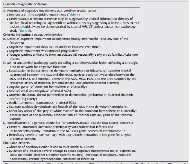
meet the criteria for dementia.53,54 The current diagnostic criteria for VaD are not concordant, which makes comparisons difficult.55,56 The more recent harmonisation standards from NINDS and the Canadian Stroke Network represent substantial progress towards diagnostic criteria57 (Table 1).
1.4.1 Clinical evaluation
VCI remains a clinical diagnosis (Figure 5). The harmonisation standards57 recommend a detailed account of the complaints by the patient and informant on different cognitive domains, such as memory, speed of thinking or acting, mood, and functional status. Information about vascular risk factors, such as hypertension, hyperlipidaemia, diabetes, alcohol or tobacco use, and physical activity, should be sought. History taking should also include checks for atrial fibrillation, coronary artery bypass surgery, angioplasty and stenting, angina, congestive heart failure, peripheral vascular disease, transient ischaemic attacks or strokes, and endarterectomy. Other elements, including hypercoagulable states, migraine, and depression, might also be helpful.57 Finally, the history should also include details of the acuity of onset, progression, and occurrence of urinary incontinence, gait disturbance, and motor deficit.58
Physical examination should focus on blood pressure, pulse, body mass index, waist circumference, and examination of the cardiovascular system for evidence of arrhythmias or peripheral vascular disease. Neurological examination should note focal neurological signs and assess gait initiation and speed.59
1.4.2 Cognitive assessment
The pattern of cognitive deficits in patients with VCI varies considerably. Single strategic infarcts can lead to specific psychopathological profiles, whereas subcortical lesions are often associated with abnormalities of information processing speed, executive function, and emotional lability. This cluster of features (the “subcortical syndrome”) can also result from cortical lesions.60
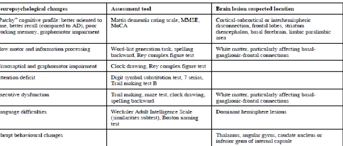
Standard cognitive assessments, such as the Mini Mental State Examination (MMSE), are usually insensitive to these abnormalities,61 even in patients followed up for one year after a confirmed stroke.62 Cognitive tests can usefully differentiate between VaD and AD, but the inclusion of mixed dementia and VCI–ND withon the VCI construct affects the specificity of the diagnosis. In patients who have had a stroke, impairment in global cognition and attention is more closely associated with worse functional outcomes than are executive dysfunction and isolated memory deficits.63 Three subsets of the Montreal Cognitive Assessment,64 a five-word immediate and delayed recall test, a six-item orientation task, and a phonemic fluency test, were selected for the 5 minute cognitive assessment, with the recommendation that the entire assessment, the trail-making test,65 or a semantic fluency test were also administered (Table 2) .
Table 2. Neuropsychological findings of vascular-related cognitive impairment.66
1.4.3 Neuroimaging
Neuroimaging studies require a clinical correlation in the assessment of VCI patients. Indeed, VCI shows no pathognomonic neuroimaging features. Infarct location often does not correlate with the cognitive profile, and neuroimaging cannot reliably confirm the chronology of lesions or disentangle the contribution of neurodegenerative and ischemic processes to the clinical presentation.2 For a diagnosis of probable VaD, the NINDS–AIREN and the California criteria require evidence of cerebrovascular disease at neuroimaging and that infarcts and leukoaraiosis fit specific criteria with regard to their location and the amount of white matter affected.67,68 These criteria have proved to be insensitive in practice, however, which makes their use problematic for the routine clinical diagnosis of VCI.53,69-71 Increasing recognition of the importance of incomplete infarction and hypoperfusion inform the understanding that VCI might be present in the absence of neuroimaging abnormalities72 (Figure 6).
Although Computed Tomography is widely available and in many parts of the world is a pragmatic choice for patients, it is less sensitive than MRI. Newer MRI-based neuroimaging techniques continue to advance our knowledge of the pathophysiology of VCI. The results of DTI-MRI studies have enhanced our understanding of lesion location in relation to clinical presentation,47 and suggest that such white matter changes are not necessarily ischaemic.73 In general, WMLs are associated with loss of neuronal integrity, which leads to higher mean diffusivity and lower fractional anisotropy. DTI-MRI technique might eventually enable measurement of the number of fibres per tract and the functional areas connected by white matter.
The importance of atrophy in VCI is increasingly recognised and might show a stronger association with disease progression and depressive symptoms than WMLs.74 Medial temporal atrophy is emerging as an important correlate of cognitive dysfunction even in VCI.40 Likewise, even small amounts of white matter abnormalities are associated with significant memory and language impairments.45
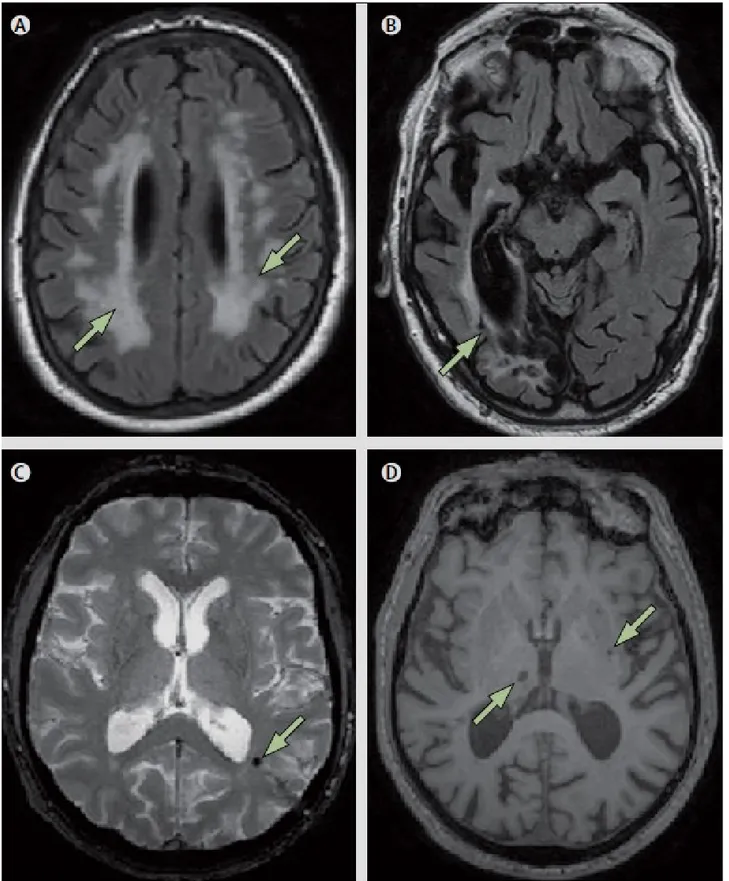
Figure 6. Vascular imaging changes on MRI.72
(A) Arrows indicate extensive (>25%) white matter lesions (Fluid-attenuated Inversion Recovery – FLAIR – image); (B) Arrow indicates large cortical infarction (FLAIR image);
(C) Arrow indicates microbleed (T2*-weighted image);
1.4.4 Neuropathology
Many researchers believe that neuropathology is the gold standard for the diagnosis of VCI, despite a considerable body of evidence that undermines it as a test of 100% sensitivity and specificity.3,4,27,69 Other researchers have accepted that neuropathology is not ideal, and propose neuroimaging as an alternative gold standard. Anyway, there is currently no single standard for neuropathology in relation to VCI, and neuropathological criteria need to be understood in relation to clinical presentation. Moreover, there is currently no neuropathological threshold of cerebrovascular disease reliably distinguishes between no cognitive impairment and dementia.3
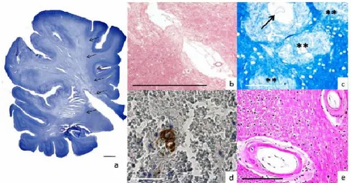
The types of neuropathological lesions associated with VCI include large and small vessel infarcts, white matter changes, haemorrhage, gliosis, and, in mixed dementia, the neuropathological changes of AD75 (Figure 7). In a population-based neuropathological study, in which 13% of participants had pure VaD without major evidence of AD, the requirement that dementia follows a known stroke resulted in high specificity but low sensitivity in autopsy-verified cases.76
Figure 7. Some of the major pathological changes underlying vascular dementia.66
A) Ischemic infarct. Note the wedge-shape border of the infarct (arrows). Both white and gray matter are involved. Nissl staining. B) Lacunar infarct characterized by an irregular cavity and a central blood vessel surrounded by a rim of gliotic, rarefied brain tissue (arrow). Hematoxylin and Eosin stain (H&E). C) Histopathological counterpart of a white matter lesion detected by MRI in a 62 years-old male. Note the regions of myelin pallor (**) and an enlarged perivascular space (arrow). Kluever-Barrera stain. D) Cerebral amyloid angiopathy (CAA). The Amyloid β-deposition in the wall of a cortical artery is colored in brown (arrow). In this case, there is a microbleed around this artery (note the anuclear red blood cells). Immunostain with an antibody against Ab17-24 (4G8; Covance). E) Small vessel disease. Two white matter arteries exhibit fibrosis and hyalinization of wall (arrows). These lesions are also referred to as arteriolosclerosis, arteriohyalinosis or lipohyalinosis (arrow). H&E. The calibration bars correspond to 100 μm.
1.4.5 Biomarkers
Biomarkers are indicators that might or might not be in the causal pathway of the disease but correlate with the disease process and its progress. Candidate biomarkers should aid early detection, discriminate the neuropathology, estimate the prognosis, and monitor disease progression or treatment response. The search for biomarkers in VCI is hampered by the clinical and pathological heterogeneity and the presence of pathology, such as white matter hyperintensities, even in healthy individuals. Furthermore, the high prevalence of mixed dementia within VCI poses challenges for the discriminative abilities of biomarkers. Indeed, to date the results of studies that compare the phenotypes associated with the presence or absence of biomarkers are inconsistent.
Markers in the cerebro-spinal fluid (CSF) have shown more discriminative ability in patients with VCI than have serological biomarkers. The CSF-albumin index is a measure of blood-brain barrier integrity, which is compromised in many types of dementia,77 particularly in subcortical vessel disease.78 Matrix metalloproteinases (MMP) attack tight junctions in the cerebral vessels, thereby opening the blood–brain barrier and contributing to demyelination. Of 26 known metalloproteinases, MMP-2 and MMP-9 are the most studied: MMP-2 is constitutively expressed, whereas MMP-9 is associated with inflammation and has variable specificity in VCI compared with AD.79,80 The light neurofilament subunit of normal myelin has been found in higher concentrations in the CSF of people with subcortical vessel disease compared with those with AD,81 and might be a marker of demyelination.82 CSF tau and phospho-tau concentrations are elevated in non-vascular dementias and therefore might be useful as negative biomarkers.83
Nevertheless, biomarkers cannot take the place of clinical diagnosis although they can inform on the relationship between risk factors and disease progression. However, their contribution to our understanding requires considerable empirical data and standardisation of collection, storage, and measurement techniques.
1.5 Disease progression and management
There is over-reliance on objective measures considered as useful and sensitive indexes for the evaluation and prediction of progression of VCI.44,45 Notwithstanding, the unifying empirical theme is that the subtype VCI–ND is an at-risk state for future dementia and should be the target of prevention models. Moreover, the progression of dementia in patients with VCI is neither linear nor unidirectional. There is still much plasticity in VCI–ND, and careful evaluation of patients with this condition who do not progress to dementia or who revert to no cognitive impairment might enhance our understanding of how exposures interact with neuropathology.
Although some cross-sectional evidences suggest that VCI lies on a spectrum between normal cognition and VaD,39,84 little is known about the progression of VCI–ND. It is true that VCI-ND does not always progress to dementia, and data from epidemiological and clinical series indicate that improvement is even possible.85 Moreover, in clinic-based longitudinal studies, VCI– ND showed less progression compared with other VCI subtypes,52 which might also be the case for post-stroke VCI–ND.86
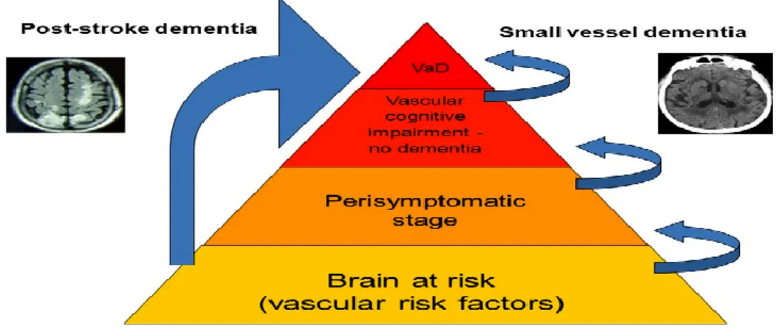
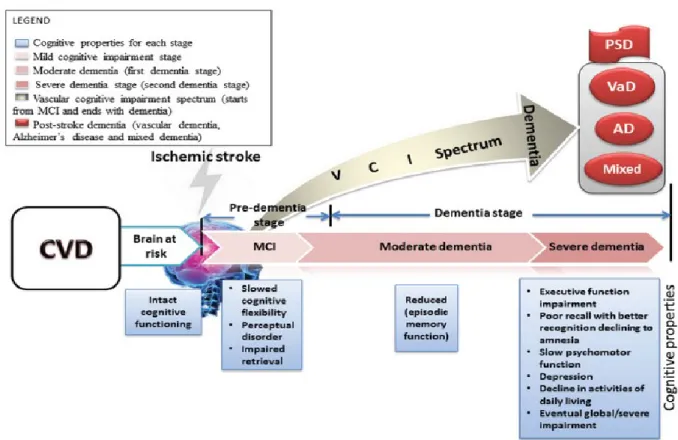
In a population-based study, progression from VCI–ND to incident dementia was not associated with a particular neuropsychological profile but occurred at the highest rate in those patients with both memory and functional impairment at baseline.37 There was no difference in the rates of progression between those patients with predominant functional impairment and those patients with predominant memory impairment, and a half of the patients with VCI–ND were later diagnosed with AD. However, in the same study, 52% of people with VCI–ND died, and 42% developed dementia within 5 years.17 VCI–ND also carries a high risk of PSD.18 Figure 8 illustrates the course of patients with cerebrovascular disease, from the “brain at risk” stage to an overt dementia or different origin (PSD, VaD, AD, mixed).
Similar to VCI–ND, considerable variability is seen in the progression of PSD; up to a third of people change diagnostic category (no cognitive impairment; cognitive impairment without
dementia; overt dementia) within one year after a stroke.87 Age, previous cognitive impairment, polypharmacy, hypotension during acute stroke,88 and depression89 are risk factors for progression and functional decline. In general, infarct location is less important in predicting cognitive decline than medial temporal atrophy,40,74 and medial temporal atrophy is more important than are WMLs to predict dementia after stroke.74
Figure 8. Course of cerebrovascular disease, from the “brain at risk” stage to a picture of overt dementia.
AD = Alzheimer’s disease; CVD = cerebral vascular disease; MCI = Mild Cognitive Impairment; PSD = post-stroke dementia; VaD = Vascular Dementia; VCI = Vascular Cognitive Impairment.
The available data on vascular risk factors for the progression of VCI show surprisingly equivocal support for primary and secondary prevention. Pre-stroke hypertension is associated with post-stroke dementia;90 treatment of hypertension with perindopril + indapamide reduced the dementia and cognitive decline associated with recurrent stroke.91 However, a recent Cochrane review found no convincing evidence that lowering of blood pressure prevents the development of dementia or cognitive impairment in patients with hypertension without prior cerebrovascular
disease, although methodological difficulties cast doubt on the true effects.92 Diabetes has been associated with risk of stroke, cognitive decline, and dementia in several population studies.93-95 Hypercholesterolemia is a well recognised risk factor for stroke, and the recent Stroke Prevention by Aggressive Reduction in Cholesterol Levels (SPARCL) study found a reduction in recurrent stroke in those patients with a history of stroke or transient ischemic attack who were treated with high doses of statins.96
To date, however, apart from controlling vascular risk factors, the overall effect of treatment on patients with VaD is modest. There is variable but generally limited enthusiasm for the drugs used to treat AD; the results of a meta-analysis concluded that only small benefits of uncertain clinical meaningfulness were available from cholinesterase inhibitors or memantine.97 Even so, some patients (who had probably a mixed dementia) experience clinical benefit, although the responders cannot be easily identified. Despite reasons to believe that cholinesterase inhibitors might benefit people without concomitant AD,98 the most persuasive studies, which assembled patients with VaD into groups,90 had small effect sizes, no dose-response effect, limited evidence for convergence of treatment effects within and across trials, and no clear translation into responses that physicians might look for in routine clinical care.99
Neuroimaging, particularly the DTI-MRI technique, gives some insight into disease mechanisms, although their relationship with clinical effects is not always clear. In a secondary analysis of the results of the Study on Cognition and Prognosis in the Elderly, patients treated with the angiotensin receptor blocker candesartan showed reduced risk of WMLs.100 Similarly, physical activity was not associated with a slower progression of WMLs seen on MRI,101 and a relationship with whole brain volume is not clear from the published literature. Perhaps physical activity prevents VCI through enhancement of cognitive reserve,101 which refers to the ability to maintain cognitive function despite increasing neuropathological burden.
CHAPTER 2
“Vascular Depression”, the hypothesis linking vascular
disease and late-life depression
2.1 Definition
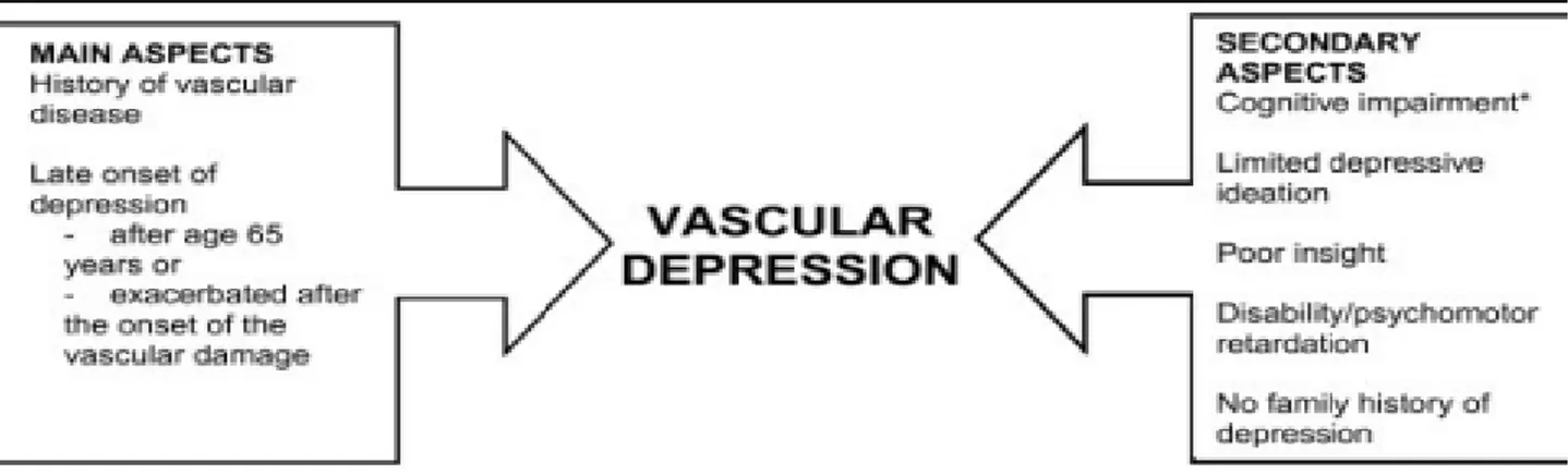
The “Vascular Depression” (VD) hypothesis proposed that cerebrovascular disease may predispose, precipitate, or perpetuate late-life depression (LLD).102 First, Alexopoulos and colleagues proposed a working definition based on the presence of vascular risk factors. The clinical presentation of VD was characterized by cognitive deficits, psychomotor retardation, lack of insight, and disability disproportional to the depression severity.102,103 Investigators subsequently focused on the cognitive dysfunction occurring in LLD and its relationship to response to antidepressants.104 Second, Krishnan and colleagues proposed a MRI-based definition requiring evidence of vascular changes on neuroimaging, referred to as MRI hyperintensities (WMLs).105 Both definitions are relevant as subsequent work demonstrated the validity of a VD diagnostic subtype characterized by peculiar MRI findings and involvement of executive functions.106,107
2.2 The role of white matter lesions: “MRI-defined Vascular Depression”
The hallmark of MRI-defined VD is the presence of WMLs identified as white matter hyperintensities on T2-weighted or fluid attenuated inversion recovery (FLAIR) MRI.107 WMLs are associated with advanced age108 and cerebrovascular risk factors, including diabetes, cardiac disease, and hypertension.109-113 Vascular dysregulation contributes to WML development as white matter is sensitive to transient ischemia114 and many larger WMLs are ischemic in origin.115,116
Hypertension and blood pressure variability are associated with LLD117-119 and also contribute to WML development,120,121 particularly when accompanied by impaired cerebral vasomotor reactivity and altered autoregulatory processes.122,123 Such deficits reduce cerebral blood flow (CBF) and may lead to WMLs.124,125 LLD is consistently associated with greater WML severity126-129 and volumes.111,130-132 Compared with early-onset depression, individuals with a later onset (e.g., after 50 year old) exhibit greater WML severity105,119,132,133-137 and cognitive impairment.138-139
However, VD as a potential diagnostic entity may not be limited to late-onset patients. Individuals with an earlier onset are at increased vascular risk as precedent depression is associated with increased risk of vascular disease and stroke.140-144 Depression in early and mid-life may promote inflammation145-147 or epigenetic modifications of genes related to vascular homeostasis.148 Thus some individuals with early onset depression may be prone to VD later in life.
WMLs location is also important. In non-depressed samples, periventricular WMLs are more closely associated with cognitive impairment than are deep WMLs.32,149-150 It is possible this is due to anatomical differences, as the periventricular region has a high density of long associating fibers with cortical-subcortical connections, while subcortical deep white matter has a high density of shorter U-fibers connecting adjacent cortical regions.150,151 Others have localized WMLs associated with depression to the frontal152-155 and temporal lobe.156 More recently, several groups reported that LLD is associated with greater WML severity in specific white matter fiber tracts including the cingulum bundle, uncinate fasciculus, and superior longitudinal fasciculus.157-159
2.3 Neuropathological evidences
Studies using both neuroimaging and pathological techniques demonstrate that WMLs represent a wide range of pathological processes, including perivascular demyelination,
arteriosclerosis, ischemia, gliosis, or partial loss of myelin and axons.116,160,161 Generally, confluent deep WML but not periventricular appear to be related to ischemic processes.162
There are also regional differences in WML etiology. Deep WML and punctuate lesions of depressed older adults are most likely to have ischemic origins. In LLD ischemic lesions are also more likely to occur in the dorsolateral prefrontal cortex (DLPFC), instead of the anterior cingulate cortex (ACC) or occipital lobe.115 Similarly, depressed elders exhibit increased expression of cellular adhesion molecules (CAMs) in the DLPFC, but not the ACC or occipital lobe.163-165 CAMs are inflammatory markers whose expression is increased by ischemia, supporting a role for ischemia in LLD and highlighting the relationship between vascular and inflammatory processes. Although ischemic pathology is thus consistently localized to the DLPFC, other frontal regions may also be involved. Depressed elders exhibit decreased density of pyramidal neurons in the orbitofrontal cortex, which may be the result of vascular processes166 in arterioles and medium arteries167 or alterations in astrocyte-associated immune function.168
Neuropathological studies also demonstrate that LLD can develop in the absence of significant vascular abnormalities. In a study of late-onset depressed elders, depression was not related to either lacunes or microvascular lesions.169 Similarly, in a population-based study where depression was ascertained in a pre-mortem diagnostic interview, depression was associated neither with cerebrovascular nor AD pathology.170 However, depression was associated with the Lewy bodies,170,171 highlighting the heterogeneity of neuropathologies that present as depression.
2.4 “Depression-executive dysfunction syndrome” and other cognitive deficits
A “depression-executive dysfunction syndrome” was conceptualized as the clinical expression of frontal network impairment caused by vascular and other aging related factors.104
Accordingly, this syndrome describes depressed patients with vascular disease and evidence of impairment in networks related to mood and executive function.172
Executive function is a frontally mediated domain that encompasses cognitive processes including selective attention, response inhibition, and performance monitoring.173-175 It is clinically expressed as difficulty with planning, sequencing, organizing, and abstracting. These deficits are common in depression,176-179 particularly in LLD.139,178,180 When compared with depressed elders without executive dysfunction, patients with depression-executive dysfunction exhibit reduced fluency, impaired visual naming, suspiciousness, anhedonia, psychomotor retardation, and significant disability.181-183 Importantly, studies across eight different samples identified executive dysfunction as a predictor of poor antidepressant response.184-191
Individuals with LLD exhibit also deficit across other cognitive domains, including episodic memory, working memory, visuo-spatial ability, and processing speed.176,179,192-197 Processing speed deficits may influence other cognitive deficits195-197 and mediate in part executive task performance.198 Although cognitive performance improves with successful antidepressant treatment, such deficits can persist.192,199-201
Greater WML burden is associated with executive dysfunction, perseveration, and slowed processing speed113,158,202-205 but also episodic and visuo-spatial memory deficits.118,150,194,206 These cross-sectional observations are supported by longitudinal studies demonstrating that progression of WML severity parallels cognitive decline.207-209
2.5 Course of Vascular Depression
2.5.1 Cognitive dysfunction and antidepressant outcomes
Multiple studies demonstrate that executive dysfunction predicts poor acute response of LLD to antidepressants.191,210 Executive dysfunction is also associated with high relapse and
recurrence rates,211 although not all studies found this association.212 Much of this work used the Mattis Dementia Rating Scale Initiation-Perseveration (I/P) subtest, which yields a composite score of several executive skills.213 Notably, a recent meta-analysis examined the relationship between antidepressant response and performance across multiple executive tests, including the I/P subtest, the Wisconsin Card Sort Test, the Stroop Color-Word test, and others. The authors concluded that the I/P subtest was the only test providing reliable discrimination between antidepressant responders and non-responders.214 Further analysis of I/P subtest components showed that semantic strategy while performing the I/P verbal fluency task explained most of the variance in predicting remission.215 Effective semantic strategy appears to be associated with treatment response regardless of the probing task; during verbal learning task, it was also associated with high LLD remission rate.216 These observations suggest that preserved semantic organization, rather than fluency or verbal learning, is critical for remission during antidepressant treatment. However, impaired response inhibition is another aspect of executive dysfunction that influences antidepressant response.184,187,210,217
Other cognitive deficits can influence outcomes. Poorer performance on tests of episodic memory, language processing, and processing speed are associated with poorer acute antidepressant response190 and may predict poorer long-term course of depression.218
2.5.2 White matter lesions as a predictor of antidepressant outcomes
Most studies used cross-sectional WML measures to predict outcome. However, WML is a progressive rather than a static process,112,219 and the rate of change may be a more important predictor. The few studies in LLD cohorts examining this question found that greater increases in WML volume over two- and four-year intervals are associated with non-remission or relapse.220,221 Some have examined both WML severity and cognitive dysfunction as predictors of outcome. In these studies, cognitive measures are generally a better predictor of antidepressant response than are WML measures. The largest published study examining a 12-week response to sertraline found that
poorer response was independently associated with greater WML severity and poorer performance in cognitive domains of episodic memory, language processing, processing speed and executive function. However, in models controlling for baseline depression severity, WML severity was no longer associated with response.190 Smaller studies have found similar patterns, wherein initial associations between WML severity and response were not statistically significant in models incorporating cognitive measures.189,191
Other measures of white matter integrity are associated with antidepressant response. In voxel-wide analysis, microstructural white matter abnormalities (low fractional anisotropy – FA) in multiple frontolimbic brain areas, including the rostral and dorsal anterior cingulate, DLPFC, genu of the corpus callosum, hippocampus, and posterior cingulated, were associated with non-remission.222 In region of interest analyses, low FA in the corpus callosum, left superior corona radiata, and right inferior longitudinal fasciculum was associated with lower remission rates.223 In contrast, others found that higher prefrontal white matter FA was associated with a failure to remit to antidepressants,224 a finding that parallels reports in depression of increased functional connectivity to a dorsal nexus.225 Moreover, depressed elderly carriers of 5-HTTLPR polymorphism short allele had lower FA than long allele homozygotes in frontolimbic areas, including the anterior cingulate, posterior cingulate, dorsolateral and medial prefrontal regions. Notably, such differences in FA can be caused by several pathologies, not all of which are vascular in nature.226
2.6 Vascular risk genes in late-life depression
There is a wide literature examining genetic influences in LLD, including reports examining polymorphisms in Brain-derived Neurotrophic Factor (BDNF), Apolipoprotein E (ApoE), and serotonin transporter genes, amongst others.227,228 Within this field, there are an increasing number of reports associating LLD with genetic polymorphisms increasing vascular risk.
One example of this literature includes studies examining genetic variation of the renin-angiotensin system (RAS). Beyond influencing blood pressure, RAS polymorphisms may also increase the risk of depression via modulation of monoamines,229 or contributing to hypothalamic-pituitary-adrenal axis dysregulation.230 RAS variation is further related to differences in brain structure and function. Several studies in older adults have now associated variants in angiotensin converting enzyme (ACE) and angiotensin receptor (AGTR) blocker with altered cerebral fronto-temporal structure231-234 and abnormal default mode network activity.235 AGTR-1 variants are associated with greater WMH progression, particularly in men,236 and this effect may be localized to specific tracts.157 RAS polymorphisms may also influence antidepressant outcomes. In a general adult population, the ACE-D variant and the AGTR-1 C1166 allele are associated with better acute antidepressant response.237,238 However, this contrasts with a study of LLD, wherein AGTR-1
C1166 allele homozygous individuals exhibited a poorer antidepressant response over a longer
period.239 Such age differences could be important, reflecting stress reactivity in earlier life and allostatic effects of an overly active RAS later in life.107
2.7 Mechanistic hypotheses
According to a model by Alexopoulos,102 the clinical expression of LLD is mediated by altered brain activity in cognitive and affective circuits, characterized as hypometabolism of dorsal cortical regions and hypermetabolism of ventral limbic structures. The impact of these neurobiological contributors can be increased by vulnerability factors originating from pre-existing differences in mood circuitry. Such etiological factors may lead to depression only after crossing a certain threshold above which they acquire a dose effect relationship. In this conceptualization, single or multiple potential etiological factors may reach an initial severity threshold and contribute to LLD vulnerability. When etiological factors further increase in severity and cross a second
threshold, they may have a greater and direct effect on mood circuit function, leading to affective and cognitive symptomatology (Figure 9). Of course, instead of threshold effects, such relationships could be cumulative following linear or curvilinear patterns. This threshold model can account for multifactorial contributions to LLD.107
Figure 9. Proposed mechanisms underlying vascular depression.
2.7.1 The “Disconnection hypothesis”
Building on the concept of “Disconnection syndromes,” ischemia and WMLs may contribute to depression by disrupting neural connections among regions regulating mood-affect and cognition.240 In this model, global cerebral WML severity is less determinant to LLD than is focal damage to specific fiber tracts and neural circuits. Such focal damage would adversely affect the tract structural and functional connectivity. In turn, this state adversely affects the function of connected regions at rest and during cognitive tasks, thus contributing to neural circuitry alterations that mediate clinical symptoms and influence antidepressant response.240
This view is supported by studies examining WML location. As stated, LLD is associated with greater WML severity in the cingulum bundle, uncinate fasciculus, and superior longitudinal fasciculus.157-159 Additionally, greater WML severity in the uncinate and superior longitudinal fasciculi is associated with executive dysfunction158, 241,242 and greater depression severity.243 These macrostructural MRI findings are supported by DTI studies examining white matter microstructure. DTI changes reflect various pathologies leading to decreased myelin integrity, including demyelination secondary to cerebrovascular and inflammatory changes. WMLs occurring in fiber tracts are also associated with DTI changes.244,245
2.7.2 The “Inflammation hypothesis”
Aging- and disease-related processes promote pro-inflammatory states in older individuals.246,247 Further, immune system activation can be a characteristic of depression147,248 and precipitate depressive symptoms.249 Alexopoulos and Morimoto recently proposed that immune dysregulation may promote the development of affective and cognitive symptoms in LLD.250
Pro-inflammatory cytokines affect monoamine neurotransmitter pathways, including indoleamine 2,3-dioxygenase up-regulation and kynurenine pathway activation.251-253 This results in decreased tryptophan and serotonin and increased synthesis of detrimental tryptophan catabolites that promote hippocampal damage and apoptosis.253,254 Cytokines, including interleukine (IL)-1β,
also reduce extracellular serotonin levels by activating the serotonin transporter.255 Additionally, proinflammatory cytokines disrupt glucocorticoid receptor function and reduce neurotrophic support256,257 (Figure 10).
Figure 10. Bilateral relationships between depression, vascular disease and dementia.
Successful antidepressant treatment may reduce pro-inflammatory markers.147 This action may be a direct effect, as in vitro studies demonstrate that antidepressants reduce pro-inflammatory markers while increasing levels of anti-inflammatory cytokines.258-262 Results in clinical populations are inconsistent, but a recent meta-analysis concluded that antidepressants, particularly Selective Serotonin Re-uptake Inhibitors (SSRI), reduce IL-6, IL-1β, and tumor necrosis factor (TNF)-α.263 Furthermore, some anti-inflammatory drugs may have antidepressant properties,264-266 particularly in individuals with elevated pre-treatment pro-inflammatory markers.267
Notably, pro-inflammatory processes contribute to neurodegenerative processes: increased peripheral inflammatory marker levels are associated with increased risk for all cause dementia,268 and central inflammatory processes significantly contribute to the pathogenesis of AD.269
2.7.3 The “Hypoperfusion hypothesis”
Vascular dysregulation is common in LLD,270-275 and CBF reduction can impair regional brain function, contributing to affective and cognitive symptoms. Blood flow to the brain is influenced by systemic hemodynamic and cerebrovascular autoregulation, with cerebral arteries contracting or dilating as arterial pressure changes. However, these processes are impaired in the context of vascular disease: hypertension, diabetes, and atherosclerosis lead to vascular wall hypertrophy, reduced arterial lumen diameter, reduced arterial distensibility, and endothelial cell dysfunction.276-278 Such vascular changes, including increased intima-media thickness, increased arterial stiffness, and endothelial dysfunction, are pronounced in LLD populations,270-275 and endothelial function may be particularly poor in antidepressant non-responders.279
Perfusion deficits do not need to cause ischemia in order to influence brain function. Reduced CBF impairs protein synthesis280 crucial for cognitive processing281,282 and for maintaining the integrity of cortical functional maps.283 Thus mild CBF reduction may impair cognitive and affective processes, while greater CBF reduction in the context of autoregulatory deficits may cause ischemic injury. The subcortical white matter is particularly sensitive to these changes because it is supplied by terminal arterioles with limited collateral flow284 and so susceptible to infarction due to impaired autoregulation.285 Greater WML severity may be a marker of broader deficits in perfusion and autoregulation as individuals with greater WML severity exhibit reduced CBF in both white and gray matter regions.286-288 Depressed elders with greater WMLs severity also exhibit decreased perfusion in the cingulate gyrus,289 a region involved in cognitive and affective processing.290
Advanced age is itself associated with decreased fronto-temporal CBF,291 an effect mediated by vascular risk factors.292 In LLD, perfusion deficits are more severe, particularly in the medial and lateral prefrontal cortex and temporal structures.287,293-297 Others report no change of CBF in antidepressant non-responders,298-302 suggesting that persistently reduced regional CBF may be a biomarker of non-response.
CHAPTER 3
Dementia through the looking glass of Transcranial Magnetic
Stimulation
3.1 Introduction
Clinically introduced approximately 30 years ago as a diagnostic tool to study the central motor pathways, today Transcranial Magnetic Stimulation (TMS) goes well beyond the simple assessment of the cortical-spinal tract, providing novel insights into the pathophysiology of the neural circuitry that underlies several neurological and psychiatric diseases. TMS may give information about the excitability of the human brain cortex, the conduction along cortical-spinal tract,303 the functional integrity of intracortical neuronal structures and callosal fibers.304 TMS has also a strong talent to unveil motor system impairments in their pre-clinical phase. Moreover, integrated approaches using electrophysiological techniques together with structural and functional neuroimaging have allowed the study of connectivity across motor and non-motor areas.305 By evaluating the effects of agonists or antagonists for specific neurotransmitters, it has been shown that TMS can selectively and non-invasively explore the function of glutamatergic, gamma-aminobutyric acid (GABA)-ergic and cholinergic cortical circuits.306 Although the physiological abnormalities revealed by TMS are not disease-specific,304 there may be specific neurophysiological changes that co-segregate in each dementing illness, consistent with the involvement of distinct neurobiological substrates in the pathogenesis of each disease.307
In the last years, there has been a significant growth in the number of publications exploiting TMS techniques to aid the diagnostic approach in patients with primary dementia. Although not always clinically evident, the involvement of motor areas in dementia has been shown by clinical, neuropathological and neuroimaging studies. Changes in motor areas may be secondary to the direct
structural alterations caused by the disease process but, more often, they are the consequence of indirect remodeling mechanisms.307 In this context, increasing evidences support the hypothesis that phenomena of brain plasticity are involved in different kind of dementia, related to functional and structural components, each entailing a number of cellular mechanisms operating at different time scales, synaptic loci, and developmental phases within an extremely complex framework.308
However, the exact relationship between brain plasticity and excitability of cortical areas and their connections is still unclear.
3.2 Cortical excitability and plasticity in Vascular Cognitive Impairment
It is well known that motor cortex hyperexcitability is a relatively stable electrophysiological feature of both AD and VaD.305 This has been considered part of a plastic compensatory response to neuronal loss, supporting the concept of dementia as a dynamic condition and changes of specific TMS parameters as indexes of neural plasticity.309 The enhanced cortical plasticity might counteract cognitive decline and shed light on the reasons underlying decline or preservation of cognitive functions. This hypothesis has been demonstrated by means of TMS mapping technique in AD patients who showed a frontal and medial shift of the motor cortical output’s center of gravity (CoG), suggesting functional reorganization compensating for disease progression, at least in the early stages.310 Similarly, a clear pattern of global hyperexcitability has been observed in subcortical ischemic VaD,311 which is a homogeneous subtype of VaD of particular interest because of the relatively slow progression often making difficult the differentiation from AD.5
However, although a cortical reorganization similar to that occurring in AD was hypothesized, it has not been demonstrated yet. Based on this theoretical background, Guerra et al. proved this hypothesis with a TMS mapping study exploring the relationship between excitability
and plasticity in subcortical ischemic VaD.312 Although obtained from small sample size, they found that motor cortex had enhanced excitability in both AD and subcortical ischemic VaD patients with respect to controls, and it was plastically rearranged although with a slightly lesser CoG frontal shift of subcortical ischemic VaD compared to AD. Moreover, a significant direct correlation between parameters associated to cortical excitability and those associated to synaptic plasticity was evident, suggesting the existence of mechanisms that partially overlap and probably act in the same neurophysiological way although they are, at least in principle, different both in localization (subcortical vs cortical) and in origin (vascular vs degenerative).312 The authors conclude that AD and VaD can share a common neurophysiological platform, related to the progressive neuronal loss within motor areas and to the ischemic disconnection, respectively. This alteration finally could promote a functional rearrangement that could allow the preservation of motor programming and execution despite disease progression.312,313 Nevertheless, as stated before, vascular lesions, even in the absence of any motor deficit, give a significant contribution to the development and progression of degenerative dementia, so that it cannot be excluded that some of those patients had rather a mixed form of dementia. With this respect, it is worth to highlight that TMS is not currently able to clearly distinguish VaD from AD based only on their neurophysiological profiles or to disentangle the vascular and degenerative burden.314
A crucial issue is whether it is possible to identify non-demented patients at risk for progression. A recent study on non-demented elderly patients with subcortical vascular disease has suggested that the ischemic lesion of prefrontal subcortical loops implicated in cognition and mood-affection regulation might result in functional changes of intracortical excitatory neuronal circuits, specifically in an enhanced intracortical facilitation.315 After a 2-year follow-up, the same patients showed an increase of global cortical excitability along with a significant worsening of frontal lobe abilities, but without substantial functional impairment.316 The question here is whether these changes might represent a marker of ‘‘brain at risk’’ for dementia. It was hypothesized that the
critical point at which the excitability becomes abnormal might discriminate patients advancing to VaD from those with no conversion.316
Although it is not possible at the moment to determine whether these findings are reflected in changes of decision-making in the care of patients, an integrated approach utilizing modern neurophysiological techniques, including high-density electroencephalography (EEG), event-related potentials and TMS, together with biological markers and advanced Neuroimaging, is promising for a large-scale, affordable and non-invasive intercept of at-risk population. This approach might also guarantee the possibility of studying drug-induced changes in the electrical properties of the human cortex, probing models of brain connectivity and testing neuromodulatory techniques as therapeutic tools for cognitive rehabilitation.314
3.3 TMS: basic principles and derived parameters
3.3.1 Single-pulse TMS measures
A single TMS pulse applied over the primary motor cortex (M1) elicits a motor evoked potential (MEP) in the contralateral target muscles317 and may provide a functional assessment of the cortical-spinal conduction. In particular, the latency of MEP and the central motor conduction time (CMCT), defined as the latency difference between the MEPs induced by motor cortex stimulation and those evoked by motor root stimulation, are measures of the integrity of the cortical-spinal pathways, while the amplitude of the MEP is an aggregate measure of the excitation state of output cells in the motor cortex304 (Figure 11).
Resting motor threshold (rMT) is defined as the minimum stimulus intensity which is required to produce a MEP amplitude >50 μV in at least 5 of 10 consecutive trials at rest, and it is believed to provide information about a central core of neurons in the muscle representation of the M1.318 Resting MT is increased by drugs that block voltage-gated sodium channels,319 whereas it is
not affected by drugs with effects on GABA,320 and is lowered by drugs increasing glutamatergic transmission not mediated by N-methyl-D-aspartate (NMDA),321 suggesting that rMT reflects both neuronal membrane excitability and non-NMDA receptor glutamatergic neurotransmission. Resting MT is typically increased if a significant portion of the cortical-spinal tract is damaged, while it decreases in situations of hyperexcitable cortical-spinal system.304
Figure 11. Basic principle and measures of single-pulse Transcranial Magnetic Stimulation (TMS).
Left side (top): example of motor evoked potential (blue arrow: latency; red arrow: amplitude; green arrow: duration). Left side (bottom): example of cortical silent period. Right side: schematic registration of cortical motor response and peripheral component; method to calculate the central motor conduction time is illustrated.304
When the single magnetic pulse is delivered during a voluntary contraction of the contralateral target muscle, the MEP is followed by a suppression of the electromyographic (EMG) activity (Figure 11). This phenomenon, called contralateral cortical silent period (CSP), is indeed a measure of the suppression of the pyramidal output at a cortical level, probably due to the activation, after an early spinal phase (its first 50-75 ms), of inhibitory cortical interneurons mainly mediated by GABA-B transmission.322,323
3.3.2 Paired-pulse TMS measures
TMS may also be used to investigate the intracortical inhibitory and facilitatory mechanisms within the M1 and other non-motor areas. Some of these TMS techniques involve paired stimulation, in which a conditioning subthreshold stimulus precedes a suprathreshold test stimulus by a programmable interstimulus interval (ISI). By this way, paired-stimuli TMS has revealed the existence of a complex of inhibitory and excitatory intracortical interactions within the human brain,324,325 mainly depending on the ISI employed. At short ISIs (1-5 ms), the conditioning stimulus determines a short-latency intracortical inhibition (SICI) of motor responses with respect to the test stimulus, whereas at longer ISI (7-20 ms), the effect is an intracortical facilitation (ICF).
SICI and ICF are considered to arise from different neural circuits: SICI is thought to reflect mostly the excitability of inhibitory GABAergic intracortical circuits;206326,327 ICF is considered to depend on the activity of glutamatergic excitatory circuits,328,329 since dextromethorphan, an NMDA receptor antagonist, reduces the ICF.330 However, neurochemical networks underlying ICF seem to be more complex, supporting the hypothesis of a complex cortical-subcortical neurophysiological phenomenon, mainly mediated by the glutammatergic system.324
Figure 12. Schematic representation of paired-pulse Transcranial Magnetic Stimulation (TMS) and obtained curve of intracortical excitability at the different interstimuls intervals.304
3.3.3 TMS measures of sensory-motor modulation
Using a different TMS paradigm, it is possible to investigate the sensory-motor interaction within the cerebral cortex as wella as the cortical phenomena of the short-latency afferent inhibition (SAI) and long-latency afferent inhibition (LAI). SAI refers to the suppression of the amplitude of the MEP produced by a conditioning afferent electrical stimulus applied to the median nerve at wrist approximately 20 ms prior to TMS of the hand area of the contralateral M1.331 SAI is thought to reflect the integrity of central cholinergic neural circuits, as it has been shown to be reduced or abolished by the muscarinic antagonist scopolamine in healthy subjects,332 wheras it is positively modulated by acetylcholine.333 On the other hand, it has been suggested that SAI may depend on the integrity of circuits linking sensory input and motor output,334 although other neurotransmitters (dopamine, in particular) are supposed to play a modulatory role on the cholinergic transmission.
LAI is probably related to cortical-cortical connections involving the motor cortex and both primary and secondary somatosensory cortical areas.
3.3.4 Plasticity-related measures and repetitive TMS
TMS allows also to probe the synaptic plasticity at different levels. In healthy subjects, TMS applied after a brief period of exercise reveals the “post-exercise facilitation” and the “delayed facilitation” phenomena, providing valuable information on cortical excitability and synaptic reorganization underlying motor learning.335
Single TMS pulses delivered in trains are the principle of repetitive TMS (rTMS), an approach that can transiently influence the function of stimulated and connected brain areas,336,337 mainly depending on the frequency of stimulation. Repetitive TMS might have therapeutic and rehabilitative applications since the effects of repeated sessions may persist in time. Generally low-frequencies of stimulation (stimulus rates of <1 Hz) induces inhibitory effects on motor cortical excitability, allowing creation of a reversible “virtual lesion”,338 while high-frequencies (5-20 Hz) usually promotes an increase of cortical excitability.339,340 This modulation can last for several
minutes, depending on the duration of the train itself, thus provideing an index of plasticity. The mechanisms of these changes are not clear, but seem to be related to phenomena of long-term potentiation (LTP) and long-term depression (LTD) within the Central Nervous System.305,341
Similarly it is possible to induce LTP-like changes in the sensory-motor system at the level of the M1, by means of an experimental intervention known as paired-associative stimulation (PAS).342 PAS protocol involves a stimulus to a peripheral nerve (usually the median nerve) followed by a single TMS pulse applied over the hand area of the M1. PAS induces a lasting increase of cortical-spinal excitability, which can be considered as a marker of motor cortex plasticity, with long-term plasticity-like mechanisms thought to play a major role.342
Figure 13 shows common magnetic stimulators used for both clinical and research purposes.
Figure 13. Left-side (top): single-pulse transcranial magnetic stimulator. Left side (bottom): paired-pulse transcranial magnetic stimulator (“BiStim”). Right side: devices for repetitive transcranial magnetic stimulation (rTMS).