UNIVERSITY OF CATANIA
International PhD in Chemical Sciences
XXVI Cycle
V
ALENTINA
O
LIVERI
New Glycoconjugates for the Treatment of Diseases
related to Metal Dyshomeostasis
PhD Thesis
Tutor: Prof. Graziella Vecchio
Coordinator: Prof. Roberto Purrello
i
ABSTRACT
Metal dyshomeostasis has been involved in the etiology of a great number of pathologies such as Wilson‟s, Alzheimer‟s, Parkinson‟s and Niemann-Pick diseases, Friedreich‟s ataxia, transfusion-related iron overload diseases and cancer. Neurodegenerative diseases and cancer have long been viewed as among the most enigmatic and problematic issues in biomedicine and their multiple facets have been well-documented. The precise mechanisms responsible for triggering these disorders remain unclear; however, they share multifactorial pathogenic mechanisms related to metal dyshomeostasis and, as a consequence, oxidative stress. Considering the multifactorial nature of these diseases, it is becoming more and more evident that the next generation of therapies must have multiple functions to combat the multiple mechanisms of disease progression and metal ions represent a promising therapeutic target as they could be a starting point to hit multiple targets. On the other hand, especially in the treatment of cancer, drugs may be directed to the site of action in order to reduce their side effects on healthy tissues and enhance their uptake by targeted cells.
This PhD thesis centers on the synthesis and evaluation of multifunctional molecules to interfere with different key target points of neurodegeneration and cancer.
8-hydroxyquinoline, a metal chelator, has been chosen as a basic molecular scaffold to build selective prodrugs and multifunctional metal-binding compounds on the basis of the rekindled interest in this compound and its derivatives for the treatment of Alzheimer‟s disease and cancer.
The synthesis, characterization and biological evaluation of new covalent glycoconjugates with 8-hydroxyquinolines are reported here. Glycosylation has been a successful strategy for improving several features of these systems. Apart from the increase in solubility, sugar moiety confers selectivity and multifunctionality.
In this thesis, glucosylated and galactosylated prodrugs of hydroxyquinolines were designed and synthetized in order to exploit unique and frequent features of cancer cells such as the high expression of certain proteins (e.g. β-glucosidases and glucose transporters) and the high levels of Cu2+. The main advantages of these glycosylated compounds are targeting and prevention of side effects due to systemic chelation. The chelating function of these compounds is masked and they must be subject to hydrolysis through specific β-glucosidases
ii or β-galactosidases to liberate the active aglycone in targeted cells. The obtained results suggest that glucose- and galactose-bearing 8-hydroxyquinoline prodrugs could be suitable for prodrug monotherapy and antibody-directed enzyme prodrug therapy, respectively.
Chelating 8-hydroxyquinoline conjugates with monosaccharides and disaccharides have also been synthetized in order to compare their properties to those of the glycosylated prodrugs. Unlike these latter compounds, they are able to complex Cu2+ and Zn2+ ions with high stability constants, essentially leading to the formation of ML species. These systems are promising compounds for the treatment of diseases related to metal dyshomeostasis.
Finally, the synthetized cyclodextrin conjugates are promising multifunctional molecules that combine antioxidant, SOD-like activity, chelating, antiaggregant and inclusion abilities into one compound as demonstrated in this thesis. Cyclodextrin-hydroxyquinoline conjugates have significant antioxidant capacity and the possibility to form inclusion complexes with coformulating drugs and/or endogenous compounds such as cholesterol. The compounds can complex Cu2+ and Zn2+ ions with high conditional stability constants. Moreover, these derivatives are also able to strongly inhibit metal-induced protein aggregation and the formation of amyloid fibrils. The antiproliferative studies, carried out for this class of compounds, bode well for administering relatively high concentrations of these derivatives, for use as antioxidant and antiaggregant drugs, if necessary, without damage to healthy tissues. Overall, these conjugates compounds have great potential as therapeutic agents in the treatment of neurodegenerative diseases related to oxidative stress and metal dyshomeostasis, such as Alzheimer‟s and especially Niemann-Pick C diseases.
iii
Contents
1. INTRODUCTION 1 1.1 Copper 2 1.1.1 Copper-related diseases 4 1.2 Zinc 8 1.2.1 Zinc-related diseases 9 1.3 Iron 11 1.3.1 Iron-related diseases 13 1.4 Chelation therapy 14 1.4.1 8-hydroxyquinolines (OHQs) 18 1.5 Targeting 221.5.1 Prodrug therapy and targeting strategies 24
1.5.2 Prodrug monotherapy (PMT) 24
1.5.3 Two-step prodrug therapies (ADEPT) 25
1.6 Glycoconjugation: approaches to targeting 28
1.6.1 Gluconjugates 31
1.6.2 Galactoconjugates 34
1.6.3 Cyclodextrin conjugates 38
2. AIMS OF THE RESEARCH 41
3. EXPERIMENTAL SECTION 45
3.1 Materials 45
3.2 NMR spectroscopy 45
3.3 UV-visible and circular dichroism spectroscopy 46
3.4 Mass spectrometry 46
3.5 Competition experiments 46
3.6 Trolox equivalent antioxidant capacity assay 47
3.7 SOD-like activity (NBT assay) 48
3.8 ThT fluorescence measurements 48
3.9 Dynamic light scattering measurements 49 3.10 In vitro stabilities of glycoconjugates 49
3.11 Glucosidase cleavage assay 49
3.12 Galactosidase cleavage assay 50
3.13 Amylase cleavage assay 50
3.14 Determination of half-lives of the glucoconjugates 50 3.15 Determination of antiproliferative activity (MTT assay) 51 3.16 Inhibition assay of glucosidase activity 51 3.17 Microscopy visualization of apoptotic cells after DAPI staining 52 3.18 Evaluation of apoptosis by annexin-V staining 52
3.19 Molecular docking 52
3.20 Synthesis of 8-quinolinyl-tetra-O-acetyl-β-D-glucopyranoside 53 3.21 Synthesis of 8-quinolinyl-β-D-glucopyranoside (GluOHQ) 54 3.22 Synthesis of 5-chloro-7-iodo-8-quinolinyl-tetra-O-acetyl-β-D-glucopyranoside 54
iv 3.23 Synthesis of 5-chloro-7-iodo-8-quinolinyl-β-D-glucopyranoside (GluCQ) 55 3.24 Synthesis of 5-chloro-8-quinolinyl-β-D-glucopyranoside (GluClHQ) 56 3.25 Synthesis of 5,7-dichloro-8-quinolinyl-β-D-glucopyranoside (GluCl2HQ) 57
3.26 Synthesis of 2-methyl-8-quinolinyl-β-D-glucopyranoside (GluMeHQ) 58 3.27 Synthesis of 2-methyl-8-quinolinyl-β-D-galactopyranoside (GalMeHQ) 59 3.28 Synthesis of 2-amino-8-quinolinyl-β-D-galactopyranoside (GalAHQ) 60 3.29 Synthesis of 5-chloro-8-quinolinyl-β-D-galactopyranoside (GalClHQ) 61 3.30 Synthesis of 5-nitro-8-quinolinyl-tetra-O-acetyl-β-D-galactopyranoside 62 3.31 Synthesis of 5-nitro-8-quinolinyl-β-D-galactopyranoside (GalNHQ) 63 3.32 Synthesis of 5,7-dichloro-8-quinolinyl-β-D-galactopyranoside (GalCl2HQ) 64
3.33 Synthesis of 5-chloro-7-iodo-8-quinolinyl-tetra-O-acetyl-β-D-galactopyranoside 65 3.34 Synthesis of 5-chloro-7-iodo-8-quinolinyl-β-D-galactopyranoside (GalCQ) 66 3.35 Synthesis of
2‟-[(8-hydroxyquinolyl)-2-carboxyl]aminoethyl]-β-D-glucopyranoside (GlcHQ) 67
3.35.1 I step: synthesis of
2‟-(Fmoc-amino)ethyl-2,3,4,6-tetra-O-acetyl-β-D-glucopyranoside 67
3.35.2 II step: synthesis of 2‟-aminoethyl-β-D-glucopyranoside 68
3.35.3 III step: synthesis of GlcHQ 68
3.36 Synthesis of 6-deoxy-6-(8-hydroxyquinolyl)-2-methylamino]-α,α‟-trehalose
(THRHQ) 69
3.37 Synthesis of 6A-deoxy-6A-[[(8-hydroxyquinolyl)-2-carboxyl]amino]-β-cyclodextrin
(CD6HQ) 70
3.38 Synthesis of 6A-deoxy-6A
-[[(8-hydroxyquinolyl)-2-carboxyl]ethylamino]-β-cyclodextrin (CDOHQ) 71
3.38.1 Synthesis of 6-deoxy-6-hydroxylethylamino-β-cyclodextrin 71
3.38.2 Synthesis of CDOHQ 72
3.39 Synthesis of 6A-deoxy-6A
-[[(8-hydroxyquinolyl)-2-carboxyl]aminoethylamino]-β-cyclodextrin (CDNHQ) 73
3.39.1 Synthesis of 6-deoxy-6-aminoethylamino-β-cyclodextrin 73
3.39.2 Synthesis of CDNHQ 74
3.40 Synthesis of 2A(S),3A(R)-3A-[[(8-hydroxyquinolyl)-2-carboxyl]amino]-3A
-deoxy-β-cyclodextrin (CD3HQ) 75
3.41 Synthesis of 6A,6B-dideoxy-6A,6B
-di[[(8-hydroxyquinolyl)-2-carboxyl]amino]-β-cyclodextrin (ABCD6HQ) 76
3.42 Synthesis of 6A-deoxy-6A-[(8-hydroxyquinolyl)-2-methylamino]-β-cyclodextrin
(CD6RHQ) 78
3.43 Synthesis of 2A(S),3A(R)-3A-[(8-hydroxyquinolyl)-2-methylamino]-3A
-deoxy-β-cyclodextrin (CD3RHQ) 79
3.44 Synthesis of 6A-deoxy-6A-[(8-benzyloxyquinolyl)-2-methylamino]-β-cyclodextrin
(CD6RHQBn) 80
3.44.1 Synthesis of 2-methyl-8-benzyloxyquinoline (HQBn) 80 3.44.2 Synthesis of 2-carboxaldehyde-8-benzyloxyquinoline (HQBnA) 81
v
3.44.3 Synthesis of CD6RHQBn 82
4. RESULTS AND DISCUSSION 84
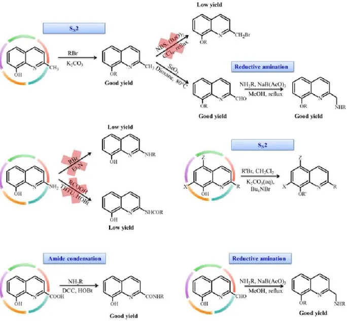
4.1 Glucosylated prodrugs 86
4.1.1 Synthesis and characterization 86
4.1.2 Copper complexes 89
4.1.3 Chemical and enzymatic stability 90
4.1.4 Biological activities 93
4.1.5 Docking studies 98
4.2 Galactosylated prodrugs 100
4.2.1 Synthesis and characterization 100
4.2.2 Chemical and enzymatic stability 105
4.2.3 Antiproliferative activity 106
4.3 Chelating OHQ conjugates with monosaccharides and disaccharides 110
4.3.1 Synthesis and characterization 110
4.3.2 Metal complexes 114
4.4 New cyclodextrin-bearing 8-hydroxyquinoline ligands 117
4.4.1 Synthesis and characterization 117
4.4.2 Metal complexes 146 4.4.3 Antiproliferative activity 167 4.4.4 Antioxidant activity 168 4.4.5 Antiaggregant activity 172 5. CONCLUSIONS 180 6. ACKNOWLEDGEMENTS 186 7. REFERENCES 187 A1. PUBLICATIONS 196 A2. PROCEEDINGS 198
1
1. INTRODUCTION
The application of inorganic chemistry in medicine is becoming more and more prominent since metals play a pivotal role in many life-related processes.
Sodium, potassium, magnesium and calcium are essential for nerve conduction, muscle contraction, stabilization of nucleic acids and a plethora of other biological systems and thus they are the dominant metal ions in a living organism. Nevertheless, the metal ions incorporated into proteins are often not these abundant metal ions but are trace elements such as iron, copper, manganese, zinc, cobalt and nickel. Proteins that bind transition metals are usually catalytic enzymes with the transition metal ion being essential for their activity. Under physiological conditions, most transition metals can exist in multiple valence states and thus can readily participate in single electron transfer reactions, making them attractive for inclusion in biological systems. Metal ions are also found as components of prosthetic groups, cofactors and complexes prior to insertion into proteins.
Homeostasis of metal ions is a complex process, maintained through tightly regulated mechanisms of uptake, storage and secretion. Several metal ion transporters participate in the conservation of the required levels of the various metal ions in the cellular compartments.
Altered metal homeostasis can lead to the metal binding to protein or replacement of other metals from their natural binding sites. Several results have provided evidence that toxic metals can interact with DNA and proteins causing oxidative deterioration of biological macromolecules. Thus the metal dyshomeostasis has been involved in the etiology of a great number of pathologies such as Wilson‟s (WD),1 Alzheimer‟s (AD),2,3 Parkinson‟s (PD)4 and Niemann-Pick5 diseases (NPC), Friedreich‟s ataxia,6 transfusion-related iron overload diseases7 and cancer.8
Furthermore, in some of these cases metals are implicated in redox reactions leading to production of ROS (Reactive oxygen Species) and thus oxidative stress.9
The different metal ions may be grouped into redox-active ions such as Fe2+, Cu2+, Co2+ and to a lesser extent Mn2+; and non-redox-active ions such as Ca2+ and Zn2+. Copper, zinc and iron have been chosen as the focus of this introduction as they are biologically relevant particularly with respect to human health.
2
1.1 Copper
Copper is an essential trace element for living organisms, taking part in several processes of metabolism, including mitochondrial oxidative phosphorylation, iron metabolism, free radical detoxification, neurotransmitter synthesis and denaturation, connective tissue synthesis and pigment formation.10 Copper is a transition metal characterized by a low redox potential between Cu2+ and Cu+ 0.158 V in water that explains its capacity to exchange electrons with other systems. The biological utility of copper mainly derives from this ability to cycle between its oxidized and reduced forms. A healthy human adult (70 Kg) contains about 110 mg of copper that is involved as cofactor of different redox enzymes. The best known among these are reported in Table 1.11 In addition to its enzymatic roles, copper plays non-enzymatic functions in angiogenesis,12 nerve myelination, activity of endorphin and brain development.13 Copper is essential for reproduction, regulation of gene expression and for normal growth and development.
Table 1. Some Cu-dependent enzymes in mammals.
Enzyme Function
Superoxide dismutase Free radical detoxification Cytochrome c oxidase Dioxygen reduction into water Ceruloplasmin Ferroxidase
Dopamine β-hydroxylase Oxygenase, converts dopa into norepinephrine Tyrosinase Monophenol oxidase, melanin synthesis Lysyl oxidase Cross-linking of collagen and elastin Metallothionein Storage of excess Copper
Amyloid Precursor Protein Normal function unknown
Angiogenin Induction of blood vessel formation
Hephaestin Ferroxidase
Despite its physiological role, copper can be toxic. Copper can catalyze the formation of ROS via a Fenton-like reaction. Cu2+, in the presence of superoxide anion radical or biological reductants such as ascorbic acid or gluthatione, can be reduced to Cu+. This latter is able to
3 catalyze the formation of reactive hydroxyl radicals through the decomposition of hydrogen peroxide via the Fenton reaction:
Cu+ + H2O2 → Cu2+ + •OH + OH− (Fenton reaction).
The hydroxyl radical is extremely reactive and can further react with practically any biological molecules in the near vicinity. •OH can abstract hydrogen atom leaving behind a carbon-centered radical (e.g. a lipid radical from unsaturated fatty acids). Copper is also capable of causing DNA strand breaks and oxidation of bases via ROS.In addition, copper can possibly displace other essential metals in proteins.
For these reasons, copper homeostasis is tightly controlled in all organisms.
Copper uptake into cells is thought to be mediated by the plasma membrane protein CTR1 (as for high affinity copper uptake) with some involvement of the divalent metal transporter and potentially other unidentified uptake systems (Figure 1). Essentially, no free copper is available in eukaryotic cells since the ion is always complexed with other molecules. Small molecules such as glutathione (GSH) and metallothioneins bind copper for storage and/or detoxification and may provide an exchangeable pool of copper.14 Futhermore, proteins called
metallochaperones15 also bind copper and target it to specific sites inside the cell. Copper chaperone for superoxide dismutase (CCS) delivers copper to Cu/Zn-SOD in cytosol and mitochondria, cytochrome c oxidase copper chaperone (COX17) mediates copper transfer within the mitochondrial intermembrane space for metallation and assembly of cytochrome c oxidase, and HAH1 transfers copper to the copper-ATPases, ATP7A and ATP7B for delivery to the secretory pathway and for efflux of excess copper from the cell (Figure 1).
4
Figure 1. Pathways of copper trafficking within a mammalian cell. The three principal chaperones, Cox17, CCS, and Atox1 are shown along with the respective protein targets, cytochrome c oxidase, SOD1
and the Wilson and Menkes transporting ATPases.
1.1.1 Copper-related diseases
Dyshomeostasis of copper has implications in a number of diseases, and deficiencies in copper uptake have a number of detrimental effects. Mutations in ATP7A and ATP7B result in Menkes and Wilson‟s diseases, respectively.
Patients with WD suffer from brain disorders and liver disease. Copper levels rise and serum Ceruloplasmin levels decrease because of the diminished function of the ATP7B protein, which is directly involved in the vesicular pathway of hepatic copper transport from the liver to bile canaliculi. Pathologic manifestations include liver failure, tremors, slurred speech, and other neurological impairments.
Untreated WD is fatal. The overall therapeutic approach is the generation of a negative copper balance. Some increase the removal of copper from the body, while others prevent the
5 absorption of copper from the diet. With chelation treatment or liver transplantation, prolonged survival has become the norm.
It is generally agreed that patients with symptoms or signs of hepatic insufficiency or chronic active hepatitis with or without neurologic manifestations should be treated with copper chelators (D-penicillamine (D-Pen) trientine hydrochloride (TETA) or tetrathiomolybdate) and/or zinc salts. Chelating agents directly bind copper and facilitate its excretion whereas zinc interferes with the intestinal uptake of copper and induces the endogenous chelator metallothionein.16
Menkes disease is an X-linked recessive disorder associated with copper deficiency due to mutations on the ATP7A gene. Since the ATP7A protein is malfunctioning in the enterocytes, there is a defect in dietary copper absorption and therefore in its distribution in most tissues.
Menkes disease is characterized by neurological defects, growth retardation, hypopigmentation, „„kinky‟‟ or „„steely‟‟ hair and deficiencies in a number of cuproproteins. The treatment consists of Cu supplementation by subcutaneous injections of Cu-His.
Light changes in copper homeostasis have strong impact on the brain, leading to the onset of neurological diseases. However, further elucidations are required to clarify the exact mechanisms of these relationships.
Prions are infectious protein particles involved in transmissible spongiform encephalopathies (TSEs) including Creutzfeld-Jakob disease (CJD), chronic wasting disease (CWD), and bovine spongiform encephalopathy (BSE). These fatal neurodegenerative diseases are caused by the misfolding of cellular prion. In its normal cellular form (PrPc) the protein is thought to transport copper into the cell, protect from oxidative stress and buffer copper in the cell. Conversion of the protein to the scrapie form (PrPsc) is induced by interaction with copper and other metal ions such as zinc, iron and manganese and altered metal binding by PrPsc contributes to the pathology. Copper shows an increase in toxicity for PrP knockout mice and exacerbates the disease in a mouse model of familial CJD. Increased copper in the diet of mice raises PrPc levels.17 Furthermore, when isolated from diseased brain, PrPSc has been found to be bound with Cu2+ ions.18 In addition, oxidative modifications of PrP, generated by copper-catalyzed Fenton reaction, have been linked to prion disease.19,20 Significantly, copper ion chelation therapy will delay onset of scrapie in mice.21
6 Huntington‟s disease (HD) is another progressive neurodegenerative disease characterized by a dominant polyglutamine expansion within the N-terminus of huntingtin protein and results in oxidative stress, energetic insufficiency and striatal degeneration. Affected individuals undergo progressive motor, cognitive, and psychiatric deterioration. HD brain has shown an accumulation of copper and iron in the striata of the patients, but the role of these metals in HD pathogenesis is unknown. It has recently hypothesized that increased amounts of copper bound to low affinity sites could contribute to pro-oxidant activities and neurodegeneration.22 Copper, but not other metal ions, is able to interact with both normal and mutant huntingtin. It also promotes the aggregation of mutant huntingtin fragments probably through the induction of structural changes, obviously metal chelation prevents these effects.
Another highly debilitating progressive neurodegenerative disorder is AD. This latter is characterized by extra cellular deposition of Aβ peptides in senile plaques and intracellular accumulation of hyperphosphorylated tau protein in neuronal cells as neurofibrillary tangles. Generally, AD affects people over 65 or as young as 40–50 years. It is the most prevalent form of dementia and is characterized by severe cognitive impairment due to degeneration of brain cells. For many years, the most popular hypothesis for AD neuropathogenesis was the “Amyloid Cascade Hypothesis,” which proposes that all pathology in the AD brain occurs downstream of the excessive accumulation of Aβ in the CNS. Recently, Bush et al have posited the “Metal Hypothesis of Alzheimer‟s Disease,” which stipulates that the neuropathogenic effects of Aβ in AD are promoted by (and possibly even dependent on) Aβ-metal interactions.23 Copper (390 µM), zinc (1055 µM), and iron (940 µM) have been reported to be elevated by several-fold in AD brain as compared with normal age-matched samples (copper [70 µM], zinc [350 µM] and iron [340 µM]). Aβ directly coordinates copper and zinc, but not iron or other metal ions, within the cores of plaques. Both zinc and copper are able to promote the aggregation of oligomers in the synaptic cleft. Moreover, copper and iron can also cause the neurotoxic redox activity of Aβ and induce oxidative cross-linking of the peptide into stable oligomers.
Copper is also associated with cancer and angiogenesis. Serum and tumor tissue copper levels in cancer patients are significantly elevated compared to healthy subjects. The most elevated levels of copper have been documented in cancer patients suffering from breast, cervical, ovarian, lung, prostate, stomach cancer and leukemia.24,25 Elevated copper levels
7 have been shown to be directly linked to cancer progression. In fact, copper is important for angiogenesis that is required for invasive tumor growth and metastasis. Angiogenesis is a multi-step process, involving degradation of the endothelial cell basement membrane, endothelial cell migration to the perivascular stroma and capillary sprouting. Avascular tumors are severely limited in their growth because of the lack of an adequate blood supply. Tumors have to perturb the local balance of proangiogenic and antiangiogenic factors in order to trigger angiogenesis. Therefore, tumors overexpress proangiogenic factors, such as vascular endothelial growth factor. It has been recently demonstrated that extracellular translocation of the cytosolic copper occurs during angiogenesis processes in vitro,26 suggesting that the metal coordination by an extracellular protein involved in angiogenesis, such as angiogenin (hAng) and/or vascular endothelial growth factor (VEGF), could play a pivotal role. Copper or its complexes have been shown to directly stimulate angiogenesis in several model systems27 as well as in vivo.28 Furthermore, copper chelation inhibits angiogenesis in numerous animal and xenograft models.29 Such findings have led to clinical trials for the treatment of solid tumors by copper chelators with some results of efficacy in disease stabilization, however the molecular basis and the effects that copper ion exerts are still incompletely understood.
Niemann–Pick type C (NPC) is an autosomal recessive disorder characterized by progressive neurological degeneration. The most commonly mutated gene in this pathology, NPC1, is a membrane protein localized in the late endosomes and regulates intracellular cholesterol trafficking. New insight into this disease suggests a possible role of copper. Conflicting results have been reported. Recent findings suggest that ATP7B localizes in the late endosomes where copper is transported to the secretory compartment via an NPC1-dependent pathway and incorporated into Ceruloplasmin.30 Futhermore, it has been observed copper accumulation in the liver31 and plasma, along with a decrease of copper excretion into the bile of NPC mice.9
Copper has been also associated with other diseases such as Parkinson‟s Disease, diabetes, atherosclerosis, cardiovascular disease and amyotrophic lateral sclerosis. To sum up, it is clear that the regulation of copper homeostasis is essential for the survival and proper functioning of all organisms.
8
1.2 Zinc
Zinc is essential for all living organisms and is the second most abundant transition metal in both seawater and humans.32
The adult human body contains approximately 1.5–2.5 g of zinc, present in all organs, tissues and fluids. The level of free intracellular Zn2+ is as low as 0.5 nM and so this makes zinc the most prevalent trace metal found in the cytoplasm. Zinc is a redox inert metal ion and does not participate in oxidation/reduction reactions because of the Zn2+ ion has a full d shell. It does not exist in multiple valences under physiological conditions rendering it unique amongst biologically-relevant transition metals. For these reasons, zinc is generally thought to be less toxic than the redox active metals such as copper and iron.
The combination of structural and thermodynamic properties with the abundant bioavailability have led to the incorporation of zinc in a variety of biological systems. Zinc is an element present in more than 300 enzymatic reactions that function in many aspects of cellular metabolism, involving metabolism of proteins, lipids and carbohydrates. Within the zinc proteins a number of distinct functional classes can be identified: for example, matrix metalloproteases (with roles in embryonic development, cell motility, wound healing and reproduction), peptidases (digestive enzymes) and zinc finger proteins (DNA or RNA binding proteins).
Intracellular uptake and efflux of zinc are mediated by two class of mammalian zinc transporters. The first is the ZnT family of transporters, which act to decrease intracellular zinc levels by transporting zinc from the cytoplasm to the lumen of organelles or the extracellular space. The second is the ZIP family responsible for increasing intracellular zinc levels by either transporting the metal from the extracellular space or lumen of organelles into the cytoplasm (Figure 2).
9
Figure 2. Cellular localization and function of ZIP and ZnT zinc transporter family members in mammalian cells. Arrows indicate the direction of zinc mobilization. ZIP1, 2 and 4 are induced in zinc
deficient conditions, whereas ZNT-1 and 2 by zinc accumulation.
1.2.1 Zinc-related diseases
Over one billion people in developing countries are nutritionally deficient in zinc. Its deficiency is associated with a range of pathological states, including skin changes, alopecia, diarrhea, slowed growth, delayed wound healing, hypogonadism, impaired immunity, and brain development disorders, all of which are reversible with zinc supplementation. Zinc deficiency occurs as a result of insufficient dietary zinc uptake (which can be exacerbated by consumption of foods such as cereal grains which are rich in phytates, inhibitors of zinc dietary absorbtion), malabsorption syndromes and other gastrointestinal disorders, chronic liver and renal diseases, excessive alcohol use, malignancy, pancreatic insufficiency, rheumatoid arthritis, and other chronic conditions.33
Acute and chronic forms of zinc toxicity have been observed. Acute zinc toxicity is rare and usually due to consumption of contaminated food that can lead to gastrointestinal irritation, fatigue, muscle pain and fever. Chronic zinc toxicity is more common and usually
10 occurs as a result of prolonged use of oral zinc supplements. Excess zinc can also interfere with other essential transition metals such as copper and iron. 34
Because of zinc is involved in maintaining of intracellular ion homeostasis and in signal transduction in most cells, it directly affects tumor cells through its regulatory role in gene expression and cell survival.
Levels of zinc in serum and malignant tissues of patients with various types of cancer are altered indicating the involvement of zinc in oncogenesis. Studies of the role of zinc in malignant diseases have contradictory results. It is clear, however, that serum zinc levels are reduced in patients with cancers of the breast, gallbladder, lung, colon, head, neck. Interestingly, while serum zinc levels are low in the setting of most cancers, tumor tissue in breast and lung cancer have elevated zinc levels compared to the corresponding healthy tissues.35 It has been found that zinc depletion induces cell death via apoptosis (or necrosis if apoptotic pathway is blocked) while sufficient zinc levels allow the maintenance of autophagy.35 Cancer cells have upregulated zinc importers, and frequently increased zinc levels, which allow them to survive.35
Besides its role in cancer, zinc is also associated with AD. Zn2+ is involved in protein modification, misfolding and aggregation, leading to the precipitation of Aβ.36 Zinc, released through glutamatergic neurotransmission, is concentrated in the presynaptic bouton by the action of ZnT3 (Figure 3) and it manages to achieve concentrations up to 300 µM in synaptic clefts. Cu2+ is released post-synaptically after NMDA-induced activation, which causes the translocation of the Menkes ATPase and its copper release through vesicles to the synaptic cleft. Cu2+ concentrations can reach 15 µM.
In the vicinity of the neuron, amyloid-beta comes across Zn2+ and forms oligomers of increasing insolubility as the amount of protein-bound-zinc increases. Aβ can bind up to 2.5 moles of Zn2+. The soluble Aβ monomers are constitutively degraded by matrix metalloproteinases (MMPs), neprilysin and insulin degrading enzyme (IDE) but Zn2+-loaded Aβ oligomers are resistant to degradation. Although there has been considerable focus on the toxic nature of amyloid-beta and its oligomers, it is important to note that the latter act as a Zn2+ sponge. The net effect of sequestering Zn2+ from its physiological targets is the disruption of normal memory events (for example, long-term potentiation) and loss of trophic and metabotropic stimulation of the postsynaptic neuron.37
11
Figure 3. Aβ is released into the synaptic cleft where it has the potential to react with Cu and Zn to form oxidized, crosslinked soluble aggregates and precipitated amyloid.
1.3 Iron
A large amount of cellular enzymes depends on iron for their function and, consequently, this metal is essential for life. The normal iron content of the human body is 3 or 4 grams.
Many of the iron-requiring proteins are involved in enzyme catalysis and electron transport, whereas others are needed for oxygen transport and delivery. Major classes of such proteins include those with heme as the prosthetic group and those with iron–sulfur (Fe–S) clusters. However, the most abundant iron proteins in the body are the hemoglobin and myoglobin. Iron can have two thermodynamically stable oxidation states, ferric iron (Fe3+) and ferrous iron
12 (Fe2+). This makes it suitable for the catalysis of biochemical reactions, but it also means that it is able to catalyze, similarly to copper, reactions with the production of toxic oxygen radicals.
Thus, individual cells and whole organisms must tightly controlled iron homeostasis. This latter is a complex process and there are several proteins that respond to the total body burden of iron and to hypoxia, anemia and inflammation. Most iron is delivered to cells bound to plasma transferrin via a process that involves transferrin receptor 1, divalent metal-ion transporter 1 and several other proteins (Figure 4). Non-transferrin bound iron can also be taken up efficiently by cells, although the mechanism is not completely clarified. The efflux of iron can occur via the iron export protein ferroportin and an iron oxidase. The coupling of an oxidoreductase to a membrane permease is a common feature in membrane iron transport. At the systemic level, iron transport is regulated by the liver-derived peptide hepcidin which acts on ferroportin to control iron release to the plasma (Figure 4).38
Figure 4. Iron transport in mammalian cells. Arrows indicate the direction of iron mobilization. Hepcidin blocks iron efflux into plasma inducing Ferroportin internalization and degradation in lysosomes.
13
1.3.1 Iron-related diseases
The most common outcome of systemic iron deficiency is anemia. Anemias are characterized by a deficiency in the number of mature erythrocytes in the circulation, which inevitably lowers the oxygen-carrying capacity of the blood resulting in clinical symptoms such as fatigue, weakness, increased cardiac output as well as increased morbidity and mortality.
On the contrary, there are also iron-overload diseases that can be inherited or acquired, and are typically characterized by the accumulation of iron in the body. Obviously, in these pathology increased levels of oxidative stress have been observed. Among iron-overload diseases, there are hereditary hemochromatoses and β-thalassemia major.39 The former is characterized by an accelerated rate of intestinal iron absorption and the progressive iron deposition in various tissues. Beta-thalassemia major is a hereditary disease affecting the hemoglobin. The treatment of this disease consists of an opportune transfusion program that prevents death from anemia in childhood and permits a normal growth and development. Nevertheless, transfusion-dependent patients will develop iron overload and require chelation therapy to remove the excess iron.39
Iron has been implicated in the onset of a number of neurodegenerative disorders, for example Friedreich's ataxia that is an autosomal recessive disease characterized by degeneration of neurons in the cerebellum. This pathology results in mitochondrial iron accumulation, decreased Fe/S cluster synthesis and an altered antioxidant response. Furthermore, a significant increase in brain iron levels has been found in both PD and AD. The mechanisms responsible for iron accumulation are still unknown in PD. Similarly, how iron is delivered to its major recipients in the cell (e.g. mitochondria) has yet to be elucidated. However, chelation of iron seems to protect from dopaminergic degeneration,40,41 typical feature of PD.
To sum up, iron is implicated in a large number of diseases and the most mechanisms responsible in the iron-involvement have to be clarified. However, iron overload disorders are successfully treated with chelation therapy.
14
1.4 Chelation therapy
Metal chelators can be used either to deliver metal ions for medical application (i.e. diagnostic application) or to remove unwanted metals from human body. This latter one is the topic of this section.
The manipulation of the metal distribution in biological systems is very complicated and it requires to consider all possible factors influencing the process.
When a ligand is introduced into a biological system, it is necessary to consider the reactivity and distribution characteristics of all parts of the coordination complex:
metal ion
ligand or pro-ligand complex
Whereas some neutral chelators are able to permeate biological barriers, other charged systems will achieve significant permeation only upon formation of an uncharged complex. In fact in some cases the corresponding metal complexes are more lipophilic and mobile throughout the biological system than ligands. Metal binding selectivity is very important otherwise one risks affecting the concentrations of other essential metals. In order to design a selective metal ion chelator, one must consider the hard-soft acid-base (HSAB) theory.
Another consideration in ligand design is the chelate effect describing the favorable entropy change upon exchange of many monodentate ligands for fewer multidentate ligands binding a given metal ion. The higher denticity the ligand, the more thermodynamically stable will be its complex.
Furthermore, the introduced metal-binding therapeutic system must participate in a series of ligand exchange reactions to form the new, desired complex and so its affinity must be comparable with that of the endogenous ligands. A final consideration must be made about the redox activity of the metal ion in the complexed form; in fact ligands could modify their redox potential promoting production of reactive oxygen species (ROS).
First, an overview of the major compounds for metal ion binding and new strategies to design multifunctional chelator will be discussed, to be followed with the discussion about specific metal chelators or metal ionophores.
15 The use of small-molecule chelators began in 1941 with the administration of citrate to relieve accidental overexposure to lead. The most obvious medicinal application of metal chelators is in the treatment of metal overload conditions. Among the first used metal chelators, there is D-penicillamine (D-pen, Figure 5) that has been introduced in 1956 for treatment of copper-overloaded WD patients.
Ethylenediaminetetraacetic acid (EDTA, Figure 5) was the next chelator in clinical use (1950s) for treatment of lead toxicity and also used in cases of accidental radionuclide dosing. Later, EDTA was substituted for diethylenetriaminepentaacetic acid (DTPA, Figure 5) in order to remove radionuclide excess. Triethylenetetraamine (TETA, Figure 5), although considered less effective, was introduced in 1980s for use in WD patients intolerant of D-pen.
Figure 5. Metal chelators.
Currently, D-Pen is the most widely used treatment across the world; TETA is used in the USA but is hardly accessible in Europe. Tetrathiomolybdate (TM) has been recently used in the USA as a copper chelator for patients who are intolerant to D-Pen and TETA, but it has not approved by FDA.16 However, D-Pen has severe side effects including bone marrow suppression, anorexia, vomiting and diarrhea. Both TETA and TM have fewer potential side effects, but must still be carefully monitored. Therefore, it is clear that new drugs have to be investigated in order to obtain more efficient and selective therapy in the treatment of WD.
O H N N OH O O OH O O H O N N N OH O O H O O H O O H O O O H O OH NH2 C H3 CH3 S H NH NH NH2 N H2 EDTA DTPA D-Pen TETA
16 In thalassemia major, iron accumulates in the liver, heart, pancreas and other organs with long-term consequences including fibrosis, cirrhosis, hepatocellular carcinoma, diabetes and heart disease. To prevent this progressive deterioration, iron has to be removed by chelation therapy. Deferoxamine (DFO, Figure 6) a naturally occurring microbial siderophore, is a trihydroxamic acid that efficiently binds Fe3+. It has been used to treat iron overload since the 1970s whereas deferiprone (DFP, Figure 6) has been used orally for the same purpose since the late 1980s.
DFP is currently approved in Europe and in India. It is a bidentate chelator and forms a neutral stable 3:1 ligand iron complex at pH 7.4, which is water soluble and readily excreted by kidneys.42
Figure 6. Metal chelators used in the treatment of iron-overload diseases.
Its relative lipophilicity gives it high oral activity and Blood Brain Barrier (BBB) penetration. Because of its side effects, DFP is approved for use only as a second-line treatment after DFO. Nevertheless, deferiprone offers a significant advantage over DFO in its ability to remove cardiac iron and is now in consideration for combination therapy with DFO in iron-overloaded thalassemia patients. Combination therapies are thought to provide higher efficacies and lower toxicities than current monotherapies. Deferiprone and DFO have been used together in iron-overloaded patients; not only was combination therapy well-tolerated, but it caused significant improvement in at least one additional observed end point (such as cardiac function) vs treatment with DFO alone.43 The smaller bidentate chelator acts as a
N CH3 C H3 O O H N H N O O N OH NH O O N OH NH3+ C H3 O OH DFO DFP
17 “shuttle” to first bind the biological metal ion then transfer it to the multidentate chelator, which acts as a “sink” for the ion.
In addition to these properties, the structure of DFP can be easily functionalize by N-substituent variation, which can alter the physicochemical properties of the drug without significant alteration of the metal binding ability. For these reasons, a large amount of derivatives has been synthetized and studied with different applications. Some of these have been applied to the treatment of AD, because of it has been recently demonstrated that deferiprone and a series of its derivatives such as 2-amido-3-hydroxypyridin-4-one are neuroprotective in mouse cortical neuron cultures.44 Deferiprone prevents neuronal death in cells exposed to AD-related insults such as Fe3+, H2O2, and Aβ 1-40.44
Other conditions for which chelation therapy and then DFP derivatives have been proposed or tested include cancer. Even if cancer does not involve iron overload, it is a requirement for cell cycle progression and for DNA synthesis; thus, cancer cells have a higher demand for the ion and are particularly susceptible to its depletion. Iron chelators (including DFO and DFP) and other hexadentate naturally occurring siderophores have been investigated as antiproliferatives for cancer therapy as reported by Richardson and coworkers.45
However, the chelation of copper has been also proposed in order to have antitumor activity.
Regulation of the copper levels as an anti-cancer strategy is currently under intense investigation and significant efforts were undertaken aiming at the control of the angiogenesis. Obviously, copper chelators utilized in WD were first checked as anti-angiogenic drugs. D-pen, TM, TETA have been shown to inhibit angiogenesis both in vitro and in vivo.46-48 Several animal studies have supported the hypothesis of employing copper chelators as anti-angiogenic agent. TETA was used to treat mice bearing hepatocellular carcinoma xenografts showing significant inhibition of the tumor growth associated with suppression of tumor angiogenesis.49 Analogously, TM displayed encouraging anti-angiogenic and antitumor effects in animal models. Pan et al. have reported that copper deficiency induced by tetrathiomolybdate resulted in impairment of tumor growth and angiogenesis in two animal models of breast cancer (an inflammatory breast cancer xenograft model in nude mice and HER2/neu cancer prone transgenic mice).47
18 TM was then selected for clinical trials in humans. In a phase I clinical trial, TM was administrated to patients with metastatic cancer. A reduction of ceruloplasmin without toxicity was observed and five of six patients displayed stable disease.50 In a following phase II clinical trial in patients with advanced kidney cancer, TM was well tolerated and a significant depletion of copper was observed in all patients. Furthermore, 31% patients exhibited stable disease for at least 6 months.51
Amongst the most important metal binding compounds, 8-hydroxyquinolines have recently rekindled interest as promising therapeutic agents in the treatment of diseases related to metal dyshomeostasis and oxidative stress such as cancer, Alzheimer‟s and Huntington‟s diseases.
1.4.1 8-hydroxyquinolines (OHQs)
8-Hydroxyquinoline (OHQ) is a quinoline derivative that has been used as a fungicide in agriculture and a preservative in the textile, wood, and paper industries. OHQ possesses a good metal chelating ability and thereof it is widely used for analytical and separation purposes as well as for metal chelation.
Derivatives of OHQ are systems of great interest in the field of inorganic and bioinorganic chemistry. As metal-binding compounds they have recently been viewed with interest as anticancer agents (especially in the presence of Cu2+)52 and as therapeutic agents for Alzheimer‟s disease (AD).53 A series of OHQ derivatives, such as 5-chloro-7-iodo-8-hydroxyquinoline or clioquinol (CQ), 5-((4-(prop-2-ynyl)piperazin-1-yl)methyl)quinolin-8-ol (HLA-20, Figure 7), 5-((methyl(prop-2-ynyl)amino)methyl)quinolin-8-ol (M30, Figure 7) and 5-((4-(2-hydroxyethyl)piperazin-1-yl)methyl)quinolin-8-ol (VK-28, Figure 7) have been reported to exert potent antineurodegenerative effects. HLA-20 is a strong Fe3+ chelator and radical scavenger with moderate inhibitory effect on monoamine oxidase, MAO-B. VK-28 is a strong iron chelator that can penetrate the mitochondrial membrane and M30 is a multimodal iron chelator that sequesters iron and inhibits MAO-A and MAO-B.54 In particular, combining the antioxidant chelating moiety of 8-hydroxyquinoline of the brain permeable iron chelator VK-28, with the propargyl moiety found in the MAO inhibitors, HLA-20 became a lead
19 compound for neuroprotective studies in PD and AD. Among these metal chelators used in neurodegeneration, CQ has reached pilot Phase II of clinical trials in AD patients.
N OH N N CH N OH N CH3 CH N N N OH OH HLA-20 M30 VK-28
Figure 7. Fe3+ chelators with possible application in AD and WD.
Clioquinol (5-chloro-7-iodo-8-hydroxyquinoline, CQ, PBT1, Figure 8) the most successful member of this family, was developed by Ciba–Geigy as an oral antibiotic to treat amoebic dysentery. N Cl I OH Figure 8. Clioquinol.
In the 1970s, it was associated with a wave of neurotoxic effects in Japan known as subacute myelo-opticoneuropathy (SMON)55 and was withdrawn from the market. The explanation for such side effects is not clear, but many of these cases may have been related to the ability of CQ to function as a “carrier of heavy metals” to the central nervous system (CNS) or perturbing the homeostasis of vitamin B12. Several recent studies have rekindled interest in CQ as a modulator of metal homeostasis in neurodegenerative disorders, such as AD.56,57A pilot phase II study of orally dosed CQ reported that the drug improved the
20 cognition and behavior in AD patients.58 Prana Biotechnology, who developed this drug for AD, discovered that the manufacturing process for CQ led to certain mutagenic “di-iodo” impurities and decided to suspend its development.59
However, since these results, a new OHQ derivative PBT2 (Prana Biotechnology, phase II completed) was developed. PBT2 (Figure 9) outperforms CQ, avoiding its side effects and offers even better results in the treatment of AD.
N Cl Cl OH N C H3 CH3 Figure 9. PBT2.
PBT2 has successfully completed phase IIa trials in patients with early Alzheimer's Disease and it is currently in phase IIb trials60. Furthermore, PBT2 has been also investigated as a therapeutic agent in Huntington‟s disease61 and has recently completed phase IIa clinical trials. The action mechanism of this family of compounds has been investigated and experimental evidence suggests that they could remove copper and other metals from extracellular amyloid.62 CQ or PBT2 encounter the extracellular pool of metals that are in a dissociable equilibrium with Aβ; e.g. in senile plaques and oligomers, CQ and PBT2 bind the metals (ionic copper or zinc) and facilitate plaque dissociation. The dissociate metal ions may be in a ternary complex with the drug itself or in a complex with dissociated Aβ. These complexes are taken up by neighboring cells, where the elements are separated. The metal ions (copper or zinc) can activate phosphoinositol 3-kinase and JNK and then MMP2 and 3, inducing peptidolytic degradation of Aβ (Figure 10).23
21
Figure 10. Hypothesized action mechanism of CQ. The figure shows only copper and CQ for clarity, but this mechanism has been also proposed for zinc and PBT2.
Moreover, as for CQ, it directly induces cell death in tumor cells at micromolar concentrations. Currently, CQ is in phase I clinical trial in patients with hematologic malignancy, such as leukemia, myelodysplasia, non-Hodgkin‟s and Hodgkin‟s lymphoma and multiple myeloma.63
For CQ and other OHQs it has been reported that the interaction with Cu2+ ions is a prerequisite for anticancer activity64 and, although the mechanism of action is not completely understood, experimental evidence suggests that OHQs act as anticancer agents as they are 20S proteasome inhibitors in the presence of Cu2+.65,66 CQ induces apoptosis in cancer cells through a caspase-dependent apoptotic pathway (Figure 11).67
Nevertheless, other mechanism have been reported to explain the cytotoxicity of CQ, such as the induction of the cytoplasmatic clearance of X-linked inhibition of apoptosis protein (Figure 11)68 and the induction of DNA double strand-breaks.69
22
Figure 11. Clioquinol inhibits the proteasome through metal dependent and independent mechanisms. The proposed mechanisms include: (i) Cu+ or during its reduction from Cu2+ to Cu+, copper interacts with
thiol and amino groups outside the active site of the proteasomal enzyme and thereby induces a conformational change in the protein structure causing inhibition of the proteasomal enzymes; (ii) Clioquinol directly inhibits the enzymatic activity of the proteasome by binding to regions of the alpha
subunits outside of the active site; (iii) Clioquinol forms a 2:1 complex with copper and this complex inhibits the enzymatic activity of the proteasome.
1.5 Targeting
Although metal chelating agents represent a necessary therapeutic strategy in metal overload diseases such as WD and hemochromatosis, they are associated with severe side-effects. Long-term use of strong chelators that are not selective (e.g. DFO), can be anticipated affecting the homeostasis of numerous biometals and perturb normal physiological functions of essential metal-requiring biomolecules (e.g. metalloenzymes). The ability to target tissues
23 selectively, through the coupling of chelation agents with targeting moieties, may minimize the toxicity of metal binding agents and enhance their effectiveness.
The concept of site-selective drug delivery or drug targeting has evolved since Nobelist Dr. Paul Ehrlich launched his idea of the “magic bullet”, an active chemical substance delivered only at the disease site and left the healthy tissues unharmed, over a century ago. Since then, the goal to develop safer therapeutical agents70,71 that are efficiently and selectively delivered to the desired site of action without affecting non-target cells has begun an attractive and actively pursued objective in pharmaceutical research.
For example, Mints et al have proposed to specifically target intracellular copper in liver because it is the main organ where copper accumulates in WD.16 Since the pool of intracellular copper is in the +1 oxidation state, they have designed a prochelator that releases the Cu+ chelator only once inside the reducing medium of the targeted cells. This strategy is represented in Figure 12.
Figure 12. Cu+ prochelators for the selective targeting of hepatocyte.
In prodrug approach, targeted drug delivery can be obtained by site-specific drug bioactivation or site-directed drug delivery. With site-directed drug delivery, the intact prodrug is primarily transported to the action site; for example, when prodrug is applied directly to the target organ (ocular and dermal drug delivery). Site-directed drug delivery after systemic administration constitutes very challenging task due to various unpredictable barriers in the body, but has demonstrated some success, for example, in brain delivery.72 With
24 bioactivation and exerts its own activity primarily at the desired site. The active agent is typically detached from a carefully designed promoiety, which can act as tissue/organ-selective carrier, or results from a prodrug itself after series of biotransformations in situ.73
1.5.1 Prodrug therapy and targeting strategies
Site-selective drug delivery has originated from cancer chemotherapy where drugs are highly reactive and poorly separate their cell-destroying action between healthy cells and tumor cells. Many antitumor drugs possess narrow therapeutic index, indicating that there is only minor difference between the dose needed for therapeutic response and the dose causing potentially life-threatening hazardous adverse effects to the patient.
To overcome the drawbacks of conventional cancer chemotherapy, it has been recently realized that the selectivity of drugs can be substantially increased by conjugating (directly or by using a complex drug delivery system) an anticancer drug with a targeting moiety, which provides two major functional roles. First, it directs the anticancer drug specifically to tumor cells, enhancing drug accumulation in tumor and therefore preventing adverse side effects. Second, it works as a penetration enhancer increasing the drug influx by tumor cells. In these cases non-cytotoxic prodrugs are designed to locally deliver the antitumor agent by tumor-specific activation. This may occur by hypoxic environment of solid tumors, by pH differences, by tumor-associated proteases or by enzyme selectively present in the tumor. In the case of tumor-associated enzyme activation, the active enzyme may be present in the tumor, like in PMT (prodrug monotherapy),74,75 or targeted to the tumor site such as ADEPT
(antibody directed enzyme prodrug therapy). The ADEPT and PMT approaches have been widely investigated as means to deliver drugs specifically to cancerous tissue and have shown promise in both animal studies and clinical trials.76,77
1.5.2 Prodrug monotherapy (PMT)
In PMT, prodrugs are designed for direct activation or recognition by tumor-associated factors (a phenotypic difference between tumor and normal tissue). These factors can be a low pH, the overexpression of certain enzymes or receptors in cancer, or the existence of regions in solid tumor where low oxygen tension is present and thereby there is an increased activity of reductive enzymes. In the past, a great number of carrier-linked prodrugs has been
25 developed with enzyme-specific substrates such as peptides or sugars. An overview of enzymes overexpressed in tumors and examples for respective substrates is given in Table 2.
Because the enzymes that are used for prodrug activation are also present in normal cells, activation of an enzymatically cleavable prodrug can be observed in healthy tissue. This stresses the need to improve tumor uptake through active or passive targeting and to characterize the overexpression of the respective enzymes in the individual tumor.
Table 2. Overview of enzymes that are overexpressed in tumors.
Enzyme Tumor Function Substrate Ref.
β-galactosidase Breast, colon Hydrolysis of galactose moieties β-Galactose
moieties 78
β-glucuronidase Breast, pancreas Hydrolysis of glucuronide moieties
β-Glucuronide
moieties 79-81
Carboxylesterases Hydrolysis or transesterification of drugs
Ester or carbamate
moieties 82,83
α-mannosidase Breast, colon Hydrolysis of mannose moieties mannose moieties 78
Chatepsin B Chatepsin L Breast, gastric, colon Lysosomal degradation of proteins Arg-Arg, Ala-Leu, Gly-Leu-Phe-Gly. 84
Chatepsin D Breast, gastric, ovary Degradation of extracellular matrix Phe-Ala-Ala- Phe(NO2)-Phe-Val-Leu-OM4P 85
Plasmin Degradation of blood plasma proteins Ala-Phe-Lys, D-Ala-Trp-Lys, D-Val-Leu-Lys 86 Matrix Metalloproteases Degradation of extracellular matrix and collagens
Ac-Pro-Leu-Gly-Leu, Gly-Pro-Leu-Gly-Ile-Ala-Gly-Gln
87,88
1.5.3 Two-step prodrug therapies (ADEPT)
Enzyme-activating prodrug therapy is a two-step approach. In the first step, a drug-activating exogenous enzyme is targeted or expressed in tumors. In the second step, a nontoxic prodrug, a substrate of the exogenous enzyme, is administered systemically.
26 Currently, delivery methods for an enzyme/prodrug strategy can be divided into two major classes:
delivery of genes that encode prodrug-activating enzymes into tumor tissues (GDEPT, VDEPT, etc.);
delivery of active enzymes conjugated to a vehicle that is capable of target tumor tissues (such as an antibody in ADEPT, ligand in LIDEPT).
This introduction focuses only on antibody-directed enzyme prodrug therapy ADEPT (Figure 13).
Figure 13. Scheme of antibody-directed enzyme prodrug therapy (ADEPT).
In this approach, a monoclonal antibody is initially employed to selectively deliver an enzyme (for example β-galactosidase) to the tumor cells, which subsequently converts an inactive prodrug to active drug at tumor site. Thus, the selective activation of the prodrug at the tumor site would result in improved antitumor activity with minimal side effects.89
Monoclonal antibodies have been used to deliver potent toxins and radionuclides to tumors. Many successful conjugates such as Mylotarg and Zevalin for the treatment of leukemia and
27 B-cell lymphomas have been developed.90 However, there are no clinically approved immunoconjugates for the treatment of solid tumor because they suffer from insufficient density of antigens on malignant cells and low cellular internalization. ADEPT approach offers two advantages over chemoimmunoconjugates. The first is the amplification effect since one molecule of enzyme-antibody conjugate, acting as a catalyst, is able to activate a large number of prodrug molecules. The second is the induction of the Bystander effect. The latter occurs when not only the tumor cells that are bound to the antibody enzyme conjugate but as well as the neighboring cells antigen are killed (Figure 14).
Figure 14. Bystander effect: cells that are killed because they are in close proximity to the targeted cells, rather than being targets themselves.
There are a several general considerations for ADEPT.
The requirements for the prodrug are low cytotoxicity, suitable Km and kcat parameters and
good pharmacokinetic properties. Furthermore, the prodrugs have not to be substrates for endogenous enzymes. Instead, high cytotoxicity and diffusion across cell membrane are pivotal features of the released active drugs.
The target antigen should be either expressed on the tumor cell membrane or secreted into the extracellular matrix of the tumor, and the use of a high affinity monoclonal antibody is
28 very important. The enzyme should be able to exert its optimal activity at a pH close to that of the tumor extracellular fluid. Because antibody-enzyme conjugate may be immunogenic, circulating host anticonjugate antibodies may interfere with treatment. Ideally, the enzyme system should not have a human homologue to avoid prodrug activation outside the tumor site. However there are many clinical limitations associated with ADEPT. For example, it is difficult to generate effective quantities of active drug since the levels of exogenous enzyme are low. Other drawbacks of ADEPT include cost and difficulties related to development and purification of antibodies, immunogenicity of antibodies, accessibility of tumor to the enzyme/antibody conjugate and the conversion of prodrugs in healthy tissues.91 For these reasons, a more convenient strategy could be the use of an already present enzyme (PMT).
1.6 Glycoconjugation: approaches to targeting
A good drug is a target-specific agent; it would have high efficiency and few, if any, side effects. Target specificity also means recognition, and this is where carbohydrates come in. While many drugs contain carbohydrates as part of their molecules, other non-saccharidic drugs can be guided by them. Carbohydrates‟ role is that they provide a guidance mechanism for sick cells, enabling drugs to selectively arrive there.
A few dozen FDA-approved drugs contain carbohydrate moieties as part of their structures. Typically, removal of the sugar eliminates the therapeutic value of the drug.
Among saccharidic drugs there are monosaccharide conjugates that include four groups of systems:
Anthracycline antibiotics and agents such as Doxorubicin, Daunorubicin, Epirubicin and Idarubicin (Figure 15);
Nucleotides, nucleosides and their analogs such as Fludarabine Phosphate, Stavudine, Adenosine, Ribavirin;
Polyene: Amphotericin B;
Other agents: Etoposide, Lincomycin, Clindamycin, Pentostatin.
The first group (Figure 15) includes cytotoxic anthracycline antibiotics of microbial origin (Doxorubicin and Daunorubicin) or their semi-synthetic derivatives (Epirubicin and
29 Idarubicin). These drugs are potent neoplastic agents consisting of a naphthacenequinone nucleus linked through a glycosidic bond at ring atom 7 to an amine sugar, daunosamine.
Figure 15. Structures of Doxorubicin (DOX), Daunorubicin (DNR), Epirubicin (EPI) and Idarubicin (IDA).
The second group of monosaccharide drugs is represented by nucleotides and nucleosides and their synthetic analogs. Among them, there are potent neoplastic agents, such as Fludarabine Phosphate (fluorinatedarabinofuranosyladenine 5′-monophosphate) and Gemcitabine (2′-deoxy-2′,2′-difluorocytidine, whose metabolic products inhibit DNA synthesis. The third group, polyenes, is represented by Amphotericin B, which is an antifungal antibiotic of microbial origin. Amphotericin B is a 3-amino-3,6-dideoxy-β-D-mannopyranosyl derivative of an octahydroxypolyene containing seven carbon-carbon double bonds in a macrocyclic 38-member ring.
O O OCH3 OH OH O OH O OH O C H3 OH NH2 O O OCH3 OH OH O OH O C H3 O C H3 OH NH2 O O OCH3 OH OH O OH O OH O C H3 O H NH2 O O OH OH O OH O C H3 O C H3 OH NH2 DOX DNR EPI IDA
30 Finally, the fourth group of monosaccharide drugs contains a number of different compounds, among them Etoposide. It is an anticancer agent which belongs to the topoisomerase inhibitor class (Figure 16).
Figure 16. Structure of Etoposide (semi-synthetic β-D-glucopyranoside derivative of podophyllotoxin).
An essential component of these drugs is their sugar moiety. Removal of the sugar residue typically leads to elimination of the drug therapeutic properties. On the other hand, addition of a certain sugar moiety sometimes enhances the recognized potential of the drug at the target level.
For these reasons, it has recently developed the concept of saccharides/anti-tumor conjugates. These compounds contain sugar moieties that allow the binding of the anti-tumor drugs with specific receptors such as glucose transporters or galectin, which transfer the drugs to the tumor cells. The rationale behind the development of glucoconjugates resides on the glucose avidity and elevated glycolysis of tumor cells. Whereas the galactoconjugates could be recognized by lectins which are overexpressed in a number of tumors.92
Glucose (Glc) and galactose (Gal) are not the only saccharidic compounds that are used to target cancer cells. Prodrugs of the glucuronic acid that might be activated by glucuronidases
O O O O O O O O C H3 OH O CH3 O O H O H CH3 H H H H
31 are particularly appealing because the levels of glucuronidase have been reported to be elevated in different tumors. It is possible, therefore, that glucuronide prodrugs may be preferentially activated in tumors. Moreover, glucuronidases have also been used in ADEPT strategy.
Disaccharides (lactose) and oligosaccharides are also used in targeting strategies. Cyclodextrins are also noteworthy among the oligosaccharides applied for targeting.
1.6.1 Gluconjugates
Glucoconjugate approach has been used in an effort to enhance targeting of analgesics,93 dopamine derivatives,94 anti-cancer agents,95 and anti-Alzheimer drugs.96,97
Glucoconjugates could target tumors exploiting glucose avidity of cancer cells. It is now clear that the majority of tumor cells in vivo and transformed cells in vitro, exhibit elevated levels of glucose transporter and elevated rates of glycolysis. This phenomenon was observed for the first time by Otto Warburg.98 Furthermore, glucose-conjugated compounds can enhance uptake of a drug in cancer cells99 as tumor cells require a significant amount of glucose and this demand is met by the high density of hexose transporters GLUTs, a family of membrane proteins. Cancer cells generally express higher levels of GLUTs than normal cells. The ratio of GLUT expression in malignant versus normal cells is about 100-300.100 This is because the efficiency of ATP production in normal cells does not satisfy the high energy requirements of cancer cells, so GLUTs are overexpressed in cancer cells to improve glucose uptake. This differential rate of glucose uptake has been used in positron emission tomography (PET), whereby 2-[18F]-2-deoxy-D-glucose (FDG) accumulates in cancer cells and indicates the tumor.101 It has been reported102-104 that β-glucosidases have a crucial role in the activation of the glycosylated prodrugs to release the active aglycone moieties. The level of expression of these enzymes into cancer cells remains an essential point to clarify. A good balance between the β-glucosidase activity and the transmembrane glucose transporters may ensure the right and effective activation of glycosylated prodrugs.105 In practice, high cellular β-glucosidase levels might be effective only when transport proteins are expressed as well.
It has been recently reported the synthesis and preliminary antiproliferative activity of potential DNA bisintercalators that include quinoline and glucose moieties.106 Binding
32 experiments have suggested that these compounds could bind DNA by intercalation and glucose moiety could regulate their interaction with DNA. Preliminary MTT assay showed a good anticancer activity of these compounds but experiments of nuclear permeation were not performed.
β-glucosides of curcumin, perillyl alcohol and diethylstilbesterol were also tested in cytotoxicity enzymatic cleavage studies in order to demonstrate the involvement of β-glucosidase and caspases in glucoconjugate antiproliferative activity in colon cancer cell lines.107 Thus, following glucoconjugate entrapment within cancer cells, they are presumably subject to hydrolysis by specific β-glucosidases to liberate the active aglycone, that exerts its anti-cancer activity.107
The glucoconjugates have been also used by various groups to impart elevated brain uptake exploiting the glucose transport system in the BBB (Figure 17).108
Figure 17. Deferiprone glucoconjugates: possible mechanism of action.
Orvig‟ s group has recently reported glucosylated tetrahydrosalens (Figure 18) as potential drugs for Alzheimer‟s therapy.109,110 These derivatives can be divided in two compound classes: one in which the metal binding site is free110 and another one in which metal binding
33 or antioxidant functionality is masked.109 The molecule in this form is referred to as a prodrug and it requires the cleavage of the carbohydrate moiety to exert its activity. These derivatives are very promising as antioxidant agents and Aβ aggregation inhibitors.
O OH O HO H OH N R O N R O O OH OH OH O H R1 R1 N C H3 N CH3 R R OH OH O OH O HO H OH O O O OH OH OH O H
Figure 18. Glucosylated tetrahydrosalens.
The same approach was utilized to design multifunctional pyridinone ligands (Figure 19).97,111
Figure 19. N-aryl-substituted 3-(β-D-glucopyranosyloxy)-2-methyl-4(1H)-pyridinones as agents for Alzheimer’s therapy.


![Table 3. ESI-MS characterization of the Cu 2+ complexes of MeHQ, ClHQ, Cl 2 HQ ([Cu 2+ ] = [ligand] = 3.0 ](https://thumb-eu.123doks.com/thumbv2/123dokorg/4520132.34886/96.918.181.761.396.830/table-esi-ms-characterization-complexes-mehq-clhq-ligand.webp)