International Ph.D Program in Neuropharmacology
XXVII Cycle
Department of Biomedical and Biotecnological Sciences
University of Catania
Modulation of Stress Granules formation:
Role of mGlu5 receptor and FMRP and implications for
pathophysiology of Fragile X Syndrome
Ph.D. Thesis
Barbara Di Marco
Coordinator: Prof. S. Salomone
Tutor: Prof. U. Spampinato, Inserm, U862, Neurocentre Magendie,
Physiopathology of Addiction group, Bordeaux, F-33000, France
Co-Tutor: Dr. M. V. Catania, ISN-CNR, Catania, Italy
2
3
ACKNOWLEDGEMENTS
I would like to express my gratitude to Prof. Filippo Drago and Prof.
Salvatore Salomone for giving me the opportunity to take part to this
prestigious Ph.D program.
I would like to thank Prof. Umberto Spampinato for his excellent
supervision, constructive comments and discussion, as well as for
welcoming me in his groups and leading me working on diverse exciting
projects.
I would like to thank Dr. Maria Vincenza Catania, who has supported me
during my Ph.D. Dr. Catania taught me how to approach “Research”. Her
immense knowledge, enthusiasm and guidance, allowed me to reach this
important goal.
Sincerely thank to Dr. Paola Dell’Albani for her crucial contribution, caring,
patience, that made this thesis possible; it would have been a lonely and
poor work without her.
I warmly thank Dr. Rosalia Pellitteri, for each constructive scientific advice
and comments. She always encouraged me and she made me feel in the
laboratory as in a family.
I would like to thank Dr. Michela Spatuzza, good friend who always gave
me her help, her best suggestions and with whom I spent the best moments
in the lab.
I want to thank CNR-ISN, Oasi M.S Troina and especially my colleagues
Dr. Melania Bonaccorso, Dr. Simona D’Antoni and Dr. Eugenia Ranno,
with which I spent these years learning and enjoying research.
4
I’m grateful to Francesco Marino for technical support.
I thank my Ph.D. colleagues and all members of the Ph.D. Program with
which I shared this experience pleasantly.
I would like to thank Celine Devroye and Adeline Cathala for their infinite
kindness and hospitality show me during my time in Bordeaux. With them I
learned techniques that I had never even thought of doing!
Words cannot express how grateful I am to my beloved parents and to my
sister Roberta, who have always believed in me, supporting and
understanding me during these years.
Last but not least, my thanks go to my darling Luigi, who pushed and
encouraged me to pursue a career in research, supporting me in the best and
worst moments of my life.
5
TABLE OF CONTENTS
ABBREVIATIONS ... 7 ABSTRACT ... 11 INTRODUCTION ... 13 1. FRAGILE X SYNDROME (FXS) ... 13 1.1 PHENOTYPE OF FXS ... 13 1.2 SYNAPTIC PLASTICITY IN FXS ... 15 1.3 CAUSES OF FXS ... 16 1.4 FMR1 GENE ... 181.5 FMRP: EXPRESSION, STRUCTURE AND FUNCTIONS ... 19
1.6 FMRP AND PROTEIN SYNTHESIS ... 21
1.7 FXR1P AND FXR2P ... 25
1.8 FXS MOUSE MODEL ... 26
2. METABOTROPIC GLUTAMATE RECEPTORS ... 29
2.1 Metabotropic Glutamate Receptors and FXS ... 32
3. THERAPEUTIC STRATEGIES IN FXS ... 35
4. OXIDATIVE STRESS AND FRAGILE X SYNDROME ... 38
5. STRESS GRANULES (SGs) ... 39
5.1 SGs Assembly ... 40
6
5.3 SGs and neurodegeneration ... 50
5.4 SGs and Fragile X Syndrome ... 52
CHAPTER I ... 54
CHAPTER II ... 85
GENERAL DISCUSSION AND CONCLUSIONS ... 113
REFERENCES ... 117
7
ABBREVIATIONS
82-FIP 82 kDa-FMRP Interacting Protein
AGO1 Argonoute 1
AGS Audiogenic seizures
ALS Amyotrophic Lateral Sclerosis
AMPA α-Amino-3-hydroxy-5-methyl-4-isoxazolepropionic acid
BC1 Brain Cytoplasmic RNA 1
BRF1 Butyrate Response Factor 1
BTF Bcl-2-associated transcription factor 1
CaMKII Calcium Calmodulin-dependent Kinase II
cAMP amp cyclic
CC Coiled Coil
CC coiled coil
CNS Central Nervous System
CPEB Cytoplasmic Polyadenylation Element Binding Protein
CREB cAMP response element binding protein
CYFIP1 Cytoplasmic FMRP Interacting Protein
CYFIP2 Cytoplasmic FMRP Interacting Protein 2
DAG Diacylglycerol
DIS1 Disrupted In Schizophrenia
eIF2α Eukaryotic Translation Initiation Factor 2 α
eIF4E Eukaryotic Initiation Factor 4E
ERK Extracellular Signal-Regulated Kinase
FAST TIA1 Interacting Protein
FMR1 Fragile X Mental Retardation 1
FMRP Fragile X Mental Retardation Protein
FTD Front temporal Dementia
FUS Fused in Sarcoma
8
FXR1P Fragile X Related Protein 1
FXR2P Fragile X Related Protein 2
FXS Fragile X Syndrome
FXTAS Fragile X Associated Tremor/Ataxia Syndrome
G3BP GTPase Activating Protein Binding Protein
GCN2 General Control Nonderepressible 2
Gp1 mGluRs Group I mGlu Receptors GPCR C G-Protein-Coupled Receptor
HITS CLIP High-Throughput Sequencing–Cross-Linking Immunoprecipitation
HPA Hypothalamic-Pituitary-Adrenal
HRI Heme-Regulated Initiation F ctor 2α Kinase
Htt huntingtin
ID Intellectual Disability
iGlu Ionotropic Glutamate
IP3 Inositol-1,3,4-Trisphosphate IQ Intelligence Quotien KA Kaina e KH K Homology 2 KH1 K Homology 1 KH2 K Homology 2
LTD Long Term Depression
LTP Long Term Potentiation
MAP1B Microtubule-Associated Protein-1B
MAPK Microtubule Associated Protein Kinase
mGlu Metabotropic Glutamate
MMP9 Matrix Metallopeptidase 9
MPEP 2-Methyl-6-(Phenylethynyl)-Pyridine
mRNPs Messenger Ribonucleoprotein Particles
mTOR Mammalian Target of Rapamycin
9
NLS Nuclear Localization Signal
NMDA N-Methyl-D-Aspartate
NoS Nucleolar Localization Signal
NRF1 nuclear respiratory factor 1
NRF2 nuclear respiratory factor 2
NTMs Normal Transmitting Males
NUFIP1 Nuclear FMRP Interacting Protein 1
PABP-1 Poly(A) Binding Protein 1
PERK PKR Like Endoplasmic Reticulum Kinase
PI3K Phosphoinositide 3 Kinase
PIP2 Phosphatidylinositol-4,5-Bisphosphate PKB Protein Kinase B
PKC Protein Kinase C
PLC Phospholipase C
PP2A Protein Phosphatase 2A
PSD Postsynaptic Density
Rac1 Ras related C3 botulinum toxin substrate 1
RGG Arg-Gly-Gly
RISC RNA-Induced Silencing Complex
SAPAP4 SAP90/PSD-95-associated protein 4
SGs Stress Granules
SMA Spinal Muscular Atrophy
SMN Survival of Motor Neurons
SOD1 Superoxide Dismutase 1
Sp1 specificity protein 1
TDP-43 TAR DNA-Binding Protein of 43 kDa
TIA-1 T Cell Internal Antigen 1
TIAR TIA-1 Related Protein
TRAF2 Tumor Necrosis Factor Receptor Associated Factor 2
TRAL Trailer-Hitch Protein
10
USF1 Stimulatory Factor 1
11
ABSTRACT
Fragile X syndrome (FXS) is the most common form of inherited intellectual disability and autism. The genetic defect in FXS is a CGG trinucleotide repeat expansion (>200) in the promoter region of the FMR1 (fragile X mental retardation 1) gene; this amplification causes the absence of the encoded protein FMRP (Fragile X Mental Retardation Protein). FMRP is an RNA-binding protein involved in mRNA transport and translation. Despite numerous studies, the available treatments are only symptomatic. There is no cure to replace FMRP expression, yet. FMRP can interact with RNA-binding proteins such as FXR1P, FXR2P, NUFIP and 82-FIP, and with proteins that do not bind RNA, like CYFIP1 and CYFIP2. The interaction with these different proteins may modulate FMRP functions and its RNA affinity. A new role of FMRP in mRNA metabolism as component of stress granules (SGs) has been identified. FMRP seems to lead mRNAs in SGs upon cellular stress, during which protein synthesis is blocked. SGs are ribonucleoproteic aggregates containing translation initiation components and RNA binding proteins, like eIF2α and FMRP. Several data also indicate that some of the FXS symptoms are a consequence of a defect in group-I metabotropic glutamate receptor, namely mGlu5; pharmacological blockade of mGlu5 receptors provide a therapeutic target in FXS. mGlu5 receptor, like FMRP, regulates protein synthesis but in a functionally opponent manner: mGlu5 receptor activates protein synthesis, FMRP suppresses it. In the absence of FMRP, mGlu5-dependent protein synthesis is unchecked, with consequent excessive translation. Activation of mGlu5 receptors stimulates FMRP-mediated mRNA transport and protein synthesis, but its role in SGs formation is unknown.
The aim of this PhD thesis was to better investigate FMRP function studying the relationship of FMRP with its interacting proteins and the role of FMRP in stress response under activation of mGlu5 receptor. In Paper I, we analyzed the expression pattern of FMRP and its interacting proteins in different brain areas, at different ages in wild type (WT) mice to better define the interplay between FMRP
12
and its interacting proteins during development. FMRP was strongly expressed at P3, peaked at P7-P14 and gradually decreases thereafter. The analysis of expression pattern of several proteins carried out, indicate that FMRP and its interacting proteins have distinct developmental patterns of expression and suggest that FMRP may be preferentially associated to certain proteins in early and late developmental stages. We found that the RNA binding and cytoskeleton remodeling functions of FMRP may be differently modulated during development.
In Paper II we studied FMRP under stress condition using WT and Fmr1
knockout (KO) astrocytes. We have demonstrated that the lack of FMRP impairs SGs formation and furthermore that activation of mGlu5 receptor affects SGs formation through a FMRP-mediated mechanism in WT. Interestingly, the mGlu5 receptor blockade restores SGs formation in Fmr1 KO. Also, mGlu5 receptor activation before stress reduced FMRP recruitment in SGs and phosphorylation of eIF2α and FMRP. In contrast, mGlu5 receptor activation did not affect SGs formation in Fmr1 KO astrocytes. Since phosphorylation of eIF2α and FMRP are two crucial key events in SGs formation and modulation of protein synthesis, mGlu5 receptors may act by shifting the balance from inhibition to activation of protein synthesis during stress. These findings suggest a potential novel role for mGlu receptors in SGs formation. We suggest that FMRP may have a positive role in stress response, facilitating and enhancing SGs formation to prevent stress damages. This process is useful to understand what happens in FXS, in which can occur abnormal modulation of different proteins during development with consequent abnormal response during adversal conditions, like oxidative stress that represent a frequent component in FXS and neurodegenerative disorders.
13
INTRODUCTION
1. FRAGILE X SYNDROME (FXS)
Fragile X Syndrome (FXS) is the most common form of inherited intellectual disability (ID) and a leading cause of autism, affecting 1/4000 males and 1/8000 females (Rousseau et al., 1995; O’Donnel & Warren, 2002). FXS, also called Martin-Bell syndrome in the past, belongs to a large group of human X-linked mental retardation syndromes (Chiurazzi et al., 2004).
FXS is mainly caused by the amplification of CGG trinucleotide repeat in the promoter region of the FMR1 gene. This abnormal triplet expansion leads to transcriptional silencing of the FMR1 gene and absence of the FMR1 encoded protein FMRP (Fragile X Mental Retardation Protein) (Devys et al., 1993; O’Donnel and Warren, 2002; Penagarikano et al., 2007). FMRP is an RNA binding protein involved in the regulation of target mRNA metabolism. FMRP mainly acts as a negative regulator of translation in association with polyribosomes and it plays an important role in synaptic plasticity (Li et al., 2001; Qin et al., 2005; Dolen et al., 2007). FMRP is also a component of RNA granules, the mRNPs (messenger ribonucleoprotein particles) that escort mRNAs in repressed conditions from soma to synapses. The absence of FMRP causes abnormal distribution of its mRNAs cargos and is the basis for the FXS phenotype (Miyashiro et al., 2003).
1.1 Phenotype of FXS
The clinics of FXS is characterized by moderate to severe cognitive impairment (IQ<70), which particularly affects short-term memory for complex information, visuospatial skills, and language acquisition (reviewed by Panagarikano et al., 2007). Moreover, FXS is a frequent cause of autism, indeed 15-50% of FXS individuals show autistic behavior such as poor visual contact, tactile
14
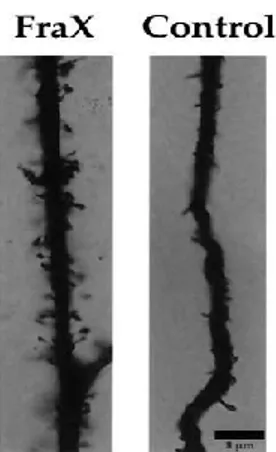
defensiveness, and repetitive behaviors (Cornish et al., 2004). FXS patients also show epilepsy, hyperactivity, hypersensitivity to sensorial stimuli, attention deficit, sleep disorders; they also manifest physical characteristics such as dysmorphic long face with prominent mandibular and large ears, arched palate, mitral valve prolapse, hypotonia, increased joint laxity and macroorchidism (The Dutch-Belgian Fragile X Consortium, 1994; Hagerman, 2002). Autopsy of brains from FXS patients showed no significant abnormalities (Bakker et al., 1994; Reyniers et al., 1999), although in some brain areas such as cortex and hippocampus, dendritic spines appear immature, like filopodia (Rudelli et al., 1985), longer and thinner than healthy, suggesting a misregulation in dendritic development (The Dutch-Belgian Fragile X Consortium, 1994; Irwin et al., 2001; Nimchinsky et al., 2001; O’Donnel et al., 2002; Penagarikano etal., 2007). During development, an exceeded number of dendritic spines occurs but eventually, a lot of them undergo to elimination by maturation and pruning mechanisms that establish the final synaptic phenotype. An abnormal dendritic spines development has been reported in Fmr1 knockout (KO) mice, the animal model for FXS (Nimchinsky et al., 2001). Abnormalities in dendritic and spines are associated with neurological problems in FXS such as epileptic seizures (Incorpora et al., 2002). Abnormal dendritic spines phenotype has been evident in FXS since the first autopsy and spine density appeared increased compared to control patients supporting the hypothesis that defects in spine maturation and elimination may underlie ID (Rudelli et al., 1985). Spine abnormalities have long been associated with mental retardation as well as with Down and Rett syndromes (Kaufmann and Moser, 2000) and the spine phenotype observed in FXS patients and Fmr1 KO mice suggests that FMRP is involved in synaptogenesis early in development.
15
Figure 1. Dendritic spines of fragile X neurons. Adapted from Irwin et al., 2001
1.2 Synaptic plasticity in FXS
FXS is considered a synaptic disease in which FMRP and several proteins/biochemical pathways associated with FMRP and with spine morphogenesis are involved. FMRP may influence synthesis of proteins involved in dendritic spine morphology; among them Rho GTPase, Rac1, microtubule-associated protein-1B (MAP1B), calcium calmodulin-dependent kinase II (CaMKII), calbindin, cadherins (Grossman et al., 2006). Spines are dynamic structures with an electrondense region, the postsynaptic density (PSD), that consists of dense area containing receptors, channels, scaffolding proteins and enzyme involved in synaptic transmission (Tada and Sheng, 2006). During development, dendritic spines morphology changes from small, immature like filopodia dendritic spines, which are more frequent in early age, to mature mashroom-shape dendritic spines that are characteristic postdevelopmentally (Hinton et al., 1991; Ivanco and Greenough, 2002). The spine structural changes are related to synaptic plasticity. Synaptic plasticity is a phenomenon during which a change, persistent or transient, of morphology, composition, or signal transduction efficiency occurs at a neuronal synapse in response to intrinsic or extrinsic signals (Mosbacher, 2014). The discovery of altered spine morphology
16
supports a defective synaptic plasticity in FXS but does not elucidate the cause of mental retardation. The most direct evidence about this aspect that underlines FMRP’s role in synaptic plasticity comes from studies on long-term depression (LTD) and long-term potentiation (LTP) in wild-type (WT) and Fmr1 KO mice. LTD and LTP are two forms of synaptic plasticity essential for cognitive functions, memory and learning processes; LTP is associated with synapse creation of new spines and enlargements of existing spines associated with the strengthening of the connection between a presynaptic and post-synaptic neuron, whereas LTD is associated with elimination, shrinkage of spines (reviewed by Panagarikano et al., 2007). One form of LTD is dependent on metabotropic glutamate receptor and requires activation of new protein synthesis (Huber et al., 2000). This form of LTD is enhanced in Fmr1 KO mice (Bear et al., 2004), and this finding represents a direct evidence that the absence of FMRP alters synaptic plasticity. Indeed FMRP is possibly involved in the assembly and functioning of neuronal circuits and in the regulation of the dendritic spines turnover.
1.3 Causes of FXS
The gene implicated in the pathology of FXS is FMR1 (fragile X mental retardation 1) gene. It is located on the long arm of the X chromosome at position 27.3. This region cytogenetically displays a fragile site from which the name of the syndrome takes origin. The most common genetic defect in FXS is a CGG trinucleotide repeat expansion in the 5’ untranslated region of the FMR1 gene (Verkerk et al., 1991). This triplet amplification is associated with methylation of the FMR1 promoter region and transcriptional silencing of the FMR1 gene with consequent loss or significant reduction of the FMR1 encoded protein FMRP (fragile X mental retardation protein) (Devys et al., 1993; O’Donnel & Warren, 2002). In the normal population, this CGG repeat is polymorphic, with a repeat length ranging from 6 to 53 units (Fu et al., 1991). During meiosis, expansion of CGG repeats is instable and an increase in lenght from one generation to next may
17
occur. Carriers show a repeat length of 55-200 CGGs repeats, condition defined premutation, and are asymptomatic; in fact, up to 200 CGG repeats there isn’t
FMR1 methylation and reduction in FMRP expression, but there is a higher
predisposition to the development of other pathologies such as fragile-X associated tremor/ataxia syndrome (FXTAS) and premature ovarian failure, in males and females respectively (Hagerman and Hagerman, 2003). The full mutation is characterized by a large repeat of > 200 CGGs in the 5’ untranslated region of
FMR1 gene. As a result of this repeat amplification, the FMR1 promoter region and
the CGG expansion become methylated, leading to silencing of transcription and translation of the FMR1 gene (Pieretti et al., 1991; Verheij et al., 1993). Males with a full mutation are affected, and 50-70% of females with a full mutation shows mild to moderate mental impairment (Rousseau et al., 1991). The transition from premutation to full mutation occurs only by transmission through a female carrier, and the probability of triplet expansion increases with the size of the premutation and then with the passing of generations, phenomenon called “Sherman paradox” (Devys et al., 1993). FXS inheritance is more than a simple X-linked recessive model, because data reported in a several number of pedegrees shows different irregularities. The main conflicting datum was the existence of nonaffected male carriers and affected female carriers (Nielsen et al., 1981). Sherman et al. defined nonaffected male carriers “normal transmitting males” (NTMs); in addition, 30% of carrier females showed some form of mental impairment (Sherman et al., 1984, 1985). They also noticed that the risk of inheriting the syndrome depended on the position of an individual within the pedigree: the mothers of NTM showed three times less risk of having affected sons than their daughters. The Sherman paradox was only resolved in the 1991, when the gene responsible for the syndrome was identified and, at the same time, a new mutational mechanism revealing the particular inheritance model: trinucleotide repeat expansion.
CGG repeat expansion cause over 95% of FXS cases, but it is not the only cause for FXS (O’Donnell and Warren, 2002). It has been reported that several deletions and point mutations affecting the FMR1 gene also cause FXS (De Boulle et al., 1993; Lugenbeel et al., 1995; Hammond et al., 1997). In fact a point mutation may
18
lead to the expression of non-functional protein and an example of missense point mutation was reported at amino acid site 304 of FMRP, in the nuclear ribonucleoprotein K Homology 2 (KH2) domain. Esactly, a substitution of isoleucine (I) to asparagine (N) (I304N) was reported at this site (De Boulle et al., 1993; Siomi et al., 1993). Thus, the absence or loss-of-function mutation of FMRP causes the FXS phenotype.
1.4 FMR1 gene
The FMR1 gene is 38 kb in length and contains 17 exons encoding 4.4 kb transcripts. FMR1 gene encodes fragile X mental retardation protein (FMRP) composed of 632 amino acids and a molecular mass of 80 kDa, although alternative splicing of exons can produce different isoforms (O’Donnel and Warren, 2002). FMR1 has two autosomal paralogs, FXR1 and FXR2 (Siomi et al., 1995, Zhang et al., 1995) and FMR1 orthologs are highly conserved in mammals, mouse, chicken, and Drosophila melanogaster (Ashley et al., 1993, Price et al., 1996, Wan et al., 2000; Bardoni et al., 2001). FMR1 mRNA and protein are highly expressed in neurons and testis of fetal and adult brain (Abitbol et al., 1993; Devys et al., 1993).
The transcription start point is located 69 base pairs (bp) downstream the repetitive region (CGG)n that was mapped in the 5’untranslated region (UTR) of FMR1 (Ashley et al. 1993a). As results of CGG repeat expansion (full mutation), the CpG island and the surrounding sequence become hypermethylated with consequent gene silencing and absence of FMRP encoded protein (Pieretti et al., 1991). FMR1 promoter activity is regulated by several transcription factors. The FMR1 gene promoter shows four sites of binding for transcription factors, including a palindrome, two GC like boxes, and an overlapping E-box-cAMP response element (CRE) site. Among transcription factors that regulated FMR1 promoter activity, stimulatory factor 1/2 (USF1 and USF2), nuclear respiratory factor 1/2 (NRF1 and NRF2), specificity protein 1 (Sp1) and cAMP response element binding protein (CREB) have been identified (Kumari et al., 2005; Smith et al., 2006). FMR1 gene
19
methylation acts at two levels by inhibiting the binding of transcription factors, and by inducing chromatin condensation. The FMR1 5’ region is normally associated with histone proteins H3 and H4 in acetylated form; this acetylation is reduced in FXS cells (Coffe et al., 1999). Furthermore changes in histone methylation have also been described. Generally histone 3 showing methylated lysine 4 and unmethylated lysine 9, but in FXS an opposite methylation pattern has been found (Coffe et al., 2002). In addition, the transition from active to inactive state of FMR1 gene display different broader chromatin conformations. Overall these data suggest a strong implication of translation modification on FMR1 to cause FXS. However, the cause-and-effect relationship among promoter activation, local histone modifications, and broader changes in chromatin remains to be determined.
Figure2. FMR1 gene. KH1/2, nuclear ribonucleoprotein K Homology 1/2; NES, nuclear export signal; NLS, nuclear localization signal; RGG, an
Arginine-Glycine-Glycine box. From Penagarikano et al., 2007
1.5 FMRP: expression, structure and functions
Fragile X Mental Retardation protein (FMRP), encoded by FMR1 gene, is an RNA binding protein involved in the regulation of target mRNA translation, transport and stability. FMRP with its paralogs, FXR1P and FXR2P (Fragile X Related Protein 1/2), belongs to a small family of highly conserved RNA binding proteins
20
referred to as the fragile X–related (FXR) proteins; it is expressed in several tissues and organs and it is particular abundant in the brain and testis. FMRP major expression is in neurons, but it has also been detected in non-neuronal cells (Devys et al., 1993). It is associated with translating polyribosomes and mRNPs in the cytoplasm, in dendrites and dendritic spines where it regulates mRNA translation, indeed, FMRP is found to selectively bind ~4% of the mRNA in the mammalian brain (De Diego Otero et al., 2002; Bassel and Warren, 2008; Darnell et al., 2011). Alternative splicing of FMR1 gene can generate 12 different FMRP isoforms between 67–80 kDa (Devys et al., 1993; Siomi et al., 1993), and they show the same expression pattern in different tissues (Verkerk et al., 1993). The most common isoform lacks exon 12, instead the least expressed isoform lacks exon 14 (Sittler et al., 1996). In general, FMRP is mainly localized in the cytoplasm, but the isoforms lacking exon 14 were localized to the nucleus, being absent the exon encoding for the nuclear exportation signal (NES). FMRP also contains a nuclear localization signal (NLS), indicating that FMRP shuttles between nucleus and cytoplasm (Devys et al., 1993; Feng et al., 1997a). The analysis of the structure of FMRP has revealed the presence of several functional motifs, useful to elucidate FMRP functions. FMRP contains three different RNA binding domains: two heterogeneous nuclear ribonucleoprotein K Homology (KH1, KH2) domains, an Arg-Gly-Gly (RGG) box (Siomi et al., 1993), which bind sequence–specific elements such as the U-rich sequences called FMRP kissing complex and G-quartet, and an RNA-binding domain in the amino-terminal region of the protein (Darnell et al., 2001, 2005). The identification of three RNA-binding domains in the sequence of FMRP strongly suggest a direct interaction between FMRP and RNAs (Asley et al., 1993; Siomi et al., 1993). KH domains are thought to recognize “kissing-complex” tertiary motifs in RNA (Darnell et al., 2005), indeed a missense mutation in the second hnRNP KH binding domain (I304N) abolishes FMRP association with polyribosomes and causes FXS. The RGG box binds RNA G-quartet loops (Blackwell et al., 2010), whereas another motif called SoSLIP, found in Sod1 mRNA, is able to bind the C-terminal RGG region (Bechara et al., 2009). In addition, U-rich sequences have been isolated as potential RNA binding
21
motifs, although the precise binding domain within FMRP is unknown (Bhakar et al., 2012).
FMRP presents also two coiled coil (CC) domains involved in protein-protein interactions. Using large mass spectrometry analysis, several FMRP interacting proteins have been identified including its two close paralogs, FXR1P and FXR2P (Fragile X Related Protein 1/2), NUFIP1 (Nuclear FMRP Interacting Protein 1), 82-FIP (82 kDa-FMRP Interacting Protein) and the two closely related proteins CYFIP1 and CYFIP2 (Cytoplasmic FMRP Interacting Protein 1/2). The role and importance of these interacting proteins in the function of FMRP is not clear; it is possible that the interaction with these proteins might modulate the function of FMRP in different cellular compartments (Bardoni et al., 2006).
Figure 3. FMRP. Nuclear localization signal (NLS), two K-homology domains (KH1 and KH2), an RGG (arginine-glycine-glycine) box and a nuclear export sequence
(NES). FromBhakar et al., 2012
1.6 FMRP and protein synthesis
There is a general consensus that FMRP acts mainly as a negative regulator of translation. Many symptoms of FXS are correlated with a modest increase in synaptic protein synthesis, therefore, how FMRP interacts with mRNAs to regulate synaptic protein synthesis is a major interest in the field. Several in vitro studies have suggested that FMRP is implicated in the mRNA transport being a component of RNA granules, dynamic escorts mRNAs aggregates that traffic from the soma to dendrites and axons. In the absence of FMRP some of its RNAs cargoes, as well as their encoded proteins, show differential subcellular distribution (dendritic spines vs soma). In the nucleus, FMRP binds RNAs and proteins to form the mRNP
22
complexes and in this form FMRP transports mRNAs to the cytoplasm (Eberhart et al., 1996; Khandjian et al., 1996; Corbin et al., 1997; Feng et al., 1997b). The mRNP complex can stay in the neuronal cell body or it can move to the dendritic spines through microtubule structures. In this way, FMRP can control the local protein synthesis at the synapses, influencing synaptic function, structure and plasticity (Feng et al., 1997b; Miyashiro et al., 2003; Bardoni et al., 2006; Zukin et al., 2009).
The identification of FMRP mRNA targets has been achieved, using a variety of in
vitro assays. Darnell et al. has been identified 842 transcripts cross-linked to FMRP
in mouse brain polysome using a stringent high-throughput sequencing-cross-linking immunoprecipitation (HITS CLIP) method (Darnell et al., 2011). In vivo ligands that are translationally altered by FMRP include a number of transcripts involved in synaptic function (Brown et al., 2001). For example, translation of both UNC-13 and SAPAP4 is downregulated in patient cell lines; UNC-13 is involved in presynaptic vesicle fusion, whereas SAPAP4 is associated with PSD-95 at the postsynaptic density (O’Donnel and Warren, 2002).
In addition to mRNPs there are other different complexes in which FMRP is involved; FMRP is a component of stress granules, the cytoplasmic structures where mRNA is recruited and protected under stress condition, during which cap-dependent translation is blocked (Anderson and Kedersha, 2006). FMRP is also a component of processing bodies, in which RNA is silenced or stocked. Furthermore, FMRP leads its RNAs in different compartment and once localized to the synapse, mRNAs are released from the granules and subsequently translated in response to stimuli (Krichevsky and Kosik, 2001).
Over the years a number of studies have tried to explain the function of FMRP like a repressor of translation (Laggerbauer et al., 2001; Li et al., 2001) and in vivo and
in vitro measurements of protein synthesis performed in Fmr1 KO mouse, where
FMRP is absent, show a global increase in brain protein synthesis (Qinet al., 2005; Dolen et al., 2007; Osterweil et al., 2010). Although a repressor translation role of FMRP has been demonstrated, the underlying mechanism remains controversial and different possibilities through which FMRP could inhibit translation it has been
23
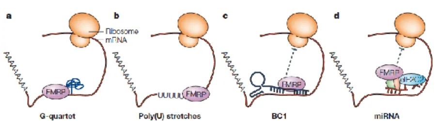
proposed. FMRP could repress translation by blocking elongation (Khandjian et al., 1996; Ceman et al., 2003; Bardoni et al., 2006). The presence of FMRP in stress granules suggest that FMRP represses translation throughout blocking translation initiation (see more ahead). Studies of co-sedimentation have found an association of FMRP with polyribosomes, with BC1 (brain cytoplasmic RNA 1), with CYFIP1 (cytoplasmic FMRP interacting protein) and with translation initiation factors (Centonze et al., 2008; Gabus et al.. 2004; Johnson et al., 2006; Lacoux et al., 2012; Laggerbauer et al., 2001; Napoli et al., 2008; Zalfa et al., 2007); in this model FMRP represses translation by inhibiting cap-dependent initiation (Napoli et al., 2008). Other data have suggested that association with the microRNA (miRNA) machinery may be involved too. miRNAs are small noncoding RNAs that inhibit translation by association with an RNA-induced silencing complex (RISC). FMRP associates with miRNAs, with RISC proteins and with the mammalian ortholog of Argonoute 1 (AGO1) that work together to silence mRNAs, either by direct cleavage of transcripts or by translational repression (Jin et al., 2004; Didiot et al., 2009).
Figure 4. FMRP recognizes different RNA sequences. G-quartet structure (a), or poly(U) stretch (b) to bind directly mRNA; BC1 (c) or miRNAs (d) to bind
indirectly mRNA. From Claudia Bagni and William T. Greenough, 2005
Although the exact mechanism by which FMRP stalls ribosomes remains elusive, several authors suggest that it is a dynamic and reversible mechanism related with plastic changes occurring both in the cytoplasm and at synapses (Darnell et al., 2011). Interestingly, FMRP may promote and not only inhibit translation of target
24
mRNAs, such as Trailer-Hitch (protein TRAL) and Superoxide Dismutase 1 (SOD1) transcripts (Bechara et al., 2009; Monzo et al., 2006). Thus, the translation and expression of FMRP targets can be either positively or negatively affected by FMRP expression, indicating that the potential role of FMRP as a translational regulator is much more complex than it was originally believed. In the regulation of FMRP function several post-translational modifications give also a contribute. FMRP is normally methylated in RGG box, mainly on 544 arginine residues and this methylation seems to regulate its protein-protein and protein-RNA interactions (Dolzhanskaya et al., 2006; Stetler et al., 2006). FMRP can also be phosphorylated on a series of serine on the N-terminal side, mainly at serin 499. It has been suggested that the phosphorylated form of FMRP is associated to stalled polyribosome and in this state the association of FMRP with its mRNA is preserved; whereas the unphosphorylated form of FMRP has been observed in association with actively translating polysomes (Ceman et al., 2003; Coffee et al., 2011; Muddashettyet al., 2011). Thus, also phosphorylation of FMRP is implicated in FMRP-mediated protein synthesis.
25
1.7 FXR1P and FXR2P
Fragile X related protein 1 and 2 (FXR1P and FXR2P), like their autosomal homolog FMRP, are RNA binding proteins which belong to “FXR family”. They are encoded by autosomal genes, named FXR1 and FXR2, which are located in the chromosomal regions 3q28 and 17p13.1, respectively (Coy et al., 1995). FXR1P and FXR2P show a 86% of similarity with FMRP and a similar FMRP structure, being characterized by the presence of two KH, one RGG box RNA binding domains and nuclear localization and export signals (NLS and NES), suggesting that these two proteins may play a similar role than FMRP (Siomi et al., 1995; Zhang et al., 1995; Eberhart et al., 1996; Corbin et al., 1997; Feng et al., 1997). FXR proteins are also present in brain, testis and skeletal muscle tissue of mice (Bakker et al., 2000). The brain and testis isoforms of FXR1P have a molecular mass of 70 and 78 kDa, 80-84 kDa in skeletal muscle, instead FXR2P is of 95 kDa (Bakker et al., 2000; Khandjian et al., 1998; Zhang et al., 1995). In the absence of FMRP, like in Fmr1 KO mice, no change in FXR1P and FXR2P distribution was observed; it is possible that both proteins compensate for the absence of FMRP, which would suggest functional redundancy (Bakker et al., 2000), but this aspect is controversial (Coffe et al., 2010). FXR1P, FXR2P with FMRP are highly expressed in the neurons of adult human brain (Tamanini et al., 1997). They are mainly localized to the cytoplasm associated with polyribosomes, but they are also present in the nucleus; for example, they have been observed in the nucleus of hippocampal neurons (Bakker et al., 2000). They also shuttle between the nucleus and cytoplasm, and FXR2P and certain isoforms of FXR1P show a nucleolar localization signal (NoS) and shuttles between the cytoplasm and the nucleolus (Tamanini et al., 1999, 2000). These data suggest that while the FXR proteins are associated in the cytoplasm with FMRP in an mRNP, they may be playing different functional roles in the nucleus.
FMRP interacts with the two paralogs FXR1P and FXR2P, although the significance of this interaction is not clear. It has been proposed that the interaction with these proteins might modulate the RNA binding function of FMRP, for
26
istance FXRP1 can modulate the FMRP affinity for the G-quartet RNA structure (Bechara et al., 2007). Using an in vitro RNA binding assay, Bechara and collaborators have indeed showed that the brain isoforms of FXR1P negatively regulate the affinity of FMRP for target RNAs, suggesting that FMRP works in the context of its multimolecular complex (Bechara et al., 2007).
Figure 6. FXR family. From Pop et al., 2013
1.8 FXS mouse model
One of the most important advancement for the investigation and a better understanding of the molecular mechanisms implicated in FXS is represented by the development of FXS animal models. For these purposes, mouse and
Drosophila melanogaster genetic models organisms have been generated. Both the
fly and the mouse models exhibit a phenotype with similarities to humans (O’Donnel and Warren, 2002). FMR1 gene is highly conserved between human and mouse: the murine Fmr1 gene shows nucleotide sequence homology of 95% to human gene, the CGG repeat in the promoter region (Ashley et al., 1993) and a similar temporal tissue expression. Also protein sequence show a 97% of similarity between mouse and human (Hinds et al., 1993; Hergersberg, 1995). The mouse model is not a perfect representation of the human desease because it lakcs the trinucleotide repeat expansion mutation in FMR1 gene, without which cannot mimic the timing of methylation and inactivation of the FMR1 gene seen in
27
humans; however, it is characterized by a total loss of FMRP that is sufficient to cause the FXS phenotype. In the 1994, a consortium of different labs created an accurate animal model for the human condition: by inserting a neomycin cassette, that causes gene inactivation, into exon 5 of the murine gene by homologous recombination (The Dutch-Belgian fragile X consortium, 1994; O’Donnel and Warren, 2002). The Fmr1 KO mouse lacks normal Fmr1 RNA and FMRP in any of the tissues. Adult mutant mice show symptoms similar to those found in the human syndrome: significant macroorchidism, hyperactivity, spatial learning defect, altered sensorimotor integration (The Dutch-Belgian fragile X consortium, 1994; Van Dam et al., 2000; Chen and Toth 2001). Furthermore, Fmr1 KO mice brains, like those of FXS patients, show an increased density of long and tortuous dendritic spines suggesting a delay in spine maturation (Dolen et al., 2010). To evaluate aspects of cognition and behavior in mice is particular difficult, but to overcome this issue a number of paradigms have been designed like approved sostitutes of IQ test (Crawley and Paylor, 1997). For istance, Morris water maze and radial arm maze are both test to measure spatial learning in which Fmr1 KO mouse shows a defect (The Dutch-Belgian fragile X consortium, 1994; D’Hooge et al., 1997; Peier et al., 2000; Van Dam et al., 2000). Fmr1 KO mice also exhibit increased susceptibility to audiogenic seizures (AGS) (Musumeci et al., 2000), which is specifically reverted by the introduction of constructs codifying the human FMR1 gene (Musumeci et al., 2007). Fmr1 KO mice is currently considered one of the leading animal models of autism (Bernardet and Crusio, 2006).
To study the function of FXR2P and FXR1P and their possible implication in FXS,
Fxr1 and Fxr2 KO mouse models have been also generated. Fxr1 KO mice display
a phenotype completely different to those observed in Fmr1 KO mice because they show abnormalities in muscle development. Fxr1 KO neonates die early by cardiac or respiratory failure; this mouse model expresses very low levels of FXR1P, reduced limb musculature and has a reduced life span, suggesting a role for Fxr1 gene in muscle mRNA transport/translation control similar to the function of Fmr1 gene in neuronal cells but in muscle instead of neurons (Mientjes et al., 2004). In contrast, Fxr2 KO mouse shows no pathological defects in brain or testes, and a lot
28
of similarities with Fmr1 KO mouse (Bontekoe et al., 2002); these mice are hyperactive in the open-field test, show learning defect in the Morris water maze task, have reduced levels of prepulse inhibition, display less contextual conditioned fear and are less sensitive to a heat stimulus (Bontekoe et al., 2002). These features suggest a similar but not identical function for the Fmr1 gene and the Fxr2 gene. A double Fmr1/Fxr2 KO has also been created. These double Fmr1/Fxr2 KO mouse exhibits an exaggerated behavioural phenotype in open-field activity, prepulse inhibition of acoustic startle response and contextual fear conditioning when compared with Fmr1 KO mice, Fxr2 KO mice or WT (Spencer et al., 2006). This is in line with the hypothesis that these homologous genes play a similar role in the regulation of locomotor activity, sensorimotor gating and cognitive processes (Zhang et al., 2009).
Also Drosophila melanogaster is often used like a valuable FXS model. In the fruit fly the dFMR1 gene, a structurally and functionally well-conserved ortholog for the human FMR1 gene is present (Wan et al., 2000). Several studies on Drosophila
melanogaster have provided insight into the molecular biology of FXS (Zhang et
al., 2001; Dockendorff et al., 2002; Inoue et al., 2002; Morales et al., 2002). To summarize, many animal models have been created to study FXS, which have proved really useful and without which it would be impossible obtain a clear dissection of the molecular, physiological, cognitive, and behavioral phenotypes of FXS.
29
2. METABOTROPIC GLUTAMATE RECEPTORS
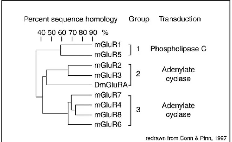
Most evidence over the past 15 years supports a role of group I metabotropic glutamate (mGlu) receptors in the pathophysiology of FXS. Metabotropic glutamate (mGlu) receptors are key players in excitatory transmission and important regulators of synaptic plasticity. Glutamate, the major excitatory neurotransmitter in the mammalian central nervous system (CNS), exerts its action interacting with ionotropic (iGlu) and metabotropic (mGlu) receptors. iGlu receptors are multimeric ion channels responsible for fast synaptic transmission and are subdivided into three distinct subtypes: AMPA (α-amino-3-hydroxy-5-methyl-4-isoxazolepropionic acid), kainate (KA), and NMDA (N-methyl-D-aspartate) receptors (Monaghan et al., 1985; Cincotta et al., 1989). mGlu receptors are members of a C G-protein-coupled receptor (GPCR) superfamily that consists of eight receptor subtypes (mGlu1-mGlu8) categorized into three groups, Group 1, 2 and 3, on the basis of their sequence homology, G-protein coupling specificity similarities and different pharmacological response (Abe et al., 1992; Nakanishi, 1992; Tanabe et al., 1992; Conn and Pin, 1997; Nicoletti et al., 2011).
Group I mGlu receptors includes mGlu1 and mGlu5 receptor subtypes which are coupled to Gq/G11 proteins and are mainly localized at postsynaptic level. Their stimulation activates phospholipase C (PLC) that hydrolyzes phosphatidylinositol-4,5-bisphosphate (PIP2) with consequent production of inositol-1,3,4-trisphosphate
(IP3) and diacylglycerol (DAG), responsible for the release of intracellular Ca2+ and
activation of protein kinase C (PKC) respectively (Kawabata et al., 1996; reviewed by Hermans and Challiss, 2001).
Group II and group III include mGlu 2, 3 and mGlu 4, 6, 7, 8 receptor subtypes respectively, they are coupled to Gi/Go proteins and are mostly presynaptic. Their activation negatively regulates adenylyl cyclase activity and voltage-sensitive Ca2+
channels. Because of their distribution, mGlu1 and mGlu5 receptors generally modulate postsynaptic efficacy, instead mGlu 2, 3, 4, 7, and 8 receptors regulate neurotransmitter release (Luján et al., 1997; Schoepp, 2001).
30
Figure 7. Classification of mammalian mGlu1-8 receptors. From Dolen et al., 2010
Although hydrolysis of PIP2 represent the canonical trasduction pathway coupled to
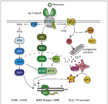
mGlu5 receptor by PLC, actually mGlu5 receptor signalling occurs also throught other two cascades: ERK cascade and PI3K/Akt/mTOR cascade.
In the first case, the tyrosine kinase Src phosphorylates and activates MEK kinase, which in turn phosphorylates and activates ERK (also known as microtubule associated protein kinase, MAPK) (Ferraguti et al., 1999; Garcia et al., 2008; Dolen et al., 2010). In the second case, phosphorylation of the phosphoinositide 3-kinase (PI3K) activates Akt (also known as protein 3-kinase B, PKB), which turns on the mammalian target of rapamycin (mTOR) (Ronesi and Huber, 2008; Dolen et al., 2010). ERK1/2 MAP kinase and/or PI-3-K pathways are involved in cell proliferation, differentiation, survival, and in synaptic plasticity (Ferraguti et al., 1999; Rong et al., 2003). The activation of these pathways directly or indirectly regulates protein synthesis; for istance, activation of mTOR phosphorylates 40S ribosomal protein S6 kinase (Hou and Klann, 2004; Klann and Dever, 2004; Banko et al., 2006; Antion et al., 2008), ERK is responsible for the phosphorylation of the eukaryotic initiation factor 4E (eIF4E) (Banko et al., 2006). Both activated mTOR and ERK phosphorylate eIF4E binding proteins (4E-BPs) (Klann and Dever, 2004). The binding of glutamate to mGlu5 receptor stimulates translation of several mRNAs with consequent increase in protein synthesis throught the activation of
31
these pathways (Proud, 2007; Klann and Dever, 2004;). The activation of ERK and PI3K requires the interaction of group-I mGlu receptors with Homer proteins, a class of scaffolding proteins that cross-link group-I mGlu receptors to inositol triphosphate (IP3) receptors and to other proteins of the post synaptic density such as SHANK (Tu et al., 1998, 1999; Rong et al., 2003; Mao et al., 2005). Homer proteins are also involved in the regulation of several properties of group-I mGlu receptors functions such as constitutive activity (Ango et al., 2001), cell surface expression and trafficking (Coutinho et al., 2001; Ango et al., 2002;), lateral mobility (Sergé et al., 2002) and coupling to ion channels of the cytoplasmic membrane (Kammermeier et al., 2000).
Figure 8. Signaling cascades coupling to group-I mGlu receptors. From Dolen et al., 2010
mGlu1 and mGlu5 receptors show different regional and developmental expression profiles; mGlu5 receptor expression is elevated in hippocampus, neocortex and striatum (Shigemoto, 2000) and its expression is elevated during the first three
32
postnatal weeks while declines afterwards; mGlu1 receptors is maximally expressed in the cerebellum (Catania et al., 1994; Catania et al., 2007, reviewed in D’Antoni et al., 2014), it is also expressed in olfactory bulb, thalamus, and pars compacta of the substantia nigra. Its expression is higher in adulthood, instead it is barely expressed during early development (Lopez-Bendito et al., 2002). Both mGlu1 and mGlu5 receptors are present in cortical and hippocampal interneurons (van Hooft et al., 2000), where they partecipate to regulation of brain connectivity. mGlu5 receptors are also found in astrocytes under physiological and pathological conditions (D’Antoni et al., 2008), but they are also present in oligodendrocytes, microglia, stem progenitor cells, and a variety of peripheral cells (Nicoletti et al., 2011). In dendritic spines, mGlu1 and mGlu5 receptors are localized in the perisynaptic region (Baude et al., 1993; Nusser et al., 1994), but are also present at extrasynaptic sites with a higher frequency for mGlu5 than mGlu1 receptors (Lujan et al., 1997). Expression studies of mGlu receptors suggest that mGlu5 receptors have a crucial role in plastic remodelling during post-natal development (Catania et al., 2007).
2.1 Metabotropic glutamate receptors and FXS
In the last years, several evidence suggested a strong implication of mGlu5 receptor in the pathogenesis of FXS. An interplay between FMRP and mGlu5 receptor has emerged, particularly the role of mGlu5 receptor in several FMRP-mediated function. (Huber et al., 2002; Bear et al., 2004). The first indication for a link between mGlu5 receptor and FXS was the evidence that activation of group-I mGlu receptors in synaptoneurosomes stimulates the rapid translation of pre-existing mRNA, including the mRNA encoding FMRP (Weiler et al., 1997). Furthermore the activation of group-I mGlu receptors is necessary for FMRP trafficking from the cell body into dendrites (Antar, 2004) and enhances the dendritic transport of several FMRP target mRNAs, such as Map1b, CaMKII and also FMRP mRNA, in hippocampal cultured neurons (Antar et al., 2004;
33
Dictenberg et al., 2008; Ferrari et al., 2007). Dictenberg showed that, upon stimulation with the selective agonist of group-I mGlu receptors (S)-3,5-Dihydroxyphenylglycine (DHPG), FMRP interacts more efficiently with kinesin and that this group-I mGlu receptor mediated transport is substantially attenuated in the absence of FMRP. These data suggest that, as a consequence of the lack of FMRP, levels and distribution of several synaptic and non synaptic proteins are altered and also that key biochemical pathways might be dysregulated in FXS. Subsequent studies revealed the finding that group-I mGlu receptors has an influence on LTD and LTP. In particular, group-I mGlu receptor-dependent long term depression (LTD), that requires mGlu5 receptor activation and local protein synthesis (Huber et al., 2000), is increased in Fmr1 KO hippocampus, whereas NMDA receptor-dependent LTD is not (Huber et al., 2002). Curiously, mice lacking mGlu5 receptor show impaired learning and reduced LTP in the hippocampal CA1 region (Lu et al., 1997). Hippocampal epileptogenesis, another form of synaptic plasticity that depends on group-I mGlu receptor activation and protein synthesis, is also altered in Fmr1 KO mice (Chuang et al., 2005; Dolen et al., 2010). Furthermore, the mGlu receptor-dependent LTD in Fmr1 KO is insensitive to inhibitors of protein synthesis (Hou et al., 2006; Nosyreva and Huber, 2006), suggesting that the abnormal expression of synaptic proteins alters long-term responses to mGlu5 receptor activation in FXS. The finding that mGluR-LTD is exaggerated in Fmr1 KO mice suggested that FMRP and mGlu5 receptor might work in functional opposition, where mGlu5 receptor activates protein synthesis and FMRP suppresses it (Dolen and Bear, 2008). Excitement for the proposed mechanistic link between FMRP and mGlu5 receptor in the regulation of protein synthesis, led Bear and collaborators to formulate the “mGlu theory” of FXS, which postulates that mGlu5 receptor and FMRP regulate translation of mRNAs at the synapse in a functionally opponent manner: activation of mGlu5 receptors stimulates protein synthesis and FMRP blocks it. In the absence of FMRP, like in FXS, mGlu5 receptor-dependent protein synthesis proceeds unchecked, and consequent excessive translation leads to development of FXS clinical features. According to ‘the mGlu Theory”, this defect can be corrected
34
using mGlu5 receptor antagonist, like MPEP, or by genetic reduction of mGlu5 receptor activity (Bear et al., 2004; Dolen and Bear, 2008).
Figure 9. The mGluR theory: Opponent regulation of protein synthesis by group-I mGlu receptor and FMRP is disrupted in the absence of FMRP. Reduction of mGlu
receptor signaling restores the balance and corrects FXS phenotype. From Dolen et al. 2010
To validate “The mGluR Theory”, Dolen and Bear generated double mutant mice by crossing Fmr1 mutant mice with Grm5 mutant mice, the gene that encodes for mGlu5 receptor. In this study they observed that 50% genetic reduction of mGlu5 receptor is able to rescue Fmr1 KO phenotypes to levels closer to WT; the mGlu5 receptor genetic reduction in the Fmr1KO/Grm5 heterozygote rescued altered ocular dominance plasticity, increased density of dendritic spines, increased basal protein synthesis, audiogenic seizure susceptibility, but not macroorchidism. These data confermed the opponent regulatory role for mGlu5 receptor and FMRP (Dolen et al., 2007; Dolen and Bear, 2008; Dolen et al., 2010).
FMRP suppresses translation of several proteins implicated in mGlu-LTD; upon group-I mGlu receptor activation, FMRP is dephosphorylated, ubiquitinated and degraded. It is known that when FMRP is associated with its mRNAs is in the phosphorylated form (Ceman et al., 2003; Narayanan et al., 2007; Bassel and
35
Warren, 2008). Activation of mGlu5 receptors stimulates FMRP dephosphorylation by activation of protein phosphatase 2A (PP2A), an FMRP phosphatase that can rapidly dephosphorylate FMRP in response to stimulation. Unphosphorylated FMRP loses the affinity for its mRNAs with consequent increase in translation (Narayanan et al., 2007; Bassel and Warren, 2008). This derepresses translation of FMRP mRNA targets contributes to rapid translational activation of proteins necessary for LTD such as Arc (Nalavadi et al., 2012; Niere et al., 2012). Recently, 831 mRNAs directly interacting with FMRP have been identified; among these 1/3 encode synaptic proteins and mGlu5 mRNA is higly represented (Darnell et al., 2011). These data all together illustrate that group-I mGlu receptor dysfunction is a large contributor to the pathophysiology of FXS (reviewed by D’Antoni et al., 2014).
3. THERAPEUTIC STRATEGIES IN FXS
Over the years, several studies have been aimed at ultimately achieving of a good treatment for FXS, which is currently merely symptomatic. However, the knowledge of countless targets involved in FXS suggests that is not simple to find a single therapy. Unfortunately, there is not yet a treatment to compensate the absence of FMRP and the therapy commonly used is designed specifically for each patient and is based on his/her specific behavior symptoms (reviewed by Penagarikano et al., 2007). One potential therapeutic approach in FXS consists in the reactivation of the silenced FMR1 gene to restore the production of FMRP (Chiurazzi et al., 1999; Pietrobono et al., 2002). For this purpose two compounds have been suggested, such as 5-Azadeoxycytidine (Chiurazzi et al., 1999; Tabolacci et al., 2005) and valproic acid (Tabolacci et al., 2008). Unfortunaly, reactivation processes are too general and not specific for FMR1 gene and also toxicity of these approaches is too high. The possibility of gene therapy is currently not possible due to difficulties in the restoration of the normal gene into neurons
36
(reviewed by Penagarikano et al., 2007). Thus, common medications include stimulants, antipsychotic, anti-depressant and anticonvulsant. Patients with FXS also seem to benefit from behavioral intervention and special educational programs. As demonstrated in the FXS mouse model, an enriched environment can improve behavior, and thus this therapy might also be beneficial for patients (Restivo et al., 2005).
A Significant progress in the treatment of FXS was obtained by understanding the mGlu5 receptor role in the pathophysiology of FXS. Many studies have been aimed use of drugs to correct the abnormal activity of the mGlu receptor in FXS using specific mGlu5 receptor antagonists and nowadays mGlu5 receptor is considered a valid target to treat FXS. The first potent and selective, noncompetitive antagonist 2-methyl-6-(phenylethynyl)-pyridine (MPEP) was used in several drug discovery programs in industrial and academic research laboratories (Pop et al., 2013). Treatment with MPEP resulted in suppression of audiogenic seizure susceptibility in Fmr1 KO mice (Chuang et al., 2005) and reduction in repetitive-like behavior (Thomas et al., 2012). These data indicate that the interaction between mGlu receptor signaling and FMRP function is responsible for some of the symptoms associated with FXS (reviewed by Penagarikano et al., 2007). Another drug commonly used is Fenobam; previously investigated as an anxyolitic, was later tested as a negative modulator of mGlu5 receptor (Porter et al., 2005). Beneficial effects included reduced anxiety and improvement of prepulse inhibition (Berry-Kravis et al., 2009). Both MPEP and fenobam restored dendritic spine morphology in hippocampal cell cultures from Fmr1 KO mice (De Vrij et al., 2008). Recently, another novel mGlu5 receptor antagonistis, CTEP, has proven effective in restoring cognitive defects, auditory seizures, abnormal dendritic spine density; it is also able to stimulate ERK and mTOR signaling and partially to correct macroorchidism (Michalon et al., 2012). mGlu5 receptor antagonists appeared promising during preclinical studies; however preclinical studies did not translate into a broadly effective treatment for FXS. mGlu5 receptor antagonists, such as AFQ056 compound by Novartis, have been tested in clinical studies; however, this trial has been discontinued after Phase II because treatment
37
did not improve phenotypes or showed side effects. Currently a few preclinical and clinical studies for FXS are still in progress, for example RO4917523, another mGlu5 receptor negative modulator is in trials by Roche in U.S.A. Further experiments should help to understand discrepancies between outcomes obtained in pre-clinical and clinical studies using mGlu5 antagonists.
Another approach aimed to reduce excessive mGlu5 receptor signaling inhibiting glutamate release via the presynaptic activation of GABAB receptors. (Dolen et al.,
2010; Pop et al., 2013). The most frequent GABAB receptors agonist used are
baclofen and its enantiomer arbaclofen. Additional approaches consist of drugs like lithium, that reduces group-I mGlu receptor activity by attenuating GSK3β activity and probably phosphatidyl inositol turnover. Also Minocycline, an antibiotic that inhibits MMP9 (Matrix metallopeptidase 9), normalizes dendritic spine phenotypes and improves anxiety and exploratory behavior in the Fmr1 KO (Bilousova et al., 2009). Finally, a preclinically intervention in the Fmr1 KO mouse model, aim to modulate intracellular targets such as PI3K, (Gross et al., 2010), mTOR (Hoeffer et al., 2012), or MEK to investigate the effects of new agents without compromising patient safety (Wang et al., 2012). These agents represent potential target in emerging cellular models to realize the reprogramming of patient tissue samples in inducible pluripotent cells with a subsequent differentiation in neuronal cells (Sheridan et al., 2011).
Figure 10. Candidate drugs in preclinical and clinical trials for the treatment of FXS. Form Berry-Kravis, 2014
38
4. OXIDATIVE STRESS AND FRAGILE X SYNDROME
Several evidences suggest a role of oxidative stress in FXS. Oxidative stress is defined as damage to cellular tissue caused by free radicals such as reactive oxygen species (Maurin et al., 2014). Oxidative stress is implicated in a wide variety of neurodegenerative disorders and psychiatric diseases such as autism (Chauhan and Chauhan, 2006; James et al., 2006; Rossignol and Frye, 2012). In FXS an increased sensitivity to oxidative stress has been detected, with possible impacts on neuronal and glial function (Davidovic et al., 2011). For istance, in the brain of Fmr1 KO mice higher levels of reactive oxygen species, nicotinamide adenine dinucleotide phosphate (NADPH)-oxidase activation, lipid and protein oxidation have been found (El Bekay et al., 2007). This suggests that the oxidative stress in the brain may play a role in the pathophysiology of FXS. Recently, a study of metabolomic analysis performed on different brain regions of 12-day-old newborn Fmr1 KO mice, has identified multiple metabolic abnormalities in Fmr1 KO mice brains. Metabolites implicated in neurotransmission, osmoregulation, energy metabolism and oxidative stress response are altered (Brown et al., 2001; Miyashiro et al., 2003; Davidovic et al., 2011). Fmr1 KO mice show altered mRNA profiles in glutathione transferase and SOD1; levels of SOD1 protein are reduced in the absence of FMRP suggesting that increased oxidative stress in Fmr1 KO brain might be due to the altered SOD1 expression (Bechara et al., 2009).
In addition, FXS patients display an increase in adrenocortical activity and an altered hypothalamic-pituitary-adrenal (HPA) axis (Hessl et al., 2004); adrenal hormones is a source of oxidative stress in the brain, causing oxidation of molecules and depletion of antioxidants such as glutathione (Herman & Cullinan, 1997).
Anti-oxidant agents may be useful in the treatment of FXS and are supported by recent results obtained in Fmr1 KO mice after treatment with alpha-tocopherol and melatonin (de Diego-Otero et al., 2009; Romero-Zerbo et al., 2009). Chronic pharmacological treatment with alpha-tocopherol reverses free radical overproduction, oxidative stress, macroorchidism, and also behaviour and learning
39
deficits (de Diego-Otero et al., 2009). Chronic administration of melatonin protects
Fmr1 KO mouse from the oxidative stress reverting several behavioural and
learning deficits, normalizes free-radical production in macrophage cells and brain slices, and normalizes carbonyl content in proteins and lipid peroxidation (Romero-Zerbo et al., 2009). As mentioned before, a promising clinical trials in FXS is based on minocycline treatment (Leigh et al., 2013). This drug acts like an inhibitor of metalloproteases, the expression of which is increased in FXS cells (Siller and Broadie, 2011).
5. STRESS GRANULES (SGs)
Stress granules (SGs) are multimolecular cytoplasmic aggregates composed of non-translating messenger ribonucleoproteins (mRNPs) that rapidly aggregate when cells are exposed to adverse environmental conditions (Kedersha et al., 2005).
The first evidence about SGs dates back to 1989, when they were observed in tomato cell cultures (Nover et al., 1989). Subsequently, reversible aggregates of mRNPs were discovered in yeast (such as Saccharomyces pombe), protozoa (Trypanosoma brucei) and metazoa (such as Homo sapiens and Caenorhabditis elegans. At the beginning, SGs were described as large cytoplasmic mRNA aggregates, microscopically visible only in response to different types of stress. Then, it was discovered that SGs are composed not only by mRNAs, but also by abortive preinitiation translation complexes and RNA binding proteins (Stoecklin and Kedersha, 2013). Different types of stress such as heat shock, oxidative stress, UV irradiation and viral infection cause polysomes disassembly and therefore inhibit translation and promot SGs formation (Anderson and Kedersha, 2002). Translational initiation is regulated through different pathways, but under stress condition the pathways most frequently involved is the phosphorylation of the eukaryotic translation initiation factor 2 alpha (eIF2α). eIF2α phosphorylation is
40
the trigger for SGs formation after which several RNA binding protein are recruited to contribute to SGs formation (Kedersha et al., 2013). SGs show a dynamic nature that suggests that they are sites of mRNA triage, wherein mRNAs are arranged for storage, degradation, or translation during stress and after during recovery. SGs are formed to protect cells from stress and to favore cell survival by synthesizing stress protective proteins, such as heat shock proteins, and transiently blocking house-keeping proteins translation (Anderson and Kedersha, 2008). It has been established that more than 100 proteins regulate SGs assembly (Ohn et al., 2008), suggesting that SGs are the main sites that under stress condition play in helping cells respond to adverse environmental conditions. Furthermore, the cytoprotective effects of SGs is corroborated by the finding that multiple interventions that prevent SGs assembly are associated with higher cells susceptibility (Arimoto et al., 2008; Buchan and Parker 2009; Kedersha et al., 2013), but specifically how SGs exert a protective role is obscure.
5.1 SGs assembly
SGs formation is a complex mechanism related to stalled translational initiation, polysome disassembly and mRNPs aggregation, which occurs through a number of reversible steps. It is possible to identify different phases in the process of SGs assembly:
1) SGs initiation
Generally, the first step in SGs formation is the phosphorylation of eIF2α. Eukaryotic cells express a family of eIF2α kinases (eg, PKR, PERK-PEK, HRI, GCN2) that are activated in response to distinct types of environmental stress (Anderson and Kedersha, 2002). Of these kinases, PKR (protein kinase R) is a double-stranded RNA-dependent kinase activated by viral infection, heat, UV irradiation and oxidative stress (Williams, 2001), whereas PERK (PKR like endoplasmic reticulum kinase, also called PEK, or pancreatic eIF2α kinase) is activated by endoplasmic reticulum stress, such as unfolded proteins accumulated
41
in the ER lumen (Harding et al., 2000). HRI (heme-regulated initiation factor 2α kinase) is activated by oxidative stress and regulates changes in the availability of heme during erythrocyte differentiation (Han et al., 2001; Lu et al., 2001; McEwen et al., 2005; Anderson and Kedersha 2008); GCN2 (general control nonderepressible 2) is a protein that controls amino acid levels in the cell and responds to amino acid deprivation (Wek et al., 1995). Activation of one or more of these eIF2α kinases results in the phosphorylation of eIF2α at serine 51, a crucial component of the ternary eIF-2 complex that loads the initiator tRNA (Met-tRNAMet) onto 40S ribosomal subunit to initiate protein translation.
Phosphorylation of eIF2α results in abortive initiation complexes with consequent arrest in translation initiation (Anderson and Kedersha, 2002). Generally, the ternary complex is composed by eIF2αβγ bound to tRNAiMet and GTP that loads
initiator tRNAiMet onto the small ribosomal subunit (40S) to assembly the 43S
preinitiation compex. The 43S complex, with other initiation factors (eg, eIF4E, eIF4G, and poly(A)-binding protein), recruits a 7-methyl guanosine-capped mRNA to makes up the canonical 48S preinitiation complex. At this point, the 48S complex scans the 5’ untranslated region (UTR) of the mRNA transcript, starting by classical AUG codon that is recognized by the tRNAMet anticodon. Recognition
of the initiation codon triggers hydrolysis of eIF2-GTP, a reaction catalyzed by eIF5. After this event, eIF2–GDP, eIF3, eIF5, eIF1A dissociate from the 40S subunit, and the 60S ribosomal subunit can take part to form a functional 80S ribosome (see figure 11A) (Anderson and Kedersha, 2008). Under stress condition, phosphorylated eIF2α inhibits eIF2B function, the GTP/GDP exchange factor that converts inactive ternary complex (GDP-associated) to active ternary complex (GTP-associated) (Krishnamoorthy et al., 2001). Thus, eIF2α phosphorylation inhibits protein synthesis by reducing the availability of the active eIF2-GTP-tRNAiMet ternary complex that is required for cap-dependent translation initiation
(Anderson and Kedersha, 2002). When ternary complex levels are reduced, TIA-1 and TIAR (RNA-binding proteins) promote the assembly of a noncanonical preinitiation complex that lacks eIF2-GTP-tRNAiMet becoming SGs core proteins.