UNIVERSITY OF CATANIA
International Ph.D. Program in Neuropharmacology
XXVIII Cycle
TARGETING DOPAMINE D3 RECEPTOR:
NEW INSIGHTS INTO THE
PATHOPHYSIOLOGY OF ALCOHOL
ADDICTION AND ANXIETY
PhD Thesis
Sebastiano Alfio Torrisi
Coordinator: Prof. Salvatore Salomone Tutor: Prof. Salvatore Salomone Co-Tutor: Prof. Gian Marco Leggio
The experiments for this PhD thesis were carried out in the
laboratories of:
Home institute:
Prof. Filippo Drago
Department of Biomedical
and Biotechnological Sciences
Section of Pharmacology
Via S. Sofia, 64
95125
University of Catania
During the 4
thyear of my PhD program, I spent six months
working as visiting PhD student at University of Cambridge.
I worked on an original project entitled: Noradrenergic
mechanisms in the nucleus accumbens shell: regulation of
impulse control and the transition to compulsive behaviour.
Guest institute:
Dr. David Belin
University of Cambridge,
Department of Psychology
Downing Street
Cambridge CB2 3EB
Table of contents
Acknowledgements
9
List of abbreviations 11
Preface 13
Chapter I General Introduction
1. Dopaminergic system in the central nervous system 17
2. Dopamine D3 receptor 20
3. Dopamine D3 receptor and alcohol addiction 22 4. Dopamine D3 receptor and anxiety 26 5. Design of the present research 30
Chapter II 31
Chapter IV
6. General Discussion
6.1 Both genetic deletion and pharmacological blockade
of D3R inhibit ethanol intake 126
6.2 Both genetic deletion and pharmacological blockade of
D3R accelerate the development of tolerance to diazepam 129
Concluding Remarks 131
Chapter V
References 135
Chapter VI
Acknowledgements
I wish to express my sincere gratitude to Professor Filippo Drago that gave me the opportunity to join his lab during these years of PhD Program and discover the fascinating and challenging world of neuroscience.
I would like to thank my tutor and mentor Professor Salvatore Salomone, a great scientist that guided and taught me during my PhD with his patience and infinite knowledge.
I would also like to express my deepest thanks to my co-tutor Professor Gian Marco Leggio. His essential support and his great knowledge allows me to reach optimal objectives during these years.
I wish to thank Dr. Francesco Papaleo that gave me the chance to join Dr David Belin’s lab in Cambridge. In this regard, I am also greatful to David and his team. I spent six months in one of the most important behavioural neuroscience laboratory in the world.
Last, but certainly not least, I would like to thank my colleagues and my family, for their love and support.
List of abbreviations
7-OH-DPAT ((+/-)-7-hydroxy-N,N-(di-n-propyl-2-aminotetralin)) AD aldehyde dehydrogenase
AD4.2 Autodock 4.2
ADE alcohol deprivation effect ADT AutoDock Tools
ANOVA analysis of variance
BDNF brain development neurotrophic factor BLA basolateral amygdala
CNS central nervous system
COMT cathecol-O-methyl transferase CSD Cambridge Structural Database D3R dopamine D3 receptor
D3R-/- dopamine D3 receptor deficient mice
DA dopamine
DID drinking in the dark DMSO dimethyl sulfoxid
DSM-IV Diagnostic and Statistical Manual of Mental Disorders, 4th edition
EPM elevated plus maze test GABA γ-aminobutyric acid
GABAA γ-aminobutyric acid receptor A
GAD generalized anxiety disorder i.p. intraperitoneal injection
ICD-11 International Classification of Diseases, 11th edition MAO monoamine oxidase
NAc nucleus accumbens
P alcohol Preferring rats PDB Protein Data Bank PKA protein kinase A
SAS-potential Solvent Accessible Surface potential SI single injection
VEH vehicle
VMAT-2 the vesicular monoamine transporter 2 VTA ventral tegmental area
Preface
The dopaminergic neurotransmission in the central nervous system (CNS) is mediated by two different classes of G protein-coupled receptors, the “D1R-like”
receptors (D1R and D5R) and the “D2R-like” receptors (D2R, D3R and D4R)
(Seeman et al., 1994). Since its discovery in the early 90’s, the dopamine D3 receptor (D3R) has aroused great interest in the scientific community. Indeed, its
limited distribution in the limbic brain areas involved in the control of cognitive and emotional functions has made this receptor a promising target for the treatment of several neuropsychiatric disorders such as drug addiction, depression and schizophrenia (Leggio et al., 2016).
Several data suggest that D3R, likely acting as autoreceptor, modulates the
activity of dopaminergic neurons throughout the mesolimbic, mesocortical and nigrostriatal dopaminergic pathways (Gobert et al., 1995; Tepper et al., 1997; Diaz et al., 2000). Yet, D3R-deficient mice (D3R-/-) ehibit extracellular levels of
dopamine (DA) twice as high as their wild-type (WT) littermates suggesting that D3R could play a inhibitory role in the control of basal extracellular DA levels
(Koeltzow et al., 1998; Joseph et al., 2002).
The central hypothesis of my PhD research project has been that D3R
exerting a pivotal role in the control of the mesolimbic dopamine pathway, is involved in the pathophysiological mechanisms subserving neuropsychiatric
disorders linked to dysfunctionality of this dopaminergic pathway. In particular, the present thesis aimed to: 1) investigate the role of D3R in the mesolimbic DA
control of ethanol reward; 2) assess the involvement of a γ-aminobutyric acid receptor A (GABAA)/D3R interaction in the mesolimbic DA modulation of
C
C
h
h
a
a
p
p
t
t
e
e
r
r
I
I
1. Dopamine system in the central nervous system
In 1957, Arvid Carlsson, a Swedish neuropharmacologist, discovered that DA was a neurotransmitter and not only the precursore of noradrenaline. Since this discovery, DA has attracted a great amount of attention leading to numerous breakthroughs in neuroscience. Four main dopaminergic pathways have been mapped in the brain: the nigrostriatal pathway originating in the substantia nigra and projecting to the dorsal striatum; the mesolimbic pathway that arises from the ventral tegmental area (VTA) and sends dopaminergic fibers to the nucleus accumbens (NAc); the mesocortical pathway that also arises from the VTA and projects to the cerebral cortex and the tuberoinfundibular pathway that connects the hypothalamus to the pituitary gland (Anden et al., 1964; Dahlstroem and Fuxe, 1964). DA activity (figure 1) is mediated by five dopaminergic receptors. These receptors are divided in two subfamilies: the D1-like receptor subtypes (D1R and
D5R) coupled to Gαs proteins and the D2-like subfamily (D2R, D3R, and D4R)
coupled to Gαi proteins (Missale et al., 1998). D1Rs and D2Rs are the most
abundant subtypes in the central nervous system, but D1R is the most widespread
(Jaber et al., 1996). D1R mRNA has been detected in striatum, nucleus
accumbens, olfactory tubercule, hypothalamus and thalamus (Jaber et al., 1996). D5R is expressed at much lower level than the D1R dopamine receptor and its
nucleus and the parafascicular nucleus of the thalamus). D2Rs are localized mainly
in striatum, olfactory tubercule, nucleus accumbens, substantia nigra pars compacta, VTA and the pituitary gland. D2Rs are both pre- and post-synaptic
receptors contrary to D1-like receptors which are mainly post-synaptic receptors
(Jaber et al., 1996). D4Rs have been found with a low expression in basal ganglia
and a higher expression in frontal cortex, medulla, amygdala, hypothalamus and mesencephalon. However, this high expression is weak in comparison with other dopamine receptors (Jaber et al., 1996). D3Rs are primarily confined in the limbic
system (nucleus accumbens, olfactory tubercule and islands of Calleja). However, D3R mRNA has been found in the medial prefrontal cortex (mPFC)–NAc–ventral
pallidum loop (Koob and Le Moal, 1997) as well as in the ventral striatal, ventral pallidal, thalamic, and orbitofrontal loops (Everitt and Robbins, 2005).
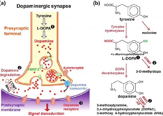
Figure 1: Dopaminergic synapse and dopamine metabolism. (a,b) In the presynaptic terminal of
dopaminergic neurons, tyrosine is converted in L-DOPA by the activity of tyrosine hydroxylase. L-DOPA is subsequently transformed to the neurotransmitter DA by action of DOPA decarboxylase. (b) DA is then transferred in vesicles by the vesicular monoamine transporter 2 (VMAT-2). After exocytosis of the DA vesicles, DA binds to DA receptors on the postsynaptic membrane, leading to the transduction of the signal in the postsynaptic neuron. (a,b) DA is then recycled by reuptake via the DA transporter, or catabolized by the action of monoamine oxidase (MAO), cathecol-O-methyl transferase (COMT) and aldehyde dehydrogenase (AD) enzymes. (modified from Jones et at., 2012)
2. Dopamine D3 receptor
In 1990, for the first time, the rat D3R was cloned and characterized
(Sokoloff et al., 1990). Among DA receptors, D3R exhibits the highest affinity for
DA (70-fold higher than D2R receptors), suggesting that DA may occupy D3R in
vivo for extended periods of time, leading to high spontaneous activation of D3R
(Richtand et al., 2001; Vanhauwe et al., 2000).
In rat, the largest D3R densities have been found in granule cells of the
islands of Calleja and in medium-sized spiny neurons of the rostral and ventromedial shell of nucleus accumbens (Diaz et al., 1994, 1995; Le Moine and Bloch, 1996). PET studies, carried out on baboons and mice by using the D2/3 PET agonist (with preferential selectivity and affinity for D3R) [11C]-(+)-PHNO,
have revealed a high expression of D3R in ventral pallidum, substantia nigra,
thalamus, and habenula (Rabiner et al., 2009). Its primary sequence is similar to that of D2R, and to a lesser extent, to the D4R. Activation of D3R expressed in a
transfected mesencephalic cell line inhibits dopamine release (Tang et al., 1994) and synthesis (O’Hara et al., 1996). Moreover, agonists with limited preference for the D3R (Sautel et al., 1995) likewise inhibit dopamine release, synthesis and
neuron electrical activity giving support to the hypothesis that D3R could operate
as autoreceptor. However, since DA agonists produce analogous inhibition of dopamine neuron activities in both WT and D3R-/- mice, their selectivity towards
of its autoreceptor function arise from immunocytochemical experiments showing that D3R is expressed in all dopaminergic neurons (Diaz et al., 2000). Yet,
dopamine extracellular levels in NAc (Koeltzow et al., 1998) and striatum (Joseph et al., 2002) are higher in D3R-/- compared to their WT littermates, suggesting a
D3R-mediated control of dopamine neurons activity. These convergent results
supported the fact that D3R-/- mice seem to be more responsive in several
physiological situations compared to their WT littermates (Le Foll et al., 2005). By contrast, the study by Simpson et al. (2014) has been demonstrated that mice with a striatal overexpression of D3Rs have less marked, but still noteworthy
phenotype. Indeed, these mice exhibit a disrupted motivation, suggesting that targeting D3R might have effect on motivational symptoms.
3. Dopamine D3 receptor and alcohol addiction
Alcohol addiction is a chronic relapsing disorder and shares many features of other chronic diseases such as hypertension and diabetes. Indeed, it is charachterized by a strong component of genetic susceptibility and is under influence of environmental factors (Heilig et al., 2011). Alcoholism produces about 10% of total disability-adjusted life years lost (measure of disease burden) in industrialized countries. Genetic and environmental factors in alcohol addiction may flow in very dissimilar sorts of vulnerability, ranging from amplified impulsivity and reward from alcohol to heightened stress responses and anxious personality traits (Goldman et al., 2005). Alcoholics are very heterogeneous in terms clinical features such as age of onset and family history (McLellan et al., 2000). The pathophysiology and etiology of alcohol addiction is still poorly understood and there are no effective pharmacological treatments.
It is well established in literature tha DA neurotransmission is involved in the pathophysiology of drug addiction. The mesolimbic DA pathway (figure 2) modulates the rewarding properties of drugs of abuse (Ikemoto and Bonci, 2013), such as ethanol and opiates (Pierce and Kumaresan, 2006). Alcohol induces an increase of DA release in the shell, but not in the core of NAc. (Bassareo et al., 2003; Cadoni et al., 2000). Moreover, in rats, intravenous administration of
a dose-dependent manner (Gessa et al., 1985). In line with these preclinical evidence, it has been reported that intoxicating doses of alcohol trigger dopamine release in the ventral striatum of humans (Boileau et al., 2003) and an activation of this brain area by alcohol-associated cues in abstinent high-risk drinkers and alcohol-dependent individuals has been found as well (Braus et al., 2001; Kareken et al., 2004). It is well demonstrated that D3R, which is highly expressed in the
shell of NAc, regulates the mesolimbic DA pathway and is involved in the neural mechanisms underlying drug seeking behavior (Heidbreder et al., 2005). Numerous studies have investigated the involvement of D3R in ethanol-drinking
paradigms (Cohen et al., 1998; Harrison and Nobrega, 2009; Heidbreder et al., 2007; Rice et al., 2012; Silvestre et al., 1996; Thanos et al., 2005). In this regard, the pharmacological manipulation of D3R seems to produce different behavioural
effects compared to the genetic manipulation of this dopaminergic receptor. Heidbreder and colleagues (2007) demonstrated that the selective D3R antagonist
SB277011A reduces alcohol intake and prevents relapse to alcohol-seeking behavior of male C57BL/6N mice exposed to oral operant self-administration. It has also been reported that the preferential DA D3R antagonist S33138 decreases
the binge drinking of ethanol without significantly affect the consumption of water (Rice et al., 2012). In agreement with these preclinical evidence, the dopamine receptor agonist with reasonable selectivity for the D3R 7-OH-DPAT
preference at the dose of 0.01 mg/kg (Silvestre et al., 1996). The study by Vengeliene et al. (2006) reported that the selective D3R antagonist SB277011A
induces a dose-dependent decrease of relapse-like drinking in the alcohol deprivation effect (ADE) model as well as a reduction in cue-induced ethanol-seeking behavior. Moreover, SB277011A significantly diminishes ethanol preference, intake and lick responses both in alcohol Preferring (P) and Non-Preferring (NP) rats tested in the two bottle choice paradigm (Thanos et al., 2005). Regarding the genetic manipulation of D3R, D3R-/- mice are resistant to ethanol
sensitization (Harrison and Nobrega, 2009) and seem to have similar levels of ethanol intake (McQuade et al., 2003) compared to their WT littermates. Despite several studies have investigated the involvement of D3R in ethanol reward, its
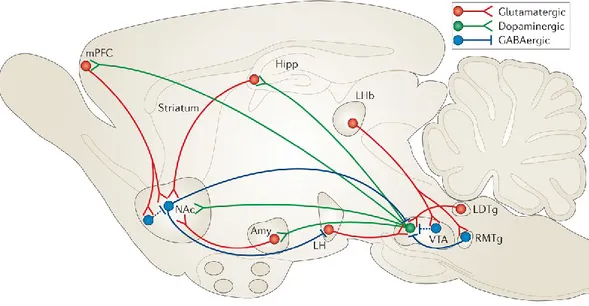
Figure 2: VTA– NAc reward circuit. The major reward circuit consists of dopaminergic fibers originating
from the VTA and projecting to the NAc (in green), which release dopamine in response to reward-related stimuli. (from Russo and Nestler, 2013)
4
.Dopamine D3 receptor and anxiety
Anxiety is a physiologic emotion under stressful and dangerous situations and is believed to be part of the evolutionary “fight or flight” reaction of survival. In many circumstances, the presence of anxiety may become maladaptive and constitutes a psychiatric disorder. By using significant prognostic tools, it is possible to classify anxiety disorders by their diagnostic subtype (obsessive-compulsive disorders, panic disorder, social phobia, generalized anxiety disorder [GAD] etc.). Diagnostic criteria for these several subtypes are given in the Diagnostic and Statistical Manual of Mental Disorders, 4th edition (DSM-IV, text
revision) and the International Classification of Diseases, 11th edition (ICD-11).
The contribution of GABA system and GABAA receptor complex in the
modulation of emotional processes is well known (Clement et al., 2002; Mehta and Ticku, 1999). However, the involvement of GABA-ergic innervation of particular brain structures in the regulation of anxiety is not satisfactorily documented. The mechanism of action of the most used anxiolytic drugs in clinic and preclinic research, benzodiazepines, relies on an enhancement in the affinity of the recognition site of GABAA receptors for GABA, ultimately potentiating its
inhibitory action in the limbic system (Mehta and Ticku, 1999; Mohler et al., 2002).
A large body of behavioral and biochemical data indicate the involvement of DA neurotransmission in the pathophysiology of anxiety (Kienast et al., 2008). It is well established that stress triggers the mesocorticolimbic DA system activation and induces an increase of DA extracellular levels in the nucleus accumbens and medial prefrontal cortex, generating anxiolytic-like behavioral effects (Cabib et al., 1994; Dunn, 1988; Salamone, 1994; Simon et al., 1993). It is also demonstrated that anxiolytic drugs, such as diazepam and ICS 205930 (Finlay et al., 1995; Imperato et al., 1990), can attenuate stress-induced increase in DA concentration. However, behavioral evidences of the involvement of D1R in
anxiety are week and inconsistent (Bartoszyk, 1998; Rodgers et al., 1994). By contrast, animal studies described the anxiolytic-like effects of D2R antagonists
such as haloperidol or sulpiride (Costall et al., 1987; Pich and Samanin, 1986). Biochemical studies have indicated that haloperidol, sulpiride and quinpirole show affinity not only for D2R but also for D3R (Sokoloff et al., 1990). It has been
demonstrated that D3R-/- mice display low levels of anxiety tested in the open field
arena and plus-maze tests (Steiner et al., 1997). Putative D3R antagonists such as
PNU-99194A and nafadotride showed anti-anxiety effects in the conflict drinking test in rats and exploration models in rats or mice (Gendreau et al., 1997; Rogoz et al., 2000). Furthermore, D3R agonists, used at low doses, have been suggested to
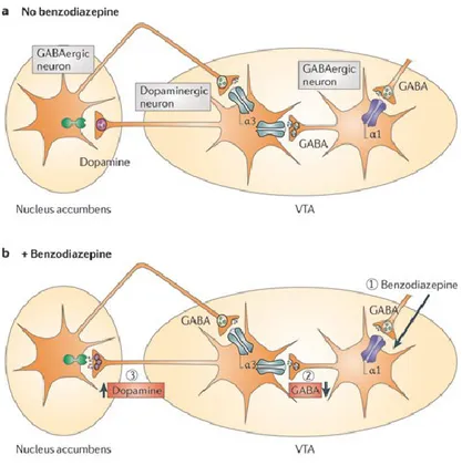
A dopaminergic-GABAergic interaction in the mesolimbic DA pathway is a well-documented phenomenon (figure 3). Indeed, binding of benzodiazepines to the α1-containing GABAA receptors on GABAergic VTA neurons leads to a
reduction of the activity of these cells and a consequent decrease of GABA release, which results in a disinhibition of the dopaminergic VTA neurons and a resulting increase in DA release in the ventral striatum (Rudolph and Knoflach, 2011). Growing data suggest a D3R mesolimbic modulation of GABA system.
Indeed, dopamine via D3R, may control the expression of innate anxiety-like
behaviors through a down-regulation of GABAergic control over lateral/basolateral amygdala neurons (Diaz et al., 2011). A dynamic-dependent inhibition of GABAA modulated by D3R receptor has also been found in NAc
(Chen et al., 2006) and hippocampus (Hammad and Wagner, 2006; Swant et al., 2008). D3R-/- mice exhibit low baseline anxiety levels and acute administration of
diazepam is more effective in D3R-/- than in WT littermates when tested in the
elevated plus maze test (EPM) (Leggio et al., 2011). However, the precise role of the D3R/GABAA systems interaction in both the modulation of anxiety-like
Figure 3: GABAA receptor subtypes in the mesolimbic dopaminergic systems. VTA GABAergic neurons
express the α1 subunit, whereas dopaminergic neurons predominantly express the α3 subunit. Binding of benzodiazepines to the α1-containing GABAA receptors on GABAergic VTA neurons leads to a reduction of
the activity of these cells and consequently to a decrease of GABA release, which in turn disinhibits the dopaminergic VTA neurons leading to an increase of DA release in the ventral striatum. In principle, benzodiazepines likely have functionally opposing actions via the α1-containing GABAA receptors on
GABAergic neurons and on α3- containing GABAA receptors on the dopaminergic neurons of the VTA.
However, the effect on the α1-containing GABAA receptors on the dopaminergic neuron is functionally
5. Design of the present research
Based on the reviewed data present in literature, the aim of the present thesis has been to assess the role of D3R in the mesolimbic DA control of ethanol reward
and to evaluate the recruitment of GABAA/D3R interaction in the mesolimbic DA
modulation of anxiety-like behavior. The following aspects were investigated: 1. Evaluating the basal behavior of D3R-/- mice and their WT littermates in
experimental models of anxiety and ethanol reward [two bottle choice, drinking in the dark (DID) and EPM]
2. Assessing the behavioural response of D3R-/- mice and their WT littermates
to selective D3R antagonists, at different doses and testing time in different
models of ethanol reward
3. Investigating the possible involvement of RACK1/BDNF/D3R pathway in
ethanol seeking behavior and the activation of dopaminergic neurotransmission in striatum of our mice.
4. Assessing the sensitivity of D3R-/- mice and their WT littermates tested in
the EPM test to repeated administration of diazepam.
5. Testing the hypothesis that genetic deletion or pharmacological blockade of D3R affect GABAA subunit expression.
C
Dopamine D3 Receptor Is Necessary for Ethanol
Consumption: An Approach with Buspirone
Gian Marco Leggio1, Giovanni Camillieri1, Chiara BM Platania1, Alessandro
Castorina2, Giuseppina Marrazzo1, Sebastiano Alfio Torrisi1, Christina N Nona3,
Velia D’Agata2, Jose ´Nobrega4, Holger Stark5, Claudio Bucolo1, Bernard Le
Foll6, Filippo Drago1 and Salvatore Salomone*1
1Department of Clinical and Molecular Biomedicine, Section of Pharmacology and Biochemistry,
Catania University, Catania, Italy;
2Department of Bio-Medical Sciences, Catania University, Catania, Italy; 3Behavioral Neurobiology Laboratory, Centre for Addiction and Mental Health and
Department of Pharmacology and Toxicology, University of Toronto, Toronto, Canada;
4Imaging Research Centre and Campbell Family Research Institute, Centre for Addiction and
Mental Health and Departments of Psychiatry, Psychology and Pharmacology, University of Toronto, Toronto, Canada;
5Institute for Pharmaceutical and Medicinal Chemistry, Heinrich-Heine-University Duesseldorf,
Duesseldorf, Germany;
6Translational Addiction Research Laboratory, Centre for Addiction and Mental Health and
Departments of Family and Community Medicine, Psychiatry and Pharmacology, University of Toronto, Toronto, Canada
Abstract
Mesolimbic dopamine (DA) controls drug- and alcohol-seeking behavior, but the role of specific DA receptor subtypes is unclear. We tested the hypothesis that D3R gene deletion or the D3R pharmacological blockade inhibits ethanol
littermates, treated or not with the D3R antagonists SB277011A and U99194A,
were tested in a long-term free choice ethanol-drinking (two-bottle choice) and in a binge-like ethanol-drinking paradigm (drinking in the dark, DID).
The selectivity of the D3R antagonists was further assessed by molecular
modeling. Ethanol intake was negligible in D3R-/- and robust in WT both in the
two-bottle choice and DID paradigms. Treatment with D3R antagonists inhibited
ethanol intake in WT but was ineffective in D3R-/- mice. Ethanol intake increased
the expression of RACK1 and brain-derived neurotrophic factor (BDNF) in both WT and D3R-/-; in WT there was also a robust overexpression of D3R. Thus,
increased expression of D3R associated with activation of RACK1/BDNF seems
to operate as a reinforcing mechanism in voluntary ethanol intake. Indeed, blockade of the BDNF pathway by the TrkB selective antagonist ANA-12 reversed chronic stable ethanol intake and strongly decreased the striatal expression of D3R. Finally, we evaluated buspirone, an approved drug for anxiety
disorders endowed with D3R antagonist activity (confirmed by molecular
modeling analysis), that resulted effective in inhibiting ethanol intake. Thus, DA signaling via D3R is essential for ethanol-related reward and consumption and
Keywords: Dopamine D3 receptor; Knockout mice; Animal models of ethanol
reward; Buspirone; BDNF
* Published in Neuropsychopharmacology (2014) 39, 2017–2028; doi:10.1038/npp.2014.51
1. Introduction
The mesolimbic dopamine (DA) pathway mediates the rewarding effects of drugs of abuse (Bowers et al., 2010; Ikemoto and Bonci, 2013; Koob, 1992; Robbins and Everitt, 1996; Wise and Bozarth, 1987), including ethanol and opiates (Pierce and Kumaresan, 2006; Wise and Bozarth, 1987). Both oral self-administration (Weiss et al., 1992) and systemic self-administration of ethanol increase the firing rate of mesolimbic dopaminergic neurons (Gessa et al., 1985; Mereu et al., 1984) and stimulate extracellular DA release in the striatum and in the nucleus accumbens (Imperato and Di Chiara, 1986; Yoshimoto et al., 1992). In a recent metaanalysis on published data sets of in vivo microdialysis in rat brain, the acute administrations of ethanol appear to increase the level of monoamines, including DA, globally and independent of the brain sites up to 270% of the basal concentrations (Brand et al., 2013). DA exerts its action through five receptor subtypes (D1–5R); the D3 receptor (D3R) subtype has an important role in the
modulation of the mesolimbic DA pathway and in the control of drug-seeking behavior (Heidbreder et al., 2005; Joyce and Millan, 2005). The D3R is located
both at pre- and post-synapses, in the ventral striatum (nucleus accumbens and island of Calleja) (Bouthenet et al., 1991; Murray et al., 1994); in these structures, stimulation of presynaptic D3R may modulate DA synthesis and release (Levant,
paradigms (Cohen et al., 1998; Harrison and Nobrega, 2009; Heidbreder et al., 2007; Rice et al., 2012; Silvestre et al., 1996; Thanos et al., 2005), but their precise role remains unclear. Indeed, pharmacological studies generally report that D3R blockade decreases ethanol consumption (Heidbreder et al., 2007; Rice et al.,
2012; Silvestre et al., 1996; Vengeliene et al., 2006); in contrast, genetic manipulation studies did not find a change in ethanol intake following D3R gene
deletion (McQuade et al., 2003). In the present study, we tested the hypothesis that D3R gene deletion or the D3R pharmacological blockade inhibits the ethanol
preference and the voluntary intake in mice. Mice D3R-/- and their wild-type (WT)
littermates, treated or not with D3R selective antagonists, were tested in a
long-term free choice ethanol-drinking paradigm (two-bottle choice) (McQuade et al., 2003; Wise, 1973) and in a binge-like ethanol-drinking paradigm (drinking in the dark, DID). Activation of the RACK1/BDNF (brain-derived neurotrophic factor)/D3R pathway (Jeanblanc et al., 2006) and activation of DA transmission
were assessed at the end of behavioral experiments. The RACK1/BDNF/D3R
pathway was here considered because D3R expression is related to BDNF (Guillin
et al., 2001; Le Foll et al., 2005b) and ethanol exposure is able to increase RACK1 translocation into the nucleus of neurons, which increases expression of BDNF (Jeanblanc et al., 2006; McGough et al., 2004). Finally, the effect of buspirone was evaluated in the drinking paradigms. Because buspirone is an already
approved drug for anxiety disorders, endowed with D3R antagonist activity, it may
2. Materials and methods
2.1. Animals
D3R null (D3R-/-) mice and their WT littermates (males, 8–12 weeks old)
were individually housed, with free access to chow and water (except in the ethanol-drinking procedures), in an air-conditioned room, with a 12-h light–dark cycle. D3R-/- mice were 10th–12th generation of congenic C57BL/6J mice,
generated by a back-crossing strategy (Accili et al., 1996). All experiments were carried out according to the Directive 2010/63/EU and to the Institutional Animal Care and Use Committee of Catania University.
2.2. Drugs and treatment
Ethanol, U99194A maleate, SB277011A hydrochloride, buspirone hydrochloride, 8-OH-DPAT and ANA-12 were from Sigma (St Louis, MO). All drugs were dissolved in saline and intraperitoneally (i.p.) injected (in a volume of 10 ml/kg), except ANA-12 that was dissolved in 10% dimethyl sulfoxide. U99194A was used at 10 mg/kg (Harrison and Nobrega, 2009), SB277011A was used at 10 mg/kg (Song et al., 2012), buspirone was used in the range 0.1–10 mg/kg (Martin et al., 1992), 8-OH-DPAT was used at 1 mg/kg (Martin et al., 1992), and ANA-12 was used at 0.5 mg/kg (Cazorla et al., 2011).
alcohol-experimental groups (n= 6/10 per group): WT/vehicle, WT/U99194A, WT/SB277011A, WT/buspirone, D3R-/-/vehicle, D3R-/-/U99194A, D3R
-/-/SB277011A, and D3R-/-/buspirone. Animals were i.p. injected once a day, for 14
consecutive days. On day 14, animals were sacrificed 1 h after the last administration and brain tissues were taken. In another set of experiments, after 30 days of voluntary alcohol-drinking procedure, mice were randomly allocated to five experimental groups (n= 5/7 per group): WT naive, WT/vehicle, WT/ANA-12, D3R-/-/ vehicle, and D3R-/-/ANA-12. Animals were i.p. injected once a day, for
4 consecutive days with the selective Trkb antagonist ANA-12 at 0.5 mg/kg (Cazorla et al., 2011; Vassoler et al., 2013). On day 4, animals were sacrificed 1 h after the last administration and brain tissues were taken.
In the DID paradigm, mice were allocated to 10 experimental groups (n= 5/6 per group): WT naive, D3R-/- naive, WT/vehicle, D3R-/-/vehicle, WT/SB277011A,
D3R-/-/ SB277011A, WT/buspirone 0.1 mg/kg, WT/buspirone 1 mg/ kg,
WT/buspirone 3 mg/kg, and WT/buspirone 10 mg/kg. In another set of experiments, mice were allocated to four experimental groups (n= 5/6 per group): WT/vehicle, WT/8-OH-DPAT, D3R-/-/vehicle, and D3R-/-/8-OH-DPAT, and they
were tested in the DID paradigm. Animals were i.p. injected 1 h before the behavioral procedure.
2.3 8-OH-DPAT-Induced Hypothermia
Body temperature was measured intrarectally using a lubricated probe inserted B2 cm and a digital thermometer (CEM advanced thermometer; DT-610B). Mice were moved to the behavioral room and two baseline temperature measurements were taken. After 10 min, animals received an i.p. injection of vehicle or 1 mg/kg 8-OH-DPAT or 3 mg/kg buspirone. The body temperature was recorded every 15 min for a total of 45 min.
2.4. Behavioral tests
2.4.1. Two-Bottle Choice Paradigm
D3R-/- (n=30) and WT (n=30) mice received 24 h free access to tap water
and 10% ethanol solution (v/v), contained in 100 ml graduated tubes with stainless steel drinking spouts; the position of tubes was interchanged (left/right) every 24 h, to prevent acquisition of position bias. Ethanol and water intake was measured as daily consumption in grams. The experiments lasted 59 days. For the first 15 days, (habituation period) animals received 24 h free access to two tubes containing only tap water (time 0 in Figure 1a). After the habituation period (from 15 to 59 days), 10% ethanol solution was available in one of the bottles. In the forced alcohol-drinking procedure, D3R-/- (n=12) and WT (n=18) received for the
first 15 days (habituation period) tap water only (time 0), followed (from 15 to 59 days) by 10% ethanol only.
2.4.2. Drinking in the dark (DID)
The 4th version of the behavioral paradigm was used, as described by
Rhodes et al. (2005). The procedure started 3 h after lights off in the animal room. Water bottles were replaced with graduated tubes with stainless steel drinking spouts containing 20% (v/v) ethanol in tap water. This was done in home cages where animals were singly housed (Rhodes et al., 2005). The ethanol tubes remained in place for 2 h. After the 2-h period, intakes were recorded, and the ethanol tubes were replaced with water tubes. This procedure was repeated on days 2 and 3. On day 4, the procedure was again repeated except that the ethanol tubes were left in place for 4 h, and intakes were recorded after 4 h.
2.5 Analysis of mRNA Expression by Real-Time Quantitative RT-PCR
Total RNA was isolated by TRIzol (Invitrogen, Carlsbad,CA). Single-stranded cDNA was synthesized with SuperScript III (Invitrogen), by priming with oligo-(dT) 20. Aliquots of cDNA were amplified in parallel reactions with external standards at known amounts, using specific primer pairs for D3R,
RACK1, BDNF, and S18 ribosomal RNA (reference gene). Each PCR reaction (20 ml final volume) contained 0.5 mM primers, 1.6 mM Mg2+ , and 1 x Light
Cycler-Fast Start DNA Master SYBR Green I (Roche Diagnostics, Indianapolis, IN). Amplifications were carried out in a Light Cycler 1.5 instrument (Roche Diagnostics). Quantification was obtained by the DCt comparative method.
2.6 Western Blot Analysis
Protein extracts from striatum and cerebellum were run in SDS-PAGE, blotted, and probed for non-phosphorylated and phosphorylated forms of DARPP-32, GSK-3b, and Trkb, with primary antibodies (Cell Signalling Technology, Beverly, MA), diluted at 1:1000, and secondary antibody (goat anti-rabbit IRDye; Li-Cor Biosciences, Lincoln, NE). Blots were scanned with an Odyssey Infrared Imaging System (Li-Cor Biosciences) and analyzed with ImageJ software (NIH, Bethesda, MD; http://rsb.info.nih.gov/ij/index.html).
2.7. Statistical analysis of data
Data were analyzed using one- or two-way analysis of variance (ANOVA). The post hoc Newman–Keuls test was used for multiple comparisons; p-values <0.05 were considered as significant.
3. Results
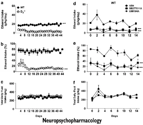
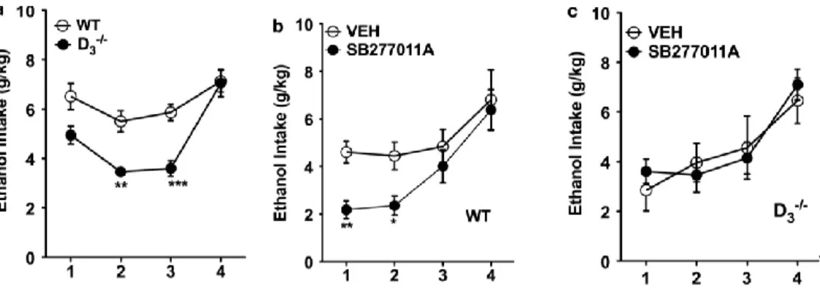
3.1 D3R-/- mice exhibited lower ethanol intake
As shown in Figure 1a and b, WT mice exhibited a high intake of ethanol-containing solution. In contrast, D3R-/- mice showed a low ethanol intake (Figure
1a and b). During the entire period of observation (44 days), WT mice maintained their preferential intake of ethanol, whereas D3R-/- mice maintained a preferential
intake of water (F(1,307)=1170.08, p<0.001). There was no difference between WT
and D3R-/- mice in terms of total amount of fluid intake (ethanol + water) (Figure
1c). In the DID paradigm, D3R-/- mice also showed a lower ethanol intake
compared with their WT counterparts (F(3,97)= 13.90, p<0.01, 2nd day;
F(3,97)=21.04, p<0.001, 3rd day; Figure 2a).
3.2 Blockade of D3R Inhibited Ethanol Intake
In the two-bottle choice paradigm, after 30 days of stable ethanol/water intake, mice were treated with D3R antagonists (U99194A or SB277011A). As
shown in Figure 1d and e, treatment of WT with each D3R antagonist decreased
voluntary ethanol intake (F(2,56)= 55.23 p<0.01, for both U99194A and
SB277011A). Treatment of D3R-/- mice with U99194A and SB277011A did not
change ethanol intake (data not shown). Neither in WT nor in D3R-/- mice total
SB277011A also significantly decreased ethanol intake in WT mice tested in the DID (F(3,48)= 8.67, p<0.01, 1st day; p<0.05 2nd day; Figure 2b), while it did not
change ethanol intake of D3R-/- in the DID paradigm (Figure 2c).
3.3 RACK1, BDNF, and DA D3R expression were increased in the striatum of WT
mice following chronic ethanol intake
BDNF induces D3R expression in the ventral striatum, both during
development and in adulthood (Guillin et al., 2001). RACK1, a mediator of chromatin remodeling, regulates in an exon-specific manner the expression of the BDNF gene (He et al., 2010) and the RACK1/BDNF pathway is activated upon exposure to ethanol (McGough et al., 2004). We therefore assessed D3R, BDNF,
and RACK1 mRNA expression in striatum of WT that had free access to either water only or to both water and ethanol. Figure 3a shows that chronic ethanol intake increased D3R mRNA expression in striatum (F(3,23)= 170.4, p<0.05).
Long-term access to ethanol also increased BDNF (Figure 3b, F(7,47)= 48.05, p<0.01)
and RACK1 (Figure 3c, F(7,47)= 21.14, p<0.01) mRNA in striatum of WT mice.
Long-term ethanol exposure appeared to be associated with BDNF/RACK1 overexpression, but interpretation of these data was made difficult by the different ethanol intake in the two genetic groups, as it was very high in WT and very low in D3R-/- mice. To address this issue, some WT and D3R-/- mice were subjected to
overexpression of BDNF (F(7,47)=48.05, p<0.05, p<0.01) and RACK1 (F(7,47)=
21.14, p<0.05, p<0.05) mRNAs in striatum of both WT and D3R-/- mice. We also
tested the effects of the D3R antagonists SB277011A and buspirone (see also
below) on mRNA expression of D3R, BDNF, and RACK1. None of these values
were changed by a 14-day treatment with SB277011A or buspirone (Figure 3f–h).
3.4 Blockade of the BDNF receptor TrkB inhibited ethanol intake and decreased D3R expression
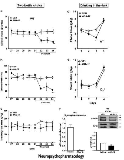
TrkB is the high affinity receptor for BDNF, belonging to the family of tyrosine kinase receptors, which undergo autophosphorylation upon agonist binding (Soppet et al., 1991). In order to assess the role of BDNF pathway in ethanol intake, we used the recently available TrkB selective antagonist ANA-12 (Cazorla et al., 2011). After 30 days of stable ethanol/water intake, mice received daily i.p. injections of either vehicle or ANA-12 (Figure 4a and b). ANA-12 reversed the stable ethanol intake of WT mice (F(7,42)=30.53, p<0.001) but did not
change the voluntary and the forced ethanol intake of D3R-/- mice (data not
shown). Neither in WT nor in D3R-/- mice total fluid intake was affected by
treatment with ANA-12 (Figure 4c and data not shown). Also in the DID paradigm ANA-12 was effective in reducing ethanol intake in WT mice (F(3,55)=6.64, P<0.05, Figure 4d), whereas it did not change ethanol intake in D3R-/-
striatum by ANA-12, we determined, by immunoblot, the abundance of phosphorylated TrkB. As shown in Figure 4f, treatment of WT with ANA-12 significantly decreased phosphorylation of TrkB (F(3,35)=184.5, p<0.01). Finally
and more interestingly, ANA-12 strongly decreased D3R mRNA expression in the
striatum of WT mice exposed to voluntary ethanol intake (Figure 4f, F(3,35)=184.5,
P<0.001).
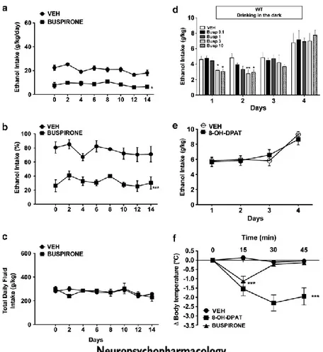
3.5 Buspirone Inhibited Ethanol Intake
In the two-bottle choice paradigm, after 30 days of stable ethanol/water intake, mice were treated with buspirone (1 mg/kg/day). As shown in Figure 5a and b, treatment of WT with buspirone significantly decreased voluntary ethanol intake (F(1,28)=20.88, p<0.05). Treatment of D3R-/- mice with buspirone did not
change ethanol intake (data not shown). Neither in WT nor in D3R-/- mice total
fluid intake was affected by treatment (Figure 5c and data not shown). The treatment with buspirone also significantly decreased ethanol intake in WT mice when tested in the DID. Dose ranging of buspirone (0.1, 1, 3, and 10 mg/kg) showed that treatment of WT with buspirone at the doses of 3 and 10 mg/kg significantly decreased ethanol intake both in the 1st day (F(4,75)=31.24, p<0.05)
and in the 2nd day (F(4,75)= 31.24, p<0.01 3 mg/kg; p<0.05 10 mg/kg) of the
mg/kg buspirone did not change ethanol intake in D3R-/- mice (data not shown).
Because buspirone is also known as a 5-HT1A agonist, the D3R specific effect of
buspirone in decreasing ethanol intake was confirmed by using the selective 5-HT1A agonist, 8-OH-DPAT. As shown in Figure 5e, treatment with 8-OH-DPAT (1 mg/kg, i.p.) in WT and D3R-/- mice mice did not affect ethanol intake (Figure 5e
and data not shown). As expected, the 5-HT1A selective agonist 8-OH-DPAT decreased the body temperature of WT mice (F(2,39)=14.99, p<0.001) (Figure 5f).
Buspirone (3 mg/kg) decreased the body temperature of WT mice only transiently (Figure 5f).
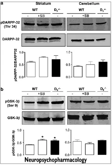
3.6 DA receptor signaling in striatum of WT and D3R-/- mice exposed to ethanol
Activation of D1 receptor results in activation of adenylyl cyclase/cAMP/protein kinase A (PKA) signaling; a major substrate for PKA in the striatum is DARPP-32. D2-like receptors regulate the activity of the protein kinases Akt and GSK3b; stimulation of either D2R or D3R results in
phosphorylation of Akt and GSK3b (Mannoury la Cour et al., 2011). In order to assess activation of dopaminergic transmission in striatum, we determined, by immunoblot, the abundance of phosphorylated DARPP-32 (Thr 34) and of phosphorylated GSK3b (Ser 9). As shown in Figure 6, posphoGSK3b was more abundant in striatum of D3R-/- mice than in WT mice, whereas phosphoDARPP-32
Treatment of WT mice with SB277011A induced phosphorylation of DARPP-32 and GSK3b, up to the level seen in D3R-/- mice. In contrast, in cerebellum, there
was no difference in the level phosphoDARPP-32 and posphoGSK3b between WT e D3R-/- mice, nor it was influenced by SB277011A treatment in WT.
4. Discussion
This study demonstrates that D3R is necessary for ethanol consumption in
mice, because either D3R gene deletion or D3R pharmacological blockade by
selective D3R experimental antagonists or the approved drug buspirone, inhibits
alcohol intake. The D3R overexpression induced by ethanol intake associated with
the activation of RACK1/BDNF may represent the basis for a reinforcing mechanism of ethanol intake. Indeed, although selective blockade of the TrkB reversed stable intake of ethanol in WT mice and decreased D3R expression levels
in their striatum, it was ineffective in D3R-/- mice. It seems that D3R, among
D2-like receptors, is the key player in addiction, particularly in reward mechanisms. Indeed, although the D2R is associated with mesocortical and mesohippocampal
DA pathway, the D3R is associated with the ventral mesolimbic DA system
(Sokoloff et al, 1990). Previous studies reported low levels of D2R both in animal
models and in patients addicted to cocaine, alcohol, metamphetamine, and nicotine (Volkow et al., 2009). Conversely, upregulation of D3R expression has
been reported following exposure to DA elevating drugs (Boileau et al., 2012; Heidbreder and Newman, 2010; Le Foll et al., 2005b; Mash, 1997; Segal et al., 1997; Staley and Mash, 1996). An important interpretative issue is the genetic background on which the D3R null mutation was placed. Specific behavioral
Young, 1998). The D3R-/- mice we used are on the C57BL/6J background (Accili
et al., 1996), a strain where ethanol preference and sensitivity is well documented (Crabbe et al., 1983). Interestingly, D3R-/- mice have extracellular DA levels twice
as high as their WT littermates (Joseph et al., 2002; Koeltzow et al., 1998); this enhanced DA tone and the resulting adaptations may reflect removal of the inhibitory influence of D3R in the control of basal extracellular DA levels (Le Foll
et al., 2005a), giving support to an autoreceptor role for D3R in the mesolimbic
areas of the brain (Diaz et al., 2000). The increased DA activity in D3R-/- mice is
consistent with their phenotype, including higher basal levels of grooming behavior, hyper-locomotion, and reactivity to drug-paired environmental cues (Accili et al., 1996; Le Foll et al., 2005a; Le Foll et al., 2002). Here we found that D3R-/- mice chronically exposed to the voluntary ethanol intake paradigm, drink
very low quantities of ethanol in comparison with their WT littermates. This observation cannot be attributed to differences in metabolism (McQuade et al., 2003), locomotor activity (Harrison and Nobrega, 2009), or taste reactivity (McQuade et al., 2003) between WT and D3R-/- mice. The lower ethanol intake of
D3R-/- mice in comparison with their WT control mice seems apparently in
contrast with the only two previous studies testing D3R-/- mice in the ethanol
voluntary intake paradigm (Boyce-Rustay and Risinger, 2003; McQuade et al., 2003). This may be due, at least in part, to some important differences in
difference between D3R-/- mice and WT in the 24-h access paradigm, used a
different experimental procedure in the two-bottle choice paradigm. First, they used just 4 days of adaptation period before ethanol exposure. Second, they tested both D3R-/- mice and WT animals with increasing concentrations of ethanol in
subsequent 7-day steps. In the first step, 3% ethanol, in the second step 6%, in the third step 10%, in the 4th 15%, and finally, in the 5th 20% ethanol. Thus, the behavioral paradigm used by McQuade and co-workers is quite different from our paradigm. From our experience, for these mice it is to have a long period of habituation in the two-bottle paradigm (15 days) before to start with the ethanol access procedure. It is likely that the progressive increase of the ethanol concentration every 7 days, may induce an adaptation to the ethanol that damps the difference between D3R-/- and WT mice. Furthermore, in the McQuade’s
study, the relative positions of the ethanol and water bottle were determined randomly each day, whereas in our experiments the position of tubes was interchanged (left/right) every 24 h, to prevent acquisition of position bias. The random change of bottles may expose a given animal to access the same solution (either ethanol or water) in the same position for two/three days consecutively, which may interfere with the results of the experiment during a short period of observation (7 days). In the study by Boyce-Rustay and Risinger (2003), C57 animals were used as control of D3R-/- mice. These experiments are not
in this study increasing concentrations of ethanol were used in 8-day steps (3 and 10%). Thus, (i) the behavioral procedure is different; (ii) an adaptation to ethanol may occur and damp the difference between genotypes. To obtain pharmacological evidence for a functional role of D3R in the control of voluntary
ethanol intake, we tested two D3R antagonists, U99194A and SB277011A at
doses reported to selectively target the D3R (Carr et al., 2002; Reavill et al., 2000).
Before administering these drugs, we performed a molecular modeling study to gain information on the interaction of U99194A and SB277011A with D3R. As
illustrated in Supplementary Information, in silico analysis showed that the two D3R antagonists were (i) highly selective for the D3R subtype and (ii) displayed a
distinct interaction (different binding energy, different interaction patterns) with D3R, consistent with their distinct chemical structure. We found that both
U99194A and SB277011A induced a significant decrease in voluntary ethanol intake in WT but not in D3R-/- mice. This pharmacological evidence reinforces the
view that the D3R is necessary for ethanol consumption in mice and is consistent
with rat data showing that D3R antagonism reduces relapse-like drinking and
cue-induced ethanol-seeking behavior (Vengeliene et al., 2006).
We confirmed the primary role of D3R in the control of ethanol-drinking
behavior in a binge-like ethanol-drinking paradigm (Crabbe et al., 2011; Rhodes et al., 2005; Rhodes et al., 2007). Here, again, D3R-/- mice exposed to DID drank
blockade by SB277011A decreased ethanol intake in WT but not in D3R-/- mice.
No differences were recorded in the DID at day 4. Indeed, there was neither a genotype effect between WT and D3R-/- mice nor a treatment effect with the
SB277011A in WT mice. In general, the binge-like behavior is captured by the 2 h time window that detects differences between treatments/genotypes better than the 4 h window, because the cumulative intake over 4 h makes smaller the proportion of differences (Rhodes et al., 2005). Thus, it is likely that, the lack of differences on day 4 is due to the longer lasting access to ethanol that produced overall a higher consumption, potentially masking the genotype/treatment effect on binge-like drinking behavior occurring in the first 2 h. Enhanced D3R expression in
striatum following long-term alcohol consumption has been previously reported in both mice and rats (Jeanblanc et al., 2006; Vengeliene et al., 2006). Our data show and confirm that chronic voluntary ethanol intake upregulated D3R mRNA
expression in the striatum of WT mice. Interestingly, D3R expression is increased
by exposure to other addictive drugs, such as nicotine and cocaine, in caudate– putamen (Neisewander et al., 2004) and in nucleus accumbens of rats (Le Foll et al., 2003, 2005b) and humans (Staley and Mash, 1996). Expression of D3R
therefore appears to be a potential basis for a reinforcing mechanism in reward-related behavior associated with voluntary intake of addictive drugs and ethanol. A number of studies have linked D3R expression in the nucleus accumbens to
furthermore, ethanol exposure increases both BDNF and D3R within the striatum
itself (Jeanblanc et al., 2006; McGough et al., 2004). The scaffolding protein RACK1 is a key regulator of BDNF expression; RACK1 translocates to the nucleus after exposure of neurons to ethanol and increases expression of BDNF (McGough et al., 2004). Jeanblanc et al. (2006) proposed that the RACK1/BDNF/D3R pathway is involved in the control of ethanol consumption in
mice. Our hypothesis is that activation of RACK1/BDNF by ethanol may induce expression of D3R, which in turn controls and maintains ethanol consumption.
This hypothesis is supported by the data we generated showing that: (i) ethanol intake is negligible in D3R-/- mice and robust in WT; (ii) increase in
RACK1/BDNF/D3R is maintained during chronic ethanol intake in WT; (iii)
forced ethanol intake increases RACK1/BDNF even in D3R-/- mice. Furthermore,
chronic voluntary ethanol intake increased D3R expression in striatum
concomitant with increased expression of BDNF. It is noteworthy that, in the basal condition, D3R-/- mice exhibited higher BDNF than WT, consistent with a
tendency reported in a recent study (Xing et al., 2012). When subjected to forced ethanol intake, D3R-/- mice showed a robust increase in BDNF expression in the
striatum. Therefore, chronic ethanol intake increases BDNF independently of D3R
receptor stimulation. The finding that chronic ethanol intake increased RACK1 in striatum of both WT and D3R-/- mice provides additional evidence for the role of
RACK1/BDNF pathway leading to D3R overexpression and addictive behavior in
WT, but not in D3R-/- mice, because this latter lacks D3R. To provide additional
evidence, we blocked the BDNF pathway by using the TrkB specific antagonist, ANA-12. We found that ANA-12 reversed ethanol intake both in the twobottle choice and DID paradigms and strongly decreased the expression of D3R in the
striatum of WT-treated mice. Recently, D3R on VTA-SN dopaminergic neurons
were found to mediate neuroplasticity effects of several addictive drugs (Collo et al., 2012; Collo et al., 2013). Therefore, our conclusion about the engagement of striatal RACK1, BDNF, and D3R in mediating ethanol consumption may be only a
part of a more complex mechanism, whose elucidation may require an assessment of the effects of ethanol intake in the VTA-SN dopaminergic neurons.
Finally, in a translational perspective, we tested buspirone, a drug marketed for anxiety disorders, endowed with D3R antagonist (Bergman et al., 2013; Le
Foll and Boileau, 2013; Newman et al., 2012) and 5-HT1A partial agonist activity (Wong et al., 2007). Notably, buspirone shows also high affinity for other D2-like receptors (Bergman et al., 2013; Kula et al., 1994; Tallman et al., 1997). D3R
antagonists may be effective for treating substance use disorders and buspirone has proven effective in several preclinical model of drug abuse (Heidbreder and Newman, 2010; Higley et al., 2011; Song et al., 2012), but no studies have, so far, investigated its D3R antagonist action in ethanol consumption. By both
Information), we found that buspirone: (i) shows slight higher affinity at D3R than
at D2R (Ki, 29 vs 62 nM, respectively) and may form interactions comparable
with those of SB277011A in D3R, having the antagonist binding mode at D3R, (ii)
displays a distinct interaction from the other two antagonists SB277011A and U99194A (different binding energy, different interaction patterns) with D3R,
consistent with their distinct chemical structure. Thereafter, we found that buspirone induced a significant decrease in ethanol intake in both two-bottle choice and DID paradigms. The dose of 1 mg/kg inhibited ethanol intake in both paradigms, though its effect did not reach statistical significance in DID; 3 and 10 mg/kg, however produced a significant effect in DID. We confirmed the specificity of D3R effect by using a selective 5-HT1A agonist, 8-OH-DPAT, in the
DID. Treatment with 8-OH-DPAT did not impact ethanol intake, whereas, as expected, decreased the body temperature in a stable manner. In a translational perspective, an important issue is the actual availability of buspirone to bind D3R
in human CNS. Reported buspirone’s affinity toward human recombinant D3R
ranges from 3.5 to 98 nM (Bergman et al., 2013; Newman et al., 2012), which partially overlaps its affinity for 5-HT1A receptors; because buspirone binding to 5-HT1A is considered the basis of its anxiolytic activity in humans, it is likely that anxiolytic doses are sufficient to occupy also D3R in human CNS. However, the
including measurements of D3R receptor occupancy in human PET studies, as an
essential prerequisite to clinical application.
Finally, as D3R-/- mice have been shown to exhibit extracellular DA levels
substantially higher than WT, as assessed by microdialysis (Koeltzow et al., 1998), a phenomenon related to the lack of autoreceptor function (Joseph et al., 2002), we hypothesized that ethanol intake effectively stimulates DA release and transmission in WT, but not in D3R-/- mice, presumably because this latter already
displays high extracellular DA levels. To test the hypothesis that treatment with D3R antagonists mimicked the high DA phenotype documented in D3R-/- mice
(Koeltzow et al., 1998), we assessed phosphorylation of DARPP32, that is increased by different addictive drugs, including ethanol (Nuutinen et al., 2011; Svenningsson et al., 2005), and of GSK3b, that is linked to D2-like receptors signaling cascade (Beaulieu et al., 2007; Li et al., 2009), particularly under hyper-DAergic conditions (Li et al., 2009). Treatment with SB277011A increased phosphorylation of DARPP32 and of GSK3b to a level similar to that of D3R-/-
mice. Thus, chronic blockade of the D3R or its genetic deletion increased DA
transmission in striatum, consistent with increased extracellular DA (Joseph et al., 2002; Koeltzow et al., 1998).
In conclusion, either D3R gene deletion or D3R pharmacological blockade
inhibit ethanol intake. Thus, pharmacological antagonism selectively targeting D3R may provide a basis for novel weaning treatments to inhibit ethanol
consumption. In this context, buspirone, a drug marketed as anxiolytic since more than 25 years and endowed with D3R antagonist activity, exhibits, translational
potential for treating alcohol addiction.
5. Funding and disclosure
This work was supported in part by a National Grant PON01-00110. Dr Camillieri, Dr Platania and Dr Torrisi were supported by the International PhD Program in Neuropharmacology, University of Catania Medical School, Catania, Italy; they declare no potential conflict of interest. Dr Leggio and Dr Marrazzo were full-time research fellows of Catania University; they declare no potential conflict of interest. Dr Castorina, Dr D’Agata, Dr Bucolo, Dr Drago, and Dr Salomone were full-time employees of Catania University. Dr Drago was member of the board of the Italian Medicinal Agency (AIFA). Dr Castorina, Dr D’Agata, and Dr Drago declare no potential conflict of interest. Dr Bucolo has received unrestricted research funding from Novartis. Dr Salomone has received unrestricted research funding from Novartis, Bayer, Gilead. Dr Le Foll has received grant and salary support from Pfizer and was a consultant for Richter Pharmaceuticals, Lundbeck, Mylan, Ethypharm and Pfizer. Dr Le Foll research is supported by CAMH, the Campbell Family Mental Health Research Institute, CIHR and the National Institute on Drug Abuse at the National Institutes of
(NSERC). Christina N. Nona was the recipient of a Vanier Canada Graduate Scholarship. Dr Stark is an employee of Heinrich-Heine-University Duesseldorf. None of the authors have competing financial interests in relation to the work described.
6. Acknowledgements
We are indebted to Dr G. Tanda for reading the manuscript and giving his invaluable advice.
References
Accili D, Fishburn CS, Drago J, Steiner H, Lachowicz JE, Park BH et al (1996). A targeted mutation of the D3 dopamine receptor gene is associated with hyperactivity in mice. Proc Natl
Acad Sci USA 93: 1945–1949.
Beaulieu JM, Gainetdinov RR, Caron MG (2007). The Akt-GSK-3 signaling cascade in the actions of dopamine. Trends Pharmacol Sci 28: 166–172.
Bergman J, Roof RA, Furman CA, Conroy JL, Mello NK, Sibley DR et al (2013). Modification of cocaine self-administration by buspirone (buspar(R)): potential involvement of D3 and D4 dopamine receptors. Int J Neuropsychopharmacol 16: 445–458.
Boileau I, Payer D, Houle S, Behzadi A, Rusjan PM, Tong J et al (2012). Higher binding of the dopamine D3 receptor-preferring ligand [11C]-( þ )-propyl-hexahydro-naphtho-oxazin in methamphetamine polydrug users: a positron emission tomography study. J Neurosci 32: 1353– 1359.
Bouthenet ML, Souil E, Martres MP, Sokoloff P, Giros B, Schwartz JC (1991). Localization of dopamine D3 receptor mRNA in the rat brain using in situ hybridization histochemistry: comparison with dopamine D2 receptor mRNA. Brain Res 564: 203–219.
Bowers MS, Chen BT, Bonci A (2010). AMPA receptor synaptic plasticity induced by psychostimulants: the past, present, and therapeutic future. Neuron 67: 11–24.
Boyce-Rustay JM, Risinger FO (2003). Dopamine D3 receptor knockout mice and the motivational effects of ethanol. Pharmacol Biochem Behav 75: 373–379.
Brand I, Fliegel S, Spanagel R, Noori HR (2013). Global ethanolinduced enhancements of monoaminergic neurotransmission: a meta-analysis study. Alcohol Clin Exp Res 37: 2048–2057.
Carr KD, Yamamoto N, Omura M, Cabeza de Vaca S, Krahne L (2002). Effects of the D(3) dopamine receptor antagonist, U99194A, on brain stimulation and d-amphetamine reward, motor activity, and c-fos expression in ad libitum fed and foodrestricted rats. Psychopharmacology (Berl) 163: 76–84.
Cazorla M, Premont J, Mann A, Girard N, Kellendonk C, Rognan D (2011). Identification of a low-molecular weight TrkB antagonist with anxiolytic and antidepressant activity in mice. J Clin Invest 121: 1846–1857.
Cohen C, Perrault G, Sanger DJ (1998). Preferential involvement of D3 versus D2 dopamine receptors in the effects of dopamine receptor ligands on oral ethanol self-administration in rats.
Psychopharmacology (Berl) 140: 478–485.
Collo G, Bono F, Cavalleri L, Plebani L, Merlo Pich E, Millan MJ et al (2012). Pre-synaptic dopamine D(3) receptor mediates cocaine-induced structural plasticity in mesencephalic dopaminergic neurons via ERK and Akt pathways. J Neurochem 120: 765–778.
Collo G, Bono F, Cavalleri L, Plebani L, Mitola S, Merlo Pich E et al (2013). Nicotine-induced structural plasticity in mesencephalic dopaminergic neurons is mediated by dopamine D3 receptors
Crabbe JC, Kosobud A, Young ER, Janowsky JS (1983). Polygenic and single-gene determination of responses to ethanol in BXD/ Ty recombinant inbred mouse strains. Neurobehav Toxicol Teratol 5: 181–187. preference drinking in a mouse line selectively bred for high drinking in the dark. Alcohol 45: 427–440.
Diaz J, Pilon C, Le Foll B, Gros C, Triller A, Schwartz JC et al (2000). Dopamine D3 receptors expressed by all mesencephalic dopamine neurons. J Neurosci 20: 8677–8684.
Gessa GL, Muntoni F, Collu M, Vargiu L, Mereu G (1985). Low doses of ethanol activate dopaminergic neurons in the ventral tegmental area. Brain Res 348: 201–203.
Guillin O, Diaz J, Carroll P, Griffon N, Schwartz JC, Sokoloff P (2001). BDNF controls dopamine D3 receptor expression and triggers behavioural sensitization. Nature 411: 86–89.
Harrison SJ, Nobrega JN (2009). A functional role for the dopamine D3 receptor in the induction and expression of behavioural sensitization to ethanol in mice. Psychopharmacology (Berl) 207: 47–56.
He DY, Neasta J, Ron D (2010). Epigenetic regulation of BDNF expression via the scaffolding protein RACK1. J Biol Chem 285: 19043–19050.
Heidbreder CA, Andreoli M, Marcon C, Hutcheson DM, Gardner EL, Ashby CR Jr. (2007). Evidence for the role of dopamine D3 receptors in oral operant alcohol self-administration and reinstatement of alcohol-seeking behavior in mice. Addict Biol 12: 35–50.
Heidbreder CA, Gardner EL, Xi ZX, Thanos PK, Mugnaini M, Hagan JJ et al (2005). The role of central dopamine D3 receptors in drug addiction: a review of pharmacological evidence. Brain Res
Brain Res Rev 49: 77–105.
Heidbreder CA, Newman AH (2010). Current perspectives on selective dopamine D(3) receptor antagonists as pharmacotherapeutics for addictions and related disorders. Ann N Y Acad Sci 1187: 4–34.
Higley AE, Kiefer SW, Li X, Gaal J, Xi ZX, Gardner EL (2011). Dopamine D(3) receptor antagonist SB-277011A inhibits methamphetamine self-administration and methamphetamineinduced reinstatement of drug-seeking in rats. Eur J Pharmacol 659: 187–192.
Ikemoto S, Bonci A (2013). Neurocircuitry of drug reward. Neuropharmacology 76: 329–341.
Imperato A, Di Chiara G (1986). Preferential stimulation of dopamine release in the nucleus accumbens of freely moving rats by ethanol. J Pharmacol Exp Ther 239: 219–228.
Jeanblanc J, He DY, McGough NN, Logrip ML, Phamluong K, Janak PH et al (2006). The dopamine D3 receptor is part of a homeostatic pathway regulating ethanol consumption. J
Neurosci 26: 1457–1464.
Joseph JD, Wang YM, Miles PR, Budygin EA, Picetti R, Gainetdinov RR et al (2002). Dopamine autoreceptor regulation of release and uptake in mouse brain slices in the absence of D(3) receptors. Neuroscience 112: 39–49.
Joyce JN, Millan MJ (2005). Dopamine D3 receptor antagonists as therapeutic agents. Drug
Discov Today 10: 917–925.
Koeltzow TE, Xu M, Cooper DC, Hu XT, Tonegawa S, Wolf ME et al (1998). Alterations in dopamine release but not dopamine autoreceptor function in dopamine D3 receptor mutant mice. J
Neurosci 18: 2231–2238.
Koob GF (1992). Drugs of abuse: anatomy, pharmacology and function of reward pathways.
Trends Pharmacol Sci 13: 177–184.
Kula NS, Baldessarini RJ, Kebabian JW, Neumeyer JL (1994). S-( þ )-aporphines are not selective for human D3 dopamine receptors. Cell Mol Neurobiol 14: 185–191.
Le Foll B, Boileau I (2013). Repurposing buspirone for drug addiction treatment. Int J

![Table 1S. Recombinant hD 3 versus hD 2 receptor binding. Compound hD 2 Ki [nM] ± SD hD 3 Ki [nM] ± SD U2991941 2639 ± 410 102.3 ± 37.3 SB277011 A 1914 ± 329 10.04 ± 2.65 Buspirone 62.26 ± 17.6 29.98 ± 11.5 Haloperidol 1.91 ± 0.15](https://thumb-eu.123doks.com/thumbv2/123dokorg/4532980.35392/75.748.75.687.108.378/table-recombinant-versus-receptor-binding-compound-buspirone-haloperidol.webp)
