International PhD Program in Neurosciences
XXIX Cycle
DYNAMIC EXPRESSION OF AQUAPORINS IN
PHYSIOLOGICAL AND PATHOPHYSIOLOGICAL
IN VITRO MODELS
PhD Thesis
Rosanna Avola
Coordinator: Prof. Salvatore Salomone Tutor: Prof. Venera Cardile
Department of Biomedical and Biotechnological Sciences
University of Catania - Medical School
2
To Gloria
With encouragement to aim as high as possible
and with the belief that everything in life
can be achieved if you believe and work
hard enough for it.
3
TABLE OF CONTENTS
Acknowledgements ... 4 List of abbreviations ... 5 Abstract ... 7 Introduction ... 9 Discovery of aquaporins ... 10Structural features of aquaporins ... 11
Aquaporin classification and selectivity ... 16
Aquaporin distribution and physiological functions ... 17
Aquaporin in brain ... 20
AQP1 ... 21
AQP4 ... 22
AQP9 ... 23
Other brain aquaporins ... 24
In vitro models for neurological research ... 25
Stem cells ... 28
Mesechymal stem cells ... 30
Mesechymal stem cells from adipose tissue ... 32
Neural differentiations of mesenchymal stem cells ... 33
Stem cells and aquaporins
...
35Aims of the research ... 37
Chapter I - Krabbe’s leukodystrophy: approaches and models in vitro ... 39
Chapter II - Human mesenchymal stem cells from adipose tissue differentiated into neuronal or glial phenotype express different aquaporins ... 65
Chapter III - New insights on Parkinson’s disease from differentiation of SH-SY5Y into dopaminergic neurons: the involvement of aquaporin 4 and 9 ... 100
General discussion and conclusion ... 132
References ... 134
4
A
CKNOWLEDGEMENTS
The years of PhD program have been a precious experience, an exciting journey that I could not have undertaken without the enduring support of my Tutor, Colleagues and Family.
I wish to thank my Tutor, Prof. Venera Cardile, University of Catania, for welcoming me in her laboratory, who introduced me to the world of cells physiology and the complexities of stem cells science. I have greatly appreciated her support, without which,I would have never successfully completed this project.
I am very grateful to Prof. Filippo Drago and Prof. Salvatore Salomone, University of Catania; they gave me the opportunity to take part to this PhD program.
I wish to thank Dr. Adriana Carol Eleonora Graziano and Dr. Giovanna Pannuzzo, for sharing with me their knowledge, they were able to give me good advices and technical supports.
I want to thank also all Professors, colleagues and friends of the Section of Physiology of University of Catania.
Finally, I would like to thank my family and Giuseppe for their love and support kept me going, especially those difficult days when things do not seem to work.
Thank you everyone for making my PhD such a wonderful journey!
5
LIST OF ABBREVIATIONS
6-OHDA ADH Ag ANP AQP1 AQP2 AQPs ar/R ASCs AT-MSCs BBB BDNF BHA BM- MSCs BME CFU-F CGMP CHIP 28 CNS CP CSF DA DMSO ECS EGF ER ESCs FAK FGF- FSCs G1-G3 6-hydroxydopamine Antidiuretic hormone AntigenAtrial natriuretic peptide Aquaporin-1
Aquaporin-2 Aquaporins Aromatic/arginine Adult stem cells
Mesenchymal stem cells from adipose tissue Blood Brain Barrier
Brain-derived neurontrophic factor Butylated hydroxyanisole
Bone marrow mesenchymal stem cells -mercaptoethanol
Colony-forming-unit fibroblastic cells Cyclic guanosine monophosphate Channel like Intrinsic Protein of 28 kDa Central Nervous System
Choroid plexus Cerebro Spinal Fluid Dopamine transporter Dimethyl sulfoxide Brain extracellular space Epidermal growth factor Reticulum endoplasmatic Embryonic stem cells Focal adhesion kinase Basal fibroblast growth factor Fetal stem cells
6
GALC GLD GlpF HGF IGF ISCT KD kDa Kir4.1 MIP MPA MPP+ MPTP NGF NPA NSCs OECs P PAS PD PDGF PLA PNS RA RBC ROS RT-PCR SNpc SVF TM VEGF Enzyme galactosylceramidase Globoid cell leukodystrophy Glycerol facilitatorHepatocyte growth factor Insulin-like growth factor
International Society for Cellular Therapy Krabbe disease
Kilodalton
Rectifyng potassium channel 4.1 Major Intrinsic Protein
Phorbol 12-Myristate 13-acetate 1-methyl-4-phenylpyridinium
1-methyl-4-phenyl-1, 2, 3, 6-tetrahydropyridine Nerve growth factor
Asn-Pro-Ala conserved motif Neural stem cells
Olfactory ensheathing cells Passage
Periodic acid-Schiff Parkinson’s disease
Platelet-derived growth factor Lipoaspirate
Peripheral nervous system Retinoic acid
Red blood cells Reactive oxygen species
Reverse transcriptase-polymerase chain reaction Substantia nigra pars compacta
Stromal-vascular fraction Transmembrane domains
7
ABSTRACT
Water is the main component of biological fluids and a prerequisite of all organisms living. In 1987, Agre and coworkers isolated a new integral membrane protein acting as a channel that mediates the water flux and uncharged solutes across biological membranes. This protein was called aquaporin1 and ever since its discovery, more than 300 homologues have been identified in many phyla, including animal, bacteria and plant. So far, in human have been discovered 13 aquaporins (AQPs) isoform (AQP0-AQP12) widely distributed in various epithelia and endothelia where are important actors of fluid homeostasis maintenance in secretory and absorptive processes in response to an osmotic or pressure gradient. In the human brain nine aquaporin subtypes (AQP1, 2, 3, 4, 5, 7, 8, 9, and 11) have been recognized and partially characterized, but only three aquaporins (AQP1, 4, and 9) have been clearly identified in vivo. This discovery highlighted the concept of the important role of AQPs in all brain functions and of the dynamics of water molecules in the cerebral cortex. Additionally, AQPs releaved an important role in glial control and neuronal excitability, such as in brain structure and general development. However, a clearer understanding of specific function and distribution of water channels in adult or in development brain requires a more detailed elucidation. Some of these findings are limited from the complexity of direct investigation, inaccessibility of the neural tissue, and hence difficulty in obtaining a brain biopsy, until after the death of an individual. In this sense, several past and present in vitro models have been used to provide important clues about many processes, such as brain development, neurotoxicity, inflammation, neuroprotection, pathogenic mechanisms of the diseases and potential pharmacological targets. In the Chapter I,we have reviewed some in vitro approaches used to investigate the mechanisms involved in Krabbe disease with particular regard to the cellular systems employed to study processes of inflammation, apoptosis and angiogenesis. Moreover, in this study, we used some in vitro methods with the aim to update the knowledge on stem cells biology and to provide a relationship between aquaporins expression and cellular differentiation. In particular, we have analysed the differentiation of human mesenchymal stem cells from adipose tissue (AT-MSCs) into neural phenotypes and SH-SY5Y neuroblastoma cell line into physiological and pathophysiological dopaminergic neurons.
8
Thus, in the Chapter II, we have reported the results of the expression of AQP1, 4, 7, 8 and 9 at 0, 14, and 28 days in AT-MSCs during the neural differentiation by performing immunocytochemistry, RT-PCR and Western blot analysis. Our studies demonstrated that AT-MSCs could be differentiated into neurons, astrocytes and oligodendrocytes, showing reactivity not only for the typical neural markers, but also for specific AQPs in dependence from differentiated cell type. Our data revealed that at 28 days AT-MSCs express AQP1, astrocytes AQP1, 4 and 7, oligodendrocytes AQP1, 4 and 8, and finally neurons AQP1 and 7. In the Chapter III, we have examined the possible involvement of AQPs in a Parkinson’s disease-like cell model. For this purpose, we used SH-SY5Y, a human neuroblastomacell line, differentiated indopaminergic neurons with retinoic acid (RA) and phorbol 12-myristate 13-acetate (MPA) alone or in association. The vulnerability to dopaminergic neurotoxin 1-methyl-4-phenyl-1, 2, 3, 6-tetrahydropyridine (MPTP) and H2O2 was evaluated and compared in all cell groups. We found that the vulnerability of cells was linked to dynamic changes of AQP4 and AQP9. The data described here provides fundamental insights on the biology of the human mesenchymal stem cells and significant evidences on the involvement of AQPs in a variety of physiological and pathophysiological processes. This suggests their possible application as markers, which may be helpful in diagnosing as well as in the understanding of neurodegenerative diseases for future therapeutical approaches.
9
INTRODUCTION
Water is a prerequisite of all organisms living and it is an essential component for the biologic activity of proteins [1].Water accounts for approximately 60% of our body weight, (which translates to about 42 L in a 70 kg person). Of this, 65% is found inside the cells, while the remaining 35% constitutes the extracellular fluid. At the extracellular level, water is the main component of biological fluids, allowing, for instance, the long distance trafficking of important solutes such as sugars and ions in human blood. At the extracellular/intracellular interface, water exchange through the plasma membrane maintains the osmolality of the cytoplasm and thus the integrity of the cell. At the molecular level, water is involved in the configuration of some important molecules. Indeed, water molecules are polar, which allows them to easily form hydrogen bonds with each other and with other molecules. They serve as excellent solvents for a variety of polar substances in the cells. Water provides solvent shells around charged groups of biopolymers. A striking property of most human tissues is their capacity for extremely rapid and highly regulated transport of water through cellular membranes, processes essential to human health [2, 3]. Until the 1990, little was known about the molecular mechanism regulating total body water content and the distribution of water between the extracellular and intracellular space.
The discovery of the plasma membrane in the 1920s started the discussion on how water can be transported across this membrane. Such trans-tissue water flow is possible by two routes: transcellular water flow across both basal and apical membranes, which occurs in response to the osmotic stimuli [2], created by salt transport [4] or paracellular flow across cell–cell junctions into intercellular spaces, driven by salt or solute gradients [4]. Transcellular water flow is dependent on the permeability of the plasma membrane to water molecules. The biological membrane surrounding living cells is not a pure lipidic bilayer and although have a measurable permeability to water, the simple diffusion of water is insufficient for the high flux rates needed in specialized tissue throughout the body, including organs such as brain, kidney, vascular system, lungs and others. Long-standing experimental evidence suggests that cellular membranes had a higher permeability to the water, that not could be explained by diffusion alone [5], nor the low energy activation observed for such phenomena [6, 7]. Historically, these observations led to the
10
hypothesis that the specialized water permeable cells must have ‘water channels’. The discovery of intrinsic membrane proteins acting as water channels aquaporins (AQPs) mediate the water flux across biological membranes is the fundamental discovery in biology of the twentieth century [8-10]. Water movement by osmosis may be through the lipid bilayer, by passive co-transport with other ions and solutes [11] or through aquaporins (AQPs) water channels [12]. This discovery became a very hot area of research in molecular cell biology with increasing physiological and medical implications.
Discovery of aquaporins
The possible existence of water channels was predicted for a long time. The first studies on water transport started in the late 1950s on mammalian red blood cells (RBC). These pioneer studies demonstrated that water permeability in these cells was much higher than predicted by simple water diffusion through the bilayer [13], and water flux could be inhibited by addition of mercuric chloride in a reversible fashion by adding a reducing agent [14]. However, in 1986 Benga's group discovered the presence and location of the water channel protein among the polypeptides migrating in the region of 35-60 kDa on the electrophoretogram of RBC membrane proteins and labeled with 203Hg-PCMBS in the conditions of specific inhibition of water diffusion [10]. In 1987, Agre and coworkers isolated a new integral membrane protein from the RBC membrane, having a non-glycosylated component of 28 kDa and a glycosylated component migrating as a diffuse band of 35-60 kDa [15]. Agre’s team suggested that the new protein, called CHIP 28 (Channel like Intrinsic Protein of 28 kDa), may play a role in linkage of the membrane skeleton to the lipid bilayer. In the 1992s, using a Xenopus oocyte expression assay, Agre and co-workers [10] demonstrated that CHIP28, a functional unit of membrane water channels abundant in RBC and renal proximal tubules, was water permeable [9]. By reconstitution in liposomes, it was demonstrated that CHIP28 is a water channel itself rather than a water channel regulator. In 1993, CHIP28 was renamed aquaporin-1 (AQP1) [16]. In parallel, studies on the antidiuretic hormone (ADH) responsive cells in amphibian urinary bladder led to the discovery of the second water channel protein, called today aquaporin-2 (AQP2). The corresponding cDNA was cloned and the deduced amino acid sequence related to the ancient family of membrane channels, MIP for Major Intrinsic Protein [17].
11
Ever since their discovery, more than 300 homologues of MIP members have been identified in many phyla, including animal, bacteria and plant. The increase of the identified member has supported their importance for life [18]. In October 2003, The Royal Swedish Academy of Sciences awarded the Nobel Prize in Chemistry “for discoveries concerning channels in cell membranes”, with one-half of the prize to Peter Agre for the discovery of aquaporins (AQPs). The seminal contributions from 1986 of the Benga's group were mentioned in several comments on the 2003 Nobel Prize.
Structural features of aquaporins
The MIP family has fundamental significance in biology and comprise around 1000 members with a widespread distribution in all kingdoms of life. MIPs members have a relatively small size are less than 300 amino acids in length, with average molecular weights between 28 and 30 kDa [19]. Electronic crystallography and, more recently, molecular dynamics revealed that AQPs are relatively high homology and common general structure [20] indicating that they all may descend from an ancestral aquaporin prototype and probably have appeared in a very early state of evolution. MIPs share a specific three-dimensional structure embedded in the phospholipidic bilayer. In the native membrane, MIP are organized in 6 transmembrane (TM) domains connected by five loops, the N- and C- termini being cytoplasmic and grouped into homotetramers [21], where each monomer is a functional unit and contains a single water channel. This creates a central pore in the middle of the four monomers (Figs. 1 and 2).
Figure 1. Aquaporin structure
Generalized schematic of AQP family proteins expanded to show connectivity.
(From Huber et al. 2012).
12
Figure 2. Atomic model of AQP1
(A) AQP1 monomer viewed in the direction parallel to the membrane. Membrane-spanning helices are denoted as H1–H6, loops are denoted as A–E, and the two pore helices formed by loops B and E as HB and HE, respectively.
(B) AQP1 tetramer viewed from the extracellular surface, with monomers labelled 1–4, based on the X-ray structure of bovine AQP1 (Protein Data Bank (PDB) code: 1J4N).
(Adapted from Gonen et al., 2006 and Verkman et al., 2014).
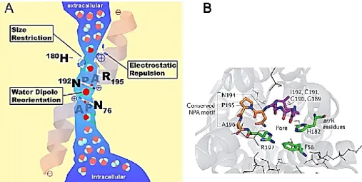
At the center of the pore, highly conserved NPA (Asn-Pro-Ala) motifs, located at the loops B and E that form short hydrophobic helices, in which the asparagine is bonded back with the NH group of the main chain by a carbonyl group, and it enters the membrane from the extracellular side or cytoplasmic side in an opposite orientation. [22, 23]. This organization formed what has come to be known as the ‘hour-glass model’ (Fig. 3A), characterised by wide external openings to the channel with a narrow central constriction where the NPA motifs interact, forming the functional water pore. The primary structure can be divided into 2 similar halves (TM1–3, hemipore-1 and TM4–6, hemipore-2) that probably arose by gene duplication during evolution [24, 25].
In spite of a general highly conserved structure, differences at key areas of the MIP sequences provide a channel selectivity and two constriction points within the pore,
13
referred to as the NPA constriction and the aromatic/arginine (ar/R) selectivity filter (Fig. 3 B), respectively and the selection is done through mechanisms of charge, polar and size exclusions [26, 27].
Figure 3. ‘Hour-glass model’
(A) Schematic diagram representing channel pore in same orientation.
(B) The constriction region (in green) is made up of aromatic and arginine residues (known as the ar/R constriction; residues Phe58, His182 and Arg197); extracellular Asn-Pro-Ala (NPA) residues (Asn194, Pro195 and Ala196) are shown in orange.
(Adapted from Agre et al., 2003 and Verkman et al., 2014).
In the center of the pore, located at the end of loop B and E, as previously described, two conserved Asn-Pro-Ala motifs (NPA filter) are responsible of generation of electric field and provoke the reorientation of 180 degree of the water molecule that destroyed H-bonds between adjacent water molecules and promote the proton exclusion (Fig. 4).
The aromatic/arginine (ar/R)-constriction region is the second filter located at the extracellular side, it impairs the entrance of high molecular weight substrates (∼2.8 Å in AQPs) but it is also a checkpoint site for uncharged molecules in AQPs [27]. The ar/R constriction is formed by the interaction of four amino acids within the pore.
14
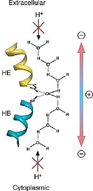
Figure 4. The proton exclusion mechanism
The asparagine residues of the two NPA motifs orient the water molecule in the center of the pore with the two hydrogen atoms perpendicular to the pore axis, dividing the pore into two half channels. The electrostatic field in the pore causes the water molecules in the two half channnels to be oriented in opposite directions, thus preventing proton conduction through the pore.
(From Gonen et al., 2006)
In 1990s was recognized a new AQP family the GlpF is the glycerol facilitator in E. coli. GlpF has a much lower water permeability, but it is permeable to glycerol, urea and glycine [28, 29]. The structure of GlpF was determined to 22 Å resolution by X-ray crystallography [30, 31] showing the same right-handed bundle of six -helices seen in AQP1 [32-34], but also revealed three glycerol molecules (G1–G3) in the pore. The main differerences in the structures of AQP1 and GlpF are found in the extracellular loops (Fig. 5).
15
Figure 5. Glycerol molecules in the GlpF pore
GlpF containing three glycerol molecules shown in space-filling representation. The glycerol molecule in the center occupies the constriction, while the lowest one is located at the height of the two NPA motifs.
(From Gonen et al., 2006)
Loop A is much shorter in GlpF than in AQP1. Loop E, the NPA-carrying loop in the second tandem repeat, is longer in GlpF than in AQP1 and forms a one-turn helix before connecting with transmembrane helix 6. The ar/R constriction region of GlpF pore has to be larger and more hydrophobic than that in the pore of AQP1. This is accomplished by a number of substitutions in residues forming the constriction site of GlpF. In particular Trp48, Gly191 and Arg 205 instead of Phe56, His180, Cys189 and ARg195 in AQP1. A much smaller glycine residue Gly191 replaces His182, which is preserved among water-specific AQPs. The much more hydrophobic Phe200 and Phe58 replace Cys191 of AQP1. These substitutions have critical effects on the characteristics of the GlpF ar/R constriction region: they increase the size in GlpF pore (3.4 Å), increase the hydrophobicity [34] and the conduction is not sensitive to mercurials.
The GlpF channel lining is strongly amphipathic, with oxygens and nitrogens lined up on one side and carbons on the opposite side of the lumen surface.
16
This amphipathic channel uniquely matches the chemical structure of glycerol, which is a composite of the polar hydroxyl group arranged on a non-polar alkyl backbone [30].
Aquaporin classification and selectivity
The AQPs family arose by tandem gene duplication [24]. Since bacteria such as E. coli contain both a glycerol facilitator (GlpF) and a specific water pore (AQPZ), the gene duplication appears to have occurred early in evolution. So far, in mammalian have been discovered 13 AQPs isoform (AQP0-AQP12). Phylogenetic analysis showed that the members of the AQP family have sequence identity that ranges from about 25 to 40%, and can be classified into three major functional subfamily according to their transport capability: aquaporins, aquaglyceroporins and S-aquaporins (Fig 6) [35-37]. AQPs (AQP0, AQP1, AQP2, AQP4, AQP5, AQP6 and AQP8) are a mainly water selective or specific water channels, also named by various authors as "orthodox", "ordinary", "conventional", "classical", "pure", "normal", or "sensu strictu" aquaporins. AQP0, AQP1 and AQP6 are considered water channels, but they are also permeate nitrate and chloride ions, and AQP8 ammonia. Water diffusion through AQPs is inhibited by mercury, except AQP4, which is mercury-intensive aquaporin [38]. The aquaglyceroporins (AQP3, AQP7, AQP9 and AQP10) are permeable to water, but also to other small-uncharged molecules, in particular glycerol and urea. AQP10 is an aquaglyceroporin permeable to water, but not to glycerol and urea [39]. AQP9 was also named “neural channel” that facilitates the water diffusion, but also polyols (glycerol, mannitol, and sorbitol), purines (adenine), pyrimidines (uracil and chemotherapeutic agent 5-fluorouraricil) and monocarboxylates (lactates and -hydroxybutirrate). In addition, AQP9 facilitates metalloid transport, suggesting that AQP9, like AQP7may be a major route of arsenite intake uptake into mammalian cells [40]. S-aquaporins (AQP11 and AQP12), the third subfamily of little conserved amino acid sequences around the NPA boxes, unclassifiable to the first two subfamilies. They are also named "superaquaporins", "subcellular aquaporins", or "sip-like aquaporins". Both unusual AQPs are intracellular proteins, which display a crucial role in endoplasmic reticulum (ER) integrity maintenance; in particular, AQP11 has a C-terminal extremity with a putative ER-related retention signal even AQP12 that lacks this ER retention signal.
17
Figure 6. Phylogenetic tree of mammalian AQPs and AQPs of known atomic structure
(Adapted from Gonen et al., 2006)
Aquaporin distribution and physiological function
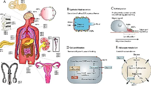
In human, aquaporins are widely distributed in various epithelia and endothelia of tissues involved in fluid transport such as kidney, brain, eye, skin, fat and exocrine glands, suggesting their involvement in major organ functions and disease processes, including urinary concentrating, brain swelling, epilepsy, glaucoma, cancer and obesity [41]. In general, AQPs increase water membrane permeability, they are important actors of fluid homeostasis maintenance in secretory and absorptive processes in response to an osmotic or pressure gradient. However, their specific distribution in certain cell types of an organ often reflects a precise function (Fig. 7A). Tissue distribution and regulation studies have provided indirect evidence for the involvement of AQPs in a variety of physiological processes.
Fluid secretion
AQPs are express in many epithelia, such as kidney tubules, glands, choroid plexus, ciliary body and alveoli, where they increase transepithelial water permeability and are responsible of fluid secretion and absorption.
18
Reduced epithelial cell osmotic water permeability can consequently impair active fluid transport and osmotic water equilibration, resulting in the secretion (or absorption) of a reduced volume of inappropriately hypertonic fluid [42] (Fig. 7B).
Figure 7. Aquaporins distribution in human body and their physiological functions
(A) Schematic rappresentation of AQPs in human body.
(B) Expression of AQPs in epitelium of secretion: AQP deficiency reduces transepithelial water permeability, preventing osmotic equilibration of lumenal fluid and impairing urinary concentrating ability.
(C) Proposed mechanism of AQP-facilitated cell migration, showing water entry into protruding lamellipodia in migrating cells.
(D) Proposed mechanism of AQP3-facilitated cell proliferation involving increased cellular glycerol and consequent increased ATP energy and biosynthesis.
(E) AQP7 facilitates glycerol escape from adipocytes: adipocyte hypertrophy is seen in AQP7 deficiency, possibly becauseof impaired AQP7-dependent glycerol escape from adipocytes, resulting in cellular glycerol accumulation and increased triglyceride content.
(Adapted from Day et al., 2013 and Verkam et al., 2014)
Cell migration
Migration is a fundamental property of cells that occurs during many physiological and pathological processes including organogenesis in the embryo, repair of
19
damaged tissue after injury, the inflammatory response, formation of new blood vessels, and the spread of cancer. In migrating cells, AQP1 and AQP4 become polarized to the front end of cells and they are associated with increased turnover of cell membrane protrusions, suggesting an important role for AQPs at the leading edge of migrating cells [43, 44] (Fig. 6C).
Glycerol metabolism
For many years, the physiological significance of glycerol transport by the aquaglyceroporins was unclear. AQP7 is expressed in the plasma membrane of adipocytes where are involved in efflux of glycerol, in AQP7 deficiency, the plasma membrane of adipocytes showed a reduced glycerol permeability contemporary with an accumulation of glycerol in adipocyte cytoplasm, resulting in increased glycerol-3-phosphate and triglyceride biosynthesis. AQP9 expressed in liver (which facilitate the uptake of glycerol and thereby the availability of glycerol for de novo synthesis of glucose and triglyceride), have an important role in controlling glycerol metabolism in both adipose tissue and liver [45] (Fig. 6D).
Cell proliferation
Another important aspect is the implication of same AQPs, in particular AQP3 and AQP5 to enhance cell proliferation in epidermal cells and the cell growth in cancer condition [46-48] (Fig. 6E).
20
Aquaporin in brain
Considering that about 80% of the brain is water, AQPs play an important role in the production, circulation and homeostasis of the cerebrospinal fluid (CSF). Fluid balance (secretion, removal, fluxes and homeostasis of salts) is important for the brain survival but also neuronal excitability [49].The knowledge of the distribution and regulation of water channels in the brain is important to understand water homeostasis and their correlation with physiopathological condition. To date, nine aquaporin subtypes (AQP1, 2, 3, 4, 5, 7, 8, 9, and 11) have been recognized and partially characterized in human brain cell function, but only three aquaporins (AQP1, 4, and 9) have been clearly identified in brain cells in vivo [50].
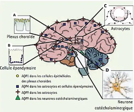
Figure 8. Aquaporin distribution in brain
A) AQP1 is mainly express in the apical pole of choroid plexus (CP). B) AQP4 and AQP9 are co-express in astrocytes.
C) AQP4 is express in ependymal surfaces. D) AQP9 is express in catecolaminergic neurons. (Form Guérin et al., 2005)
21
AQP1
In the central nervous system, AQP1 is mainly express in the apical pole of choroid plexus (CP) (Fig. 8A) in the lining of the cerebral ventricles in primate and in human astrocytes [51-53]. This protein was proposed to be the major water-transporting protein in choroid plexus and to play a role in cerebrospinal fluid formation and in governing the rate of net fluid secretion. The CP is essential for the brain from the earliest stages of the development through the passages into old age. One might speculate that AQP1 is express contemporary with the CP formation and is enables the CP cells to sense the osmolality of the CSF and thereby initiate the appropriate adjustments of transport rates [54]. The ion channel activity of AQP1 is dependent on intracellular cGMP. Activation of AQP1 cation channel by cGMP signaling (activated by the atrial natriuretic peptide, ANP) decreased the rate of net fluid flow from basolateral to apical in confluent layers of cultured rat choroid plexus, and the effect was reversed by application of a blocker of the AQP1 cation channel [55]. CFS traditionally has been considered a cushion providing a physical protection of the brain, a reservoir of salt and nutrient, a drainage system of removal waste products and metabolites, and in generally have a role into the development, homeostasis and repair of CNS. During the life, a progressive functional decline of CP in the aging increase the risk for development of late-onset diseases such as Alzheimer’s disease [56]. This could suggest a hypothetical relationship between AQP1 expreesion and increased risk of Alzheimer’s disease. In the peripheral nervous system, AQP1 is expressed in a subpopulation of primary sensory neurons of dorsal root and nodose ganglia in mice [57] plus enteric neurons and it has been proposed to play a role in cerebrospinal fluid formation and in mice and rats [58-60]. Moreover, AQP1 mRNA or immunoreactivity has been observed in trigeminal ganglion neurons in rodents [58, 61-65]. These results indicate that AQP1 could play a relevant role in trigeminal neurotransmission. Interestingly, AQP1 is express in capillary endothelial cells in the systemic circulation [64], but it is not found in the capillaries of cerebrovascular system in normal condition [66]. Conversely, vascular AQP1 immunolabeling was observed in various tumor cell lines (mammary carcinomas and glioblastomas) implanted in the rat brain [43].
22
AQP4
Aquaporin-4 (AQP4) is the main water channel in the neuropil of the central nervous system and is highly polarized in expression [67]. It is primarily found on astrocytes (Fig. 8C), particularly on the astrocytic end-feet surrounding capillaries and the blood brain barrier as well as the glia limitans [68-70]. AQP4 exists in the end-feet adjacent to the capillary, with lower density found on the end-feet adjacent to neurons; the large surface area of the end-feet near neurons compensates for this lower density. AQP4 is highly abundant at sites of fluid transport, including pial and ependymal surfaces (Fig. 8B) in contact with cerebrospinal fluid (CSF), subarachnoid space, and the ventricular system [67]. AQP4 is also present in vasopressin-secretory neurons in supraoptic and paraventricular nuclei of the hypothalamus, and in Purkinje cells of cerebellum [67, 71, 50]. AQP4 expression has also been reported in neural stem cells obtained from human and murine subventricular zone and in their glial progeny in culture [72]. Based on its location and expression, the first functional role of the AQP4 has been suggested to be an involvement in bidirectional fluid exchange between both the blood and CSF compartments and the brain [68]. In fact, the highly polarized AQP4 expression (in glial membranes that are in direct contact with capillaries and pia) indicates that it mediates the flow of water between glial cells and the cavities filled with CSF and the intravascular space (i.e. across the BBB and blood–CSF barrier). The location of the AQP4 follows the distribution of the inwardly rectifying potassium channel 4.1 (Kir4.1) [68]. Although no functional relationship was clearly shown between AQP4 and Kir4.1, the absence of the AQP4 resulted in delaying the reuptake of the extracellular potassium in epileptic conditions possibly due to an increase of the extracellular space [73]. Water transport via AQP4 in brain is involved in the control of neuronal activity and linked to this activity is the spatial K+ buffering/K+ siphoning [74, 75]. In AQP4 deleted mice was observed an altered neuronal activity explained by altered K+ kinetics in brain extracellular space (ECS) [76, 50].
It was found that AQP4-null mice have delayed K+ clearance from brain ECS after local electrical stimulation [73] and during cortical excitation by spreading depression [77, 50]. AQP4 similarly with other AQPs enhance cells migration, in particular AQP4 deficiency impairs the migration of cultured astrocytes [78].
23
AQP9
AQP9 is an aquaglyceroporin with a broad selectivity transporting carbamides (urea, thiourea), ammonia, polyols (glycerol, mannitol, and sorbitol), purines and pyrimidines (adenine, uracil) and monocarboxyates (lactate, -hydroxybutyrate) [79, 80]. In the brain, AQP9 was first detected in astrocytic cultures [79] and confirmed by immunocytochemical studies in rodent brain [81, 82]. To date, AQP9 expression has been observed in three cell types: glial cells, in particular tanycytes and astrocytes (Fig. 8C) [81-83], endothelial cells of sub-pial vessels [82] and in neurons (Fig. 8D) [82, 84]. The AQP9 expression was observed both on neuronal cell bodies and their processes as well as on the noradrenergic processes present in the adventitia of pial vessels [82]. The distribution of AQP9-ir in neurons and astrocytes is indicated on the schematic drawing of a sagittal rat brain section. The presence of AQP9 in the ependymal lining of the cerebral ventricles raises the possibility that this water channel may be involved in the extrachroideal production and the extra arachnoid reabsorption of the cerebrospinal fluid. AQP4 is also present in these regions in the mouse, suggesting that AQP9 contributes with AQP4 to the facilitation of water movements between CSF and brain parenchyma. Indeed, these channels can facilitate the diffusion of glycerol and lactate, which can serve as energetic substrates [85-89]. In the “lactate shuttle model”[88], glucose is converted into lactate in astrocytes and can then be used as fuel for neurons. In addition, AQP4 and AQP9 labeling on astrocytes was found in white matter tracts, hippocampus and several hypothalamic nuclei. In general, AQP9 is involved in brain energy metabolism as a metabolite channel. Since AQP9 is abundant in glucose-sensing neurons in the brainstem, it could also participate in the regulation of the whole body energy balance. In agreement with this hypothesis, the presence of AQP9 protein was recently demonstrated in mitochondria of astrocytes and dopaminergic neurons [90].
24
Other brain aquaporins
The presence of other members of AQPs family in the brain has been reported, but their functional role remains to be established [37].
AQP7 protein was found during perinatal development of mouse brain and in particular was observed in choroid plexus in parallel with the AQP1 expression. Moreover, despite it was detected in ependyma, pia and some blood vessels of the brain few functional information are yet available [91].
AQP8 was discovered in 1997 and the first studies in the brain showed its expression only by Northern blot analysis, AQP8 mRNA was not detected. However, Yamamoto et al., [92] observed AQP8 expression in oligodendrocytes. Later, Oshio et al., [69] found the AQP8 expression in the spinal cord, in particular in the ependymal cells lining the central canal and observed a faint staining in cells surrounding the canal suggesting that AQP8 could play a role in concert with AQP4 and AQP9 facilitating the water transport into the central canal [93].
25
In vitro models for neurological researchThe studies of the central nervous system are beset with the complexity of direct investigation because of the inaccessibility of the neural tissue, and hence the difficulty in obtaining a brain biopsy, until after the death of the affected individual. Neuronal cell development is a complex and dynamic processes, which generates new neurons of different phenotypes. The neurogenic function from undifferentiated stem and progenitor cells starts at very early stages of development and continues in discrete regions of the mammalian nervous system throughout life. Understanding mechanisms underlying neuronal cell development, biology, function, and interaction with other cells, especially in the neurogenic niches of fully developed adults, is important to provide fundamental insight into the neuroscience. On the other hand, many common and rare neurological diseases can be modelled in rodents, in many cases these animal models do not faithfully reproduce the human syndrome at either the molecular or anatomical levels - perhaps owing to important species differences. Therefore, the employ of in vitro models would be extremely important to understand the brain development, the normal brain functions, the pathological processes involved in neurodegeneration, and finally, to explore therapeutic options for disorders of the CNS, including cells replacement. In vitro models offer advantages over in vivo models in several aspects. First, it is possible to study the role of isolated cells of one particular type in a controlled environment that simulates the disease allowing the exploring in a fast and reproducible manner the single pathogenic mechanisms a possible deleterious or protective role of specific molecules and compounds related damage to the brain. Second, screening for potential actions of drugs is also facilitated [94]. In this sense, in vitro models of neurodegenerative processes have been used to provide important clues about mechanisms of the diseases and potential pharmacological targets. Several studies with neuronal and non-neuronal primary cultures and cell lines have investigated many processes, such as neurotoxicity, inflammation, neuroprotection, and therapeutic approaches. For example, several past and present approaches in vitro have allowed identification of some pathogenic mechanisms involved in Krabbe’s leukodystrophy (Fig.9). Krabbe disease (KD), also known as Globoid cell leukodystrophy (GLD, OMIM #245200), is a metabolic disorder of the white matter of the central and peripheral nervous systems (CNS and PNS) caused by inborn genetic defects. In particular, it is an autosomal recessive inherited
26
neurodegenerative disorder caused by deficient lysosomal enzyme galactosylceramidase (GALC) [95, 96], that leads to a progressive deterioration of oligodendrocytes in the CNS and Schwann cells in the PNS [97]. In any case, studies with primary cultures and cell lines of neurons, microglia, astrocytes, and oligodendrocytes have been used to investigate many processes such as neurotoxicity, inflammation, and neuroprotection and to screen for novel therapies to treat neurodegenerative disorders, including KD [98].
Figure 9. Pathways involved in Krabbe’s leukodystrophy
The in vitro systems described the direct and/or indirect roles of psychosine in the release of cytokines, ROS and NO and in the activation of kinases, caspases, and angiogenic factors. (From Avola et al., 2016).
The in vitro approaches have been widely used, not only to increase knowledge on rare neurodegerative disorders, but also to analyze molecular pathways involved in most common neurodegenerative disease, like Parkinson’s disease.
Parkinson’s disease (PD) is a common progressive neurodegenerative disease clinically characterized by motor impairment, namely bradykinesia, rigidity, resting tremor, and postural instability [99]. Synaptic and axonal degeneration within the striatum followed by loss of dopaminergic neurons in the substantia nigra pars compacta (SNpc) leads to reduced levels of dopamine in the nigrostriatal circuitry [100]. Despite the several information about the clinical progression, the pathogenesis of PD and the involved-molecular mechanisms are not yet completely understood. To clarify these two aspects and consequently provide the means for therapeutic strategies remain indispensable. In the recent years a growing interest
27
has been addressed to neuroinflammation and oxidative stress as key aspects of pathogenesis of disease-associated nigrostriatal degeneration in PD animal models as well in clinical [99, 100]. Usually, PD studies are limited because of the inaccessibility of the neural tissue and hence the difficulty in obtaining a brain biopsy, until the death of the affected individual. The perfect cell culture system needs to be homogeneous, easily expanded in order to generate large numbers of neuronal precursor cells and easily directed towards a post-mitotic state in a synchronized manner with a mature neuronal (dopaminergic) phenotype. In this kind of system, the specific molecular pathways and the genes/proteins at the basis of the progression of PD should be studied (Fig 10) [101]. Up to now, an in vitro model widely used in the field of PD research is the SH-SY5Y human neuroblastoma cell line. These cells are easy to differentiate into a post-mitotic mature dopaminergic state and widely used to study mechanisms of neurodegeneration [102].
Figure 10. Parkinson’s disease in vitro model
Many molecules are currently used in cellular models of PD, including pesticides as paraquat or rotenone and neurotoxins such as 6-hydroxydopamine (6-OHDA) and 1-methyl-4-phenylpyridinium (MPP+).Paraquat, 6-OHDA and MPP+ easily cross cell membrane through the dopamine transporter (DA) thus inducing the formation of α-synuclein aggregates and mitochondrial impairment with the subsequent production of ROS, inhibition of mitochondrial complex I and the release of proapoptotic molecules. (FromCabezas et al., 2013).
28
Stem Cells
The term “Stem cells” appears for the first time in the scientific literature as early as 1868 by Ernst Haeckel (German biologist), that used the term “Stammzelle” (German for stem cell) to designate an unicellular organism that he considered the evolutionary ancestor of multicellular organism from which he presumed all multicellular organisms evolved. Subsequently he used the same term to designate the fertilized egg distinguishing it sharply from maternal egg. (Fig. 11) [103].
Figure. 11 First representations of an early embryo (bottom) from which the term Stammzelle, stem cell.
(From Federico Calegari, Claudia Waskow 2014)
Today, the term “stemness” includes the ability of self-renewal (replication capacity), clonality, and potency. These capabilities make the stem cells, the heart of some of the most fascinating questions in biology and medicine and can be defined as single cells capable of generating daughter cells identical to their mother as well as long-term survival. They have the potential to divide asymmetrically and eventually becoming a functionally mature cell to differentiating into multiple specific cell types, including neuronal and glial cell lineages [104]. They were found in all multicellular organisms. A hierarchy classification of stem cells can be obtained based or on their time of onset or their differentiation potency. According to their time of onset may be sourced from the blastocyst in the developing embryo prior to implantation, derived from inner cell mass (embryonic stem cells, ESCs) or derived from the fetus (fetal stem cells, FSCs) or, from blood and tissues postnatally (adult or somatic stem cells, ASCs) [105, 106]. Stem cells can best understood in terms of how committed they are to becoming any particular type of cells. Their developmental versatility includes cells capable of differentiating into any cell type
29
in the body including embryonic and extra-embryonic tissues, defined totipotent. According to this definition, the zygote (fertilized egg) is the only totipotent stem cell. Pluripotent stem cells, are descendants of the totipotent stem, and include embryonic stem cells, embryonic germ cells and cord blood-derived stem cells capable to differentiate into cell types of all three germ layers: ectoderm, mesoderm and endoderm. Substantially, they can develop into each of the more than 200 cell types of the adult body [107] but cannot produce the extra-embryonic tissues or the placenta [108]. The offspring of the pluripotent cells become antecedents of specialized cells able to differentiate into a limited range of cells within a tissue type. At this stage, they are multipotent with a more constraint differentiation potential and capacity for self-renewal; this category includes adult stem that are undifferentiated cells present in differentiated tissue and used to replace cells that have died or lost function. It renews itself and can specialize to yield all cell types present in the tissue from which it originated. So far, adult stem cells have been identified for many different tissue types such as hematopoetic (blood), neural, endothelial, muscle, mesenchymal, gastrointestinal, and epidermal cells. Finally, progenitor cells with a very limited differentiation potential and able to produce only one cell type are definited unipotent such as the erythroid progenitor cells that differentiate only into red blood cells (Fig.12).
Figure 12. Potency and source of stem cells
30
Mesenchymal stem cells
Mesenchymal stem cells (MSCs) are ASCs with mesodermal and neuroectodermal origin [109].The hypothesis about their existence goes back to 1867, but MSCs were first isolated and defined by Friedenstein and co-workers as plastic-adherent, colony-forming-unit fibroblastic cells (CFU-F) [110]. Later, these cells were named “marrow stromal cells” due to their possible use as a feeder layer for hematopoietic stem cells [111]. MSCs possess an extended degree of plasticity compared to other ASCs populations. In fact, they have been shown to differentiate under appropriate conditions, into adipocytes, chondrocytes, osteocytes or muscle and might be able to differentiate also into endodermal and ectodermal cell lineages with neuronal cells electrically excitable [112] and into glial phenotypes [113]. In addition, it seems that few cells that were capable of engrafting into the nervous tissue fused with endogenous cells and thereby acquired the phenotype of the partner host cell [114]. Human MSCs are generally isolated from bone marrow (BM) aspirate harvested from the superior iliac crest. Moreover, they have also been found in other BM cavities such as vertebrae bodies [115]. BM-MSCs have been considered the gold standard, but many more recent reports describe the presence of MSCs in a variety of fetal, perinatal and adult tissues, including peripheral blood, umbilical cord Wharton’s Jelly and blood, fetal liver and lung, adipose tissue, skeletal muscle, amniotic fluid, synovium and circulatory system where they work as supportive cells and maintain tissue homeostasis. Recently it was shown that MSCs are recruited from perivascular niches, which represent a tight network throughout the vasculature of the whole body. Current research on MSCs is mainly focused on their self-renewal capacity, multi-lineage differentiation potential, surface markers, and immune regulation. Considering the rising interest in the biology of MSCs, the International Society for Cellular Therapy (ISCT) describes a set of standards to define human MSC for both laboratory-based scientific investigations and for pre-clinical studies [116]:
1) Adherence to plastic in standard culture condition;
2) Specific surface antigen (Ag) expression: phenotype positivity (≥ 95 %+) for CD105, CD73, CD90; phenotype negative (≤ 2 %+) for CD45, CD34, CD14 or CD11b, CD79 α or CD19, and HLA-DR;
3) Multipotent differentiation in vitro potential to endodermal and ectodermal cell lineages (Fig. 13).
31
MSCs from different tissues also display significant differences in proliferative potential and their capacity of mature differentiation and integration in the host tissue are not yet clear. This suggested that the functional difference between MSCs from different sources might be related to the origin of the cells.
Figure 13. Mesenchymal from bone marrow and adipose tissue
Cell fate that have been demonstrated for adult stem cells regarding the mesodermic, endodermic or ectodermic cell lineage.
32
Mesenchymal stem cells from adipose tissue
Recent studies have shown that subcutaneous adipose tissue provides a clear advantage over other MSCs sources due to the ease with which adipose tissue can be accessed as well as to the ease of isolating stem cells in abundant quantities [117, 118]. One gram of adipose tissue yields approximately 5,000 stem cells, whereas the yield from BM-MSCs is 100 to 1,000 cells/mL of marrow [119]. AT-MSCs were first isolated by Zuk et al. [120] through an initial enzymatic digestion of the harvested adipose tissue, which yields a mixture of stromal and vascular cells (preadipocytes, fibroblasts, vascular smooth muscle cells, endothelial cells, resident monocytes/macrophages, lymphocytes, and AT-MSCs [121], referred to as the stromal-vascular fraction (SVF) [122]. SVF is a rich source of pluripotent AT-MSCs [123, 120], which were first named processed lipoaspirate (PLA) cells [120, 124]. Morphologically, AT-MSCs are fibroblast like cells and preserve their shape after in
vitro expansion [125, 126, and 120]. Average doubling time of tissue-cultured
AT-MSCs is between three [120] to five days [127]. AT-AT-MSCs in long-term culture showed the manifestation of senescent feature at passage (P) 15 and P20 and all the results showed that in vitro culture beyond P10 favors senescence pathways and therefore limits their clinical use [128]. They phenotypically express over 90 % of MSCs markers CD13, CD29, CD44, CD71, CD90, CD105/SH2, SH3, and STRO-1. AT-MSCs also share cell surface antigens with fibroblasts and pericytes, in contrast, no expression of the hematopoietic lineage markers: CD14, CD16, CD31, CD34, CD45, CD 56, CD 61, CD 62E, CD 104, and CD106 and for the endothelial markers: CD31, CD144, and von Willebrand factor was observed [129, 130, 124]. This observation results not only in a more homogeneous cell population with extended culturing but also in changes in AT-MSCs features: CD34+ cells have a greater proliferative capacity whereas CD34- cells have a higher plastic adherence. Moreover, telomerase activity seems to be superior in AT-MSCs compared with BM-MSCs that indicates maintenance of the capacity for self-renewal and proliferation ability in culture and after transplantation [131]. AT-MSCs are of particular interest because they demonstrated properties that could be helpful in cell therapy: angiogenicity [132-134], immunomodulation [137], and promotion of tissue remodeling [134-136]. In fact, they are able to secrete growth factors promoting angiogenesis (VEGF, HGF, PDGF, FGFb, IGF, ecc.) [134, 138-140]. Therefore, the use of AT-MSCs is highly justified to induce the tissue revitalization or
33
reconstruction. Furthermore, AT-MSCs have anti-inflammatory, low immunogenic characteristics [141] making them useful also in allotransplant without immunosuppression [142, 143]. They not display transformation in teratoma [144], and there are few ethical issues surrounding their clinical application [145-147]. These unique features make AT-MSCs the ideal candidate for very different neurological diseases associated with degeneration and inflammation. Anyway, the complexity of the mechanisms involved in survival, differentiation and immunomodulation is not yet completely understood.
Neural differentiation of AT-MSCs
AT-MSCs are traditionally considered capable of differentiating into cell types of their own original lineage, but also in ectodermal and endodermal tissues or organs in the fields of gastroenterology, neurology, orthopedics, reconstructive surgery, and related clinical disciplines [124]. We and many other groups have showed that in
vitro AT-MSCs can be capable of chrondrocyte, adipocyte, and neural phenotypes
differentiations [148-153]. Given their ability to differentiate both morphologically and functionally into neurons, astrocytes and oligodendrocytes they are currently under investigation for neurological diseases, for brain injury, stroke, neuronal protection, and peripheral nerve injury. In general, the processes of proliferation, allocation, and lineage-specific terminal differentiation are regulated by a complex interplay involving stem cell transcription factors (molecular rheostats), cell-specific transcription factors, and a wide variety of cellular kinases, growth factors, and receptors. Neural differentiation of AT-MSCs has been reported in numerous studies, by using a variety of protocols. Some used simple chemical agent, treated AT-MSCs with conditioned media or mixture of growth factors, or maintained in co-culture with other neural cells. Earlier studies that utilized certain agent such as -mercaptoethanol (BME), dimethyl sulfoxide (DMSO), and butylated hydroxyanisole (BHA) [154] reported that AT-MSCs could differentiate into cells of neuronal morphology within few hours, and therefore, these cells may be feasible for transplantation. However, the reversible morphological nature of differentiated neuronal cells [155] and the toxicity of the chemical substance employed limit their use in clinical trials [156]. Differentiation of AT-MSCs towards the neuronal lineage was also induced using conditioned media obtained from cultured rat olfactory ensheathing cells (OECs) or human B104 neuroblastoma cells [150]. In both cases,
34
AT-MSCs acquire morphological features of neuronal-like cells and, in a time-dependent manner, they express nestin, protein gene product 9.5 (PGP 9.5) and MAP-2. In other studies, AT-MSCs were turned into multipotent stage and then induced into neural cell lineages, by exposing them to appropriate neural differentiation conditions [156-158]. Different protocol using growth factors, as basal fibroblast growth factor (bFGF), FGF-, nerve growth factor (NGF), brain-derived neurontrophic factor (BDNF) has been translated from the neural stem cells (NSCs) culture method [159-161]. NSCs are traditionally grown in induction media added of EGF and FGF- forming floating aggregation called neurosphere [162]. After neurosphere dissociation, culture medium was supplemented with BDNF and retinoic acid. Following this treatment, about half of AT-MSCs showed morphological, immunocytochemical and electrophysiological evidence of initial neuronal differentiation. Neural modified AT-MSCs by the cultivation in neurodifferentiative conditions, in contrast to native AT-MSCs, express several neural progenitor and mature neural markers demonstrated by real time RT-PCR, Western blot and immunocytochemistry. The most cited proteins include S100, SOX-2, GFAP, O4, -tubulin-III, PGP 9.5 and NeuN. Moreover, with appropriate neural induction protocols, AT-MSCs could produce mature neuran-like cells that exhibit multiple neuronal properties and traits, such as action potential, synaptic transmission, secretion of neurotrophic factors and dopamine, and demonstration of spontaneous post-synaptic current.
35
Stem cells and aquaporins
SCs pass through a complex gauntlet of cell behaviors, such as proliferation, differentiation, and migration. Recently, a growing interest has been addressed to highly controlled movement of several ions and small molecules by AQPs that trigger numerous, complex signaling pathways that underscore the regulation of these behaviors. Despite it is clear that AQPs governing the water homeostasis and the considerable amount of water into or out of the cells to achieve rapid volume regulation during stem cells differentiation [163], only a small number of studies indicated the expression and function of AQPs in stem cells [72, 164-167]. AQP3 was found in human and rat progenitor cells surface and glandular airway epithelium [165, 168]. AQP4, AQP8, and AQP9 were expressed in murine adult neural stem cells [164-72] that normally replace neurons and/or glia in the adult brain and spinal cord. AQP8 is a general feature characterizing stem cells observed in mouse embryonic stem cells [169] and in intracellular membrane systems, probably corresponding to mitochondria of murine adult neural stem cells where might play a critical role in proliferation and differentiation [164]. In fact, AQP8 are involved in the mediations of water between the cytoplasm and the mitochondrial compartment, thus adjusting mitochondrial volume homeostasis pivotal for the activity of the electron-transport chain [170]. AQP4 and AQP9 expression was found in the adult forebrain periventricular region [171, 172], where the NSCs pool resides, but their expression levels and cellular localization were differentially regulated during murine adult neural stem cells differentiation into neurons and glial cells [72]. Following cell differentiation there was an increase in the overall levels of AQP4 mRNA and a concomitant redistribution of the protein, which localized in a high proportion of differentiated glial cells. In contrast to AQP4, only 30% of the cells in the differentiated cultures showed AQP9 positivity. AQPs protein levels seem to be progressively downregulated in the NSCs post commitment phases, on the contrary, using AQP4 knockout (KO) mice, it was demonstrated that AQP4 deletion reduced the proliferation, migration, survival, and neuronal differentiation of neural stem cells of adult mice [166]. An observed impairment in neurosphere formation in AQP4 KO mice was attributed to both increased cell apoptosis and decreased cell proliferation due to cell cycle arrest in G2/M phase [173]. AQP1 and AQP5 were expressed in mouse BM-MSCs in the plasma membrane pattern [167]. AQP1 promotes MSCs migration into the fracture sites by upregulating the expression of
36
-catenin and focal adhesion kinase (FAK) [174]. AQP5-mediated high plasma membrane water permeability enhances the apoptosis rate of differentiating mouse BM-MSCs, thus decreasing their differentiation capacity [167].
37
AIMS OF THE RESEARCH
AQPs are expressed in a specific age-dependent manner, concomitantly with the intracellular and extracellular water content changes reaching a crucial role during the early postnatal period. In particular, water balance involving AQPs might be important during the neurogenesis, accompaning the entire developmental age and maintaining cell and tissue homeostasis. In the brain the electrical activity and the survival of neurons is strictly correlated to water, glycerol and lactate balance; alteration or genetic defect in AQPs expression are associated with several human disease and neurodegenerative disorders.
AQPs may contribute also in stem cell differentiation, as stem cells transport considerable amount of water into or out of the cells to achieve rapid volume regulation during differentiation. Nevertheless, until now, only a small number of studies has been published on expression and function of AQPs in stem cells. Thus, the aim of the present study was to identify the gene and protein expression profile of AQPs in two different cell models, which allowed reproducing in vitro some physiological and pathophysiological conditions of nervous cells.
Additionally, during the initial phase of this research to look for the most appropriate experimental protocol to use for the proposed aims, a preliminary study of protocols about the in vitro models of some neurodegenerative diseases such as Krabbe leukodystrophy was carried out.
As regards the experimental part of the proposed objectives, the first model evaluated the dynamic expression of aquaporins during the differentiation of human mesenchymal stem cells from adipose tissue (AT-MSCs) toward neural phenotypes. The objective was to determine when and which AQPs are produced during differentiation of AT-MSCs into astrocytes, oligodendrocytes and/or neurons. The second model has reproduced in vitro oxidative and inflammatory conditions of the neurons present in the brain of patients suffering from Parkinson's disease (PD). Usually, PD researches are limited by the inaccessibility of the neural tissue and hence the difficulty in obtaining a brain biopsy, until the death of the affected individual. Therefore, a human neuroblastoma cells line (SH-SY5Y) was differentiated into dopaminergic neurons with retinoic acid (RA) and phorbol 12-myristate 13-acetate (MPA) alone or in association and subjected to treatment with the neurotoxin 1-methyl-4-phenyl-1,2,3,6-tetrahydropyridine (MPTP) and the
38
hydrogen peroxide to strongly mimic the oxidative environment that characterize PD neurons in vivo. The aim was to verify a primary involvement of AQP4 and AQP9 in the behavior of dopaminergic neurons in presence of oxidative stress and PD neurotoxin suggesting their possible application in diagnosing as well as in the understanding of PD during life.
39
40
J Neurosci Res. 2016 Nov; 94(11):1284-92. doi: 10.1002/jnr.23846.
KRABBE’S LEUKODYSTROPHY: APPROACHES AND MODELS IN VITRO
Rosanna Avola, Adriana Carol Eleonora Graziano, Giovanna Pannuzzo, Elisa Alvares, Venera Cardile
Department of Biomedical and Biotechnological Sciences, Section of Physiology, University of Catania, Via S. Sofia 64, 95125 Catania, Italy
Running title: Cell models and Krabbe disease
Key words: Apoptosis; Angiogenesis; Cell models; Globoid Cell Leukodystrophy;
Inflammation; In vitro experiment.
Abstract
This Review describes some in vitro approaches used to investigate the mechanisms involved in Krabbe’s disease, with particular regard to the cellular systems employed to study processes of inflammation, apoptosis, and angiogenesis. The aim was to update the knowledge on the results obtained from in vitro models of this neurodegenerative disorder and provide stimuli for future research. For a long time, the nonavailability of established neural cells has limited the understanding of neuropathogenic mechanisms in Krabbe’s leukodystrophy. More recently, the development of new Krabbe’s disease cell models has allowed the identification of neurologically relevant pathogenic cascades, including the major role of elevated psychosine levels. Thus, direct and/or indirect roles of psychosine in the release of cytokines, reactive oxygen species, and nitric oxide and in the activation of kinases, caspases, and angiogenic factors results should be clearer. In parallel, it is now understood that the presence of globoid cells precedes oligodendrocyte apoptosis and demyelination. The information described here will help to continue the research on Krabbe’s leukodystrophy and on potential new therapeutic approaches for this disease, that even today, despite numerous attempts, is without cure.
41
Significance Statement
This Review describes some past and present approaches in vitro that have allowed identification of the pathogenic mechanisms involved in Krabbe’s leukodystrophy. Several studies with neuronal and non-neuronal primary cultures and cell lines have investigated many processes, such as neurotoxicity, inflammation, neuroprotection, and therapeutic approaches. Here particular attention is focused on some cellular systems used to investigate processes of inflammation, apoptosis, and angiogenesis in Krabbe’s disease. The knowledge on models and mechanisms obtained through in vitro experiments can be a stimulus for further research both in vitro and in vivo.
42
Introduction
Krabbe’s disease (KD) is an autosomal recessively inherited neurodegenerative disorder characterized by demyelination in the central nervous system (CNS) and peripheral nervous system (PNS) with the consequent loss of all cognitive and nerve functions until death (Suzuki and Suzuki, 1970). Diffuse demyelination, gliosis, loss of oligodendrocytes, and presence of globoid cells are the “hallmarks” of the pathology. For this reason, KD is also called globoid cell leukodystrophy (GLD) for the presence in the white matter of phagocyte-lineage cells, the globoid cells, characteristic of galactocerebroside accumulation, which can be identified by the presence of periodic acid–Schiff (PAS)-positive material (Wenger et al., 1997). Ranked among the lipidosis, KD is caused by genetic defects in the activity of galactosylceramidase (GALC), the lysosomal enzyme (hydrolase) that degrades two important components, galactosylceramide (gal-cer) and galactosylsphingosine (psychosine; Hideki and Suzuki, 1984; Graziano and Cardile, 2015). The inadequate degradation of these glycosphingolipids involves an accumulation of uncatabolized products. Despite the GALC deficiency, gal-cer does not dramatically increase in the brain of patients with KD. On the contrary, psychosine accumulates in the brain (Miyatake and Suzuki, 1972; Vanier and Svennerholm, 1976; Svennerholm et al., 1980; Suzuki, 1998), causing apoptotic death of myelingenerating cells, oligodendrocytes in CNS, and Schwann cells in PNS (Hannun and Bell, 1987; Tanaka and Webster, 1993). Until now, the exact mechanism of psychosine cytotoxicity has not yet been fully elucidated, and several different mechanisms have been postulated to account for its cytotoxic action.
Over the last years, many advances in understanding the normal brain functions and the pathological processes involved in neurodegeneration have emerged from work undertaken in in vitro models. Thus, studies with primary cultures and cell lines of neurons, microglia, astrocytes, and oligodendrocytes have been used to investigate many processes such as neurotoxicity, inflammation, and neuroprotection and to screen for novel therapiesy to treat neurodegenerative disorders, including KD (Gibbons and Dragunow, 2010).
To update the knowledge on cell models and methods used to study molecular mechanisms and so to provide specific stimuli for future research, this review presents a mix of past and present in vitro investigations on KD. We focus our