Contents lists available atScienceDirect
Journal of Pediatric Surgery Case Reports
journal homepage:www.elsevier.com/locate/epscVolvulus secondary to cystic ileal duplication: Importance of prenatal
imaging and prompt post-natal intervention
F. Molinaro
a, E. Bindi
a,∗, C. Pellegrino
a, F.M. Severi
b, C. Bocchi
b, F. Carbone
c, M. Messina
a,
R. Angotti
aaDepartment of Medical Sciences, Surgical Sciences and Neurosciences, Section of Pediatric Surgery, University of Siena, Italy bDepartment of Medical Sciences, Surgical Sciences and Neurosciences, Section of Gynecology and Obstetrics, University of Siena, Italy cDepartment of Medical Sciences, Surgical Sciences and Neurosciences, Section of Radiology, University of Siena, Italy
A R T I C L E I N F O
Keywords: Prenatal ultrasound Volvulus Ileal duplicationA B S T R A C T
Duplications of the alimentary tract are rare malformations, and ileum is the most involved intestinal tract. During fetal life, they are usually occasionalfindings and may be asymptomatic until adulthood. We present the clinical case of volvulus on ileal cystic duplication diagnosed by ultrasound during prenatal follow-up. Case report: Diagnosis of cystic ileal duplication was made by ultrasound at our center at 23 weeks of gestational age, which was confirmed with fetal MRI. During follow-up, at 36 + 4 weeks, the presence of dilated loops with reduction of peristalsis was shown by ultrasound, so that, despite stable fetal parameters, an emergency cae-sarean delivery was programmed for suspected volvulus. After being stabilized, the patient underwent to la-parotomy that revealed ileal volvulus at 2 cm from the ileo-cecal valve on a cystic duplication. Cyst and necrotic loop (10 cm) was resected and was performed an anastomosis. Patient started feeding on VII post-operative day, and was discharged on the X day. Patient is on follow up and present good conditions.
Discussion: Diagnosis of intestinal cystic mass in prenatal period requires a close follow-up because the risk of a volvulus. According to the literature, even in the presence of stable fetal parameters, the presence of dilated loops and the absence of peristalsis are signs that must suspect the presence of a mechanical occlusion for which timely treatment is necessary to reduce mortality and morbidity.
1. Introduction
Abdominal cystic masses are one of the most frequently mal-formations discovered by prenatal ultrasound. The involvement of bowel, in sense of intestinal duplication, can cause volvulus of the small intestine, which represents a life-threatening condition for the fetus. The ultrasound (US) represents a routine examination in the current prenatal follow up; recent advances in this technology make possible to diagnose many malformations and plan a precise follow-up of women to ensure a good outcome for mother and fetus. In this paper we de-scribe our experience in a case of prenatal volvulus by ileal duplication. 2. Case report
At our prenatal counseling clinic, a cystic mass was diagnosed in a male fetus of 23 weeks of gestational age. In the suspect of an ileal cystic duplication the women underwent to magnetic resonance that confirmed the ileal duplication. Therefore, based on our previous
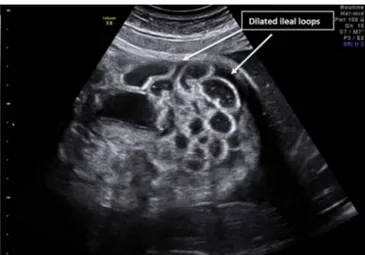
experience of two volvulus on prenatal duplication, we planned a close follow up by ultrasound (weekly). At 36 weeks, ultrasound showed bowel dilation and a peristalsis slowdown (Fig. 1). Despite the absence of signs of fetal distress, we decided to perform an urgent caesarean delivery in suspicion of volvulus. After birth, the patient was admitted to intensive care to be stabilized. The objective examination highlighted a meteoric abdomen. An abdominal x-ray has been performed, showing the presence of gastrectasis and the absence of air in the small bowel (Fig. 2a and b). Given the suggestive clinical and radiological signs of intestinal volvulus, we performed an urgent laparotomy. Abdominal exploration discovered a volvulus of small bowel by ileal duplication at 2 cm from ileocecal valve (Fig. 3). Volvulus derotation was performed and resection of necrotic loops was performed for a length of about 10 cm (Fig. 4). An end-to-end anastomosis was performed. The patient started feeding on the seventh postoperative day. He was discharged in the tenth day. He is currently attending follow up at our center and has good general conditions.
https://doi.org/10.1016/j.epsc.2018.02.011
Received 27 January 2018; Received in revised form 12 February 2018; Accepted 13 February 2018
∗Corresponding author. Ospedale Santa Maria alle Scotte, Viale Bracci 16, 53100 Siena, Italy.
E-mail address:[email protected](E. Bindi).
Journal of Pediatric Surgery Case Reports 32 (2018) 72–74
Available online 17 February 2018
2213-5766/ © 2018 Published by Elsevier Inc. This is an open access article under the CC BY-NC-ND license (http://creativecommons.org/licenses/BY-NC-ND/4.0/).
3. Discussion
Ultrasound has become the basic examination in the follow-up of all prenatal counseling centers. With the current advances in this tech-nology it is possible to highlight the presence of a wide variety of anomalies, especially abdominal [1]. In this last group, one of the most frequent discovered malformations are the cystic masses.
The presence of a cystic mass requires a more precise diagnostic definition. The cyst shape, size and relation to the adjacent organs to-gether with the timing of diagnosis are useful parameters to formulate a correct prenatal diagnosis [2]. Although, according to many studies, ultrasonography represents the gold standard in cystic mass definition and it is known a correlation up to 90% between prenatal and postnatal diagnosis, in some cases, other radiological examinations are required [3].
In our case, indeed, due to the impossibility of defining eco-graphically the cyst's anatomical origin, it was necessary to perform a magnetic resonance imaging that diagnosed an ileal duplication cyst.
Such a diagnosis made it necessary to determine, in agreement with gynecologists, an ultrasound follow-up weekly to discover early warning signs.
In fact, the presence of a cystic duplication in the prenatal age re-presents a risk factor for the onset of a volvulus. This is an emergency which has a high incidence in thefirst year of life, but there are few reports in the literature in the prenatal period, particularly in combi-nation with other congenital malformations [4] as ileal duplication.
In our center we have experienced two cases of volvulus in asso-ciation with ileal cystic duplication.
This is a life-threatening condition and late diagnosis contributes to high rate of morbidity and mortality. Survival of these infants is in-fluenced by the gestational age at birth, amount of compromised bowel, and the ability of neonate to tolerate surgery. Although not the most common outcome, stillbirth has been reported in the literature in cases of fetal volvulus. Since 2004 there have been reported three cases of term fetal demise, whit a diagnosis of volvulus by autopsy [5]. In the
Fig. 1. Ultrasound at 36 week: dilated and aperistaltic loop of bowel.
Fig. 2. a, b: Abdominal x-ray, performed at birth, showing gastroparesis and absence of air in the bowel.
Fig. 3. Intraoperative image. Intestinal infarction by volvulus of bowel by duplication.
Fig. 4. Bowel resected with ileal duplication.
F. Molinaro et al. Journal of Pediatric Surgery Case Reports 32 (2018) 72–74
Literature there are no many cases of prenatal diagnosis and early treatment of volvulus. By making a review, from 1999 to 2013, 16 cases of ultrasound diagnosis of volvulus were reported (Table 1). The most common sign at ultrasound in this series was, as in our case, the pre-sence of dilated intestinal loops and only in one case was detected the presence of aperistaltic bowel [6,7]. Thisfinding, in association with a previous diagnosis of ileal cystic duplication, resulted of great im-portance in our management and it brought to decision of urgent de-livery, despite the vital parameters of the fetus were stable.
Indeed, it is important to note that severe and abrupt fetal bowel dilatation in a previously stable fetus with hypoperistalsis and poor fetal activity are strongly associated with fetal volvulus.
This can result in a poor prognosis for perinatal outcome without prompt and appropriate intervention [8].
Currently there are no precise and reproducible guidelines re-garding the prenatal follow-up of these malformations. The American College of Obstetricians and Gynecologists and the American College of Radiology recommend routine sonograms in the first and/or second trimester, but state that no evidence exists to recommend routine so-nograms in the third trimester [9,10]. According to our experience, ultrasound is the most reliable and most sustainable exam, given the good cost-benefit ratio, as regards the follow up of fetuses with such malformations [18]. The timely identification of signs of alarm, even with stable parameters, makes it possible to program a safe delivery, to guarantee safe post-natal stabilization procedures and to perform sur-gery with the least possible risk ensuring the sparing of bowel as more possible.
4. Conclusion
The volvulus of the small intestine represents a life-threatening condition. It is known as postnatal condition, but it must be considered as a possible prenatal condition in fetus with bowel malformation as intestinal duplication. A close follow up by ultrasound and the knowl-edge of signs of alarm (bowel dilatation, aperistaltic bowel) in these situations ensures a correct perinatal and postnatal management and increases the chances of survival of fetus with low sequels.
Patient consent
Consent to publish the case report was not obtained. This report does not contain any personal information that could lead to the identification of the patient."
Funding
No funding or grant support. Authorship
All authors attest that they meet the current ICMJE criteria for Authorship.
Conflict of interest
The following authors have nofinancial disclosures: MF, BE, PC, SFM, BC, CF, MM, AR.
Appendix A. Supplementary data
Supplementary data related to this article can be found athttp://dx. doi.org/10.1016/j.epsc.2018.02.011.
References
[1] Gerscovich E, Sekhon S, Loehfelm T, Wootton-Gorges S, Greenspan A. A reminder of peristalsis as a useful tool in the prenatal differential diagnosis of abdominal cystic masses. J Ultrasonogr 2017;17(69):129–32.
[2] Olutoye O, Ohuoba E, Fruhman G, Zacharias N. Perinatal survival of a fetus with intestinal volvulus and intussusception: a case report and review of the literature. Am J Perinatol Rep 2013;03(02):107–12.
[3] Leopold S, Al-Qaraghouli M, Finck C, Hussain N. Magnetic resonance imaging di-agnosis of volvulus through mesenteric defect in neonate. Am J Perinatol Rep 2016;06(02):e239–42.
[4] Praveen C, Hota P, Kumar C, Babu A. Intestinal duplication cyst presenting as volvulus: a rare case report. Bali Med J 2014;3(2).
[5] Durand M, Coste K, Martin A, Scheye T, Creveaux I, Vanlieferinghen P, et al. Fetal midgut volvulus as a sign for cysticfibrosis. Prenat Diagn 2008;28(10):973–4. [6] Uerpairojkit B, Charoenvidhya D, Tanawattanacharoen S, Manotaya S,
Wacharaprechanont T, Tannirandorn Y. Fetal intestinal volvulus: a clinico-sono-graphicfinding. Ultrasound Obstet Gynecol 2001;18(2):186–7.
[7] Lee J, Im S, Lee G. Evolution of sonographicfindings in a fetus with ileal atresia. J Clin Ultrasound 2011;39(6):359–62.
[8] Park J, Cha S, Kim B, Kim Y, Choi Y, Chang I, et al. Intrauterine midgut volvulus without malrotation: diagnosis from the‘coffee bean sign’. World J Gastroenterol 2008;14(9):1456.
[9] ACOG Practice Bulletin No. 98: Ultrasonography in pregnancy. Obstet Gynecol. 2008;112(4):951–962.
[10] Molvarec A, Bábinszki Á, Kovács K, Tóth F, Szalay J. Intrauterine intestinal ob-struction due to fetal midgut volvulus: a report of two cases. Fetal Diagn Ther 2006;22(1):38–40.
[18] Trachsel D, Heinimann K, Bösch N, Hammer J. Cysticfibrosis and intrauterine death. J Perinatol 2007;27(3):181–2.
Table 1
Review of the literature: in the table are shown studies that reported prenatalfindings of fetal volvulus.
Papers Number of case Gestational age (wk) of diagnosis Findings by ultrasound
Molvarec et al., 2007 2 32/36 Dilated loops of bowel
Durand et al., 2008 3 34/34/39 Dilated loops of bowel
Leung et al., 2001 1 34 Polyhydramnios and dilated loops
Crisera et al., 1999 1 37 Polyhydramnios and dilated loops
Usmani et al., 1991 1 30 Polyhydramnios and dilated loops
Samuel et al., 1984 1 34 Polyhydramnios and dilated loops
Black et al., 1994 2 36/34 Polyhydramnios and dilated loops
Has et al., 2002 1 34 Dilated and aperistaltic loops
Park et al., 2008 1 33 Dilated loops
Noreldeen et al., 2008 1 31 Dilated loops
Yu et al., 2013 1 37 Dilated loops
Ohuoba et al., 2013 1 37 Dilated loops
Our cases, 2017 3 36/35/37 Dilated loops, aperistaltic loops
F. Molinaro et al. Journal of Pediatric Surgery Case Reports 32 (2018) 72–74