Research Article
Long-Term Outcomes of Behçet
’s Syndrome-Related Uveitis: A
Monocentric Italian Experience
Jurgen Sota
,
1Luca Cantarini
,
1Antonio Vitale
,
1Arianna Sgheri,
2Stefano Gentileschi
,
1Valeria Caggiano,
1Viviana Gelardi,
1Bruno Frediani,
1Gian Marco Tosi,
2and Claudia Fabiani
21Research Center of Systemic Autoinflammatory Diseases and Behçet’s Disease Clinic, Department of Medical Sciences, Surgery and Neurosciences, University of Siena, Siena, Italy
2Ophthalmology Unit, Department of Medicine, Surgery and Neuroscience, University of Siena, Siena, Italy Correspondence should be addressed to Luca Cantarini; [email protected]
Received 29 February 2020; Revised 8 April 2020; Accepted 28 April 2020; Published 15 May 2020 Academic Editor: Phileno Pinge-Filho
Copyright © 2020 Jurgen Sota et al. This is an open access article distributed under the Creative Commons Attribution License, which permits unrestricted use, distribution, and reproduction in any medium, provided the original work is properly cited. Objectives. To examine demographic and clinical characteristics and long-term visual outcome in a cohort of Italian patients affected by Behçet’s uveitis (BU). Materials and Methods. Retrospective chart review of 47 patients with BU attending our unit between January 2018 and December 2019. Ophthalmologic manifestations, best-corrected visual acuity (BCVA), fluoroangiography and optical coherence tomography findings, and ocular complications were recorded. Predictive factors of a poor visual outcome and long-term complications were also investigated. Results. Forty-seven patients (23 males and 24 females) for a total of 84 eyes were enrolled. Uveitis was bilateral in 37 (78.7%) patients with panuveitis being the most frequent anatomical pattern (40 out of 84 eyes), whereas 27 eyes presented a posterior uveitis. Isolated anterior uveitis was detected in 16 eyes. A significant improvement of median BCVA between baseline and last follow-up values was detected (p = 0:042). A higher risk of poor visual prognosis was observed in patients with uveitis duration greater than 15 years (p = 0:019). A significant resolution of retinal vasculitis was detected between baseline and last follow-up evaluation (p < 0:0001) whereas the mean ± SD macular thickness did not decrease significantly between baseline (376:00 ± 97:45 μm) and last follow-up evaluation (338:08 ± 55:81 μm). Forty-two eyes developed 57 complications during the disease course. Cataract was the most frequent (n = 12), followed by epiretinal membranes (n = 11) and cystoid macular edema (n = 6). The following variables were identified as predictors of long-term complications: human leukocyte antigen- (HLA-) B51 (p = 0:006), panuveitis (p = 0:037), and a uveitis duration of more than 15 years (p = 0:049). Conclusions. In Italian patients, BU typically arises in the third decade and predominantly manifests as a bilateral posterior uveitis or panuveitis. Its duration is associated with a poor visual prognosis. Uveitis duration, the presence of HLA-B51, and panuveitis are predictors of long-term structural complications, thus representing main drivers in the treatment decision-making.
1. Introduction
Uveitis associated with Behçet’s syndrome (BS) represents a severe intraocular inflammatory condition which plays a central role in the overall disease morbidity [1, 2]. The fre-quency of uveitis may vary according to several demographic variables. However, it is widely accepted that approximately half of BS patients have uveitis [2, 3]. Authors classically describe Behçet’s uveitis (BU) as an acute, recurrent, nongra-nulomatous panuveitis associated with occlusive necrotizing
retinal vasculitis compromising both arteries and veins [3, 4] and blindness occurring in a considerable proportion of patients [5–7]. Nevertheless, it is not rare to encounter an isolated involvement of the anterior segment and less often an intermediate uveitis [4, 8].
Severe uncontrolled uveitis relapses display a high risk of developing dreadful long-term complications, thus ending up in a poor visual prognosis. A substantial percentage of patients may experience a severe visual impairment [8] which is at least partially explained by the development of https://doi.org/10.1155/2020/6872402
various irreversible structural complications such as optic atrophy, maculopathy, retinal atrophy, and neovasculariza-tion [4]. Addineovasculariza-tionally, uveitis may severely impair patients’ quality of life going beyond the impact on visual acuity alone [9]. Posterior segment involvement has been associ-ated with a poor visual prognosis and therefore is a major indication for an early and aggressive systemic treatment. More in detail, according to the EULAR recommenda-tions, any BS patients with inflammatory disease affecting the posterior segment should be on systemic treatment with a conventional disease-modifying antirheumatic drug, inter-feron-alpha, or monoclonal antitumor necrosis factor (TNF) antibodies [10].
Solid epidemiologic data are a sine qua non condition for an accurate diagnostic work-up and for the establishment of an effective targeted therapy. BS is a relevant cause of a severe form of noninfectious uveitis in Italy, thus placing BS as a crucial element of the epidemiologic landscape of in flamma-tory ocular diseases in Italy [11].
Several studies have described BU in terms of clinical ocular features, complications, and potential risk factors pre-dicting visual prognosis [6–8, 12–17]. We herein report our monocentric experience aimed at describing the uveitis char-acteristics and its clinical features focusing on long-term out-comes as well asfinding predictors of visual outcomes and ocular complications.
2. Materials and Methods
2.1. Participants and Study Design. We conducted a retro-spective analysis of medical records of a cohort of 47 pa-tients affected by BU and admitted to the Rheumatology-Ophthalmology Collaborative Uveitis Center of the Univer-sity Hospital of Siena between January 2018 and December 2019. BS diagnosis was based on the International Study Group (ISG) criteria [18] and/or International Criteria for Behçet’s Disease (ICBD) [19]. Basic demographic and clinical information included gender, age, age at systemic disease onset, systemic disease duration, human leukocyte antigen-(HLA-) B51 typing, and clinical manifestations at the onset and throughout the disease course. Therapeutic data such as past and current treatments were also collected.
Patients undergoing biologic therapy were also screened by chest X-ray, QuantiFERON test, urine culture, and liver markers for HBV and HCV infections.
2.2. Ophthalmologic Examination and Study Objectives. Patients who had ocular involvement other than uveitis were excluded. A thorough ocular examination was performed in each patient. Best-corrected visual acuity (BCVA) was measured in a Snellen chart as a decimal fraction. Posterior segment involvement was assessed also through optical coher-ence tomography (OCT), OCT angiography, and/or fluores-cein angiography (FA). Ophthalmological data collected also included age at uveitis onset, ocular complications, clinical activity, and the anatomical pattern established according to the SUN classification criteria [20]. Laterality was defined as either uni- or bilateral.
The primary aim of the study was to summarize ocular clinical features of BS patients followed up at our unit with particular focus on long-term outcomes and potential gender differences. Secondary aims consisted in detecting any possible impact of several variables on visual progno-sis by identifying predictors of visual impairment and ocu-lar complications.
2.3. Protocol Approval. The study adhered to the tenets of the declaration of Helsinki and received approval by the local Ethics Committee (Azienda Ospedaliera Universitaria Senese (AOUS); Reference No. 14951). Written informed consent was obtained from all patients or their legal guardians. 2.4. Statistical Analysis. Data were analyzed using IBM SPSS Statistics for Windows, version 24 (IBM Corp., Armonk, NY, United States). The normality of continuous data was assessed with the Shapiro-Wilk test. Descriptive statistics was used to calculate the mean and standard deviation (SD) or median and interquartile range (IQR) as appropriate. Cat-egorical variables were analyzed by Pearson’s chi-squared test or Fisher’s exact test as required, while means were com-pared with Student’s t-test for the independent variable test. McNemar and Wilcoxon tests were employed for categorical and continuous repeated measures, respectively. Variables including gender, HLA-B51 positivity, uveitis duration, reti-nal vasculitis, and panuveitis were inserted in the stepwise forward binary logistic regression to identify potential pre-dictors of structural complications and poor visual outcome, the latter defined as a BCVA lower than 0.3 on a Snellen chart at the last follow-up. Ap value of less than 0.05 was set as the threshold for statistical significance.
3. Results
3.1. Demographic Characteristics and Type of Uveitis. Forty-seven patients for a total of 84 eyes were enrolled in the study. Our cohort was composed of 24 female and 23 male patients with a male-to-female ratio of 0.96 : 1. Mean ± SD for age at uveitis onset was 33:81 ± 13:75 for the cohort as a whole, 31:78 ± 10:11 for males, and 35:75 ± 16:50 for females, with-out any statistical significance between genders (p = 0:324). Eight patients had a uveitis onset over 50 years of age. Demo-graphic data alongside extraocular manifestations are shown in Table 1. Mean systemic disease duration at uveitis onset was 2.29 years.
Uveitis represented thefirst clinical manifestation of BS in 29 patients. It was bilateral in 37 patients and unilateral in 10 of them. Concerning the anatomical pattern, the major-ity of cases presented a posterior segment involvement with panuveitis being the most frequent (40 eyes) followed by pos-terior uveitis (27 eyes). In the remaining 17 eyes, 16 exhibited an isolated anterior uveitis and 1 eye had been enucleated before the admission in our unit for the severe anatomical consequences of a refractory panuveitis. In addition to uve-itis, 2 eyes were concomitantly affected by scleritis (n = 1) and orbital inflammatory disease (n = 1). Table 2 summarizes ocular features of our cohort including complications recorded at the last follow-up visit. No significant differences
according to gender emerged among different variables taken into examination.
3.2. Visual Outcome. At the last follow-up, BCVA resulted greater than 0.5 in 69 eyes and equal or lower than 0.5 in 15 eyes, whereas 74 eyes had a visual acuity greater than 0.3. Three eyes were characterized by blindness at the last follow-up, with two of them already presenting with a BCVA equal to 0 at the baseline. A duration of uveitis of more than 10 and 15 years was significantly associated with a higher chance of having a BCVA of less or equal than 0.3 (p = 0:0413 and p = 0:0077, respectively). Regression analysis identified a higher risk of poor visual prognosis in patients with a uveitis duration greater than 15 years (OR 13.714 (C.I. 1.549-121.424),p = 0:019).
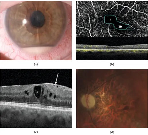
3.3. Complications. Forty-two eyes developed 57 complications during the disease course. Cataract was the most frequently recorded complication (n = 12) followed by epiretinal mem-brane (n = 11) and cystoid macular edema (n = 6). Detailed information about ocular complications is provided in Table 2. Figure 1 illustrates sight-threatening complications encountered in our cohort.
Several variables including HLA-B51 (p = 0:003), nonan-terior uveitis (p = 0:021), and the presence of retinal vasculitis at baseline (p = 0:049), were associated with a significant higher rate of complications. When complications such as cataract and glaucoma were excluded, panuveitis also dis-played a significant association with the development of structural complications (p = 0:0133). This association per-sisted also after adjusting for disease duration at regression analysis (OR 4.226 (1.182-15.112),p = 0:027). When exclud-ing patients who presented only cataract and/or intraocular hypertension, at regression analysis, the following variables were identified as predictors of long-term complications: HLA-B51 (OR 9.156 (C.I. 1.871-44.806), p = 0:006), panu-veitis (OR 4.601 (C.I. 1.094-19.348), p = 0:037), and a uveitis duration higher than 15 years (OR 4.605 (C.I. 1.002-21.168), p = 0:049).
Finally, a significant association was found between poor visual acuity and long-term complications (p = 0:048). 3.4. Treatment Impact. With regard to treatment modalities, 27 patients received only 1 biologic agent during the follow-up, 15 were treated with 2 different biologics at different time points, and 8 of them had been treated with 3 or more di ffer-ent biologics. No significant differences in terms of develop-ment of structural complications emerged between patients Table 1: Demographic and extraocular features of our cohort at
disease onset and throughout the course of Behçet’s syndrome.
Demographic features Mean ± SD expressed
in years
Female/male 23/24
Age 48:68 ± 14:38
Age at BS onset 31:17 ± 13:37
Age at uveitis onset 33:81 ± 13:75
Uveitis duration 15:21 ± 8:82
HLA-B51 27/47 (57.45%)
Clinical features at onset N (%)
Uveitis 31 (65.96%) Oral aphthosis 34 (72.34%) Genital aphthosis 9 (19.15%) Erythema nodosum 2 (4.25%) Pseudofollicular lesions 7 (14.89%) CNS involvement 2 (4.25%) Gastrointestinal involvement 4 (8.51%) Vascular involvement 2 (4.25%)
Clinical features throughout BS course N (%)
Oral aphthosis 46 (97.87%) Genital aphthosis 19 (40.43%) Erythema nodosum 8 (17.02%) Pseudofollicular lesions 24 (51.06%) CNS involvement 8 (17.02%) Gastrointestinal involvement 15 (31.91%) Vascular involvement 12 (25.53%)
List of abbreviations: BS: Behçet’s syndrome; CNS: central nervous system; HLA: human leukocyte antigen; IQR: interquartile range; SD: standard deviation.
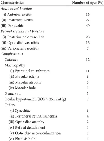
Table 2: Ocular characteristics and complications in Behçet’s uveitis in Italian patients.
Characteristics Number of eyes (%)
Anatomical location
(i) Anterior uveitis 16
(ii) Posterior uveitis 27
(iii) Panuveitis 40
Retinal vasculitis at baseline
(i) Posterior pole vasculitis 28
(ii) Optic disk vasculitis 16
(iii) Peripheral vasculitis 7
Complications
Cataract 12
Maculopathy
(i) Epiretinal membranes 11
(ii) Macular edema 6
(iii) Macular atrophy 5
(iv) Macular hole 1
Glaucoma 5
Ocular hypertension (IOP > 25 mmHg) 2 Others
(i) Synechiae 6
(ii) Peripheral retinal ischemia 4
(iii) Optic disc atrophy 2
(iv) Retinal detachment 1
(v) Optic disc neovascularization 1
(vi) Phthisis bulbi 1
treated with biologics and those receiving conventional immunosuppressive treatment (p = 0:90). Figure 2 shows a timeline graph of different biologic treatments received dur-ing the follow-up, whereas preceddur-ing and current treatments are displayed in Table 3.
Median ± IQR BCVA at baseline and last follow-up eval-uation was equal to 1:0 ± 0:38 and 1:0 ± 0:18, respectively, and the difference between these two timepoints was statisti-cally significant (p = 0:042).
A significant resolution of retinal vasculitis was detected between baseline and last follow-up evaluation (p < 0:0001). At baseline, 28 posterior pole retinal vasculitides were identi-fied on FA, and 16 of them also involved the optic disk. Figure 3 displays FAfindings of a case with severe retinal vas-culitis involving both posterior pole (Figure 3(a)) and periph-eral retinal quadrants (Figure 3(b)).
OCT findings improved from 376:00 ± 97:45 μm to 338:08 ± 55:81 μm, without any significance between base-line values and the last follow-up examination.
4. Discussion
Uveitis represents by far the most frequent and typical ocular manifestation of BS. BU and particularly posterior segment
inflammatory involvement may cause severe and irreversible damage of the optic nerve and the retinal structures causing permanent visual loss and even blindness [4]. Patients with BS-related panuveitis and visual impairment present a poorer quality of life compared to healthy controls and to BS patients without ocular involvement [21]. In our series, panuveitis and posterior uveitis taken together accounted for almost 80% of the sample, constituting approximately a similar proportion reported in previous studies [6, 7, 14– 16]. Uveitis represented thefirst clinical manifestation in 29 patients. Interestingly, among this subgroup of patients, 12 of them were free of other suggestive signs of BS at the uveitis onset. Thisfinding advocates for a close follow-up of any idi-opathic nonanterior uveitis, especially when typical ophthal-mological BS-uveitis findings are present [4]. Mean age at onset was similar to previous reports ranging from late 20s to early 30s [6, 8, 13–15].
No differences between male and female patients regard-ing age at uveitis onset, anatomical pattern, visual prognosis, and the development of long-term structural complications emerged. The lack of gender differences may be partially explained by the relatively small sample size. Indeed, our study group has recently published a large epidemiologic study where male patients were found to be significantly (a)
⁎
(b)
+
(c) (d)
Figure 1: Sight-threatening complications of Behçet’s syndrome- (BS-) related uveitis encountered in our cohort: (a) anterior chamber hypopyon in a case of BS-related panuveitis complicated with synechiae and cataract (right eye); (b) BS-related posterior uveitis complicated by macular epiretinal membrane (arrow) and chronic cystoid macular edema (plus) (right eye); (c) OCT angiography showing pathological enlargement of the foveal avascular zone (FAZ) (asterisk) along with a contiguous area of capillary dropout in the superficial retinal capillary plexus (arrowhead) as a sign of retinal ischemia (right eye); (d) optic nerve and macular atrophy due to the severe posterior pole vasculitis of the RE of a patient affected by refractory BS posterior uveitis.
more affected by posterior uveitis and panuveitis [22]. Another possible explanation may reside in the prognostic evolution of BS in recent decades. More specifically, the risk of losing useful vision has been significantly reduced over time and the BU prognosis is expected to improve in the near future also in the light of the availability of more targeted therapies [8, 23, 24]. Moreover, BS patients with poor vision in Italy have been found to be significantly fewer than those in endemic areas [15].
Innovative therapies account for a major contribution in the prognostic evolution of BU. Indeed, the advent of bio-logic agents has considerably revolutionized its management. In particular, anti-TNF-α monoclonal antibodies elicit a highly suppressive effect on ocular inflammatory attacks and have proven to be effective in the treatment of uveitis related to BS by improving visual acuity, showing a good long-term drug retention rate and a steroid-sparing effect [25–28]. In refractory cases, interleukin-1 inhibition may represent a valid alternative therapeutic approach [29]. In the present study, we observed a significant improvement of the median BCVA values and a decrease in the frequency of retinal vasculitis between baseline and last follow-up examination. The lack of significant decrease of the mean OCT values may be explained by the development of macular complications such as epiretinal membranes in a large pro-portion of patients during the disease course. Despite the ability to preserve visual function and reduce the relapses of retinal vasculitis, treatment with biologic agents was not associated with a reduced rate of long-term structural com-plications when compared with patients receiving conven-tional immunosuppressive drugs. It is possible that the lack of a significant decrease in OCT values and the already high mean disease duration at baseline may account for their inability to reduce long-term structural complications. An increased awareness along with an early diagnosis and a prompt introduction of systemic immunosuppressive ther-apy may halt the natural disease course, thus lowering the rate of long-term complications and poor visual acuity.
In our cohort 42 out of 84 eyes (50%) developed at least one complication with the most frequent being cataract, epir-etinal membranes, and cystoid macular edema. Several vari-ables were found to predict their long-term occurrence. A German study group identified a higher frequency of HLA-B51 among patients developing ocular involvement [16].
7 IFX 2 ADA 2 ANA 1 ETN 1 IFX 12 ADA 6 ANA 1 CAN 1 IFX 14 ADA 6 ANA 1 CAN 1 TCZ 2 IFX 8 ADA 6 ANA 2 CAN 8 IFX 2 ADA 4 ANA 4 IFX 1 ADA 1 IFX 2001 2005 2008 2011 2012 2013 2014 2016 2017 2018 2019 1 IFX, 16 ADA 5 ANA, 1 CAN 1 TCZ, 2 GOL 5 IFX, 5 ADA 3 ANA, 2 ETN 2 IFX, 14 ADA 6 ANA, 1 CAN 1 TCZ, 1 GOL 4 IFX
Figure 2: Timeline graph illustrating the different biologic treatments administered to our patients over time. List of abbreviations: ADA: adalimumab; ANA: anakinra; CAN: canakinumab; ETN: etanercept; GOL: golimumab; IFX: infliximab; TCZ: tocilizumab.
Table 3: Past and current treatment in our cohort.
Previous cDMARDs Current cDMARDs
CSA 16 CSA 5 AZA 12 AZA 5 MTX 14 MTX 3 SZS 4 SZS 1 LFN 1 HCQ 1 CYC 1 Thalidomide 1
Previous biologics Current biologics
ADA 18 ADA 11 IFX 10 IFX 2 GOL 2 GOL 1 ANA 9 ANA 4 CAN 1 CAN 2 ETN 4 TCZ 1 TCZ 1
List of abbreviations: ADA: adalimumab; ANA: anakinra; AZA: azathioprine; CAN: canakinumab; cDMARDs: conventional disease-modifying antirheumatic drugs; Cyc A: cyclosporin A; CYC: cyclophosphamide; ETN: etanercept; GOL: golimumab; HCQ: hydroxychloroquine; IFX: infliximab; LFN: leflunomide; MTX: methotrexate; SZS: sulfasalazine; TCZ: tocilizumab.
With regard to its association with ocular involvement and type of uveitis, current medical literature displays contro-versial results [30, 31]. However, several authors consider HLA-B51 as a marker of unfavorable prognosis [32, 33]. In line with the latter statement, our findings highlight the role of HLA-B51 as a predictor of long-term structural complications and subsequently of poor visual outcome. As expected, also posterior segment involvement and a long uveitis duration were identified as further predictors of long-term complications.
Low visual acuity at presentation and the development of a severe complication such as optic disc atrophy have been linked to a poorer visual outcome [6]. We could not evaluate these variables due to a low number of optic atrophy cases and a substantially good visual acuity at baseline evaluation. Despite its vital importance, the identification of predictive factors of poor long-term prognosis in BS is currently an understudied aspect. A deeper knowledge of the variables conferring a worse visual prognosis may allow a better patient profiling and therefore orientate a more precise and targeted therapy. Based on our findings, we suggest that HLA-B51 positivity, posterior segment involvement, and
longer uveitis duration are strong drivers in the therapeutic decision. These findings however deserve further investiga-tion and should be interpreted in the context of several limi-tations such as the retrospective design accompanied by its inherited shortcomings. Indeed, some interesting variables such as comorbidities, which may behave as confounding factors, were not collected for the purposes of this study. The relatively small sample size may also be responsible for the wide confidence intervals detected in regression analysis, which do not provide a precise magnitude of effect, highlight-ing the need for further studies with larger sample sizes. Additionally, the referral bias due to the tertiary referral nature of our center might exclude milder forms of intraocu-lar inflammation and hence be only representative of the complicated end of disease spectrum.
In conclusion, according to our experience, BU arises in the third decade of life and predominantly compromises the posterior chamber as posterior uveitis or panuveitis, without any gender influence. Its duration seems to be associated with a poor visual acuity. Uveitis duration along with the presence of HLA-B51 and panuveitis predict the development of long-term structural complications, thus
200 𝜇m 200 𝜇m
(a)
(b)
Figure 3: Fluorescein angiography (FA) findings in a patient with severe retinal vasculitis; (a) late-phase FA showing optic disc and vascular leakage and areas of nonperfusion in a Behçet’s syndrome (BS) patient with a bilateral severe occlusive vasculitis; (b) late-phase FA in a patient with active bilateral retinal vasculitis affected by BS. All retinal peripheral quadrants of both eyes of this patient showed the same diffuse fern-like capillary leakage, the most frequent FAfinding for Behçet’s uveitis.
representing main drivers in the treatment decision-making. Despite the therapeutic advances and an improved prognosis compared to the past decades, uveitis associated with BS remains a challenging and hard-to-handle condition. A bet-ter understanding of its epidemiologic landscape is essential to prevent or at least minimize irreversible and structural damage through patient profiling with the aim to identify high-risk subjects.
Data Availability
The datasets generated for this study are available on request to the corresponding author.
Conflicts of Interest
The authors declare that the research was carried out in the absence of any personal, professional, or financial relation-ships that could potentially be construed as a conflict of interest.
References
[1] D. Saadoun, B. Wechsler, K. Desseaux et al., “Mortality in Behçet's disease,” Arthritis and Rheumatism, vol. 62, no. 9, pp. 2806–2812, 2010.
[2] E. Kural-Seyahi, I. Fresko, N. Seyahi et al., “The long-term mortality and morbidity of Behçet Syndrome,” Medicine, vol. 82, no. 1, pp. 60–76, 2003.
[3] Y. Ozyazgan, D. Ucar, G. Hatemi, and Y. Yazici, “Ocular involvement of Behçet's syndrome: a comprehensive review,” Clinical Reviews in Allergy and Immunology, vol. 49, no. 3, pp. 298–306, 2015.
[4] I. Ksiaa, N. Abroug, M. Kechida et al.,“Eye and Behçet's dis-ease,” Journal Français d'Ophtalmologie, vol. 42, no. 4, pp. e133–e146, 2019.
[5] M. Sakamoto, K. Akazawa, Y. Nishioka, H. Sanui, H. Inomata, and Y. Nose,“Prognostic factors of vision in patients with Beh-çet disease,” Ophthalmology, vol. 102, no. 2, pp. 317–321, 1995. [6] K. Pathanapitoon, P. Kunavisarut, F. A. Saravuttikul, and A. Rothova,“Ocular manifestations and visual outcomes of Behçet's uveitis in a Thai population,” Ocular Immunology and Inflammation, vol. 27, no. 1, pp. 2–6, 2019.
[7] K. Janati, K. EL Omari, H. Benchiki, M. Hamdani, and H. Lakhdar, “Les manifestations oculaires de la maladie de Behçet (étude de 50 patients consultant en dermatologie),” La Revue de Médecine Interne, vol. 26, no. 10, pp. 771–776, 2005.
[8] I. Tugal-Tutkun, S. Onal, R. Altan-Yaycioglu, H. Huseyin Altunbas, and M. Urgancioglu,“Uveitis in Behçet disease: an analysis of 880 patients,” American Journal of Ophthalmology, vol. 138, no. 3, pp. 373–380, 2004.
[9] C. Fabiani, A. Vitale, I. Orlando et al.,“Impact of uveitis on quality of life: a prospective study from a tertiary referral rheumatology-ophthalmology collaborative uveitis center in Italy,” The Israel Medical Association Journal, vol. 19, no. 8, pp. 478–483, 2017.
[10] G. Hatemi, R. Christensen, D. Bang et al.,“2018 update of the EULAR recommendations for the management of Behçet's syndrome,” Annals of the Rheumatic Diseases, vol. 77, no. 6, pp. 808–818, 2018.
[11] G. Lopalco, V. Venerito, J. Sota et al.,“Epidemiological profile of non-infectious uveitis from the rheumatologist's perspec-tive: a survey from two tertiary referral centres in Italy,” Clin-ical and Experimental Rheumatology, vol. 36, 6 Suppl 115, pp. 68–73, 2018.
[12] A. Ambresin, T. Tran, F. Spertini, and C. Herbort,“Behçet's disease in Western Switzerland: epidemiology and analysis of ocular involvement,” Ocular Immunology and Inflammation, vol. 10, no. 1, pp. 53–63, 2002.
[13] F. Bazvand, M. Zarei, N. Ebrahimiadib et al.,“Ocular manifes-tations, conventional fundus fluorescein angiographic find-ings, and relationship between angiographic findings and visual acuity in Behçet's disease,” Seminars in Ophthalmology, vol. 32, no. 6, pp. 764–771, 2017.
[14] F. Sáenz-Francés, M. Elías-de-Tejada, J. M. Martínez-de-la-Casa et al.,“Ocular inflammatory signs observed in a cohort of Spanish patients with Behçet disease and ocular in flamma-tion,” European Journal of Ophthalmology, vol. 18, no. 4, pp. 563–566, 2008.
[15] N. Kitaichi, A. Miyazaki, D. Iwata, S. Ohno, M. R. Stanford, and H. Chams,“Ocular features of Behcet's disease: an interna-tional collaborative study,” The British Journal of Ophthalmol-ogy, vol. 91, no. 12, pp. 1579–1582, 2007.
[16] L. Krause, A. K. Köhler, A. Altenburg et al.,“Ocular involve-ment in Adamantiades-Behçet's disease in Berlin, Germany,” Graefe's Archive for Clinical and Experimental Ophthalmology, vol. 247, no. 5, pp. 661–666, 2009.
[17] M. Elshahaly, I. A. E. Latif, and H. Bassiouni,“Clinical charac-teristics of Behcet's disease in 453 Egyptian patients suffering from uveitis with gender comparison,” Current Rheumatology Reviews, vol. 15, 2019.
[18] Disease ISGfBs, “Criteria for diagnosis of Behçet's disease. International Study Group for Behçet's Disease,” The Lancet, vol. 335, pp. 1078–1080, 1990.
[19] International Team for the Revision of the International Criteria for Behçet's Disease (ITR-ICBD),“The International Criteria for Behçet's Disease (ICBD): a collaborative study of 27 countries on the sensitivity and specificity of the new cri-teria,” Journal of the European Academy of Dermatology and Venereology, vol. 28, no. 3, pp. 338–347, 2014.
[20] The Standardization of Uveitis Nomenclature (SUN) Working Group,“Standardization of uveitis nomenclature for reporting clinical data. Results of the First International Workshop,” American Journal of Ophthalmology, vol. 140, no. 3, pp. 509– 516, 2005.
[21] C. Fabiani, A. Vitale, I. Orlando et al.,“Quality of life impair-ment in Behçet's disease and relationship with disease activity: a prospective study,” Internal and Emergency Medicine, vol. 12, no. 7, pp. 947–955, 2017.
[22] J. Sota, D. Rigante, G. Emmi et al.,“Behçet's syndrome in Italy: a detailed retrospective analysis of 396 cases seen in 3 tertiary referral clinics,” Internal and Emergency Medicine, pp. 1–9, 2020.
[23] K. Namba, H. Goto, T. Kaburaki et al.,“A major review: cur-rent aspects of ocular Behçet's disease in Japan,” Ocular Immu-nology and Inflammation, vol. 23, no. sup1, Supplement 1, pp. S1–23, 2015.
[24] M. Accorinti, F. R. Pesci, M. P. Pirraglia, I. Abicca, and P. Pivetti-Pezzi, “Ocular Behçet's disease: changing patterns over time, complications and long-term visual prognosis,” Ocular Immunology and Inflammation, vol. 25, no. 1, pp. 29– 36, 2017.
[25] C. Fabiani, J. Sota, A. Vitale et al., “Cumulative retention rate of adalimumab in patients with Behçet's disease-related uveitis: a four-year follow-up study,” The British Journal of Ophthalmology, vol. 102, no. 5, pp. 637–641, 2018.
[26] C. Fabiani, J. Sota, A. Vitale et al.,“Ten-year retention rate of infliximab in patients with Behçet's disease-related uveitis,” Ocular Immunology and Inflammation, vol. 27, no. 1, pp. 34– 39, 2019.
[27] C. Fabiani, A. Vitale, D. Rigante et al.,“Predictors of sustained clinical response in patients with Behçet's disease-related uve-itis treated with infliximab and adalimumab,” Clinical Rheu-matology, vol. 37, no. 6, pp. 1715–1720, 2018.
[28] C. Fabiani, J. Sota, D. Rigante et al.,“Efficacy of adalimumab and infliximab in recalcitrant retinal vasculitis inadequately responsive to other immunomodulatory therapies,” Clinical Rheumatology, vol. 37, no. 10, pp. 2805–2809, 2018.
[29] C. Fabiani, A. Vitale, D. Rigante et al., “The presence of uveitis is associated with a sustained response to the interleu-kin (IL)-1 inhibitors anainterleu-kinra and canainterleu-kinumab in Behçet's disease,” Ocular Immunology and Inflammation, vol. 28, no. 2, pp. 298–304, 2020.
[30] M. Soylu, T. R. Ersöz, and E. Erken,“The association between HLA B5 and ocular involvement in Behçet's disease in south-ern Turkey,” Acta Ophthalmologica, vol. 70, no. 6, pp. 786– 789, 1992.
[31] L. Krause, A. K. Köhler, A. Altenburg et al.,“Ocular involve-ment is associated with HLA-B51 in Adamantiades-Behçet's disease,” Eye, vol. 23, no. 5, pp. 1182–1186, 2009.
[32] C. C. Zouboulis, I. Kötter, D. Djawari et al.,“Epidemiological features of Adamantiades-Behçet's disease in Germany and in Europe,” Yonsei Medical Journal, vol. 38, no. 6, pp. 411– 422, 1997.
[33] C. C. Zouboulis, J. R. Turnbull, and P. Martus,“Univariate and multivariate analyses comparing demographic, genetic, clini-cal, and serological risk factors for severe Adamantiades– Behc¸et’s disease,” in Adamantiades–Behçet’s Disease, C. C. Zouboulis, Ed., pp. 123–126, London Amsterdam, Kluwer/-Plenum, 2003.