R E S E A R C H
Open Access
Aberrant gene expression in mucosa adjacent to
tumor reveals a molecular crosstalk in colon
cancer
Rebeca Sanz-Pamplona
1, Antoni Berenguer
1, David Cordero
1, David G Molleví
2, Marta Crous-Bou
1, Xavier Sole
1,
Laia Paré-Brunet
1, Elisabet Guino
1, Ramón Salazar
2,3, Cristina Santos
2,3, Javier de Oca
4,5, Xavier Sanjuan
6,
Francisco Rodriguez-Moranta
7and Victor Moreno
1,5*Abstract
Background: A colorectal tumor is not an isolated entity growing in a restricted location of the body. The patient’s gut environment constitutes the framework where the tumor evolves and this relationship promotes and includes a complex and tight correlation of the tumor with inflammation, blood vessels formation, nutrition, and gut
microbiome composition. The tumor influence in the environment could both promote an anti-tumor or a pro-tumor response.
Methods: A set of 98 paired adjacent mucosa and tumor tissues from colorectal cancer (CRC) patients and 50 colon mucosa from healthy donors (246 samples in total) were included in this work. RNA extracted from each sample was hybridized in Affymetrix chips Human Genome U219. Functional relationships between genes were inferred by means of systems biology using both transcriptional regulation networks (ARACNe algorithm) and protein-protein interaction networks (BIANA software).
Results: Here we report a transcriptomic analysis revealing a number of genes activated in adjacent mucosa from CRC patients, not activated in mucosa from healthy donors. A functional analysis of these genes suggested that this active reaction of the adjacent mucosa was related to the presence of the tumor. Transcriptional and
protein-interaction networks were used to further elucidate this response of normal gut in front of the tumor, revealing a crosstalk between proteins secreted by the tumor and receptors activated in the adjacent colon tissue; and vice versa. Remarkably, Slit family of proteins activated ROBO receptors in tumor whereas tumor-secreted proteins transduced a cellular signal finally activating AP-1 in adjacent tissue.
Conclusions: The systems-level approach provides new insights into the micro-ecology of colorectal tumorogenesis. Disrupting this intricate molecular network of cell-cell communication and pro-inflammatory microenvironment could be a therapeutic target in CRC patients.
Keywords: Colorectal cancer, Network, Microenvironment, Molecular crosstalk, Systems biology
* Correspondence:[email protected]
1Unit of Biomarkers and Susceptibility, Catalan Institute of Oncology (ICO),
Bellvitge Biomedical Research Institute (IDIBELL) and CIBERESP, L’Hospitalet de Llobregat, Barcelona, Spain
5
Department of Clinical Sciences, Faculty of Medicine, University of Barcelona (UB), Av. Gran Vía 199-203, 08908 L’Hospitalet de Llobregat, Barcelona, Spain Full list of author information is available at the end of the article
© 2014 Sanz-Pamplona et al.; licensee BioMed Central Ltd. This is an Open Access article distributed under the terms of the Creative Commons Attribution License (http://creativecommons.org/licenses/by/2.0), which permits unrestricted use, distribution, and reproduction in any medium, provided the original work is properly credited. The Creative Commons Public Domain Dedication waiver (http://creativecommons.org/publicdomain/zero/1.0/) applies to the data made available in this article, unless otherwise stated.
Background
Colorectal cancer (CRC) is a complex disease in which many genes, proteins, and molecular processes are im-plicated. Proteins do not work independently in a tumor cell, but are organized into co-regulated units or path-ways that perform a common biological function [1]. Relevant molecular mechanisms involved in cancer are gene regulation, signaling, cell metabolism, and the con-nections between them, among others [2]. In addition to the tumor cell intrinsic complexity, increasing data support the main role of tumor microenvironment in the mechanisms of CRC progression [3-5]. Tumor mi-croenvironment is composed by a heterogeneous popula-tion of stromal cells such as fibroblasts and immune cells, extracellular matrix components and secreted factors. All these components work orchestrated by molecular trans-ducers like integrins engaging cell-cell and cell-matrix sig-naling that in turn enhance tumor growth [6].
Besides, a colorectal tumor is not an isolated entity growing in a restricted location of the body. An active communication exists not only between different cell communities within the tumor bulk but also between the tumor and the non-tumor distant mucosa. Hence, the patient’s gut environment constitutes the framework where the tumor evolves and this relationship promotes and includes a complex and tight correlation with in-flammation, blood vessels formation, nutrition and gut microbiome composition [7]. Consequently, studying the micro-ecology context of a tumor is central to under-stand colorectal carcinogenesis. The tumor influence on environment could both promote an anti-tumor and a pro-tumor response. Some microenvironments, particu-larly those associated with tissue injury, are favorable for progression of mutant cells, whereas others restrict it. Cancer cells can also instruct surrounding tissues to undergo changes that promote malignancy [8].
Field cancerization or the field-effect is a theory first described by Slaughter et al. in oral carcinoma [9]. In the initial phase of the multistep carcinogenesis, a stem cell acquires genetic alterations and forms a “patch”, a clonal unit of altered daughter cells. Further alterations convert the “patch” into a field of pre-neoplastic cells. Although only one cell becomes tumoral, the remaining field (adjacent mucosa) continues in a “pre-neoplastic-state” composed of morphologically normal, but biologic-ally altered epithelial cells. Since this field is a pre-tumor site predisposed towards development of cancer, this hy-pothesis could explain local recurrences after surgery [10].
Understanding the complex ways in which cancer cells interact with their surroundings, both locally in the tumor organ and systemically in the body as a whole has implica-tions for effective cancer prevention and therapy. In con-trast to the gene-centric view, a systems biology approach (defined as the analysis of the molecular relationship
between genes and proteins as a whole) can be useful to depict a global view of the cancer disease not only as a tumor cell but as an intricate systemic disease [11].
In this study, mRNA expression from paired tumor (T) and adjacent mucosa from CRC patients (A) and mRNA from mucosa healthy donors (H) were measured using microarrays. The inclusion of samples from healthy sub-jects has allowed us assessing whether adjacent mucosa from colon cancer patients differs from healthy donors’ mucosa possibly due to the tumor presence. Indeed, a number of differentially expressed genes (DEG) were found between these two entities (A vs. H). Considering their level of expression in tumor tissues, these DEGs were classified as “Tumor-like”, “Trend” or “Adjacent-specific” (A vs. T) patterns. To explain the mechanisms that regulate these patterns of differential expression, networks mimicking transcription regulation were used to search for those transcription factors directly influencing DEG. Then, a systems biology approach using PPIN was applied to describe a crosstalk between cytokines and other proteins secreted by the tumor and receptors acti-vated in the adjacent colon tissue; and vice versa, pro-viding new insights into the micro-ecology of colorectal tumorigenesis. Finally, relevant cytokines and receptors up-regulated in tumor tissue were identified comparing T vs. H expression (Figure 1). Further elucidation of these interactions could be helpful in the development of novel therapeutic strategies oriented to disrupt this molecular crosstalk.
Results
Characterization of differentially expressed genes between adjacent and healthy mucosa
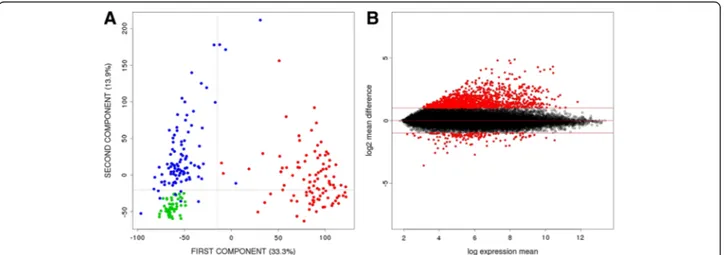
A principal component analysis (PCA) was done to ex-plore the variability of the transcriptomic data from our 246 samples (Figure 2A). As expected, tumor samples appeared as an independent cluster (T in red). Surpris-ingly, adjacent paired mucosa (A in blue) were also clearly separated from healthy mucosa (H in green), reflecting a large number of differentially expressed genes (DEG) between them. A total of 895 genes were differentially expressed at FDR < 1% and log2 mean differ-ence > 1 between adjacent and healthy mucosa (Additional file 1: Table S1). Interestingly, 88% of these genes were over-expressed in adjacent mucosa (Figure 2B).
The functional enrichment analysis of these genes identified the classical pathways involved in cancer and were highlighted by a significant enrichment of func-tions related to Inhibition of matrix metalloproteinases, Cell adhesion molecules, cytokine-cytokine receptor in-teraction, TGF-beta signaling pathway, integrin signal-ing pathway, complement and coagulation cascades, wound healing, response to external stimulus, inflam-matory responseand soluble fraction, among others (see
complete list in Additional file 2: Table S2, Additional file 3: Table S3 and Additional file 4: Figure S1). This functional analysis suggested an active reaction of the adjacent mucosa related to the presence of the tumor or a more passive reac-tion induced by factors released from the tumor.
Public transcriptomic data analyzing adjacent and healthy mucosa were used to validate the list of DEG. As a result, 60% of the genes were validated at FDR 1%. At FDR 5%, 91% of the genes were validated (Additional file 5: Table S4
and Additional file 4: Figure S2). These results should be interpreted with caution because each sample type was ana-lyzed in different experiments and, though we normalized the data jointly, we cannot exclude strong batch or la-boratory effects. We could not find a dataset like ours, in which healthy and adjacent colon mucosa were ana-lyzed simultaneously.
Figure 3A shows a hierarchical clustering performed with the set of DEG between adjacent mucosa (A) and HEALTHY MUCOSA (H)
N = 50
ADJACENT MUCOSA (A) N = 98 CRC TUMOR (T) N = 98 PAIRED Differentially Expressed Genes (DEG) Patterns of DEG expression • Tumor-like • Trend • Adjacent-specific Transcriptional Regulation Analysis Cross-talk
network over-expressed genes in CRC-tumor (Secreted and Receptors)
DEG VALIDATION (public datasets) Cellular classification (public dataset) over-expressed genes (Secreted and Receptors)
Figure 1 Work flow chart. The central core of the analysis is the comparison between adjacent mucosa and healthy mucosa at transcriptomic (gene expression data) and transcriptional (regulatory network) level. Independent public datasets were used to validate the results. In a second step, tumor tissue was used to search for different DEG patterns. Finally, a crosstalk network was inferred to decipher molecular communication between the tumor and the adjacent gut underlying DEG. Public data was used to elaborate a cellular classification of genes implicated in the crosstalk.
Figure 2 Gene expression differences between adjacent and healthy mucosa samples. A. PCA scatter plot representing the dispersion of the samples based on their gene expression levels. Tumor samples (red), adjacent mucosa samples (blue) and samples from healthy donors (green) were plotted in 1stand 2ndprincipal components. B. MA Plot representing gene expression differences between adjacent and healthy
healthy mucosa (H). Interestingly, the three different tissues were perfectly classified, including the tumors (T) that did not participate in the gene selection. Re-garding genes, three patterns of expression were identi-fied as shown in Figure 3B: a) “Tumor-like” (A = T > H or H > A = T) when genes in A had similar pattern as T (349 genes); b) “Trend” (T > A > H or H > A > T) when genes in A had an intermediate expression between H and T (132 genes); and c)“Adjacent-specific” (A < (T,H) or A > (T,H)), when genes were specifically de-regulated in A when compared to either T or H, irrespective of the relationship between T and H (414 genes). The size of this latter group was a surprise that lead us to explore in detail a crosstalk between the tumor and the adjacent mucosa.
Regarding enriched functions for these gene patterns, Tumor-like functions included AP-1 transcription factor network, COX reactions or activation of AP-1, whereas Adjacent-specific functions were enriched in axon guid-ance, PPAR signaling pathway or BMP2 signaling pathway, among others. These results suggest different functions for each gene expression pattern, though Integrin signaling pathway, complement cascade, adhesion or Interferon sig-naling were functions shared by the two patterns (see complete list in Additional file 6: Table S5).
Adjacent mucosa samples appeared divided into two groups in the hierarchical clustering analysis (Figure 3A). The smallest of them, with 24 samples, was characterized by high expression in most of adjacent-specific genes. A PCA performed with these adjacent-specific genes showed that the second component was capturing the specificity of this sample cluster and that adjacent mucosa were more similar to tumor than to healthy mucosa (Figure 3C). In fact, the original PCA analysis with all genes also identified these adjacent mucosa samples as highly variable in the second component (Figure 2A).
These clusters were not associated with the clinical pa-rameters gender, age and tumor progression neither with technical parameters RNA integrity value (RIN), 260/230 ratio and plate. In addition, a functional analysis includ-ing differentially expressed genes between these two clusters did not show specific functions but essentially those described as characteristic of adjacent mucosa. These results suggest that the smaller cluster of adja-cent samples was just an extreme phenotype of these samples. Interestingly, this pattern was also observed in
the validation dataset (see heatmap in Additional file 4: Figure S2).
Transcriptional regulation of differentially expressed genes between adjacent and healthy mucosa
We hypothesized that this differential expression could be triggered by a transcriptional program, activated only in adjacent mucosa by the presence of the tumor, and normally silenced in healthy mucosa. This hypothesis was supported by the GSEA results, in which 312 tran-scription factors motifs were found to be statistically associated with the adjacent mucosa phenotype (nom-inal p-value < 0.01) but none was found associated to healthy mucosa phenotype (Additional file 3: Table S3).
To further explore this hypothesis, transcriptional networks were inferred and compared using gene ex-pression data of adjacent and healthy mucosa (see Additional file 4: Figure S3). Venn diagram in Figure 4A shows the overlap between nodes of each network. The vast majority of healthy mucosa nodes were also active in adjacent mucosa network whereas 3120 new nodes appeared specific to the adjacent mucosa and 668 nodes disappeared from the network. As expected, DEG between adjacent and healthy mucosa were over-represented in the new active nodes of the adjacent mucosa network (empirical p-value < 10−4) suggesting that DEG are not only performing common functions but also co-regulated in a sub-transcriptional network not active in healthy mucosa samples. Out of 895 DEG, 60 (13%) were transcription factors (TF), and random sampling of genes among the complete dataset re-vealed that DEG were significantly enriched in TF (em-pirical p-value < 0.001). Among these 60 TF, 35 were specific of the adjacent mucosa transcriptional network.
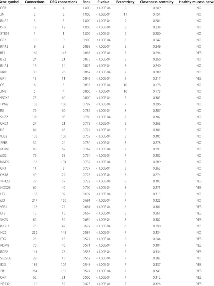
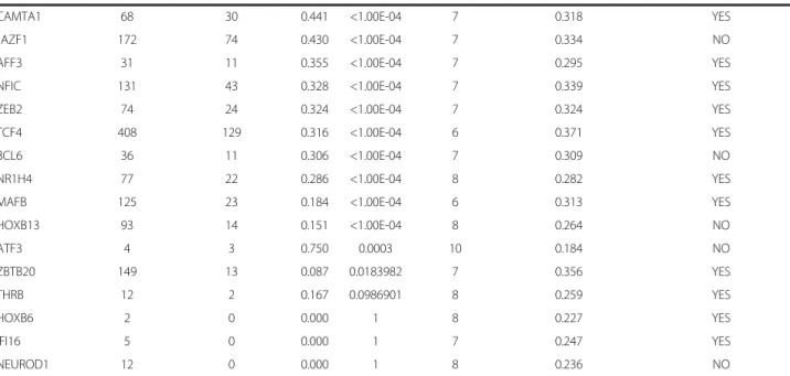
TF were ranked taking into account the total number of their targets (degree) and the proportion of targets in our DEG list. This rank suggested sub-networks spe-cifically active in adjacent mucosa tissue. TF with higher rank were more specific of adjacent mucosa, and showed higher values of eccentricity (a topological network meas-ure of the spreading of a node in the network) and lower values of closeness centrality (Table 1).
Genes from the AP-1 complex (Fosb and Jun) ranked first in the TF list. The AP-1 subunits Fos, Junb, Mafb and Atf3 also appeared in the list. Previous GSEA ana-lysis also had revealed as most significant motive“Genes
(See figure on previous page.)
Figure 3 DEG characterization. A. Hierarchical clustering of 1230 over-expressed and 136 under-expressed probes that correspond to 788 and 107 genes respectively classifying the 246 tissue samples into three clusters of healthy mucosa (green), tumors (red) and adjacent mucosa (blue). Highlighted in black, the group of 24 adjacent samples showing an extreme phenotype. B. Representative DEG patterns are displayed. DEG between adjacent and mucosa were classified as“Tumor-like”, “Trend” and “Adjacent-specific” genes. C. PCA using “Adjacent-specific” DEG. Tumor samples (T) are painted in red, adjacent samples (A) in blue and healthy mucosa (H) in green. The 24 adjacent samples showing an extreme phenotype are circled with a dot line.
A
B
C
with promoter regions [−2 kb,2 kb] around transcription start site containing the motif TGACTCANNSKN which matches annotation for JUN” (p-value = 0.002, and FDR q-value = 0.015, Additional file 4: Figure S4). A high cor-relation existed between the expression of Jun and Fos AP-1 subunits in adjacent mucosa but not in healthy mucosa (Spearman’s correlation 0.67 and 0.23 respect-ively; Figure 4B). Interestingly, these TF belonged to the “tumor-like” genes pattern (Figure 4C). Fos, Jun, Fosb and Junb did not appear in healthy mucosa transcrip-tional network highlighting their idiosyncratic role in adjacent mucosa. The family of transcription factors NR4A1, NR4A2 and NR4A3 also ranked in top posi-tions. Other TF such as GLI3, BCN2, EBF1 and ZEB1 were also significant because of their high rank in the network and large number of DEG targets.
Deciphering a crosstalk between adjacent mucosa and tumor through a protein-protein interaction network Changes in adjacent mucosa not detected in healthy mu-cosa might be a direct response in front of tumor stimulus based on a physical crosstalk between the cells (Figure 1). This molecular communication could be through the direct interaction between secreted proteins and their corresponding membrane receptors. The following strat-egy was applied to identify interactions compatible with this hypothesis: 1) Search for over-expressed genes in tu-mors compared to healthy mucosa in addition to previous DEG. 2) Identify those that code for secreted proteins and membrane receptors. 3) Construct a protein interaction network with the selected genes. 4) Identify interaction pairs that reflect cellular communication in both direc-tions: from tumor to adjacent (efferent pathway) and vice versa (afferent pathway).
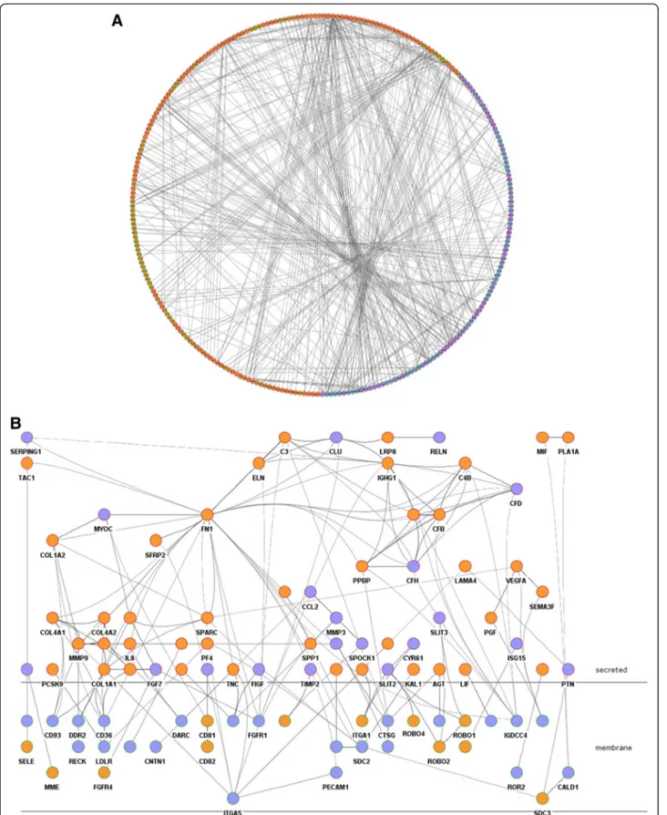
From the 788 over-expressed genes in adjacent mucosa vs. healthy mucosa, 324 (41%) corresponded to secreted (n = 111) or membrane (n = 213) genes. In addition, 442 genes (250 secreted and 192 membrane) over-expressed in tumors were included in the analysis. A level 0 (only direct interactions) protein-protein interaction network was re-trieved using the 766 up-regulated secreted/membrane genes in adjacent and tumor samples as input. The resulting network included 291 nodes connected by 596 interactions, the majority of them integrated in a giant component (Figure 5A). A functional analysis of this network revealed cell adhesion, response to exter-nal stimulus, response to wounding, and anatomical
structure development as the most statistically signifi-cant functions (Additional file 7: Table S6).
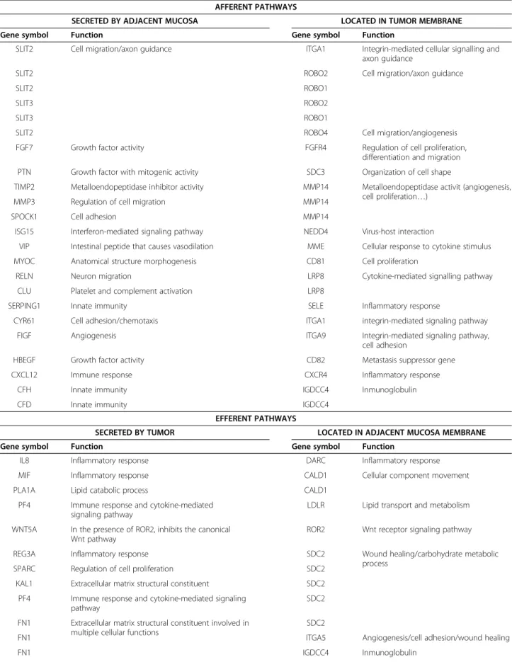
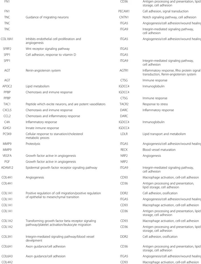
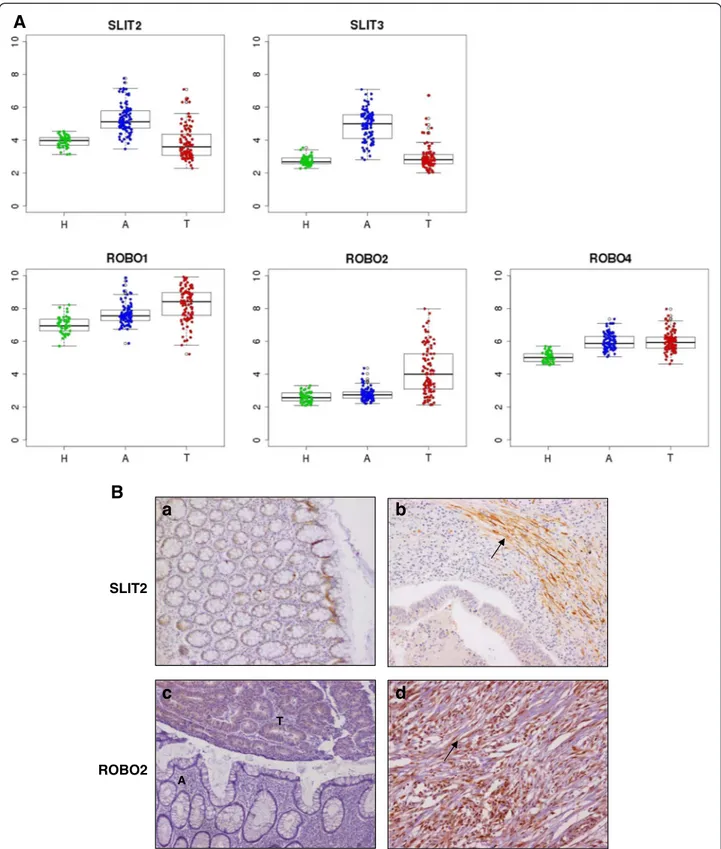
A curated analysis of the network revealed 84 crosstalk interactions (Table 2), 61 of them efferent (tumor se-creted proteins linked to a receptor in adjacent mucosa tissue), and 23 afferent (adjacent mucosa secreted pro-teins linked to a receptor in tumor). Figure 5B shows an abstraction of the original network restricted to crosstalk interactions. It is remarkable that 6 out of 23 afferent in-teractions (26%) included members of the Slit family of secreted proteins, which emerged as relevant players in tumor crosstalk determining the adjacent mucosa re-sponse. In the network, Slit2 and Slit3 were redundantly activating Robo1, Robo2, Robo4 and ITGA1 receptors in tumor. Slit family followed an adjacent-specific pattern of expression (see Figure 6A). Due to its importance, and as a proof of concept of the overall strategy of gene selection, inmunohistochemical staining was done to asses the protein expression of Slit2 and the receptor Robo2. Slit2 was expressed in adjacent epithelial cells and also in stromal cancer cells. Robo2 was expressed in both epithelial and stromal cells in cancer tissue but not in adjacent tissue (Figure 6B). Other interesting afferent crosstalk pairs involved LRP8 receptor in tumor acti-vated by a double stimulus of RELN and CLU proteins secreted by adjacent mucosa, and VIP, an intestinal pep-tide that causes vasodilatation, linked to MME receptor in tumor cells.
Efferent interactions were more numerous and in-cluded interleukins (IL-8), extracellular-matrix compo-nents (Fibronectin, Collagen) or molecules related to invasion like SPARC linked with receptors such as integ-rins or complement receptors (see Table 2 for specific pairs). Interestingly, the vascular endothelial growth factor receptor NRP2, over-expressed in adjacent mucosa, inter-acted with a plethora of candidate activating secreted fac-tors from tumors such as VEGFA or SEMA3F. Another interesting finding was the over-expression of LIF in tumor, whose receptor LIFR was over-expressed in adja-cent mucosa but not in the tumor. These results were highly indicative of an active crosstalk between cells in the gut microenvironment that triggers an intra-cellular sig-naling response. The protein-protein interaction network also revealed autocrine signals within tumor or adjacent mucosa. For example, the vascular endothelial growth fac-tor recepfac-tor FLT1 was found linked with its ligand VEGFA, both over-expressed in tumor samples.
(See figure on previous page.)
Figure 4 DEG analysis in the framework of transcriptional networks. A. Venn Diagram showing the overlap between nodes in adjacent mucosa transcriptional network (blue) and healthy mucosa transcriptional network (green). DEG were merged with the two transcriptional networks. B. Expression correlation between transcription factors Jun and Fos in adjacent (blue) and healthy mucosa (green). C. Gene expression levels of AP-1 subunits in healthy mucosa (green) adjacent mucosa (blue) and tumor tissue (red).
Table 1 List of transcription factors differentially expressed between adjacent and healthy mucosa samples
Gene symbol Connections DEG connections Rank P-value Eccentricity Closenness centrality Healthy mucosa network
FOSB 4 4 1.000 <1.00E-04 9 0.209 NO JUN 2 2 1.000 <1.00E-04 11 0.151 NO NR4A2 5 5 1.000 <1.00E-04 9 0.204 NO OSR2 12 12 1.000 <1.00E-04 8 0.234 NO ZBTB16 1 1 1.000 <1.00E-04 9 0.200 NO EGR2 10 9 0.900 <1.00E-04 8 0.247 NO NR4A3 9 8 0.889 <1.00E-04 8 0.249 NO
EBF1 162 143 0.883 <1.00E-04 7 0.294 YES
HEY2 24 21 0.875 <1.00E-04 8 0.266 NO
NR4A1 16 14 0.875 <1.00E-04 8 0.240 NO
PRRX1 30 26 0.867 <1.00E-04 7 0.289 NO
EGR1 13 11 0.846 <1.00E-04 9 0.217 YES
FOS 6 5 0.833 <1.00E-04 10 0.178 NO JUNB 5 4 0.800 <1.00E-04 10 0.178 NO MEOX2 75 60 0.800 <1.00E-04 7 0.303 NO ZFPM2 133 106 0.797 <1.00E-04 7 0.296 NO ERG 76 60 0.789 <1.00E-04 8 0.287 NO TSHZ2 109 85 0.780 <1.00E-04 7 0.302 NO FOXC1 27 21 0.778 <1.00E-04 8 0.268 NO HLF 84 65 0.774 <1.00E-04 7 0.301 NO MEIS2 133 100 0.752 <1.00E-04 8 0.305 NO CREB5 32 24 0.750 <1.00E-04 8 0.278 NO PRDM6 83 62 0.747 <1.00E-04 7 0.293 NO GLIS2 79 58 0.734 <1.00E-04 7 0.302 NO HAND2 138 101 0.732 <1.00E-04 7 0.289 NO EGR3 11 8 0.727 <1.00E-04 8 0.263 NO SOX18 40 29 0.725 <1.00E-04 7 0.274 NO ZNF423 79 57 0.722 <1.00E-04 8 0.303 YES
PHOX2B 60 42 0.700 <1.00E-04 8 0.275 YES
KLF7 133 92 0.692 <1.00E-04 7 0.313 NO
GLI3 217 150 0.691 <1.00E-04 7 0.325 NO
MEIS1 113 77 0.681 <1.00E-04 8 0.301 YES
KLF2 15 10 0.667 <1.00E-04 8 0.261 YES
TSHZ3 80 52 0.650 <1.00E-04 8 0.302 YES
NKX2-3 75 47 0.627 <1.00E-04 8 0.290 NO
BNC2 252 148 0.587 <1.00E-04 7 0.334 NO
PITX2 26 15 0.577 <1.00E-04 9 0.244 YES
PRDM8 70 40 0.571 <1.00E-04 7 0.304 YES
NR2F2 141 78 0.553 <1.00E-04 7 0.334 YES
TSC22D3 29 16 0.552 <1.00E-04 7 0.282 NO
PBX3 186 102 0.548 <1.00E-04 7 0.337 YES
ZEB1 264 139 0.527 <1.00E-04 7 0.343 YES
FOXF1 62 31 0.500 <1.00E-04 7 0.312 YES
The bulk tumor includes a mixture of epithelial and active stromal cells. In order to assess which compart-ment was predominantly expressing the proteins in-volved in the identified crosstalk interactions, we used the expression data described by Calon et al. [4] who analyzed profiles of each cell population sorted from human CRC. As a result, the vast majority of genes were over-expressed in the stromal compartment (i.e. collagens, interleukins) indicating their active role in the remodeling of the surrounding microenvironment (Additional file 8: Table S7).
To look for hypothetical relationships explaining the communication loop between TF and membrane re-ceptors activated in CRC-adjacent mucosa, a network using as seed proteins AP-1 and membrane receptors was retrieved. Only experimentally-determined interac-tions were used to construct this level 1 PPIN (includ-ing proteins work(includ-ing as bridges between seed proteins that add information to the studied system). As a result, a strong physical interaction between these two cellular components (the extracellular one and the nuclear one) was found. Twenty-one membrane receptors (out of 22) interact with each other through linker proteins to transduce a cellular signal across the extracellular matrix and membrane, finally activating TF belonging to the AP-1 complex (Additional file 4: Figure S5). It is remarkable the close relationship found between ITGA9, ITGA5, CD36, CD93, TGFBR3 and RECK re-ceptors. Also, this analysis revealed a direct path from ROR2 receptor and the AP-1 transcriptional sub-network, being the ligand WNT5A (up-regulated in tumor tissues) the activator of this signal.
Discussion
There is clear evidence of the relevance of the tumor-microenvironment crosstalk for carcinogenesis [12-15]. Here we describe altered patterns of expression of the adjacent mucosa from colon cancer patients that could be a direct response against the tumor or induced by the tumor. The analysis of transcriptional profiles and the regulatory networks derived from them allowed us identi-fying the pathways involved in tumor-microenvironment crosstalk.
We can not discard that at least part of the differences found between adjacent and healthy mucosa were ex-plained by the existence of a pre-neoplastic field in the gut. Studies of adjacent mucosa of the head and neck tu-mors indicate that such fields can expand more than 7 cm in diameter [10]. Nevertheless, a study in CRC by Jothy S. et al. reported a gradient of carcinoembryonic antigen (CEA) expression expanding only 5 cm. from the peritumor area [16]. In our study, adjacent tissue from patients was dissected from the proximal tumor resection margin, with a minimum distance of 10 cm. However, a recent paper by Hawthorn et Mojica sug-gests that the field effect cancerization could be evident up to 10 cm. from the tumor [17].
Previous studies usually have compared paired tumor and adjacent mucosa tissues, which can result in mis-leading interpretations. We have used a large sample of healthy mucosa as reference for gene expression com-parisons and have identified a large number of DEG that can be grouped into three altered patterns:“tumor-like”, “trend”, and “adjacent-specific”. Our conclusion is that adjacent normal mucosa is not so normal. In fact,
Table 1 List of transcription factors differentially expressed between adjacent and healthy mucosa samples (Continued)
CAMTA1 68 30 0.441 <1.00E-04 7 0.318 YES
JAZF1 172 74 0.430 <1.00E-04 7 0.334 NO
AFF3 31 11 0.355 <1.00E-04 7 0.295 YES
NFIC 131 43 0.328 <1.00E-04 7 0.339 YES
ZEB2 74 24 0.324 <1.00E-04 7 0.324 YES
TCF4 408 129 0.316 <1.00E-04 6 0.371 YES
BCL6 36 11 0.306 <1.00E-04 7 0.309 NO
NR1H4 77 22 0.286 <1.00E-04 8 0.282 YES
MAFB 125 23 0.184 <1.00E-04 6 0.313 YES
HOXB13 93 14 0.151 <1.00E-04 8 0.264 NO ATF3 4 3 0.750 0.0003 10 0.184 NO ZBTB20 149 13 0.087 0.0183982 7 0.356 YES THRB 12 2 0.167 0.0986901 8 0.259 YES HOXB6 2 0 0.000 1 8 0.227 YES IFI16 5 0 0.000 1 7 0.247 YES NEUROD1 12 0 0.000 1 8 0.236 NO
Figure 5 Crosstalk pathway. A. Circular layout of protein-protein interaction network representing interactions (lines) between over-expressed genes in adjacent mucosa (purple) and in tumor (orange). Nodes with a green border symbolize membrane proteins whereas red were used to represent secreted proteins B. Abstraction of the network in which only crosstalk interactions were drawn, using Cerebral view from Cytoscape.
Table 2 Afferent and efferent pairs in the crosstalk network
AFFERENT PATHWAYS
SECRETED BY ADJACENT MUCOSA LOCATED IN TUMOR MEMBRANE
Gene symbol Function Gene symbol Function
SLIT2 Cell migration/axon guidance ITGA1 Integrin-mediated cellular signalling and axon guidance
SLIT2 ROBO2 Cell migration/axon guidance
SLIT2 ROBO1
SLIT3 ROBO2
SLIT3 ROBO1
SLIT2 ROBO4 Cell migration/angiogenesis
FGF7 Growth factor activity FGFR4 Regulation of cell proliferation, differentiation and migration PTN Growth factor with mitogenic activity SDC3 Organization of cell shape
TIMP2 Metalloendopeptidase inhibitor activity MMP14 Metalloendopeptidase activit (angiogenesis, cell proliferation…)
MMP3 Regulation of cell migration MMP14
SPOCK1 Cell adhesion MMP14
ISG15 Interferon-mediated signaling pathway NEDD4 Virus-host interaction
VIP Intestinal peptide that causes vasodilation MME Cellular response to cytokine stimulus MYOC Anatomical structure morphogenesis CD81 Cell proliferation
RELN Neuron migration LRP8 Cytokine-mediated signalling pathway
CLU Platelet and complement activation LRP8
SERPING1 Innate immunity SELE Inflammatory response
CYR61 Cell adhesion/chemotaxis ITGA1 integrin-mediated signaling pathway
FIGF Angiogenesis ITGA9 Integrin-mediated signaling pathway,
cell adhesion
HBEGF Growth factor activity CD82 Metastasis suppressor gene
CXCL12 Immune response CXCR4 Inflammatory response
CFH Innate immunity IGDCC4 Inmunoglobulin
CFD Innate immunity IGDCC4
EFFERENT PATHWAYS
SECRETED BY TUMOR LOCATED IN ADJACENT MUCOSA MEMBRANE
Gene symbol Function Gene symbol Function
IL8 Inflammatory response DARC Inflammatory response
MIF Inflammatory response CALD1 Cellular component movement
PLA1A Lipid catabolic process CALD1
PF4 Immune response and cytokine-mediated signaling pathway
LDLR Lipid transport and metabolism WNT5A In the presence of ROR2, inhibits the canonical
Wnt pathway
ROR2 Wnt receptor signaling pathway
REG3A Inflammatory response SDC2 Wound healing/carbohydrate metabolic
process
SPARC Regulation of cell proliferation SDC2
KAL1 Extracellular matrix structural constituent SDC2 PF4 Immune response and cytokine-mediated signaling
pathway
SDC2 FN1 Extracellular matrix structural constituent involved in
multiple cellular functions
SDC2
FN1 ITGA5 Angiogenesis/cell adhesion/wound healing
Table 2 Afferent and efferent pairs in the crosstalk network (Continued)
FN1 CD36 Antigen processing and presentation, lipid
storage, cell adhesion
FN1 PECAM1 Cell adhesion, signal transduction
TNC Guidance of migrating neurons CNTN1 Notch signaling pathway, cell adhesion
TNC ITGA5 Angiogenesis/cell adhesion/wound healing
TNC ITGA9 Integrin-mediated signaling pathway,
cell adhesion COL18A1 Inhibits endothelial cell proliferation and
angiogenesis
ITGA5 Angiogenesis/cell adhesion/wound healing
SFRP2 Wnt receptor signaling pathway ITGA5
SPP1 Cell adhesion, response to vitamin D ITGA5
SPP1 ITGA9 Integrin-mediated signaling pathway,
cell adhesion
AGT Renin-angiotensin system AGTR1 Inflammatory response, Rho protein signal transduction, Renin-angiotensin system
AGT CTSG Immune response
APOC2 Lipid metabolism IGDCC4 Inmunoglobulin
PPBP Chemotaxis and inmune response IGDCC4
PPBP CTSG Immune response
TAC1 Peptide which excite neurons, and are potent vasodilators TACR2 Response to stress
CXCL5 Chemotaxis and inmune response DARC Inflammatory response
CCL2 Chemotaxis and inflammatory response DARC
C4A Inflammatory response IGDCC4 Inmunoglobulin
IGHG1 Innate inmune response IGDCC4
PCSK9 Cellular response to starvation/cholesterol metabolic proces
LDLR Lipid transport and metabolism
MMP9 Proteolysis ITGA5 Angiogenesis/cell adhesion/wound healing
MMP9 RECK Blood vessel maturation
VEGFA Growth factor active in angiogenesis NRP2 Angiogenesis
PGF Growth factor active in angiogenesis NRP2
ADAM12 Epidermal growth factor receptor signaling pathway ITGA9 Integrin-mediated signaling pathway, cell adhesion
COL4A1 Angiogenesis CD93 Macrophage activation, cell-cell adhesion
COL4A1 CD36 Antigen processing and presentation,
lipid storage, cell adhesion COL1A1 Positive regulation of cell migration/positive regulation
of epithelial to mesenchymal transition
DDR2 Cell adhesion, ossification
COL1A1 ITGA5 Angiogenesis/cell adhesion/wound healing
COL1A1 CD93 Macrophage activation, cell-cell adhesion
COL1A1 CD36 Antigen processing and presentation, lipid
storage, cell adhesion COL1A2 Transforming growth factor beta receptor signaling
pathway/platelet activation/leukocyte migration
CD93 Macrophage activation, cell-cell adhesion
COL1A2 CD36 Antigen processing and presentation, lipid
storage, cell adhesion COL3A1 Integrin-mediated signaling pathway/blood vessel
develpment
DDR2 Cell adhesion, ossification
COL6A1 Axon guidance/cell adhesion CD36 Antigen processing and presentation, lipid storage, cell adhesion
COL6A3 Axon guidance/cell adhesion ITGA5 Angiogenesis/cell adhesion/wound healing
studies that only compare tumor and adjacent mucosa may miss good cancer biomarkers candidates, because many genes are deregulated in adjacent mucosa mim-icking the tumor expression.
The predominant functions of DEG are mainly related to response to stimulus, extracellular matrix (ECM) remodeling, organ morphogenesis, and cell adhesion. Remodeling of the ECM network though controlled proteolysis regulates tissue tension, generate pathways for migration, and release ECM protein fragments to dir-ect normal developmental processes such as branching morphogenesis [8]. Collagens are major components of the ECM of which basement membrane type IV and in-terstitial matrix type I are the most prevalent. Abnormal expression, proteolysis and structure of these collagens influence cellular functions to elicit multiple effects on tumors, including proliferation, initiation, invasion, me-tastasis, and therapy response [18]. It has been de-scribed that integrins that connect various cell types play a vital role in the survival of a growing tumor mass by orchestrating signaling pathways activated through cell-cell and cell-matrix interactions [6]. In our system, integrins ITGA5 and ITGA9 emerged as active signal transducers, occupying central positions in the cellular networks. This result suggests that integrins are not only vital proteins in tumor cells but also in normal-adjacent cells. Moreover, our results indicate that proteins im-plicated in the described crosstalk are predominantly over-expressed by the tumor stroma. This result un-derscores the important role of this compartment in CRC carcinogenesis.
One important finding is that DEG are enriched in transcription factors. This indicates the existence of a transcriptional program driving the altered expression pattern observed in adjacent mucosa. A loop including
members of the AP-1 family of transcription factors emerged as the most significant one in the analysis. Interestingly, these TF are over-expressed in both ad-jacent mucosa and tumor tissue. AP-1 members homo or hetero dimerize to assemble the activator protein 1 (AP-1). AP-1 transcription factor acts synergistically with SMAD3/SMAD4 component and is implicated in the regulation of a variety of cellular processes including proliferation and survival, differentiation, growth, apop-tosis, cell migration, and inflammation [19,20]. Topo-logically, these nodes have a low centrality but a high eccentricity in the transcriptional network. This result can be a little controversial since it is widely accepted that the more centered a node is the more important their functional role in the studied system [21]. How-ever, a recent publication postulates that nodes with high eccentricity could be quickly activated by external factors [22]. This observation could explain the radial position of AP-1 members Jun, Fos, FosB and JunB into the transcriptional network as important fast effectors mediating response against the tumor.
We hypothesized that cytokines and other signaling proteins secreted by the tumor activate membrane re-ceptors of adjacent mucosa cells that initiate this tran-scription factor activity. Tumor-secreted growth factors act as paracrine agents distorting the normal tissue homeostasis. In turn, tumors are both maintained or attacked by signals from the surrounding microenvir-onment inducing stromal reaction, angiogenesis and inflammatory responses. To gain insight into the mo-lecular mechanisms underlying this phenomenon, a bi-tissue PPIN analysis strategy was performed to ex-tract patterns of receptor activation in both directions from adjacent mucosa to tumor and vice-versa. Robo genes appeared as the most recurrently receptors activated
Table 2 Afferent and efferent pairs in the crosstalk network (Continued)
Cellular response to transforming growth factor beta stimulus/axon guidance/angiogenesis
COL4A2 CD36 Antigen processing and presentation, lipid
storage, cell adhesion
LAMA4 Cell adhesion ITGA5 Angiogenesis/cell adhesion/wound healing
CFB Complement activation IGDCC4 Inmunoglobulin
SEMA3F Cell migration NRP2 Angiogenesis
EFNA3 Cell-cell signalling EPHA3 Cell adhesion and migration
C3 Complement activation/Fatty acid metabolism CTSG Immune response
INHBA Cell surface receptor signaling pathway TGFBR3 Negative regulation of transforming growth factor beta receptor signaling pathway
LIF Growth factor activity LIFR Cell proliferation
FN1 Cell adhesion, cell motility, wound healing FGFR1 Cell proliferation, differentiation and migration
IGHG1 Complement activation FGFR1
ELN Extracellular matrix organization, cell proliferation FGFR1
SLIT2 A T ROBO2
a
b
c
d
A
B
Figure 6 Slit2 and Robo2 expression. A. Microarray gene expression for Slit and Robo family of genes. Tumor samples (T) are colored in red, adjacent samples (A) in blue and healthy mucosa (H) in green. B. Immunohystochemical staining of Slit2 corresponding to normal epithelial cells from an adjacent mucosa from a cancer-affected patient (a). However, Slit2 antibody stained basically carcinoma-associated fibroblasts and was nearly absent in tumor cells (b). For Robo2, the staining clearly shows how this protein is restricted to tumor tissue, and depending on the patient staining only tumor cells (c) or both tumor cells and carcinoma-associated fibroblasts (d). In staining c, tumor and adjacent tissue are marked as T and A. Carcinoma-associated fibroblast are marked with an arrow in b and d photographs.
in tumor membrane by Slit family of proteins. Slits have been implicated in regulating a variety of life activities, such as axon guidance, neuronal migration, neuronal mor-phological differentiation, tumor metastasis, angiogenesis and heart morphogenesis [23]. Several studies have dem-onstrated dual roles for Slit and Robo in cancer, acting as both oncogenes and tumor suppressors [24]. This bi-functionality is also observed in their roles as axon guid-ance cues in the developing nervous system, where they both attract and repel neuronal migration [25]. In CRC, Slit2 up-regulation has been reported as beneficial for the overall survival of patients [26]. Slit is under-expressed in patients with metastatic colorectal cancer and their over-expression in cells resulted in an inhibition of cell migra-tion through AKT-GSK3β signaling pathway. In our data, no significant association between Slit2 or Slit3 level of ex-pression and prognosis was found.
CLU-RELN-LRP8 was other afferent axis to consider for further analysis. CLU codifies the protein Clusterin that has been described as both tumor suppressor and pro-survival factor in colon cancer depending on the intra- and extracellular microenvironment crosstalk [27]. In fact, it has been reported that Clusterin is a protein that shares the intracellular information with the micro-environment and it also experiences a systemic diffusion, acting as a factor that synergistically interacts with their surrounding microenvironment [28]. Moreover, it has been proposed as a diagnostic biomarker in colon cancer [29]. The other CRC-mucosa-secreted protein activating LRP8 receptor in tumor is Reelin (RELN), a glycoprotein that plays an important role in neuronal migration through the activation of lipoproteins receptors such as LRP8 [30]. Also, Reelin has been proposed as a pro-metastatic factor due to their role in cancer cell migra-tion through TGF-β pathway activamigra-tion [31].
Efferent pathways were also of interest. LIF is a member of the IL6 family of cytokines that displays pleiotropic effects on various cell types and organs [32,33]. In our sys-tem, its receptor LIFR was expressed in the colonic epi-thelium. It has been reported that LIF stimulates the Jak/STAT pathway to produce nitric oxide (NO) [34,35]. Based on this, we hypothesize that, in our model, tumor LIF activates Jak-STAT pathway in normal epithelial cells through LIFR receptor leading to NO release and the subsequent creation of a pro-inflammatory environ-ment. Moreover, in our model, Angiotensinogen (AGT) was produced by the tumor and their receptor (AGTR1) was located in membrane from adjacent tissue. Since Angiotensinogen is the precursor form of the active peptide Angiotensin, the pair AGT-AGTR1 makes up the renin-angiotensin system (RAS), usually associated with cardiovascular homeostasis but recently associated with tumor growth [36]. RAS could play a synergistic effect with LIF inducing NO production, leading to
inflammation, macrophage infiltration and tumor-induced fibrosis. In addition to their pro-inflammatory role, it has been reported that NO can activate notch-signaling path-way leading to the induction of tumors [37].
Conceptually, an active sub-network includes differen-tially expressed and connected proteins in a given pheno-type. Here we have described a sub-network including membrane receptors over-expressed in normal adjacent tissue acting together in cell-adhesion and with functions on cell surface signal transduction that finally activate the AP-1 transcription factor. ROR2 has emerged as an im-portant link in the crossroad between cell surface entering signal and Fos/Jun transcriptional role as previously de-scribed [38]. ROR2 is tyrosine-kinase receptor that plays an important role in developmental morphogenesis [39] and in our network it was activated by the tumor-secreted WNT5A, a WNT pathway signaling mediator.
We do not exclude the possibility that genes having a pivotal role in crosstalk between adjacent and tumor tis-sue also have a direct relationship with prognosis. In our data, expression of Fos and Jun were found to be pro-tective when over-expressed in adjacent but not in tumor tissue (log-rank p-value = 0.042). Further studies are needed to experimentally corroborate this hypothesis and to test the utility of these transcription factors as prognosis biomarkers. Nevertheless, a complex equilib-rium between positively pro-survival and pro-apoptotic signals given by the microenvironment ultimately influ-ences the tumor growth and their plasticity. This could be one of the reasons why prognosis signatures that only take into account tumor but not adjacent tissue expres-sion fail to accurate predict patients’ outcome [40].
The study has some methodological and technical lim-itations. Though we obtained adjacent mucosa from the farthest resection margin and usually required at least 10 cm, it is possible that some of the variability observed among adjacent mucosa might be related to the distance to the tumor that we cannot analyze. Also, despite a careful dissection of tumor blocks before RNA extrac-tion was done, a normal adjacent tissue infiltraextrac-tion can exist in some tumor samples. Regarding analytical methods, the network analysis only considered well-annotated genes. Some TFs were excluded from the transcriptional network analysis due to their low vari-ability in our data. For these reasons, some genes with a putative role in colon tissue remodeling could have been missed. In fact, we did not find TGF-β, proposed as an important microenvironment modifier [4] because its probeset had very low expression level in our microarray. Finally, our study only included colon specimens, which could raise a concern about generalizability of the results. However, we have previously analyzed that the expression levels are very similar in colon and rectal tumors [41] and this has been confirmed in the TCGA study [42].
Conclusions
In conclusion, gene expression in cells comprising normal adjacent tissue in CRC patients is not so normal and this could have important implications in colorectal cancer prognosis and progression. A systems-level approach has been useful to gain insight into the molecular mechanisms by which adjacent mucosa activates a transcriptomic pro-gram in response to cytokines and other signaling proteins secreted by the tumor. We hypothesize that a crosstalk ex-ists, not only between different cell communities within the tumor bulk, but also between colorectal tumor cells and adjacent mucosa, which reacts against the tumor like against a wound. Tumor-secreted growth factors act as paracrine agents distorting the normal tissue homeostasis. In turn, tumors are both maintained and/or attacked by signals from the surrounding microenvironment inducing stromal reaction, angiogenesis and inflammatory responses. Disrupting this intricate molecular network of cell-cell communication and signal transduction could be a therapeutic target in CRC patients.
Methods
Patients and samples
A set of 98 paired adjacent normal and tumor tissues from CRC patients and 50 colon mucosa from healthy donors (246 samples in total) were included in this work. Patients were selected to form a homogeneous clinical group of stage II, microsatellite stable (MSS) colorectal tumors. All had been treated with radical surgery, had not received adjuvant therapy and had a minimum fol-low up of three years. Adjacent normal tissue from pa-tients was dissected from the proximal tumor resection margin with a minimum distance of 10 cm. Healthy donors were invited to participate in this study when they underwent a colonoscopy indicated for screening or symptoms with no evidence of lesions in the colon or rectum (Additional file 9: Table S8). In this paper we use tumor (T), adjacent mucosa (A) and healthy mucosa (H) to designate the different tissue origins for the samples analyzed. All patients were recruited at the Bellvitge University Hospital (Spain) and the Ethics Committee approved the protocol. Written informed consent from patients and healthy donors was required for inclusion in this study.
Differential expression analysis
RNA extracted from each sample was hybridized in Affymetrix chips Human Genome U219. After a quality control assessment following Affymetrix standards, data was normalized using the RMA algorithm [43]. Both raw and normalized data are available in the NCBI’s Gene Expression Omnibus (GEO) database [44] through accession number GSE44076.
Prior to the identification of differentially expressed genes, a filter was applied to remove low variability probes (n = 15,533), which mostly corresponded to non-hybridized and saturated measures. The remaining 33,853 probes showed a standard deviation greater than 0.3 and were considered for further analysis. A t-test was used to identify differences in gene expression between apparently normal adjacent mucosa from CRC patients (A) and mucosa from healthy donors (H). A probe was considered differentially expressed when it was signi-ficant at 1% FDR (q-value method) and showed an absolute log2 mean difference higher than 1 (double expression). The same criteria were applied to identify differentially expressed genes between tumor (T) and healthy mucosa (H).
To attempt a validation of the differentially expressed genes, the same methods were applied to compare sam-ples of healthy colonic mucosa (n = 13) and adjacent mu-cosa (n = 24) extracted from public datasets GSE38713 [45] and GSE23878 [46].
Functional analysis
Pathway enrichment analysis was performed using two methods. First, Sigora R package [47] was used, which focuses on genes or gene-pairs that are (as a combination) specific to a single pathway. Sigora contains pre-computed data for human pathways in the KEGG [48], BIOCARTA [49], NCI [50], INOH [51] and REACTOME [52] reposi-tories. Second, the gene set enrichment analysis (GSEA) algorithm was also applied, which uses the ranking of differences to identify pathways from a large list of pre-specified sets [53].
Analysis of transcription factors
Transcriptional networks attempt to translate gene ex-pression correlations into transcriptional relationships to reconstruct regulatory loops between transcription factors and their target genes. Transcriptional regu-lation networks had been previously inferred using the ARACNe algorithm [54], which identifies direct regulatory associations between transcription factors and targets from mutual information measures of co-expression. The associations, represented as a tran-scriptional network, were used to identify and characterize transcription factors de-regulated in adjacent mucosa from patients when compared to healthy mucosa. Deregulated transcription factors were ranked using a score that took into account both topological parame-ters of the network and the node expression values. This score divided the number of deregulated nodes linked to each transcription factor by the total number of nodes linked to the transcription factor. To assess statistical significance, a p-value was calculated by re-sampling 1000 times random lists of genes. For each
transcription factor, the Network Analyzer module [55] from Cytoscape [56] was used to extract the to-pological parameters closeness centrality and eccen-tricity. Only those genes annotated as transcription factor based on experimental data were used in this analysis, whereas those annotated “in silico” were not considered [57,58].
Protein-protein interaction network construction and analysis
Protein interaction data can be represented as networks were nodes represent proteins and edges represent physical interactions between them. BIANA software (Biological In-teractions and Network Analysis) was used to retrieve such networks [59]. BIANA builds networks by selecting inter-acting partners for an initial set of seed proteins (i.e., the relevant proteins), combining experimentally-determined data from DIP [60], MIPS [61], HPRD [62], BIND [63] and the human interactions from two high-throughput experi-ments [64,65]. The integration of multiple sources of inter-action data into a single repository allows working with an extensive set of interactions. For our analysis, only human and experimentally-determined interactions were taken into account. Cytoscape software and its plug-ins were used to analyze and visualize the networks.
Cellular classification of tumor proteins implicated in the crosstalk
Proteins were classified as “epithelial” or “stromal” on the basis of their gene level of expression in specific cel-lular subtypes. For this classification, normalized data from the public dataset GSE39396 was used, which in-cluded 24 samples corresponding to different human CRC cell populations: epithelial, endothelial, fibroblasts and leukocytes [4].
Immunohistochemistry
Slices of paraffin-embedded tissue (4 μm thick) from 5 pairs of matched samples adjacent-mucosa tumor tissue were used. For antigen retrieval, the slides were boiled after deparaffinization in a pressure cooker for 10 minutes in citrated buffer (8.2 mM tri-sodium citrate and 1.98 mM citric acid, pH6) for Robo2 detection and in EDTA buffer (1 mM EDTA, 0.05% Tween-20, pH8) for Slit2 detection. Endogenous peroxidase was blocked with 3% H2O2during
20 minutes. After blocking during 30 minutes with 1/5 di-lution of goat serum, primary antibodies were incubated overnight at 4°C. Primary antibodies were rabbit clonal against Slit2 (Abcam, ab111128) and rabbit poly-clonal against Robo2 (Prestige Antibodies, HPA013371), diluted both 1:100 in antibody diluent (Dako, Copenhagen, Denmark). Reaction was visualized using EnVision anti-rabbit antibody system, and developed using DAB-Plus Kit
(Dako). Slides were counterstained with Harry’s modified haematoxylin. As negative control we used EnVision anti-rabbit antibody system and displayed no reactivity against any antigen.
Additional files
Additional file 1: Table S1. List of DEG between adjacent and healthy mucosa.
Additional file 2: Table S2. Sigora functional analysis results. Additional file 3: Table S3. GSEA functional analysis results. Additional file 4: Figure S1. GSEA representative results. Red and blue bar stands for adjacent and healthy mucosa, respectively. Figure S2. Venn diagram shows the intersection between DEG in our patients series and DEG in the validation series, both at FDR 1% and FC >2 (adjacent vs. healthy mucosa). The heatmap on the right shows how DEG extracted from our discovery set are able to correctly classify healthy and adjacent samples in the validation set. Highlighted in black, the group of adjacent samples showing an extreme phenotype. Figure S3. Transcriptional regulation networks of adjacent (A) and healthy mucosa (B) tissues. Figure S4. GSEA term“Genes with promoter regions [−2 kb,2 kb] around transcription start site containing the motif TGACTCANNSKN which matches annotation for JUN: jun oncogene”. Red and blue bar stands for adjacent and healthy mucosa, respectively. Figure S5. Protein-protein interaction network showing the axis membrane receptors– AP-1 transcription factors, activated in adjacent mucosa. Seed proteins are colored in green (transcription factors) or brown (membrane receptors), and highlighted in grey. Inferred interacting proteins are colored in light purple. Additional file 5: Table S4. Gene expression levels of the 895 DEG between adjacent and mucosa samples, in independent public datasets GSE38713 and GSE23878. Only 825 out of 895 genes were found in the validation serie microarray.
Additional file 6: Table S5. List of significant functions stratified by pattern.
Additional file 7: Table S6. Crosstalk network functional analysis. Additional file 8: Table S7. Origin of proteins implicated in the crosstalk which are secreted by the tumor or located in tumor membrane.
Additional file 9: Table S8. Baseline characteristics of healthy donors and CRC patients.
Abbreviations
BIANA:Biological interactions and network analysis; CRC: Colorectal cancer; DEG: Differentially expressed genes; ECM: Extracellular matrix; EMT: Epithelial to mesenchymal transition; DETF: Differentially expressed transcription factors; MSS: Microsatellite stable; PPIN: Protein-protein interaction network; TF: Transcription factor.
Competing interest
The authors have declared that no competing interests exist. Authors’ contributions
RSP and VM designed the study, conceived the experiments and wrote the article. RSP and AB carried out the experiments. DC performed transcriptional networks. DGM performed inmunohistochemistry and helped to draft the manuscript. XSo, MCB, LPB, and EG analyzed data. CS, JO, XSa, FRM and RS provided samples and clinical data. All authors critically reviewed and had final approval of the article.
Acknowledgements
We would like to thank Carmen Atencia, Pilar Medina, and Isabel Padrol for her expert assistance. This study was supported by the European Commission grant FP7-COOP-Health-2007-B HiPerDART. Also the Instituto de Salud Carlos III grants (FIS PI08-1635, PI09-01037 and FISPI11-01439), CIBERESP CB07/02/2005, the Spanish Association Against Cancer (AECC) Scientific Foundation, and the Catalan Government DURSI grant 2009SGR1489. Sample collection was
supported by the Xarxa de Bancs de Tumors de Catalunya sponsored by Pla Director d’Oncología de Catalunya (XBTC).
Author details
1Unit of Biomarkers and Susceptibility, Catalan Institute of Oncology (ICO),
Bellvitge Biomedical Research Institute (IDIBELL) and CIBERESP, L’Hospitalet de Llobregat, Barcelona, Spain.2Translational Research Lab, Catalan Institute
of Oncology (ICO), Bellvitge Biomedical Research Institute (IDIBELL), L’Hospitalet de Llobregat, Barcelona, Spain.3Medical Oncology Service,
Catalan Institute of Oncology (ICO), Bellvitge Biomedical Research Institute (IDIBELL), L’Hospitalet de Llobregat, Barcelona, Spain.4General and Digestive
Surgery Service, University Hospital Bellvitge (HUB–IDIBELL), L’Hospitalet de Llobregat, Barcelona, Spain.5Department of Clinical Sciences, Faculty of
Medicine, University of Barcelona (UB), Av. Gran Vía 199-203, 08908 L’Hospitalet de Llobregat, Barcelona, Spain.6Pathology Service, University
Hospital Bellvitge (HUB–IDIBELL), Barcelona, L’Hospitalet de Llobregat, Spain.
7Department of Gastroenterology, University Hospital Bellvitge (HUB–
IDIBELL), L’Hospitalet de Llobregat, Barcelona, Spain. Received: 14 October 2013 Accepted: 19 February 2014 Published: 5 March 2014
References
1. Hornberg JJ, Bruggeman FJ, Westerhoff HV, Lankelma J: Cancer: a Systems Biology disease. Biosystems 2006, 83:81–90.
2. Hanahan D, Weinberg RA: Hallmarks of cancer: the next generation. Cell 2011, 144:646–674.
3. Berdiel-Acer M, Bohem ME, Lopez-Doriga A, Vidal A, Salazar R, Martinez-Iniesta M, Santos C, Sanjuan X, Villanueva A, Mollevi DG: Hepatic carcinoma-associated fibroblasts promote an adaptative response in colorectal cancer cells that inhibit proliferation and apoptosis: nonresistant cells die by nonapoptotic cell death. Neoplasia 2011, 13:931–946.
4. Calon A, Espinet E, Palomo-Ponce S, Tauriello DV, Iglesias M, Cespedes MV, Sevillano M, Nadal C, Jung P, Zhang XH, Byrom D, Riera A, Rossell D, Mangues R, Massague J, Sancho E, Batlle E: Dependency of colorectal cancer on a TGF-beta-Driven program in stromal cells for metastasis initiation. Cancer Cell 2012, 22:571–584.
5. de la Cruz-Merino L, Henao Carrasco F, Vicente Baz D, Nogales Fernandez E, Reina Zoilo JJ, Codes Manuel de Villena M, Pulido EG: Immune microenvir-onment in colorectal cancer: a new hallmark to change old paradigms. Clin Dev Immunol 2011, 2011:174149.
6. Alphonso A, Alahari SK: Stromal cells and integrins: conforming to the needs of the tumor microenvironment. Neoplasia 2009, 11:1264–1271. 7. Hakansson A, Molin G: Gut microbiota and inflammation. Nutrients 2011,
3:637–682.
8. Egeblad M, Nakasone ES, Werb Z: Tumors as organs: complex tissues that interface with the entire organism. Dev Cell 2010, 18:884–901.
9. Slaughter DP, Southwick HW, Smejkal W: Field cancerization in oral stratified squamous epithelium; clinical implications of multicentric origin. Cancer 1953, 6:963–968.
10. Braakhuis BJ, Tabor MP, Kummer JA, Leemans CR, Brakenhoff RH: A genetic explanation of Slaughter's concept of field cancerization: evidence and clinical implications. Cancer Res 2003, 63:1727–1730.
11. Kitano H: Systems biology: a brief overview. Science 2002, 295:1662–1664. 12. Grizzi F, Bianchi P, Malesci A, Laghi L: Prognostic value of innate and
adaptive immunity in colorectal cancer. World J Gastroenterol 2013, 19:174–184.
13. Kipanyula MJ, Seke Etet PF, Vecchio L, Farahna M, Nukenine EN, Nwabo Kamdje AH: Signaling pathways bridging microbial-triggered inflammation and cancer. Cell Signal 2013, 25:403–416.
14. Malfettone A, Silvestris N, Paradiso A, Mattioli E, Simone G, Mangia A: Overexpression of nuclear NHERF1 in advanced colorectal cancer: association with hypoxic microenvironment and tumor invasive phenotype. Exp Mol Pathol 2012, 92:296–303.
15. Mojica W, Hawthorn L: Normal colon epithelium: a dataset for the analysis of gene expression and alternative splicing events in colon disease. BMC Genomics 2010, 11:5.
16. Jothy S, Slesak B, Harlozinska A, Lapinska J, Adamiak J, Rabczynski J: Field effect of human colon carcinoma on normal mucosa: relevance of carcinoembryonic antigen expression. Tumour Biol 1996, 17:58–64.
17. Hawthorn L, Lan L, Mojica W: Evidence for Field Effect Cancerization in Colorectal Cancer. Genomics 2013, 13:00206–1.
18. Egeblad M, Rasch MG, Weaver VM: Dynamic interplay between the collagen scaffold and tumor evolution. Curr Opin Cell Biol 2010, 22:697–706.
19. Eferl R, Wagner EF: AP-1: a double-edged sword in tumorigenesis. Nat Rev Cancer 2003, 3:859–868.
20. Vesely PW, Staber PB, Hoefler G, Kenner L: Translational regulation mechanisms of AP-1 proteins. Mutat Res 2009, 682:7–12. 21. Lu C, Hu X, Wang G, Leach LJ, Yang S, Kearsey MJ, Luo ZW: Why do
essential proteins tend to be clustered in the yeast interactome network? Mol Biosyst 2010, 6:871–877.
22. Xu K, Bezakova I, Bunimovich L, Yi SV: Path lengths in protein-protein interaction networks and biological complexity. Proteomics 2011, 11:1857–1867.
23. Mehlen P, Delloye-Bourgeois C, Chedotal A: Novel roles for Slits and netrins: axon guidance cues as anticancer targets? Nat Rev Cancer 2011, 11:188–197.
24. Legg JA, Herbert JM, Clissold P, Bicknell R: Slits and Roundabouts in cancer, tumour angiogenesis and endothelial cell migration. Angiogenesis 2008, 11:13–21.
25. Ballard MS, Hinck L: A roundabout way to cancer. Adv Cancer Res 2012, 114:187–235.
26. Chen WF, Gao WD, Li QL, Zhou PH, Xu MD, Yao LQ: SLIT2 inhibits cell migration in colorectal cancer through the AKT-GSK3beta signaling pathway. Int J Colorectal Dis 2013, 28:933–940.
27. Mazzarelli P, Pucci S, Spagnoli LG: CLU and colon cancer. The dual face of CLU: from normal to malignant phenotype. Adv Cancer Res 2009, 105:45–61. 28. Pucci S, Mazzarelli P, Nucci C, Ricci F, Spagnoli LG: CLU "in and out":
looking for a link. Adv Cancer Res 2009, 105:93–113.
29. Rodriguez-Pineiro AM, Garcia-Lorenzo A, Blanco-Prieto S, Alvarez-Chaver P, Rodriguez-Berrocal FJ, Cadena MP, Martinez-Zorzano VS: Secreted clusterin in colon tumor cell models and its potential as diagnostic marker for colorectal cancer. Cancer Invest 2012, 30:72–78.
30. Senturk A, Pfennig S, Weiss A, Burk K, Acker-Palmer A: Ephrin Bs are essential components of the Reelin pathway to regulate neuronal migration. Nature 2011, 472:356–360.
31. Yuan Y, Chen H, Ma G, Cao X, Liu Z: Reelin is involved in transforming growth factor-beta1-induced cell migration in esophageal carcinoma cells. PLoS One 2012, 7:e31802.
32. Mathieu ME, Saucourt C, Mournetas V, Gauthereau X, Theze N, Praloran V, Thiebaud P, Boeuf H: LIF-dependent signaling: new pieces in the Lego. Stem Cell Rev 2012, 8:1–15.
33. Rockman SP, Demmler K, Roczo N, Cosgriff A, Phillips WA, Thomas RJ, Whitehead RH: Expression of interleukin-6, leukemia inhibitory factor and their receptors by colonic epithelium and pericryptal fibroblasts. J Gastroenterol Hepatol 2001, 16:991–1000.
34. Park JI, Strock CJ, Ball DW, Nelkin BD: The Ras/Raf/MEK/extracellular signal-regulated kinase pathway induces autocrine-paracrine growth inhibition via the leukemia inhibitory factor/JAK/STAT pathway. Mol Cell Biol2003, 23:543–554.
35. Stempelj M, Kedinger M, Augenlicht L, Klampfer L: Essential role of the JAK/STAT1 signaling pathway in the expression of inducible nitric-oxide synthase in intestinal epithelial cells and its regulation by butyrate. J Biol Chem 2007, 282:9797–9804.
36. Ager EI, Neo J, Christophi C: The renin-angiotensin system and malig-nancy. Carcinogenesis 2008, 29:1675–1684.
37. Charles N, Ozawa T, Squatrito M, Bleau AM, Brennan CW, Hambardzumyan D, Holland EC: Perivascular nitric oxide activates notch signaling and promotes stem-like character in PDGF-induced glioma cells. Cell Stem Cell 2010, 6:141–152.
38. Nomachi A, Nishita M, Inaba D, Enomoto M, Hamasaki M, Minami Y: Receptor tyrosine kinase Ror2 mediates Wnt5a-induced polarized cell migration by activating c-Jun N-terminal kinase via actin-binding protein filamin A. J Biol Chem 2008, 283:27973–27981.
39. Kani S, Oishi I, Yamamoto H, Yoda A, Suzuki H, Nomachi A, Iozumi K, Nishita M, Kikuchi A, Takumi T, Minami Y: The receptor tyrosine kinase Ror2 associates with and is activated by casein kinase Iepsilon. J Biol Chem 2004, 279:50102–50109.
40. Sanz-Pamplona R, Berenguer A, Cordero D, Riccadonna S, Sole X, Crous-Bou M, Guino E, Sanjuan X, Biondo S, Soriano A, Jurman G, Capella G, Furlanello
C, Moreno V: Clinical value of prognosis gene expression signatures in colorectal cancer: a systematic review. PLoS One 2012, 7:e48877. 41. Sanz-Pamplona R, Cordero D, Berenguer A, Lejbkowicz F, Rennert H, Salazar
R, Biondo S, Sanjuan X, Pujana MA, Rozek L, Giordano TJ, Ben-Izhak O, Co-hen HI, Trougouboff P, Bejhar J, Sova Y, Rennert G, Gruber SB, Moreno V: Gene expression differences between colon and rectum tumors. Clin Cancer Res 2011, 17:7303–7312.
42. Network CGA: Comprehensive molecular characterization of human colon and rectal cancer. Nature 2012, 487:330–337.
43. Irizarry RA, Hobbs B, Collin F, Beazer-Barclay YD, Antonellis KJ, Scherf U, Speed TP: Exploration, normalization, and summaries of high density oligonucleotide array probe level data. Biostatistics 2003, 4:249–264. 44. Barrett T, Edgar R: Gene expression omnibus: microarray data storage,
submission, retrieval, and analysis. Methods Enzymol 2006, 411:352–369. 45. Planell N, Lozano JJ, Mora-Buch R, Masamunt MC, Jimeno M, Ordas I, Esteller
M, Ricart E, Pique JM, Panes J, Salas A: Transcriptional analysis of the intestinal mucosa of patients with ulcerative colitis in remission reveals lasting epithelial cell alterations. Gut 2013, 62:967–976.
46. Uddin S, Ahmed M, Hussain A, Abubaker J, Al-Sanea N, AbdulJabbar A, Ashari LH, Alhomoud S, Al-Dayel F, Jehan Z: Genome-wide expression analysis of Middle Eastern colorectal cancer reveals FOXM1 as a novel target for cancer therapy. Am J Pathol 2011, 178:537–547.
47. Foroushani AB, Brinkman FS, Lynn DJ: Pathway-GPS and SIGORA: identifying relevant pathways based on the over-representation of their gene-pair signatures. PeerJ 2013, 1:e229.
48. Kanehisa M, Goto S: KEGG: kyoto encyclopedia of genes and genomes. Nucleic Acids Res 2000, 28:27–30.
49. Nishimura D: A view from the web BioCarta. Biotech Software & Internet Report 2004, 2:117–120.
50. Schaefer CF, Anthony K, Krupa S, Buchoff J, Day M, Hannay T, Buetow KH: PID: the Pathway Interaction Database. Nucleic Acids Res 2009, 37:D674–D679.
51. Yamamoto S, Sakai N, Nakamura H, Fukagawa H, Fukuda K, Takagi T: INOH: ontology-based highly structured database of signal transduction pathways. Database (Oxford) 2011, 2011:bar052.
52. Vastrik I, D'Eustachio P, Schmidt E, Gopinath G, Croft D, de Bono B, Gillespie M, Jassal B, Lewis S, Matthews L, Wu G, Birney E, Stein L: Reactome: a knowledge base of biologic pathways and processes. Genome Biol 2007, 8:R39. 53. Subramanian A, Tamayo P, Mootha VK, Mukherjee S, Ebert BL, Gillette MA,
Paulovich A, Pomeroy SL, Golub TR, Lander ES, Mesirov JP: Gene set enrichment analysis: a knowledge-based approach for interpreting genome-wide expression profiles. Proc Natl Acad Sci USA 2005, 102:15545–15550.
54. Margolin AA, Nemenman I, Basso K, Wiggins C, Stolovitzky G, Dalla Favera R, Califano A: ARACNE: an algorithm for the reconstruction of gene regulatory networks in a mammalian cellular context. BMC Bioinformatics 2006, 7(Suppl 1):S7.
55. Assenov Y, Ramirez F, Schelhorn SE, Lengauer T, Albrecht M: Computing topological parameters of biological networks. Bioinformatics 2008, 24:282–284.
56. Shannon P, Markiel A, Ozier O, Baliga NS, Wang JT, Ramage D, Amin N, Schwikowski B, Ideker T: Cytoscape: a software environment for integrated models of biomolecular interaction networks. Genome Res 2003, 13:2498–2504.
57. Vaquerizas JM, Kummerfeld SK, Teichmann SA, Luscombe NM: A census of human transcription factors: function, expression and evolution. Nat Rev Genet 2009, 10:252–263.
58. Wingender E, Schoeps T, Donitz J: TFClass: an expandable hierarchical classification of human transcription factors. Nucleic Acids Res 2013, 41:D165–D170.
59. Garcia-Garcia J, Guney E, Aragues R, Planas-Iglesias J, Oliva B: Biana: a software framework for compiling biological interactions and analyzing networks. BMC Bioinformatics 2010, 11:56.
60. Xenarios I, Fernandez E, Salwinski L, Duan XJ, Thompson MJ, Marcotte EM, Eisenberg D: DIP: The database of interacting proteins: 2001 update. Nucleic Acids Res 2001, 29:239–241.
61. Pagel P, Kovac S, Oesterheld M, Brauner B, Dunger-Kaltenbach I, Frishman G, Montrone C, Mark P, Stumpflen V, Mewes HW, Ruepp A, Frishman D: The MIPS mammalian protein-protein interaction database. Bioinformatics 2005, 21:832–834.
62. Peri S, Navarro JD, Kristiansen TZ, Amanchy R, Surendranath V, Muthusamy B, Gandhi TK, Chandrika KN, Deshpande N, Suresh S, Rashmi BP, Shanker K, Padma N, Niranjan V, Harsha HC, Talreja N, Vrushabendra BM, Ramya MA, Yatish AJ, Joy M, Shivashankar HN, Kavitha MP, Menezes M, Choudhury DR, Ghosh N, Saravana R, Chandran S, Mohan S, Jonnalagadda CK, Prasad CK: Human protein reference database as a discovery resource for proteomics. Nucleic Acids Res 2004, 32:D497–D501.
63. Alfarano C, Andrade CE, Anthony K, Bahroos N, Bajec M, Bantoft K, Betel D, Bobechko B, Boutilier K, Burgess E, Cavero R, D'Abreo C, Donaldson I, Dorairajoo D, Dumontier MJ, Dumontier MR, Earles V, Farrall R, Feldman H, Garderman E, Gong Y, Gonzaga R, Grytsan V, Gryz E, Gu V, Haldorsen E, Halupa A, Haw R, Hrvojic A: The Biomolecular Interaction Network Database and related tools 2005 update. Nucleic Acids Res 2005, 33:D418–D424.
64. Rual JF, Venkatesan K, Hao T, Hirozane-Kishikawa T, Dricot A, Li N, Berriz GF, Gibbons FD, Dreze M, Ayivi-Guedehoussou N, Klitgord N, Simon C, Boxem M, Milstein S, Rosenberg J, Goldberg DS, Zhang LV, Wong SL, Franklin G, Li S, Albala JS, Lim J, Fraughton C, Llamosas E, Cevik S, Bex C, Lamesch P, Sikorski RS, Vandenhaute J, Zoghbi HY: Towards a proteome-scale map of the human protein-protein interaction network. Nature 2005,
437:1173–1178.
65. Stelzl U, Worm U, Lalowski M, Haenig C, Brembeck FH, Goehler H, Stroedicke M, Zenkner M, Schoenherr A, Koeppen S, Timm J, Mintzlaff S, Abraham C, Bock N, Kietzmann S, Goedde A, Toksoz E, Droege A, Krobitsch S, Korn B, Birchmeier W, Lehrach H, Wanker EE: A human protein-protein interaction network: a resource for annotating the proteome. Cell 2005, 122:957–968.
doi:10.1186/1476-4598-13-46
Cite this article as: Sanz-Pamplona et al.: Aberrant gene expression in mucosa adjacent to tumor reveals a molecular crosstalk in colon cancer. Molecular Cancer 2014 13:46.
Submit your next manuscript to BioMed Central and take full advantage of:
• Convenient online submission • Thorough peer review
• No space constraints or color figure charges • Immediate publication on acceptance
• Inclusion in PubMed, CAS, Scopus and Google Scholar • Research which is freely available for redistribution
Submit your manuscript at www.biomedcentral.com/submit
