A
cta
DV
A
cta
DV
A
dvances in dermatology and venereology
A
ctaD
ermato-V
enereologicaActa Derm Venereol 2017; 97: 997–998 This is an open access article under the CC BY-NC license. www.medicaljournals.se/acta
Journal Compilation © 2017 Acta Dermato-Venereologica.
doi: 10.2340/00015555-2692 997
CORRESPONDENCE
Controversies in Dermatology
*The Progressing Evidence in AK (PEAK) Working Group, formed to identify and address existing educational needs in AK
Actinic keratosis (AK) is a chronic, progressive disease of the skin that has undergone long-term sun exposure. The affected areas contain visible and subclinical non-visible sun damage resulting in epidermal keratinocyte dysplasia, known by many as ‘field cancerisation’ (1), which is prone to AKs and sun-related skin cancer (2). Thus, visible AKs are clinical biomarkers for a photo-da-maged field with subclinical damage associated with the unpredictable risk of progression to invasive squamous cell carcinoma (iSCC) (3). The aim of this multiexpert opinion article is to provide a discussion succinctly highlighting the clinical gaps for optimal management of AK: the lack of a universal definition and the need for a standardised grade assessment of AK/field cancerisation that also takes into account individual risk.
The prevalence of AK varies from 6–60%, depen-ding on age, phototype and other predisposing risk factors (most notably immunosuppressed status, out-door workers), and is increasing (1, 4), with a parallel increase in non-melanoma skin cancer (NMSC). AK presents a considerable socioeconomic burden, which will inevitably increase with an aging population (1, 4). To minimise this burden, AK should be recognised and treated, particularly in populations at high risk of NMSC. The goal of therapy should be to eliminate AK/ field cancerisation (visible and non-visible subclinical lesions) to minimise risk of AK recurrence and potential progression to iSCC (5), although evidence for the lat-ter is lacking. Some authors in specialised centres have shown the additional value of imaging methods in such management, particularly in visualising the evolution of subclinical lesions, which can be a challenge in current clinical practice (2).
Lesion-directed therapy (e.g. cryotherapy), treats only visible AKs, so field–directed treatment is necessary to treat subclinical damage, reduce AK recurrence rates and potentially minimise the risk of iSCC development (5). Recent guidelines recognise the importance of trea-ting the entire field (5–7). However, cryotherapy alone
remains the standard of care for treating AK patients with multiple lesions. This suggests that education, and growing evidence that treating the field is equally as important as treating visible AKs, will be instrumental in reducing the increasing disease burden. Shifting the treatment paradigm will require understanding the clini-cal gaps that need addressing.
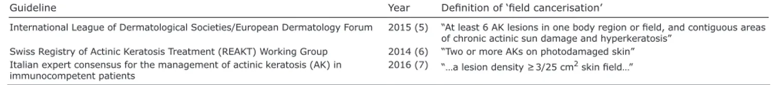
Firstly, there is a need for a standardised definition of AK field cancerisation in clinical, molecular and histo-pathological terms. Current guidelines define field can-cerisation based on number of AK lesions and presence of surrounding photo-damaged skin (5–7). However, there are wide discrepancies within these criteria (Table I). Moreover, experts have voiced concerns over using a definition based on AK counts, as existing evidence suggests that any AK should be considered a marker of field change (8). A clearer and unambiguous definition of field cancerisation that is standardised and reprodu-cible is required to support diagnosis and management, including treatment options and identification of ‘red flag’ signs of high-risk tumours. Physicians can only manage field cancerisation appropriately if they understand its characteristics and severity.
A second clinical gap is the lack of a reproducible clinical global assessment scale for grading AK/field can-cerisation. Current guidelines assess only the presence or absence of AK/field cancerisation, without a severity grading. A global assessment scale should include a clini-cal description of the key characteristics for each grade of severity to guideeffectively identification, diagnosis and treatment decisions.
This clinical grading should be considered alongside modulating risk factors:
• age
• skin phototype • lifestyle • occupation
• geographical location • history of skin cancer • immunosuppression.
Through clinical grading based on disease severity and individual patient risk factors, a therapeutic algorithm Actinic Keratosis, a Chronic, Progressive Disease: Understanding Clinical Gaps to Optimise Patient Management
Rino CERIO1, Thomas DIRSCHKA2, Brigitte DRÉNO3, Ignasi FIGUERAS NART4, John T. LEAR5, Giovanni PELLACANI6, Ketty
PERIS7, Andrés RUIZ DE CASAS8 (PEAK Working Group*)
1Department of Cutaneous Medicine and Surgery, 2nd Floor South Tower, The Royal London Hospital and QMUL, Bart’s Health NHS Trust,
Whitechapel, London E1 1BB, UK, 2CentroDerm® clinic, Wuppertal, and Faculty of Health, University Witten-Herdecke, Witten, Germany, 3Department of Dermato Cancerology, University of Nantes, Nantes, France, 4Department of Dermatology, Bellvitge Hospital, Barcelona,
Spain, 5Manchester Academic Health Science Centre, MAHSC, Manchester University and Salford Royal NHS Foundation Trust, Royal
Infirmary, The University of Manchester, Manchester, UK, 6Department of Dermatology, University of Modena and Reggio Emilia, Modena, 7Department of Dermatology, Catholic University of Rome, Rome, Italy, and 8Dermatology Unit, Virgen Macarena University Hospital, Seville,
Spain. E-mail: [email protected]
A
cta
DV
A
cta
DV
A
dvances in dermatology and venereology
A
ctaD
ermato-V
enereologica Correspondence/Controversies in Dermatology 998 www.medicaljournals.se/actathat enables physicians to make informed treatment decisions would be valuable.
A relevant clinical challenge is the lack of direct evidence to date that AK can progress to iSCC, and that treating AK may prevent the risk of SCC. This is likely to impair the uptake of field therapy despite recommen-dations by current guidelines.
However, it seems reasonable to advocate field treat-ment in appropriate patients based on expert clinical judgement supported by:
• a well-established association between AK/field can-cerisation and skin cancer (9)
• emerging evidence that field therapies treat field cancerisation (i.e. visible AK lesions and non-visible subclinical lesions) (2), and
• preliminary evidence that field therapies prevent SCC in animal models (10).
In summary, addressing the lack of expert consensus on the definition and grading of AK/field cancerisation is crucial to aid physicians in their decision making and optimise appropriate management of AK.
Aiming to address such a need, the authors propose to undertake:
• a systematic review of the literature on field cancerisa-tion to inform a robust definicancerisa-tion
• a global assessment scale for grading AK, which ta-kes into account the entire affected field and not only individual AK lesions.
ACKNOWLEDGEMENTS
Editorial assistance from Lucid Group (funded by LEO Pharma) is gratefully acknowledged.
Conflict of interests: All authors act as consultants to Leo Pharma.
GP reports receiving a research grant from Leo Pharma, and
is a member of advisory boards for Leo Pharma, Roche and Galderma.
REFERENCES
1. Dréno B, Amici JM, Basset-Seguin N, Cribier B, Claudel JP, Richard MA, et al. Management of actinic keratosis: a practical report and treatment algorithm from AKTeamTM expert clinicians. J Eur Acad Dermatol Venereol 2014; 28: 1141–1149.
2. Malvehy J. A new vision of actinic keratosis beyond visible clinical lesions. J Eur Acad Dermatol Venereol 2015; 29 Suppl 1: 3–8.
3. Werner RN, Sammain A, Erdmann R, Hartmann V, Stockfleth E, Nast A. The natural history of actinic keratosis: a syste-matic review. Br J Dermatol 2013; 169: 502–518.
4. Chetty P, Choi F, Mitchell T. Primary care review of actinic keratosis and its therapeutic options: a global perspective. Dermatol Ther (Heidelb) 2015; 5: 19–35.
5. Werner RN, Stockfleth E, Connolly SM, Correia O, Erdmann R, Foley P, et al. Evidence- and consensus-based (S3) Gui-delines for the Treatment of Actinic Keratosis – International League of Dermatological Societies in cooperation with the European Dermatology Forum – Short version. J Eur Acad Dermatol Venereol 2015; 29: 2069–2079.
6. Hofbauer G, Anliker M, Boehncke WH, Brand C, Braun R, Gaide O, et al. Swiss clinical practice guidelines on field can-cerization of the skin. Swiss Med Wkly 2014; 144: w14026. 7. Peris K, Calzavara-Pinton PG, Neri L, Girolomoni G, Malara G,
Parodi A, et al. Italian expert consensus for the management of actinic keratosis in immunocompetent patients. J Eur Acad Dermatol Venereol 2016; 30: 1077–1084.
8. Pellacani G, Neri L, Longo C. Comment to: ‘Evidence and consensus based (S3) Guidelines for the Treatment of Actinic Keratosis’. J Eur Acad Dermatol Venereol 2016; 30: e114. 9. Fernández-Figueras MT, Carrato C, Sáenz X, Puig L, Musulen
E, Ferrándiz C, et al. Actinic keratosis with atypical basal cells (AK I) is the most common lesion associated with invasive squamous cell carcinoma of the skin. J Eur Acad Dermatol Venereol 2015; 29: 991–997.
10. Cozzi SJ, Ogbourne SM, James C, Rebel HG, de Gruijl FR, Ferguson B, et al. Ingenol mebutate field-directed treat-ment of UVB-damaged skin reduces lesion formation and removes mutant p53 patches. J Invest Dermatol 2012; 132: 1263–1271.
Table I. Various definitions of field cancerisation
Guideline Year Definition of ‘field cancerisation’
International League of Dermatological Societies/European Dermatology Forum 2015 (5) “At least 6 AK lesions in one body region or field, and contiguous areas
of chronic actinic sun damage and hyperkeratosis”
Swiss Registry of Actinic Keratosis Treatment (REAKT) Working Group 2014 (6) “Two or more AKs on photodamaged skin”
Italian expert consensus for the management of actinic keratosis (AK) in
immunocompetent patients 2016 (7) “…a lesion density ≥ 3/25 cm