of Naples
PhD in Biology XXXI Cycle
(co-founding SZN/ICB-CNR
)
Toxigenic effects of benthic diatoms upon grazing
activity of the sea urchin Paracentrotus lividus
Tutors:
Dr. Maria Costantini, Stazione Zoologica Anton Dohrn, Naples
Dr. Angelo Fontana, Istituto di Chimica Biomolecolare, ICB-CNR, Pozzuoli
Dr. Valerio Zupo, Stazione Zoologica Anton Dohrn, Ischia
Table of contents
Acknowledgments ... I Abstract ... IV List of abbreviations ... VI
Chapter 1: State of the art and thesis objectives ... 1
1.1 Marine diatoms: an overview of planktonic and benthic taxa ... 1
1.1.2 Interactions of benthic diatoms with seagrasses and their grazers ... 13
1.2 Secondary metabolites in diatoms ... 16
1.2.1 Oxylipins ... 18
1.2.2 Sterol sulfates (StS) ... 44
1.3 The model organism sea urchin Paracentrotus lividus ... 51
1.4 Aims of the thesis ... 61
Chapter 2: Setting up of microcosm for feeding experiments and definition of daily feeding rate of the sea urchin Paracentrotus lividus ... 67
2.1 Introduction ... 67
2.2 Materials and methods ... 69
2.2.1 Ethics Statement ... 69
2.2.2 Sea urchin collection ... 69
2.2.3 Experimental rearing apparatus ... 70
2.2.6 Gonadal index and histological preparation ... 74
2.2.7 Gamete collection, embryo culture and morphological analysis ... 74
2.2.8 Statistical analyses ... 75
2.3 Results ... 76
2.3.1 Daily feeding rate ... 76
2.3.2 Carbon and nitrogen contents of feeds and faecal pellets ... 78
2.3.3 Adult growth and gonadal index ... 80
2.3.4 Fertility of sea urchins ... 81
2.4 Discussion ... 83
Chapter 3: RNA extraction tests ... 88
3.1 Introduction ... 88
3.2 Materials and methods ... 89
3.2.1 Fertilization, sample collection and preservation ... 89
3.2.2 RNA extraction methods and determination of quantity/quality ... 90
3.2.3 Statistical analyses ... 92
3.3 Results ... 92
3.3.1 RNA extraction protocol tests ... 92
3.4 Discussion ... 98
Chapter 4: Feeding experiments with benthic diatoms ... 102
4.1 Introduction ... 102
4.2.2 Morphological and molecular characterization ... 103
4.2.3 Preparation of diatom cultures ... 105
4.2.4 Feeding experiments, faecal pellets collection, fertilization, morphological analysis of embryos ... 106
4.2.5 RNA extraction, de novo transcriptome assembly (RNA sequencing) and Real Time qPCR ... 107
4.2.7 1H-NMR Metabolomic analysis of the gonads ... 111
4.2.8 Statistical analyses ... 113
4.3 Results ... 113
4.3.1 Morphological and molecular characterization ... 113
4.3.2 Feeding experiments ... 121
4.3.3 1H-NMR analysis of metabolites from sea urchin gonads ... 125
4.3.4 Transcriptomic assembly ... 131
4.3.5 Differentially expressed genes in P. lividus plutei after feeding experiments in RNA-seq ... 134
4.3.6 Differentially expressed genes by Real Time qPCR ... 140
4.4 Discussion ... 145
Chapter 5: Chemical analysis of benthic diatoms ... 151
5.1 Introduction ... 151
5.2 Materials and methods ... 153
5.2.4 Oxylipins extraction procedure ... 156
5.2.5 PUAs analysis by GC-MS ... 156
5.2.6 NVOs detection through LC-MS ... 157
5.2.7 FOX2 assay ... 159
5.2.8 StS extraction and LC-MS analysis ... 161
5.2.9 Statistical analyses ... 162
5.3 Results ... 163
5.3.1 GC-MS analysis of free fatty acids ... 163
5.3.2 GC-MS analysis of PUAs ... 166 5.3.3 LC-MS analysis of NVOs ... 169 5.3.4 FOX2 Assay ... 177 5.3.5 StS analysis by LC-MS ... 178 5.4 Discussion ... 180 Chapter 6: Conclusions ... 189
6.1 Contribution of the Ph. D. work ... 189
6.2 General conclusions and future perspectives ... 193
References ... 198
Appendix-A ... 241
is the final touch on my thesis. It has been a period of intense learning for me, not only in the scientific area, but also on a personal level. Writing this thesis has had a big impact on me. I would like to reflect on the people who have supported and helped me so much throughout this period.
Firstly, I would like to express my sincere gratitude to my tutors Dr. Maria Costantini, Dr. Angelo Fontana and Dr. Valerio Zupo, for the continuous support during these years and for the patience, motivation and immense knowledge. Without their precious help it would not be possible to conduct my Ph. D. experiments. Thank you for encouraging my research and for allowing me to grow as a scientist.
My deep appreciation also goes to Dr. Adrianna Ianora and Dr. Giovanna Romano, for the invaluable advices and feedbacks on my research and for always being so supportive of my work.
I am also very grateful to Dr. Genoveffa Nuzzo (Jenny) from CNR of Pozzuoli for teaching me the methodology to carry out chemical analyses and for always being positive and patient when something was wrong. Thanks to Giuliana d’Ippolito from CNR of Pozzuoli for the help in analysing chemical data. Also thanks to Dr. Susan Costantini from CROM (Istituto Nazionale Tumori “Fondazione G. Pascale”) for the great support in performing metabolomic analyses.
I am particularly indebted to Dr. Melody Clark from British Antarctic Survey for her scientific support when so generously hosted me in Cambridge. I have very fond memories of my time there.
and photos. Thanks to the members of the MB&BI (Molecular Biology and Bioinformatic) unit of SZN for providing primers for PCR experiments and for PCR product sequencing. Thanks are also due to the Fishery Service of the SZN and the MEDA (Monitoring and Environmental Data) Unit for providing sea urchins. Special thanks to the technicians of MaRe (Marine Resources for Research) Unit, especially Alberto Macina for the setting up of the continuous open flow-through system used for feeding experiments and Davide Caramiello for his valuable technical support in sea urchin maintenance and gamete collection. Many thanks to the Genomix4Life S.r.l. (Spin-Off of the University of Salerno, Baronissi, Salerno, Italy) for the support in RNA-seq and bioinformatics.
I would like to thank my labmates Annamaria, Alessio, Vincenzo, Tina and Luisa for the nice moments spent together. Special mention goes to Luisa for the chats, laughs and coffees, sharing the good and bad times. Thank you for your friendship and for all the fun we have had in the last year.
I would also to expand my acknowledgments to all people from SZN of Ischia, including researchers, Ph. D. and thesis students. In particular, I thank Francesca, Valerio, Giulia, Antonia, Marco, Anna, Sara and Cristina for always making me laugh, even in the most difficult times. Many thanks to all the Ph. D. students/fellows/technicians from CNR of Pozzuoli for being so kind with me, especially Adelaide (the sweetest woman I ever met), Lucio (I will miss your funny stories), Angela (your great humour helped me in bad times), Nunzia, Alejandro, Paolo and Marisa.
Nobody has been more important to me during these three years than the members of my family: my grandmothers, grandfather, uncles, aunts and cousins. This accomplishment would not have been possible without them.
I would like to thank my parents. I am extremely grateful to them for their love, prayers, caring and sacrifices for educating and preparing me for my future. My deep thanks to my beautiful nieces Rita and Esther for being the light of my life. I express my gratitude to my sister Linda for always supporting me as a good friend. Of course, my thanks are also due to my brother-in-law Angelo for being the elder brother I ever had.
My profound thank goes to Mirko. He is my inspiration and joy of life. I deeply love him for being so patient and kind with me. I see the future in his eyes, hoping that one day we will build a beautiful family.
Finally, I would like to dedicate this thesis to my aunt Annarita, who passed away in the first year of my Ph. D. She would have been so proud of me and I hope that she is looking down on me from Heaven. I miss you so much.
Thank you to all.
world’s ocean food web. The chemical ecology of planktonic diatoms is well documented, but few studies have reported on the effects of benthic diatoms on their consumers, also due to difficulties in the collection, quantification and massive culturing of benthic species. This study investigates, for the first time, the effects of diets based on four benthic diatoms, Cylindrotheca closterium, Nanofrustulum shiloi, Cocconeis
scutellum and Diploneis sp. isolated from the leaves of the seagrass Posidonia oceanica,
on the sea urchin Paracentrotus lividus. The results demonstrate a toxigenic effect on embryos generated by females fed for one month on these benthic diatoms by multidisciplinary approaches. (i) Morphological observations by microscope revealed a noxious effects of C. closterium, N. shiloi and Diploneis sp. on embryos deriving from adult sea urchin P. lividus fed for one month on these diets, with N. shiloi showing the strongest effects; on the contrary, C. scutellum showed no effects, producing embryos as those deriving from the control diet. Malformations of these embryos were very similar to those observed after treatment with planktonic diatom-derived oxylipins. (ii) Metabolomic analysis by Nuclear Magnetic Resonance (1H-NMR) demonstrated that feeding on these diatoms induced variations in the levels of lipids and/or amino acids in the gonads of P. lividus. (iii) Molecular analysis by de novo transcriptome and Real
Time qPCR showed that benthic diatoms were able to affect the expression levels of
several genes, involved in different cellular processes. (iv) Chemical analyses by Gas Chromatography–Mass Spectrometry (GC-MS) and Liquid Chromatography–Mass Spectrometry have been focused on two classes of secondary metabolites isolated in
produced some unknown compounds deriving from polyunsaturated fatty acids metabolism. Moreover, all four benthic diatoms showed the presence of StS.
This study is the first demonstration of the toxic effects of benthic epiphytic diatoms on embryos and larvae of the sea urchin P. lividus due to the feeding of adults during gonadal maturation. Furthermore, the present work assumes a considerable ecological relevance, opening new perspectives on the study of diatom-derived secondary metabolites influencing their grazers.
°C degree Celsius µL microlitre
11,10-EHETE 11,12-epoxy-10-hydroxy-eicosa-5Z,8Z,14Z,17Z-tetraenoic acid 11-HEPE 11-hydroxy-eicosa-5Z,8Z,12E,14Z,17Z-pentaenoic acid
14S,13R-EHETE 14S,15S-epoxy-13R-hydroxy-eicosa-5Z,8Z,11Z,17Z-tetraenoic acid 15S-HEPE 15S-hydroxy-eicosa-5Z,8Z,11Z,13E,17Z-pentaenoic acid
16,15-EHDoPE 16,17-epoxy-15-hydroxy-docosa-4Z,7Z,10Z,13Z,19Z-pentaenoic acid 17-HDoHE 17-hydroxy-docosa-4Z,7Z,10Z,13Z,15E,19Z-hexaenoic acid
19,18-EHDoPE 19,20-epoxy-18-hydroxy-docosa-4Z,7Z,10Z,13Z,16Z-pentaenoic acid 20-HDoHE 20-hydroxy-docosa-4Z,7Z,10Z,13Z,16Z,18Z-hexaenoic acid
5-HEPE hydroxy-eicosa-6E,8Z,11Z,14Z,17Z-pentaenoic acid 6-HHTrE 6-hydroxy-hexadeca-7E,9Z,12Z-trienoic acid 9-HHME 9-hydroxyhexadec-7E-enoic acid
9-KHME 9-ketohexadec-7E-enoic acid
9S,11R-EHHDE 9S,10S-epoxy-11R-hydroxy-hexadecadienoic acid 9S-HHTrE 9S-hydroxy-hexadeca-6Z,10E,12Z-trienoic acid
9S-HHTtE 9S-hydroxy-hexadeca-6Z,10E,12Z,15-tetraenoic acid AA arachidonic acid
ANOVA analysis of variance BHT butylated hydroxytoluene
BP biological process C/N carbon/nitrogen ratio CC cellular component
cDNA complementary deoxyribonucleic acid CHNS carbon, hydrogen, nitrogen, sulfur CHOS cholesterol sulfate
cm centimetre CO2 carbon dioxide
CTAB cetyltrimethylammonium bromide DE differentially expressed
DEPC diethylpyrocarbonate
DHA docosa-4Z,7Z,10Z,13Z,16Z,19Z-esenoic acid DHBS dihydrobrassicasterol sulfate
DNA deoxyribonucleic Acid DNase deoxyribonuclease
dNTP deoxynucleotide triphosphate DSP death specific proteins
dw dry weight
EAS epoxyalcohol synthase
EPA eicosa-5Z,8Z,11Z,14Z,17Z-pentaenoic acid ESI electrospray ionization
FDR false discovery rate fmol femtomole
FOX2 ferrous oxidation-xylenol orange 2 FSW filtered sea water
g gram
GC-MS gas chromatography-mass spectrometry GI gonadal index GO gene ontology GTPC guanidinium thiocyanate-phenol-chloroform h hour H20 dihydrogen monoxide HCO3¯ bicarbonate
HMDB human metabolome database hpf hours post fertilization
HPL hydroperoxide lyase HR hydroperoxide reductase
HR-MS high resolution - mass spectrometry Hz hertz
IPA ingenuity pathway analysis KCl postassium chloride
LC-MS liquid chromatography-mass spectrometry log logarithm LOX lipoxygenase m metre M molar MeOH methanol MF molecular function mg milligram min minute mL millilitre mM millimolar ms millisecond
MS/MS tandem mass spectrometry
MTT 3-(4,5-Dimethylthiazol-2-yl)-2,5-diphenyltetrazolium bromide Na+ sodium
NaCl sodium chloride
NCBI national center for biotechnology information NGS next generation sequencing
nm nanometre
NMR nuclear magnetic resonance NO nitric oxide
PAPS 3’-phosphoadenosine-5’-phosphosulfate PCA principal component analysis
PCD programmed cell death PCR polymerase chain reaction pg picogram
ppm parts per million
PUAs polyunsaturated aldehydes PUFAs polyunsaturated fatty acids
qPCR quantitative polymerase chain reaction qTOF quadrupole time of flight
REST relative expression software tool RIN RNA integrity number
RNA ribonucleic acid RNase ribonuclease RNAseq RNA sequencing ROS reactive oxigen species rRNA ribosomal RNA RT retention time s second
SCUBA self-contained underwater breathing apparatus SD standard deviation
SSH suppression subtractive hybridization STD standard
StS sterol sulfates SULT sulfotransferase
TCEP tris(2-carboxyethyl)phosphine
TUNEL terminal deoxynucleotidyl transferase (TdT) dUTP Nick-End Labeling UHPLC ultra-high-performance liquid chromatography
UVmax UV maxima
VIP variable importance in projection VOCs volatile organic compounds ww wet weight
XO xylenol orange β-ME 2-mercaptoethanol Β-SITS β-sitosterol sulfate μg microgram
μm micrometre μM micromolar
1. State of the art and thesis objectives
1.1 Marine diatoms: an overview of planktonic and benthic taxa
Diatoms are unicellular algae formally classified in the phylum Bacillariophyta, class Bacillariophyceae. They are highly abundant in nearly every habitat where water is found (oceans, lakes, streams, mosses, soils, even the bark of trees), colonizing the planktonic and benthic environments of marine and freshwater habitats. The term
plankton is referred to organisms that live in the water column and, because they are
non-motile or too weak to swim against the current, exist in a drifting state. Phytoplankton is the portion of plankton that grows autotrophically, using CO2 as its
carbon source and light as its energy source. Diatoms are considered a fundamental component within phytoplankton, because they play a key role in the global carbon cycle (Figure 1.1), constituting a rich diet for many herbivores, due to their lipid content (Pohnert, 2005). They can be placed at the bottom of food webs in marine and freshwater habitats, contributing for up to 40-50% of marine primary productivity (Nelson et al., 1995; Falkowski and Raven, 2007). In the ocean, marine phytoplankton comprises photoautotrophic organisms from 12 taxonomic divisions and 3 Kingdoms (Falkowski et al., 1998), including several algae, bacteria, protozoans, crustaceans, molluscs and coelenterates, as well as representative other organisms from almost every other phylum.
Differently from plankton, benthos consists of sessile or creeping organisms, such as prochlorophytes and cyanobacteria (Giovannoni and Rappè, 2000) and eukaryotic
microalgae, such as chromophytes (brown algae), rhodophytes (red algae), and chlorophytes (green algae) (Van Den Hoek et al., 1997).
Figure 1.1. Schematic representation of the carbon cycle in the oceans. Marine ecosystems are a major sink for atmospheric CO2 and take up similar amount of CO2 as terrestrial ecosystems, currently
accounting for the removal of nearly one third of anthropogenic CO2 emissions from the atmosphere. The
net transfer of CO2 from the atmosphere to the oceans and then sediments is mainly a direct consequence
of the combined effect of the solubility and the biological pump. While the solubility pump serves to concentrate dissolved inorganic carbon in the deep oceans, the biological carbon pump transfers both organic and inorganic carbon fixed by primary producers (phytoplankton) in the euphotic zone to the ocean and subsequently to the underlying sediments, maintaining atmospheric CO2 at significantly lower
levels.
The benthic environment of marine or freshwater habitats includes the bottom, such as the ocean floor or the bottom of a lake, the sediment surface and some sub-surface layers. Benthos can be divided in phytobenthos, the primary producers (algae, aquatic plants) living on the bottom, and zoobenthos, all consumers (from protozoa to all other benthic animals) living on/or in close relationship with the bottom. Benthic diatoms
constitute a part of phytobenthos where they represent a food source for several marine consumers (Miller et al., 1996). As already mentioned for planktonic species, benthic diatoms are implicated in the biogeochemical cycles of carbon, nitrogen, phosphorus and silicon, contributing for about the 20% of benthic primary production (Falkowski and Raven, 1997). Because of the diversity of substrates, benthic diatoms have numerous different microhabitats, as opposed to phytoplankton, whose environment is more homogenous. Benthic diatoms can range from large motile forms that are common in the intertidal sands, to sessile species that are attached to various substrates (periphyton). The latter may be attached to rocks (epilithon) and sand (episammon), as well as loosely associated with mud (epipelon), macrophytes, seagrasses and artificial substrates (epiphytic) or even growing on living animals (epizoic). Moreover, semi-aquatic habitats of temporal pools and tide pools are observed to contain some taxa of benthic diatoms. Despite the ability of benthic diatoms to adapt to different microenvironments, they tend to prefer natural substrates, especially vegetation, compared to artificial substrates, because of the selective nature of the physical and chemical properties of artificial substrates (Bere, 2010).
Considering both planktonic and benthic species, diatoms are commonly between 20-200 μm in diameter or length, although sometimes they reach 2 mm in length. They display a rigid cell wall made of silicon dioxide (SiO2), called frustule. The diatom
frustule is composed of two overlapping thecae (the larger is called epitheca and the smaller hypotheca), consisting of a valve and an accompanying series of girdle bands. Since silica is impervious, diatoms have evolved elaborate patterns of perforations in their valves to allow nutrient and waste exchange with the environment (Kröger and
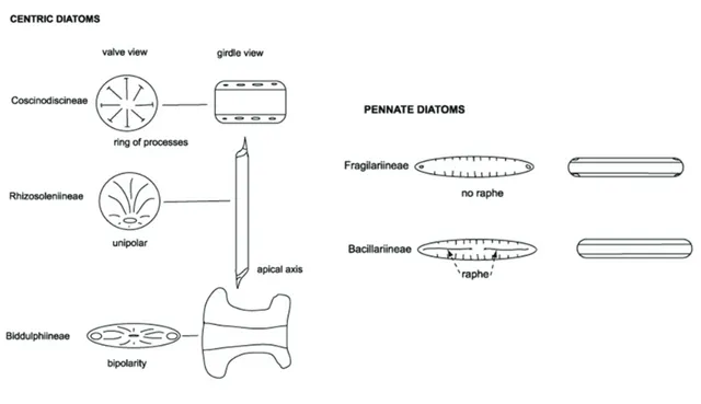
Poulsen, 2008). The valve face of the diatom frustule is decorated with pores (areolae), processes, spines, hyaline areas and other distinguishing features normally used for diatom classification. Diatoms are commonly divided into two Orders: i) the Centrales, which have valve striae arranged basically in relation to a point, an annulus or a central areola and tend to appear radially symmetrical; ii) the Pennales, which have valve striae arranged in relation to a line and tend to appear bilaterally symmetrical (Simonsen, 1979; Von Stosch, 1982; Hasle and Syvertsen, 1997; Figure 1.2).
Figure 1.2. Scanning Electron Microscopy (SEM) images of centric diatoms (A-C) and pennate diatoms (D-E). (From Brayner et al., 2011)
Centric diatoms include three sub-orders based primarily on the shape of the cells, the polarity and the arrangement of the processes: i) the Coscinodiscineae, with a marginal ring of processes and no polarity to the symmetry; ii) the Rhizosoleniineae, with no marginal ring of processes and unipolar symmetry; iii) the Biddulphiineae, with no
marginal ring of processes and bipolar symmetry (Figure 1.3; Simonsen, 1979). Pennate diatoms are generally elongated, and many of them exhibit bilateral symmetry, and even helical symmetry is possible (Round et al., 1990). They are divided into two sub-orders based on the presence or absence of a paired slit system (the raphe) in the valves:
Bacillariineae (raphid diatoms) and Fragilariineae (araphid diatoms; Hasle and
Syvertsen, 1997).
Figure 1.3. Schematic representation of centric, raphid and araphid pennate diatoms. (From Hasle and Syvertsen, 1997).
The siliceous cell wall has been used for classification of diatoms. The diatom taxonomy has been almost exclusively based upon frustule characteristics: shape, size, symmetry, structure and density of striae, nature of raphe and its position, copula and processes on the valves. However, recently ultrastructure and features of living cells, such as
chloroplastids, life cycle pattern and reproductive biology have been extensively used in systematic studies.
In fact, the scientific community acquired a greater awareness regarding the need to describe the structure of the living cells of diatoms, the protoplast, especially the plastids as part of the diagnosis of species. Plastids are the most conspicuous organelles featuring in living diatoms and their number and shape are consistent features of taxonomic importance (Cox, 1996; Round et al., 1990). Chloroplastids may be rounded or lobed discs or large plate-like with or without lobed margins, and may range from one to two, four or more. A typical centric diatom has many disc-shaped plastids, closely arranged to the periphery surrounding a large central vacuole or scattered throughout the cell. The raphid diatoms tend to have large chloroplasts (one to four), lying along the girdle with central nucleus flanked by two large vacuoles. The species concept in biology is sometimes based upon reproductive isolation but it is difficult to apply to diatoms (Round et al., 1990).
Diatom taxonomy is in a continuous state of revolutionary change, involving the splitting of genera, creation of new ones and revision of criteria for classification. This is mainly due to advancement in cultivation technologies and in molecular techniques, which have made taxonomy of diatoms a very dynamic field of research, in addition to the significance of diatoms as recognized worldwide biomonitoring tools for aquatic systems (John, 1998, 2000).
From 20.000 to 2 million species of diatoms (Round et al., 1990; Hasle and Syvertsen, 1997; Mann, 2005) are presently estimated. This range is large because scientists are still working to understand basic aspects about "what is a diatom species" and because
new and diverse forms are still being discovered and described in scientific publications (Simonsen, 1979; Von Stosch, 1982; Hasle and Syvertsen, 1997).
The diatom reproduction process can occur by either sexual or asexual mechanisms (Figure 1.4). All diatoms pass through a seed-like or a spore phase called the resting spore. The asexual reproduction occurs by binary fission. In this process, the DNA undergoes replication that causes the chromosomes to divide into two identical halves. This leads to the formation of two frustules or theca. Each daughter cell receives one of the parent cell frustules, the hypotheca, and forms a new frustule, the epitheca. The parent cell grows larger until dividing into two daughter cells by pushing out of the valve. Each daughter cell produces a new cell wall, each receiving one valve. Availability of dissolved silica limits the rate of vegetative reproduction, but also because this method gradually reduces the average size of the diatom frustule.
For this reason, the size of diatoms tends to decrease along asexual generations and there is a certain threshold at which restoration of frustule size is necessary in populations (Hasle and Syvertsen, 1997; Zhang et al., 2005; Peng et al., 2014). The algae need to restore their original cell size, and to this end they use sexual reproduction. The vegetative cells of diatoms are diploid (2n), and hence, they undergo meiosis with males and females producing gametes, respectively. The female cells tend to bend and create an opening in the cell wall helping the male gamete to enter the female cell and fertilize it. The zygote gets encased in an envelope-like structure, which produces its own shell and nucleus. Then, the diatom cell starts to grow until it reaches its full size. The parent cell and the new diatom can form auxospores, which are cells that possess a different wall structure, lacking the siliceous frustule, and enlarge to the maximum
frustule size. This helps the cells to survive for long periods under unfavourable conditions. Once the cells get proper conditions to grow, they continue with their asexual reproduction (Round et al., 1990).
Figure 1.4. Diatoms reproduce through asexual reproduction or cell division. Each half of the new cell gets half of the frustule, which then has to fit inside the other half when it is reformed. As they keep getting smaller each time, the diatoms must then produce sexually to make an entirely new frustule.
Diatoms use the pigments chlorophyll a and c to collect energy from the sun through photosynthesis. They also contain the accessory pigments, fucoxanthin and β- carotene, which give them a characteristic golden colour (Bertrand, 2010). Diatom cells store energy from photosynthesis in the form of chrysolaminarin (a carbohydrate) and lipids (fats in the form of oils). The high production of lipids in many diatom species give great interest in diatoms as a source of biofuels and prospective materials for
2015; Prabandono and Amin, 2015). Indeed, as one of the important global sources of carbon fixation, diatoms already are an important biofuel for aquatic food webs (Falkowski et al., 2008).
Diatom abundance is generally highest at the beginning of spring and in the autumn, when light intensity and day length are optimal for their photosynthesis. In some regions of the oceans, the annual production of fixed carbon can be up to 2 kg m-2 equivalent to a cereal or corn crop (Field et al., 1998). When the population of these organisms increases up to a concentration of hundreds to thousands of cells per mL, the water is coloured green-blue, yellow-brown or red: this phenomenon is known as “algal bloom” (Figure 1.5). These events mainly involve toxic phytoplankton, such as dinoflagellates of the genera Alexandrium and Karenia, or diatoms of the genus Pseudo-nitzschia. Of the over 5000 known species of marine phytoplankton, about 300 species can, under certain circumstances, proliferate in exponential numbers and only about 2% of these species have the capacity to produce potent toxins, which can negatively affect the local ecosystem, as well as fishing and aquaculture activities (Landsberg, 2002). Although these events are particularly known in planktonic species, recent evidences of benthic diatom blooms in marine and freshwater habitats were also recorded (Galland and Pennebaker, 2012; Ahn et al., 2016; Bothwell and Taylor, 2017). For example, a massive blooming of several benthic species, including Cocconeis spp., Paralia sulcata,
Achnanthes spp., Amphora sp., Eucampia antarctica, Navicula spp., Odontella litigiosa,
Pleuorosigma sp. have been detected in the shallow seafloor of an Antarctic Fjord (King George Island) (Ahn et al., 2016).
Figure 1.5. Image taken on August 14, 2011, by the Moderate Resolution Imaging Spectroradiometer (MODIS) on the Aqua satellite show brilliant shades of blue and green across the Barents Sea. The color was created by a massive bloom of phytoplankton that are common in the area each August.
Fundamentally, diatoms are unicellular, but several taxa form chains or filaments due to daughter cells, remaining connected even after cell division, often by polysaccharide pads or linking spines. The colonies may be straight, zigzagged, fan-shaped, star-fan-shaped, or radiate. The production of exopolysaccharides (EPSs) normally reaches high levels in planktonic diatoms (for example Skelertonema costatum), to cause the formation of the co-called “marine snow” (Riebesell, 1991a,b). One of the first chemical studies in benthic diatoms of mucilage and their influence on growth rate were performed by Buzzelli et al. (1996), who collected samples from the coastal area of Cesenatico (Adriatic Coast) containing cells of the diatom Amphora coffeaeformis var.
EPSs are secreted into the environment as a reserve of carbohydrates, as well as cell wall constituents within the cells, which form adhering biofilms (Figure 1.6), contributing to biostabilization of sediments (Paterson and Black, 1999). The sediment stabilization efficiency of benthic diatoms is due to the high water retention capacity of EPSs, which may protect the organisms against desiccation (Decho, 1990).
Figure 1.6. Scanning electron micrograph of Ascophyllum nodosum surface. The brown alga, collected on the rocky shores of the North Atlantic Ocean, is covered by a thin biofilm of diatoms, bacteria, and fungi. (From Canadian Science Publishing Blog).
Paterson (1994) studying the sediment-stabilizing effect of pennate diatoms on quartz-sandy tidal flats, differentiated between epipelic (immotile diatoms, living semi-permanently attached to sand grains) and epipsammic diatoms (motile diatoms, moving freely through the sediment).
In relation to the life-form of the benthic diatoms, two effects of sediment stabilization are observed:
1) Cohesive effect: the mucus, secreted by epipsammic diatoms, epipelic diatoms and bacteria, on sand grains improves the cohesion of the particles. The effect of the organic coating on the sand grains was studied in the laboratory in a small circular flume, using samples from the field. An organic coating on the grains was found to cause a 5-17% increase in critical erosion velocity, relative to sterile sediment cleaned with H2O2;
2) Network effect: when a large number of epipelic diatoms migrate to the sediment surface, they can form an extensive network of mutual attachment and mucus threads. In laboratory conditions the formation of an epipelic diatom mat leads, in 24 hours, to an increase in the sediment motion by 25% to 100%. In the field the network formation by epipelic diatoms and the physical processes are interdependent. The network protects the sediment surface against erosion by tidal currents and wave action, and conversely, the diatom population depends on these factors.
Benthic microalgae biofilms can be found in different arrangements. The structure of a biofilm is characteristic: it is primarily regulated by inundation, sediment dynamics, light, and water availability (Gerdes and Wehrmann, 2008) and depends on the degree of microalgal development. Even though the range of diatoms in biofilms is diverse, their ecology is poorly understood because of the difficulty in sampling and enumeration. Scraping or brushing are the traditional methods used for removal of diatoms from biofilms developed on solid substrata.
The biological and ecological characteristics of diatoms make them good indicators of water quality, and their use is widespread and well developed for evaluating the quality of flowing water (Prygiel et al., 1999) and standing waters, particularly lakes (Kitner and Poulícková, 2003; Blanco et al., 2004; DeNicola et al.,
2004; Denys, 2004; King et al., 2006). Analysis of diatom communities constitutes a valuable tool for general water quality monitoring and for the evaluation of more specific phenomena, such as eutrophication and acidification (Prygel and Coste, 1993; Danilov and Ekelund, 2000; Kauppila et al., 2002; Kitner and Poulícková, 2003).
1.1.2 Interactions of benthic diatoms with seagrasses and their grazers
Epiphytic community on seagrasses is constituted of various diatom species along with red, brown, green algae, cyanophyta and detritus (Frankovich and Zieman, 1994), which is called periphyton. It is obvious that benthic diatoms are the most important elements of the periphyton on seagrasses, because they take the greatest share in the epiphytic biomass (Jacobs and Noten, 1980). Most diatom species found on marine angiosperms are classified as obligate epiphytes (McIntyre and Moore, 1977) and, under nutrient limited conditions, can be strongly influenced by host metabolism. Nutrient uptake by the roots of seagrasses and, subsequent release via the leaves (McRoy and Barsdate, 1970; McRoy et al., 1972), bathe the periphyton in a nutrient-rich medium and individual diatom species exhibit a highly varied response to different nutrients (Saks et al., 1976). Biochemical, mechanical and fisical interactions may be partially responsible for the observation that the pennate diatom Cocconeis scutellum is the sole pioneer species on Zostera marina leaves, forming a uni-algal mat over newly formed blades (Sieburth and Thomas, 1973).
Benthic diatoms appear also to be specific in their selection of a suitable-sized substrate. For example, the species C. scutellum, which has a larger surface area of attachment, was shown to avoid finely branched algal thalli in preference for more thickly branched
species (Ramm, 1977). In fact, the genus Cocconeis is dominant on Z. marina (Sieburth and Thomas, 1973; Jacobs and Noten, 1980) and Thalassia testudinum (DeFelice and Lynts, 1978; Reyes-Vasquez, 1970), whereas Navicula pavillarsi, a narrow diatom, is highly abundant on Ruppia maritima leaves (Sullivan, 1977).
Both beneficial and adverse effects of epiphytes on macrophytes have been mentioned in scientific literature. Algal mats have shown to have a limiting effect on the growth of
R. maritima and have been observed to cause temperature stratification in the water
column due to shading (Richardson, 1980). Such stratification can postpone flowering, fruiting and seed production in R. maritima. If shading by algal mats is severe, active photosynthesis is restricted to the upper layer of the water column. As a result of thermal stratification, the photosynthetically oxygenated water does not reach the lower portions of the water column.
The effects of epiphytes on photosynthesis of Z. marina have been investigated by Sand-Jensen (1977). The authors have showed that epiphytes, including benthic diatoms (for example C. scutellum), form a crust over the leaves of Z. marina and reduce photosynthesis by limiting the availability of light and acting as a barrier to carbon uptake. Photosynthesis was reduced (31%) by epiphytes under optimal light conditions and environmental HCO3¯ concentrations. Light attenuation by epiphytes is therefore
responsible for premature senescence and a strong decrease in both vegetative and sexual reproductive capabilities (Richardson, 1980; Rice et al., 1983).
Trocine et al. (1981) demonstrated a protective role of epiphytes against ultraviolet-β radiation. Fouling by algal epiphytes enabled Halophila engelmannii to colonise shallow water dominated by high levels of ultraviolet-β radiation.
Colonization of seagrass blades by epiphytes commonly follows a pattern that may have further beneficial consequences for host plant. In fact, epiphytes normally grow at the tips of seagrass leaves which are the preferred food for grazers. The selective removal of highly epiphytized and senescent leaf tips cause minimal damage to basal leaf tissues and increase light penetration (Ott and Maurer, 1976; Lobel and Ogden, 1981). Thus, epiphytes may divert direct grazing from the primary photosynthetic tissue (more basal) of seagrasses to senescent leaf tips.
Grazers from diverse taxonomic groups including gastropods, amphipods, isopods, decapods, echinoderms and fish basically find most of their nutrition from seagrass epiphytes. Zupi and Fresi (1984) observed the gut content of echinoderms (23 species) living on Posidonia oceanica meadows collected in 35 sites around Ischia (Gulf of Naples, Italy). This analysis revealed the presence of P. oceanica fragments, several species of diatoms, molluscs, macrophytes, foraminiferans, poriferans, bryozoans and polychaetes, with P. oceanica dominating the gut content of the sea urchins
Parancentrotus lividus and the echinoderm Ophiura texturata individuals.
Through their feeding activities, some grazers may increase epiphytic dominance of adhesive diatoms, such as C. scutellum (van Montfrans et al., 1982), which is tightly attached and generally not removed by grazers (Lamberti and Resh, 1983). On the contrary, grazing can also reduce epiphyte abundance on seagrass leaves. For example, Caine (1980) demonstrated that periphyton biomass was dramatically reduced on ungrazed Z. marina with respect to plants grazed by the caprellid amphipod Caprella
Concluding, benthic diatoms are important food sources for benthic grazers such as sea urchins, fishes, gastropods and crustaceans. Without diatoms and other epiphytes, there will be no food for herbivores in seagrass beds, being epiphytes their main food source. Seagrass itself has poor nutritional value and is made up of cellulose, which several grazers are unable to metabolize. Consequently, some herbivorous prefer to eat only the epiphytes, eating the seagrass solely as a means for consuming the epiphytes and therefore selecting for seagrass leaves that only have an abundant amount of epiphytes and diatoms (Del Río, 2016). Contemporarily, diatoms are not able to adapt to local environmental changes becoming tolerant to drought and low light conditions and able to respond strongly to large-scale climatic variables and fluctuations by finding suitable habitats and having efficient nutrient uptake from seagrass leaves (Pajunen et al., 2016).
1.2 Secondary metabolites in diatoms
Oceans, due to the area they represent and the ecosystem services they provide, are fundamental to the planet, harbouring a huge biodiversity of life. Some estimates suggest that the probability of discovering new bioactive molecules from marine sources is approximately three times higher than that from terrestrial ones, mainly due to the great biodiversity of marine organisms and their chemical products, many of which have no terrestrial counterpart. In the last decades, metabolites produced by marine organisms, especially micro-organisms, have received increasing attention for their role in shaping interactions and communities (Leflaive and Ten-Hage, 2009). These metabolites, called secondary metabolites, are not required for the growth and maintenance of the cellular functions (primary metabolites) and are the end products of
the primary metabolism. Marine organisms, including diatoms, have been shown to produce a variety of bioactive secondary metabolites acting as chemical signals within communities (Leflaive and Ten-Hage, 2009; Stonik and Stonik, 2015).
In the plankton, marine diatoms, dinoflagellates and other green and yellow-brown flagellates are considered a potentially valuable source of new biologically active molecules that can be useful for applications in several biotechnology sectors (food, energy, health, and environment). Several studies have shown that microalgal peptides can exert a large variety of activities, such as, antioxidant, anticancer, antihypertensive, antiatherosclerotic, anti-UV radiation, and antiosteoporosis (Fan et al., 2014). However, a few of these have so far been addressed for evaluation in clinical phases and even fewer have managed to reach the market. A successful example is Dermochlorella®, an oligopeptide purified from Chlorella vulgaris which is used in cosmaceutical field. Dermochlorella® increases the expression of collagen, elastin, laminin, and the inhibitors (elafin and tissue inhibitors of metalloproteinase) of enzymes that degrade the extracellular matrix and restore skin elasticity (Martins et al., 2014). Furthermore, Lauritano et al. (2016) have recently reported that the diatom Skeletonema marinoi arrested the growth of melanoma A2058 cells, whereas the diatoms Cylindrotheca
closterium, Odontella mobiliensis and Pseudonitzschia pseudodelicatissima had
anti-inflammatory effects, and Leptocylindrus aporus and danicus and S. marinoi had antibacterial properties.
For these reasons, despite diatoms have been regarded as beneficial to the growth and survival of primary consumers (such as copepods and benthic filter feeders), several authors have placed in doubt the positive role of diatoms, demonstrating that fecundity
and/or hatching success were reduced when herbivorous copepods were fed on diatom-based diets (Ianora and Poulet 1993; Poulet et al., 1994; Chaudron et al., 1996; Buttino et al., 1999; Ianora and Miralto, 2010). Such evidences collected over the years, is known as “paradox of the diatoms-copepods interaction” in the pelagic food web (Ban et al., 1997), because the known negative plant-animal interactions are generally related to repellent or poisoning processes, but never to reproductive failure.
The present Ph. D. project has focused on two classes of diatom secondary metabolites with interesting biological activities, oxylipins and the sterol sulfates (StS), which will be described in detail in the following paragraphs.
1.2.1 Oxylipins
The first studies demonstrating the inhibitory effect of diatoms on the reproduction of copepods were reported by Ianora and Poulet (1993). They showed that when the copepod Temora stylifera was fed with mono-algal diet of diatom Thalassiosira rotula, eggs productions remained high for more than 15 days but hatching success was seriously compromised, compared to animals fed with the dinoflagellate Prorocentrum
minimum. The authors proposed that these differences were not due to the biochemical
composition of the two algae, because both T. rotula and P. minimum contained high levels of proteins, vitamins and fatty acids essential for growth and development of copepods. It has been hypothesized that the reduction of hatching rates was caused by the presence of unknown anti-mitotic compounds, which arrested embryogenesis in the copepod Calanus helgolandicus (Poulet et al., 1994). The embryonic development of copepods was blocked when eggs were exposed to diatom extracts in a dose-dependent
manner (Poulet et al., 1994; Ianora et al., 1995; Miralto et al., 1995; Uye, 1996). Depending on the incubation time, fusion of the female and male pronuclei was arrested and eggs remained either at the one-cell stage, or zygotes underwent abnormal development characterized by the presence of dispersed chromatin and accompanied by anomalous cellular division (Poulet et al., 1995). Cellular anomalies rejected erroneous synchronization between nuclear and cellular division. In a few cases, embryos developed up to hatching, but nauplii were deformed and displayed marked anatomical anomalies, such as asymmetrical bodies or small number of appendages for swimming and feeding (Poulet et al., 1995; Uye 1996; Starr et al., 1999; Ianora et al., 2004). Those nauplii did not survive long, dying a few hours after hatching because they were not able to swim or feed. The inhibition of hatching was also shown to be diatom density-dependent (Chaudron et al., 1996; Ban et al., 1997; Starr et al., 1999). The unknown compounds, responsible for these deleterious effects on copepods, were demonstrated to be produced by the diatom cells and not by bacteria associated with diatom cultures (Ianora et al., 1996). Ban et al. (1997) showed that hatching success and/or fecundity were reduced when seventeen copepod species were fed with sixteen different diatoms, representing a variety of marine, estuarine and freshwater habitats, demonstrating that this phenomenon occurs on a world-wide scale. Among the seventeen species, only one did not result in reduced egg production rates in comparison to dinoflagellate diets. Testing different diatom-copepod combinations, diatoms reduced average fecundity by 90% and hatching success by 80%. It is important to remember, however, that the bulk of evidence for diatom toxicity on copepods is based on laboratory studies.
In other studies, the attention has been focused on the effects of maternal diet on the production of eggs, the hatching success and growth and differentiation of copepods. Carotenuto et al. (2002) have shown that T. stylifera was not able to complete development from hatching to adult, after the feeding of larvae with three different species of diatoms (T. rotula, Skeletonema costatum and Phaeodactylum tricornutum), showing a high mortality rate. Ianora et al. (2004) have also analysed the effects of maternal diatom-diet on the fitness of copepod offspring. They have shown that the development was stopped in all larvae of C. helgolandicus females fed with the diatom
S. marinoi. In addition, the mortality remained high even if the larvae, generated by
females fed with S. marinoi, were nursed with only the dinoflagellate P. minimum. On the other hand, when the females were fed with P. minimum and the nauplii were grown with S. marinoi, the survival rate increased considerably. On the contrary, when females were fed with S. marinoi and then with P. minimum, the offspring mortality increased. As a result, maternal-feeding quality was demonstrated to be more important than nauplia sustenance for progeny survival.
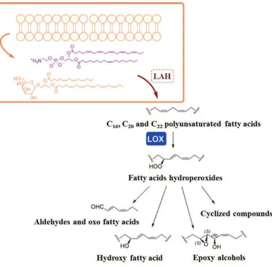
These surprising findings have led researchers to perform chemical studies in order to identify the molecules responsible for these deleterious effects. Thus, it has been demonstrated that these uncharacterized compounds were the end-products of a lipoxygenase (LOX) metabolic pathway (Figure 1.7) initiated by cell damage occurring during senescence or grazing. Cell damage activates specific lipases, such as phospholipase or lipolytic acyl hydrolases (LAHs), which release PUFAs from cell and chloroplast membranes. Then LOXs, iron non-heme enzymes, catalyse the insertion of oxygen into the 1,3-pentadienyl moiety of PUFAs to produce fatty acid hydroperoxides
(FAHs). FAHs are finally modified through several enzymatic activities catalysed by hydroperoxide lyase (HPL), epoxyalcohol synthase (EAS) and hydroperoxide reductase (HR) to form a plethora of metabolites, such as polyunsatured aldehydes (PUAs), hydroxy fatty acids and epoxyalcohols, collectively termed oxylipins (Fontana e al., 2007a,b; Romano et al., 2010).
Figure 1.7. Schematic representation of the oxylipins’ synthesis of from C16, C20 and C22 polyunsatured
fatty acids (PUFAs) through lipoxygenase enzymes (LOXs) pathway in marine diatoms. (From d’Ippolito et al., 2018).
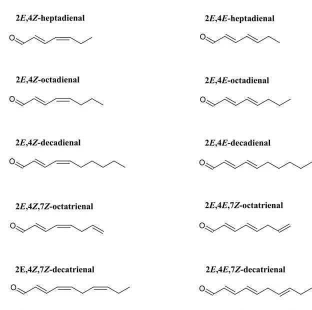
Among oxylipins, PUAs have been described only in a small number of diatom species. Firstly, Miralto et al. (1999) isolated and identified PUAs from the bloom-forming
diatoms T. rotula, S. costatum (identified as S. marinoi in Sarno et al., 2005) and
Pseudo-nitzschia delicatissima as two C10 short chain PUAs: 2E,4E/Z-decadienal and
2E,4E/Z,7Z-decatrienal. These findings have been further confirmed in several studies, using different methodologies and identifying a more complete range of PUAs from S.
marinoi and T. rotula (Pohnert, 2000, 2002; d’Ippolito et al., 2002a,b, 2003; Wichard et
al., 2005a,b). Afterwards, an expanded range of PUAs were identified including 2E,4E-heptadienal, octadienal, 2E,4E-octadienal, 2E,4E/Z,7Z-octatrienal and 2E,4Z-decadienal (Pohnert, 2000, 2002; Pohnert et al., 2002; d’Ippolito et al., 2002a,b, 2003) (Figure 1.8).
Despite PUAs have been the first oxylipins identified, almost all diatoms possess LOX pathways for the synthesis of other oxylipins, including fatty acid derivatives with hydroxy-, keto-, oxo-, and hydroxy-epoxy functionalities. In order to distinguish them from PUAs, these compounds are generically named as non-volatile oxylipins (NVOs). In addition to PUAs and NVOs, FAHs (the precursors) are also suspected to play different ecophysiological functions, including the detrimental effects on grazers and the chemical communication between diatom cells, which may contribute to regulate the oceanic phytoplankton blooms. FAHs, the primary LOX products, are reactive oxygen species (ROS) that could operate in addition to other reactive radicals to induce DNA and protein damage contributing to cell aging. These compounds also induce apoptosis and morphological abnormalities in copepods at concentrations significantly lower than PUAs and, in consideration of their almost universal presence in diatoms, it is possible that FAHs more than PUAs are responsible for the negative response and reproductive failure of copepods (Barreiro et al., 2011; Fontana et al., 2007a,b).
O O O O O O O O O O 2E,4Z-heptadienal 2E,4Z-octadienal 2E,4Z-decadienal 2E,4Z,7Z-octatrienal 2E,4E-heptadienal 2E,4E-octadienal 2E,4E-decadienal 2E,4E,7Z-octatrienal 2E,4Z,7Z-decatrienal 2E,4E,7Z-decatrienal
Figure 1.8. Structure of diatom-derived polyunsaturated aldehydes (From Ribalet et al., 2007).
Type and quantity of oxylipins are species-specific (Pohnert et al., 2002, Taylor et al., 2009), due to a variety of precursor PUFAs and enzymes (d’Ippolito et al., 2003; d’Ippolito et al., 2004; Fontana et al., 2007a,b), with variable effects on zooplankton grazers (Ianora and Miralto, 2010).
Oxylipins and LOX enzymes are classified in different groups, according to the substrate specificity, as well as the position and the stereochemistry of the oxygen addition along the PUFA carbon chains.
Oxylipin pathways of diatoms are mostly based on the oxygenation of eicosapentaenoic acid (EPA, C20:5 ω-3) (Barreiro et al., 2011; d’Ippolito et al., 2005, 2009; Fontana et al., 2007a,b), with highly diverse oxidation regiochemistry associated to species specificity (Lamari et al., 2013). The most common pathway shared by different genera of diatoms rely on a 15S-LOX activity, leading to the biosynthesis of the epoxyalcohol
trans,threo,14S,15S-epoxy-13R-hydroxy-epoxyeicosa-5Z,8Z,11Z-tetraenoic acid
(14S,13R-EHETE) from 15S-hydroperoxy-eicosa-5Z,8Z,11Z,13E,17Z-pentaenoic acid (15S-HpEPE). Other EPA-derived epoxyalcohols reported in diatoms are the products of 5-LOX in Pseudo-nitzschia delicatissima, Tenuicylindrus belgicus and S. costatum (d’Ippolito et al., 2009; Fontana et al., 2007a,b; Lamari et al., 2013; Nanjappa et al., 2014); 8-LOX in Pseudo-nitzschia pseudodelicatissima (Lamari et al., 2013); 9S-LOX in Chaetoceros socialis (Fontana et al., 2007a,b); 12-LOX in Pseudo-nitzschia
arenysensis (Lamari et al., 2013); and 14-LOX in Pseudo-nitzschia fraudulenta, Pseudo-nitzschia multistriata, Leptocylindrus aporus, and Chaetoceros affinis (Fontana
et al., 2007a,b; Lamari et al., 2013; Nanjappa et al., 2014).
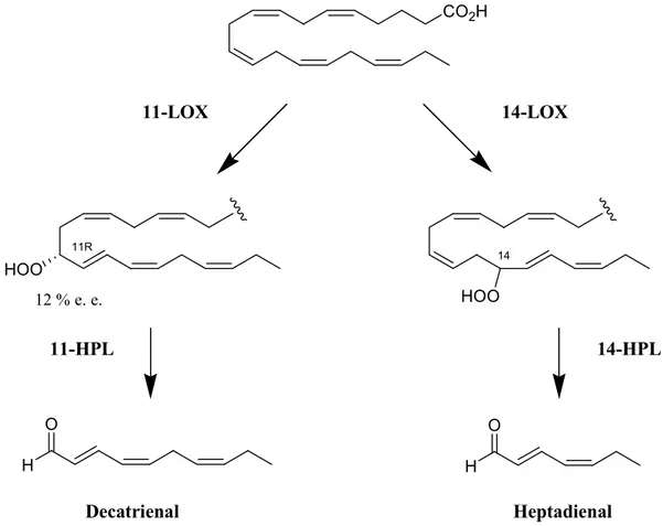
Considering PUAs, EPA oxygenation at C14 (14-LOX) and C11 (11-LOX) forms the
corresponding hydroperoxides (HpEPEs), which are then modified by HPL to heptadienal and decatrienal, respectively (Figure 1.9).
In a few species, such as, T. rotula and S. costatum, oxylipins derive from hexadeca-6Z,9Z,12Z-trienoic acid (HTrA, C16:3 ω4) and hexadeca-6Z,9Z,12Z,15-tetraenoic acid (HTtA, C16:4 ω1). In analogy to the synthesis of heptadienal and decatrienal from EPA, LOX and HPL metabolism of C16 PUFAs produce octadienal and octatrienal, whose
al., 2003, 2005; Romano, et al., 2003). Feeding experiments and stereochemical analysis of the main intermediates of C16 LOX activity proved the occurrence of a 9S-LOX,
responsible for the synthesis of 9S-hydroperoxy-6Z,10E,13Z-hexadecatrienoic acid (9S-HpHTrE) and 9S-hydroperoxy-6Z,10E,13Z,15-hexadecatetraenoic acid (9S-HpHTtE), from which the two C8 PUAs derives.
CO2H HOO HOO 11R 14 11-LOX 14-LOX 11-HPL 14-HPL H O H O Decatrienal Heptadienal 12 % e. e.
Figure 1.9. Precursors and stereochemistry of the synthesis of decatrienal and heptadienal. (From d’Ippolito et al., 2018).
Other typical oxylipins of this LOX pathway are 9S-hydroxy-hexadeca-6Z,10E,12Z-trienoic acid HHTrE), 9S-hydroxy-hexadeca-6Z,10E,12Z,15-tetraenoic acid (9S-HHTtE), and 9S,10S-epoxy-11R-hydroxy-hexadecadienoic acid (9S,11R-EHHDE).
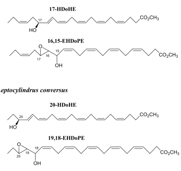
A new pathway has been described by Nanjappa et al. (2014), showing several oxylipins (epoxyalcohols and hydroxy fatty acids) that derive from docosahexaenoic acid (DHA, C22:6 ω3) from three species of Leptocylindraceae diatoms collected in the Gulf of Naples. Leptocylindrus danicus and Leptocylindrus hargravesii showed 15S-LOX
products, corresponding to 17-hydroxy-docosa-4Z,7Z,10Z,13Z,15E,19Z-hexaenoic acid (17-HDoHE) and 16,17-epoxy-15-hydroxy-docosa-4Z,7Z,10Z,13Z,19Z-pentaenoic acid (16,15-EHDoPE), while Leptocylindrus convexus displayed 18-LOX derived compounds named 20-hydroxy-docosa-4Z,7Z,10Z,13Z,16Z,18Z-hexaenoic acid (20-HDoHE) and 19,20-epoxy-18-hydroxy-docosa-4Z,7Z,10Z,13Z,16Z-pentaenoic acid (19,18-EHDoPE) (Figure 1.10).
Since Miralto et al. (1999) reported PUA production and the clear correlation between diatom diets and reproduction failure in copepods, several studies aimed to understand how diatoms regulate the rapid production of reactive PUAs. As mentioned above, PUA production is initiated upon wounding in sea water, indicating that the transcription and de novo biosynthesis of the enzymes involved in lipoxygenase pathway is quite unfeasible (Pohnert, 2000). Pohnert (2002) observed that the addition of free PUFA precursors to wounded diatom boosted the PUA production, hypothesizing that the formation of these secondary metabolites was apparently substrate limited. Nevertheless, this increase of PUA production can also be due to the fact that the equilibrium between free PUFAs and PUAs is disturbed after addition of PUFA molecules, activating again lipoxygenase pathway and the subsequent aldehyde formation (Fontana et al., 2007b).
CO2CH3 HO
17
Leptocylindrus danicus and Leptocylindrus hargravesii
O OH 17 16 15 17-HDoHE 16,15-EHDoPE
Leptocylindrus conversus
HO 20 20-HDoHE O OH 20 19 18 19,18-EHDoPE CO2CH3 CO2CH3 CO2CH3Figure 1.10. C22 oxylipins characterized in Leptocylindraceae species. (Modified from Nanjappa et al.,
2014).
These findings emphasized the importance of the availability of free PUFAs for leading to the formation of PUAs. Thus, any mechanism that regulates PUFAs discharge from cells can be involved in PUAs formation. Due to their cytotoxicity, small amounts of PUFAs are present within cells in free form but they are bound to phospholipids and galactolipids (Pohnert, 2002; d'Ippolito et al., 2004).
Mechanisms regulating the activity of phospholipase A2 and galactolipase enzymes can
A2 is modulated by many cellular factors in higher terrestrial plants such as: intracellular
Ca2+, the degree of the enzyme phosphorylation and also the presence of ROS (Chandra et al., 1996; Chapman, 1998; Narvaez-Vasquez et al., 1999; Wang, 2001). It is presumable that these cellular signals act in the same way in phytoplankton. The production of PUAs was also observed in freshwater chrysophytes under light stress, iron, nitrogen and phosphorus limitation (Watson and Satchwill, 2003). However, the influence of these environmental stresses on the mechanisms of PUAs production, including the PUFAs availability and/or the enzyme activities is still unclear and debated. Enzymes involved in the PUAs production seem to be activated only after grazer wounding and thus PUAs have been considered as “anti-predator” molecules. However, in terrestrial plants, it has been demonstrated that activation of phospholipase and galactolipase are essential to cell function and very important for regulating cell growth and differentiation (by reorganizing the cellular membrane) as well as apoptosis and senescence (by controlling the membrane entirety) (Chapman, 1998).
Several evidences indicated that oxylipins may be released not only as chemical defences against grazers, but also as signalling molecules within diatom populations. Spiteller (2003) proposed that lipid hydroperoxides (oxylipins precursors) and PUAs, induce two types of physiological responses: moderate amounts of oxylipins (nanomolar levels or lower) would allow cell division and cell proliferation, while large amounts of oxylipins (micromolar or higher) would induce necrosis and apoptosis, respectively. For example, the marine diatom Thalassosira weissflogii incubated with large amounts of decadienal (up to 6.4 μM) showed a clear growth inhibition, activating programmed cell death (PCD; Casotti et al., 2005). Recently it was hypothesized that S. marinoi (PUAs
producer) recognizes PUAs as intra-population infochemicals, while P. tricornutum (non-PUAs producer) perceives them as allelochemicals. The ability of S. marinoi to produce and use PUAs as infochemicals may modulate ecological success in natural communities (Gallina et al., 2014).
This assumption should be supported by understanding other causes, which trigger oxylipins production and their subsequent release in the marine environment without a cell disruption occurring during grazing, as observed in the marine diatom S. marinoi (Ribalet et al., 2014).
PUAs were produced by 38% of the cultivated isolates in a range of concentrations from 0.01 to 9.8 fmol cell-1 (Wichard et al., 2005b). Among them, 2E,4E-decadienal has been widely used as a model aldehyde to show deleterious effects on the reproduction of several marine invertebrates, such as echinoderms, polychaetes, ascidians, crustaceans and molluscs (Caldwell, 2009).
Several treatments with the pure molecule demonstrated that the PUA decadienal induced apoptosis in sea urchin embryos (Romano et al., 2003). As a biochemical marker for apoptotic processes in sea urchin embryos treated with decadienal, the ability of this PUA to activate caspase-like enzymes had been tested, using several commercially available kits developed for vertebrate caspases. A caspase-3-like activity was detected only in sea urchin embryos after 60 minutes of exposure to 5 µg mL-1 of decadienal, with maximum activity after incubation for 120 minutes (Romano et al., 2003). Romano et al. (2010) confirmed the deleterious effect of PUAs, such as decadienal, octadienal, octatrienal and heptadienal on early and late developmental stages of P. lividus. PUAs blocked sea urchin cell cleavage in a dose dependent manner,
but at different concentrations depending on the chain length of the molecules. The percentage blockage of cell cleavage increased with increasing chain length from C7 to
C10 PUAs, with arrest occurring at 27.27 μM with heptadienal, 16.13 μM with
octadienal, 11.47 μM with octatrienal (which was slightly more active compared to octadienal), and 5.26 μM of decadienal. The saturated aldehyde tridecanal, also found in diatoms, did not interfere with first cleavage up to 25 μM. Higher concentrations were not tested due to scarce solubility of this compound in sea water. The effect of PUAs on sea urchin hatching success were also tested, showing that all three PUAs exerted a very strong dose-dependent effect, with decadienal showing strongest effects than the other two aldehydes.
Incubation of eggs soon after elevation of fertilization membrane, with lower doses of decadienal (1.32 to 5.26 µM) than those inducing the arrest of cell division (6.58 µM), increased the number of abnormal sea urchin plutei and delayed the development of larvae or embryos which showed various degrees of malformations with increasing concentrations tested (Figure 1.11). At lower concentrations (1.32-2.63 μM), malformations were less severe with a shortening of spicules and arms. At higher concentrations (3.95-5.26 μM), larvae were similar to blastula and gastrula stages, showing severe abnormalities or blebbing associated with apoptosis.
Moreover, these studies revealed that decadienal not only slowed down development but also induced apoptosis in a dose-dependent manner. At 1.32 μM decadienal, the number of embryos, presenting a high degree of apoptosis, was more or less the same as in the control. It is interesting to note that, at this concentration, larvae completely negative to TUNEL (Terminal deoxynucleotidyl transferase (TdT) dUTP Nick-End Labeling)
staining were also recorded, suggesting the possible alteration of the cell cycle and apoptotic machinery. Already at decadienal 2.63 μM, the proportion of embryos presenting apoptotic nuclei in the whole body considerably increased, reaching almost 80% of the total number of embryos examined. At 3.95 μM decadienal, there was a stronger effect and almost all of the larvae were positively stained.
Figure 1.11. A) P. lividus embryos at the pluteus stage (48 hours post fertilization) incubated with decadienal at different concentrations: (a) control embryos in sea water without decadienal; (b) 1.32, (c) 2.63, (d) 3.95 and (e) 5.26 μM. B) Percentages of abnormal plutei (blue bars), abnormal blastulae and gastrulae (red bars), retarded larvae (yellow bars) and dead pre-hatched embryos (green bars) at different decadienal concentrations from 1.32 μM to 6.58 μM. (From Romano et al., 2010).
Recent studies confirmed the apoptotic effects of PUAs on cancer cell lines (Sansone et al., 2014). The effects of the PUAs decadienal, octadienal and heptadienal have been
tested on the adenocarcinoma cell lines lung A549 and colon COLO 205, and the normal lung/brunch epithelial BEAS-2B cell lines. Using the viability MTT (3-(4,5-dimethylthiazol-2-yl)-2,5-diphenyltetrazolium bromide)/Trypan blue assays, the results revealed that PUAs have a toxic effect on both A549 and COLO 205 tumor cells but not BEAS-2B normal cells. Decadienal was the strongest among the three PUAs tested, at all time-intervals considered, but heptadienal was as strong as decadienal after 48 hours. Octadienal was the least active of the three PUAs.
Surprisingly, although this sea urchin is extensively used as a model system for ecotoxicological studies, the complete genome is not yet available. This represents a significant limitation to the use of this sea urchin for molecular studies and explains the large number of morphological studies conducted to date. Sea urchin P. lividus has been used as a model system for the first studies on molecular response to PUAs, reported in Romano et al. (2011) and in Marrone et al. (2012). The authors treated sea urchin embryos with a low concentration of decadienal (0.25 μg mL-1 producing about 35% of abnormal embryos) and followed the expression levels of sixteen genes using Real Time
qPCR, in order to identify genes that were activated in response to this teratogen. The
results showed that at low decadienal concentrations (in the range 0.15-0.35 μg mL-1) the sea urchin places in motion different classes of genes to defend itself against this toxic aldehyde, activating heat shock proteins and several genes involved in skeletogenesis (formation of the skeleton). These molecular data were in accordance with morphological ones, demonstrating that developmental abnormalities mainly affected skeleton morphogenesis (Marrone et al., 2012). Moreover, sea urchin embryos
treated with increasing decadienal concentrations (in the range 0.15-0.35 μg mL-1) revealed a dose-dependent response of activated target genes.
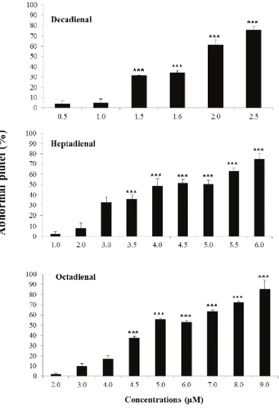
These studied have been extended to the other two commercialized PUAs, heptadienal and octadienal, the most common of the PUAs produced by diatoms, in comparison with decadienal, the most studied (Wichard et al., 2005a,b). The effect was dose-dependent for the three PUAs tested, even if the range of concentrations inducing malformations on
P. lividus embryos differed (from 0.5 to 2.5 μM for decadienal, from 1.0 to 6.0 μM for
heptadienal and from 2.0 to 9.0 μM for octadienal) (Figure 1.12; Varrella et al., 2014). Decadienal was the strongest of the three, because of the very narrow range (2.0 μM) that affected embryonic development; while heptadienal and octadienal required higher ranges of concentrations to reach the same effects as decadienal.
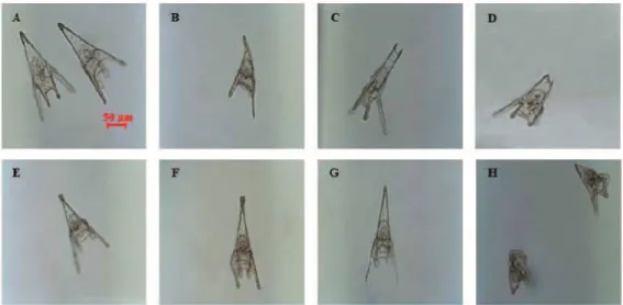
Treatments with the three PUAs induced similar malformations on sea urchin P. lividus plutei at 48 hours post-fertilization (hpf) (Figure 1.13), with a poorly-formed apex and arms appearing longer and asymmetrical or completely degenerated, compared to controls (sea urchins grown in sea water without PUAs) (Varrella et al., 2014).
Post-recovery experiments showed that embryos can recover after treatment with all three PUAs, indicating that negative effects depended both on PUA concentrations and the exposure time of the embryos to these metabolites. The time range during which PUAs exert the greatest effect on sea urchin embryogenesis has also been defined, corresponding to 10 min before fertilization and/or 10 min after fertilization. The addition of PUAs in later developmental stages does not seem to affect the embryonic development of P. lividus.
Figure 1.12. Percentage of abnormal P. lividus plutei observed after exposure to different concentrations of heptadienal, octadienal and decadienal. (From Varrella et al., 2014).
Figure 1.13. Examples of malformations of P. lividus plutei affecting: the apex (B) with spicules either parallel or disjoined (C, D) or crossed at the tip (E); the arms longer and broader or crooked and asymmetrical (F) or completely degenerated (G); the whole body plan of the plutei strongly compromised and malformed (H). All observations were compared to the control (A). (From Varrella et al., 2014).
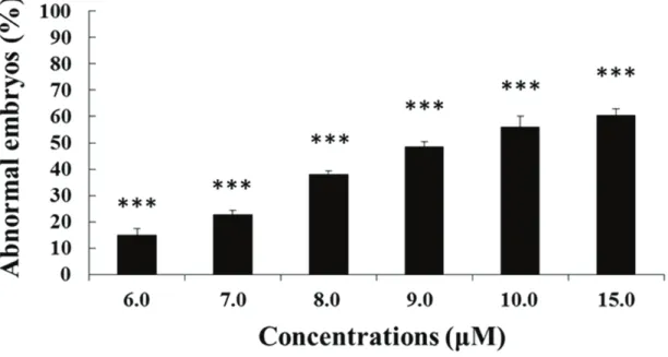
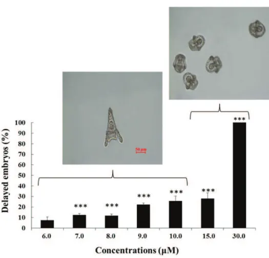
Very little information is available on the morphological and molecular effects of NVOs on sea urchin embryos. Very recently, it has been demonstrated that increasing concentrations of two hydroxy fatty acids (abbreviated as HEPEs), 5-hydroxy-eicosa-6E,8Z,11Z,14Z,17Z-pentaenoic acid (5-HEPE) and 15S-hydroxy-eicosa-5Z,8Z,11Z,13E,17Z-pentaenoic acid (15S-HEPE) induce dose-dependent developmental malformations on sea urchin embryos but at higher concentrations when compared with PUAs (range of 6-15 μM; Figure 1.14; Varrella et al., 2016b). The observed malformations were similar to those induced by PUAs (see above; Varrella et al., 2014). Interestingly, HEPEs also induced a marked developmental delay in sea urchin embryos, which has not hitherto been reported for PUAs. In particular, the percentage of delayed embryos increased with increasing HEPE concentrations to become the only class of embryos present at the highest concentrations of 30 μM (Figure 1.15; modified from Varrella et al., 2016b). Moreover, the degree of delay was different with increasing
concentrations: from 6.0 to 10 μM the delay in the development of embryos was manifested by a shortening of the body and, at the pluteus stage, the morphology of the embryo closely resembled that of the control, with only a slight reduction in body length; at 15 and 30 μM, the development of embryos was much more delayed, with embryos still at the stage of early pluteus.
PUAs and HEPEs molecular targets have also been identified by Real Time qPCR, analysing the variation in expression levels of 39 genes, implicated in a broad range of functional responses, such as stress, development, differentiation, skeletogenesis and detoxification processes (Figure 1.16; Varrella et al., 2014, 2016b).
Figure 1.14. Percentage of abnormal plutei when P. lividus newly-fertilized eggs (10 minutes after the elevation of the fertilization membrane) were exposed to different concentrations of the four HEPEs (6, 7, 8, 9, 10 and 15 μM). (From Varrella et al., 2016b).
Figure 1.15. Percentage of delayed P. lividus embryos when newly-fertilized eggs were exposed to different HEPEs concentrations (6, 7, 8, 9, 10, 15 and 30 μM) at 48 hpf respect to the control (not shown). (Modified from Varrella et al., 2016b).
PUAs had several common molecular targets in all the functional classes tested. Moreover, among PUAs heptadienal seemed to be the strongest one, affecting the largest number of genes, mainly those involved in development and differentiation processes. The results also showed that 15S-HEPE switched on fewer genes than 5-HEPE, being the weakest of the oxylipins tested. These findings highlight the differences between HEPEs and PUAs and also have important ecological implications because many diatom species do not produce PUAs, but rather these other chemicals are derived from the oxidation of fatty acids.