RESEARCH ARTICLE
A Drosophila model of myeloproliferative neoplasm reveals a
feed-forward loop in the JAK pathway mediated by p38 MAPK signalling
Ana Terriente-Félix1, Lidia Pérez1, Sarah J. Bray2, Angel R. Nebreda1,3,* and Marco Milán1,3,*ABSTRACT
Myeloproliferative neoplasms (MPNs) of the Philadelphia-negative class comprise polycythaemia vera, essential thrombocythaemia and primary myelofibrosis (PMF). They are associated with aberrant numbers of myeloid lineage cells in the blood, and in the case of overt PMF, with development of myelofibrosis in the bone marrow and failure to produce normal blood cells. These diseases are usually caused by gain-of-function mutations in the kinase JAK2. Here, we use Drosophila to investigate the consequences of activation of the JAK2 orthologue in haematopoiesis. We have identified maturing haemocytes in the lymph gland, the major haematopoietic organ in the fly, as the cell population susceptible to induce hypertrophy upon targeted overexpression of JAK. We show that JAK activates a feed-forward loop, including the cytokine-like ligand Upd3 and its receptor, Domeless, which are required to induce lymph gland hypertrophy. Moreover, we present evidence that p38 MAPK signalling plays a key role in this process by inducing expression of the ligand Upd3. Interestingly, we also show that forced activation of the p38 MAPK pathway in maturing haemocytes suffices to generate hypertrophic organs and the appearance of melanotic tumours. Our results illustrate a novel pro-tumourigenic crosstalk between the p38 MAPK pathway and JAK signalling in a Drosophila model of MPNs. Based on the shared molecular mechanisms underlying MPNs in flies and humans, the interplay between Drosophila JAK and p38 signalling pathways unravelled in this work might have translational relevance for human MPNs.
KEY WORDS: JAK, p38 MAPK, Myeloproliferative neoplasm,
Haemocyte, Hypertrophy,Drosophila
INTRODUCTION
Myeloproliferative neoplasms (MPNs) arise in patients having a gain-of-function mutation in Janus kinase 2 (JAK2) or the myeloproliferative leukaemia protein receptor (MPL). Three specific subtypes of MPN occur, polycythaemia vera, essential thrombocythaemia or primary myelofibrosis (PMF), depending on the blood cell type whose concentrations are outside the homeostatic
range. Although these subtypes are less severe than some other types of MPNs, such as chronic myelogenous leukaemia, which is caused by the translocation BCR-Abl (Philadelphia chromosome), 15% of patients exhibit PMF and a small percentage develop acute myeloid leukaemia, both of which compromise life expectancy. In 2005, JAK2V617F was identified as one of the most common
mutations causing the disease (Baxter et al., 2005; James et al., 2005; Kralovics et al., 2005; Pecquet et al., 2010). Subsequently, this mutation was shown in murine models to be sufficient to induce activation of the JAK2 pathway in the bone marrow, and to increase the rates of proliferation of myeloid cells (Lacout et al., 2006). Long before the causal role of JAK2V617F in MPNs was known,
Drosophila JAK gain-of-function mutations were shown to cause hypertrophy of the fly haematopoietic organs (lymph glands), and enhanced proliferation of circulating blood cells (haemocytes) and melanotic tumours (Corwin and Hanratty, 1976; Luo et al., 1997; Minakhina and Steward, 2006; Myllymäki and Rämet, 2014; Sorrentino et al., 2002).
In Drosophila, the conserved JAK/STAT signalling pathway is activated when ligands Unpaired (Upd) 1, 2 or 3, four-helix bundle cytokines of the Interleukin-6 family (Oldefest et al., 2013), bind to homodimers of the receptor Domeless (Dome), a type I cytokine receptor (Brown et al., 2001). This interaction promotes the anchoring of two JAK molecules at the intracellular domain of Dome, which allows JAK (also known as Hopscotch or Hop) trans-phosphorylation. Activated JAK then phosphorylates the transcription factor Stat92E, inducing its dimerization and nuclear translocation to promote transcription (Müller et al., 2005; Rivas et al., 2008). Functionally, JAK/Stat92E signalling is known to positively regulate cell proliferation. It does so in different cellular contexts under homeostatic conditions, and also, in response to stress signals. For instance, it is particularly important at sites of wound healing (Katsuyama et al., 2015; Santabárbara-Ruiz et al., 2015), in cells that lose their apico-basal polarity (Bunker et al., 2015) and in cells that experience chromosomal instability (Clemente-Ruiz et al., 2016), as well as regulating the growth of epithelial primordia (Mukherjee et al., 2005; Recasens-Alvarez et al., 2017). Similarly, the JAK pathway is required in the midgut epithelia for normal cell lineage differentiation and proliferation (Beebe et al., 2010), a requirement that is strongly evidenced under bacterial infection or stress assaults (Buchon et al., 2009; Cronin et al., 2009; Jiang et al., 2009). In the lymph gland, JAK signalling is required for the maintenance of progenitors in a naïve state (Gao et al., 2009), whereas peripheral tissues subjected to stress respond to the secretion of the ligand Upd3 by circulating haemocytes (Pastor-Pareja et al., 2008; Yang et al., 2015; Agaisse et al., 2003). Another pathway that responds to stress is the p38 mitogen-activated protein kinase ( p38 MAPK) cascade. In vertebrates, the p38 MAPK pathway can regulate cell cycle arrest, apoptosis or senescence, as well as the production of inflammatory mediators (Cuadrado and Nebreda, 2010). In Drosophila, the structurally and
Received 3 October 2016; Accepted 1 February 2017
1
Institute for Research in Biomedicine (IRB Barcelona), The Barcelona Institute of Science and Technology, Baldiri Reixac, 10, 08028 Barcelona, Spain.2
Department of Physiology, Development and Neuroscience, University of Cambridge, Cambridge CB2 3DY, UK.3ICREA, Pg. Lluı́s Companys 23, Barcelona 08010,
Spain.
*Authors for correspondence ([email protected]; marco.milan@ irbbarcelona.org)
A.T.-F., 0003-4948-6219; A.R.N., 0002-7631-4060; M.M., 0000-0002-7111-6444
This is an Open Access article distributed under the terms of the Creative Commons Attribution License (http://creativecommons.org/licenses/by/3.0), which permits unrestricted use, distribution and reproduction in any medium provided that the original work is properly attributed.
Disease
Models
&
M
functionally conserved p38 MAPK signalling pathway is activated upon heat-shock (Inoue et al., 2001; Seisenbacher et al., 2011), osmotic stress (Inoue et al., 2001; Sano et al., 2005; Seong et al., 2011; Seisenbacher et al., 2011) and oxidative stress (Vrailas-Mortimer et al., 2011; Santabárbara-Ruiz et al., 2015; Clemente-Ruiz et al., 2016), and promotes survival upon exposure to chromosomal instability (Clemente-Ruiz et al., 2016), oxidative stress (Craig et al., 2004; Cai et al., 2011; Vrailas-Mortimer et al., 2011) and pathogenic bacteria (Chen et al., 2010; Ha et al., 2009; Park et al., 2009). The physiological role of the p38 MAPK signalling pathway in the lymph gland and its potential contribution to how these cells cope with stress conditions remain to be elucidated.
Here, we report a Drosophila model of MPNs based on forced expression of JAK (hop) in the lymph gland, and identify the maturing haemocytes as the cell population susceptible to induce JAK-induced hypertrophy. We unravel a feed-forward loop in the JAK/STAT pathway that involves the ligand Upd3 and its receptor Dome, and contributes to JAK-induced hypertrophy. We also show that the p38 MAPK pathway contributes to this feed-forward loop by regulating expression of the ligand Upd3, and, most interestingly, when activated in maturing haemocytes, suffices to induce lymph gland hypertrophy and melanotic tumours.
RESULTS
Targeted expression of JAK in maturing haemocytes induces lymph gland hypertrophy
Animals bearing the JAKTum-l gain-of-function mutation, a
hyperactive form of JAK, show hypertrophic lymph glands. This hypertrophy can also be obtained by targeted overexpression of a wild-type form of JAK to this organ (Harrison et al., 1995). The Drosophila larval lymph gland is composed of five to seven
pairs of posterior secondary lobes and one pair of anterior primary lobes. Primary lobes are mainly subdivided into two domains: the medullary zone (MZ) and the cortical zone (CZ) (Jung et al., 2005). Naïve progenitors residing in the MZ progress into the CZ to differentiate (reviewed in Martinez-Agosto et al., 2007). In healthy larvae, progenitors residing in the CZ give rise to two cell types: the crystal cells (CCs, platelet-like cells) and the plasmatocytes (PLs, macrophage-like cells; Fig. 1A). In larvae parasitized by wasp eggs, progenitors differentiate into a third cell type, lamellocytes (LMs) (Jung et al., 2005). In order to identify the cell domain that is susceptible to over-proliferation upon JAK overexpression, a wild-type form of JAK was overexpressed in the MZ and CZ domains by the use of the dome-Gal4 and pxn-Gal4 drivers, respectively (Fig. 1A). The size of the resulting lymph glands and of the JAK-overexpressing domains was analysed in mid third-instar larvae [mid-L3; 91-94 h after egg laying (AEL)]. When JAK was overexpressed in the pxn+ population, lymph glands were significantly larger than controls in this developmental stage (Fig. 1B,C). By contrast, expression of JAK in the dome+ population resulted in fewer dome+ cells and smaller glands than controls (Fig. 1D). The overgrown glands in pxn>JAK primarily comprised enlarged secondary lobes, whereas primary lobes remained after apparent release of their cell contents (Fig. 1B,C, RFP, white channel, primary and secondary lobes). Such ‘bursting’ normally only occurs at metamorphosis and must be greatly accelerated in the pxn>JAK animals. In addition, the small number of pxn+ cells which are normally present at mid-L3 in wild-type glands (Fig. S1A, wild type, pxn>+) must become greatly expanded upon overexpression of JAK.
In order to identify the stage at which JAK induces growth of the pxn+ population in the primary lobes, we analysed the size of
Fig. 1. Hypertrophic lymph glands induced by JAK overexpression in the cortical zone. (A) Schematic of the primary and secondary lobes indicating the medullary zone (MZ, dome+) and cortical zone (CZ, pxn+), and the three different cell types: crystal cells (CC, Lz+), plasmatocytes (PL) and lamellocytes
(LM,βInt-ν+). (B-F) Larval lymph glands of the indicated genotypes were labelled to visualise Hemese (He, green, B,C), RFP (red or white, B-F), DAPI (D,E),
Lozenge (Lz, green or white, E) and Atilla/L1 (L1, green or white, F). CZ ( pxn-gal4) or MZ (dome-gal4) drivers were used to express RFP and/or a wild-type form of JAK in the lymph gland of mid third-instar larvae (mid-L3; 91-94 h AEL, B,C,F) or larvae at the L2-L3 transition (E). Note in C that the secondary lobes grow in JAK-overexpressing lymph glands and the primary lobes have released their content. Inset in C shows a higher magnification of an overgrown secondary lobe consisting of large and elongate-shaped lamellocytes. Single images of a larger area have been assembled in C to show an overgrown lymph gland induced by
JAK overexpression. Red arrows in E indicate Lz-positive cells. Red arrows in F highlight the presence of the lamellocyte marker L1. (G) mRNA levels ofβInt-ν and
hemese (he) measured as the mean±s.e.m. increase in JAK-overexpressing lymph glands compared with wild-type lymph glands. Expression of theβInt-ν
lamellocyte-specific gene increases (fold change=9.1, P=0.039), whereas the expression of the haemocyte-specific gene he does not change significantly
(fold change=1.55, P=0.31). Wild-type controls were given the value of 1 and are not displayed in the figure. *P<0.05. Scale bars: 40 µm (B-E), 20 µm (F).
Disease
Models
&
M
JAK-overexpressing lymph glands at early stages of larval development. We focused particularly on the transition between second- to third-instar larvae (L2-L3 transition; 69-72 h AEL) as this stage was previously shown to be critical for the generation of melanotic tumours in a JAKTum-l background (Hanratty and
Dearolf, 1993). We found that the pxn-Gal4 driver started to be expressed in wild-type lymph glands 6 h prior to the L2-L3 transition (Fig. S1B, wild type, pxn>+). Interestingly, JAK-overexpressing glands showed a faster growth rate than controls across all time points analysed, which resulted in larger glands with a larger population of pxn+ cells (Fig. S1C, pxn>JAK). Furthermore, these primary lobes did not, at this stage, show signs of having burst and released their cell content to the haemolymph. Since each primary lobe could be analysed individually, we selected the developmental stage at the L2-L3 transition for further characterisation of the lymph gland hypertrophy caused by JAK overexpression (see below).
To investigate the similarities between the JAKTum-lmutant and
JAK overexpression, we analysed the cell differentiation state. Larvae mutant for JAKTum-l showed melanotic tumours, which
consist of aggregates of lamellocytes (Minakhina and Steward, 2006), and a reduced number of crystal cells in circulation (Hanratty and Dearolf, 1993; Harrison et al., 1995). When JAK was overexpressed in the pxn+ cell population, crystal cells, visualised by the expression of Lozenge (Lz+; Jung et al., 2005), rarely differentiated (Fig. 1E, red arrows). In these lymph glands, a multitude of large, elongated lamellocytes were detected (Fig. 1C, inset). These cells were also identified by expression of the specific lamellocyte marker Atilla/L1 (Kurucz et al., 2007) (Fig. 1F). Accordingly, in the pxn>JAK glands, we detected a significant increase in the expression levels of the lamellocyte-specific gene β-integrin-ν (βInt-ν; Kwon et al., 2008) compared with the pan-haemocyte marker hemese (he) (Jung et al., 2005) (Fig. 1G). Taken together, these results indicate firstly that pxn+ cells are the most susceptible cell population to outgrow upon JAK overexpression, and secondly, that JAK induces a cell fate shift towards lamellocyte differentiation, at the expense of the crystal cells. Whether the increased number of lamellocytes observed in JAK-overexpressing lymph glands arises through the active proliferation of a normally quiescent lamelloblast population (Anderl et al., 2016) or through a programme of divisions and cell fate respecification amongst the plasmatocytes, remains to be elucidated.
An Upd3-mediated feed-forward loop contributes to JAK-induced lymph gland hypertrophy
To analyse the physiological role of JAK/STAT in the pxn+ cell population, we knocked down JAK expression using JAKRNAi and
quantified the percentage of pxn+ cells in each lymph gland at mid-L3. The resulting primary lobes displayed no significant changes in the proportion of cells in the pxn+ population when compared with wild-type controls (Fig. S2A). Similarly, when we expressed a truncated form of the receptor Dome, which lacks the intracellular domain (DomeΔCYT; Brown et al., 2001), we did not observe any changes in the percentage of pxn+ cells per gland (Fig. S2A, pxn>domeΔCYT). As control, we knocked down JAK in the dome+ cell population, and observed at mid-L3 a reduced number of dome+ cells in the MZ (Fig. S2B, MZ), which gave rise to smaller lymph glands (Fig. S2B, Total). This is consistent with the proposed role of JAK signalling in regulating the proliferation and/or survival of the cells residing in the MZ (Makki et al., 2010). Thus, our results indicate that the JAK/Dome pathway is either not required or has a redundant role with other signalling pathways during normal CZ development.
The function of the endogenous STAT (Stat92E) in the CZ was investigated by examining at mid-L3 the effect of expressing stat92ERNAi in the pxn+ cell population. As previously reported
(Minakhina et al., 2011; Mondal et al., 2011), lymph glands with stat92E knockdown resembled, although to a milder extent, the phenotype resulting from upregulation of JAK. This is shown by the expansion of the pxn+ cell population (Fig. 2A). Previous work in Drosophila has identified a non-canonical mechanism by which the unphosphorylated form of Stat92E maintains HP1a localisation and heterochromatin stability (Shi et al., 2008). We thus wondered whether the ability of JAK to induce hypertrophy of the pxn+ cell population relied, at least in part, on the release of Stat92E from the heterochromatin. To avoid JAK-overexpressing glands bursting, primary lobes were examined at the L2-L3 transition. At this developmental time, the effect of knocking down Stat92E in pxn-expressing cells was milder (Fig. 2B, compare pxn>stat92ERNAi
with pxn>+). However, we observed that the co-expression of stat92ERNAitogether with JAK did not enhance the JAK-induced
hypertrophy (Fig. 2B). By contrast, and consistent with a canonical role of Stat92E in mediating JAK activity, the downregulation of Stat92E resulted in a significant rescue of the JAK-induced expansion of the pxn+ cell population (Fig. 2B, compare pxn>JAK+stat92ERNAiwith pxn>JAK). These results indicate that
Stat92E is required downstream of JAK to sustain the growth of the CZ, independent of its non-canonical role in the repression of the CZ expansion in wild-type conditions.
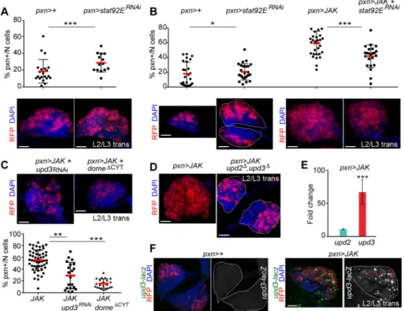
We next studied the requirement for the receptor Dome in JAK-induced hypertrophy. We observed that co-expression of the truncated receptor DomeΔCYT greatly reduced the expansion of the pxn+ cell population caused by JAK overexpression (Fig. 2C). Since the receptor Dome was apparently required for JAK-induced lymph gland hyperplasia, we investigated whether its ligands were also involved. Consistent with the requirement for Dome, JAK overexpression in larvae homozygous for a deficiency depleting the coding sequences of upd2 and upd3 resulted in a considerable reduction of the pxn+ cell population compared with JAK overexpression alone (Fig. 2D). This suggests that JAK requires both the receptor and its ligands to induce lymph gland overgrowth. Then, we directly analysed the expression of the ligands in hypertrophic glands by RT-qPCR and found that upd3 was strongly upregulated upon JAK overexpression, whereas upd2 was increased to a lesser extent (Fig. 2E). Consistent with this result, we found that an upd3 enhancer, previously shown to be activated in Drosophila neoplastic tumours (Bunker et al., 2015), was expressed in scattered pxn+ cells overexpressing JAK but not in the wild-type glands (Fig. 2F). Using an upd3-RNAi form, we confirmed that the overproliferation of JAK-overexpressing pxn+ cells requires Upd3 (Fig. 2C). Taken together, we conclude that JAK induces a feed-forward loop that triggers upd3 expression, which contributes to JAK-induced hypertrophy of the lymph gland.
A role for p38 MAPK signalling in maturing haemocytes The p38 MAPK signalling pathway is an important regulator of cytokine and chemokine expression in mammals (reviewed by Clark and Dean, 2012; Cuadrado and Nebreda, 2010). We thus investigated the possible interplay between the p38 MAPK and JAK pathways in lymph gland hypertrophy. We first analysed the effect of expressing in the lymph gland a wild-type form of Licorne (Lic), the Drosophila protein kinase that activates p38a and p38b MAPKs (Adachi-Yamada et al., 1999; Han et al., 1998; Inoue et al., 2001), or an activated form (Licact, see Materials and Methods for details).
Interestingly, when Lic was overexpressed in the cortical zone with
Disease
Models
&
M
the pxn-Gal4 driver, large melanotic aggregates were observed under the cuticle (Fig. 3A). The proportion of larvae bearing large melanotic aggregates was even higher upon overexpression of Licact
in the pxn+ cell population (Fig. 3A). Whereas lymph glands overexpressing Lic showed larger primary lobes than controls (Fig. 3B,E, compare pxn>+ with pxn>lic), the overgrowth observed in pxn>licactglands was due to the increase in size of secondary
lobes as primary lobes showed signs of having released their cell content (Fig. 3D). At earlier stages of development, the primary lobes of pxn>licactglands showed a similar growth pattern to the
JAK-overexpressing glands (Fig. S1D). Moreover, Licact-expressing
glands showed numerous lamellocytes, as detected microscopically by their large size and elongated shape (Fig. 3D, left inset), by the expression of the lamellocyte marker Attila/L1 (Fig. 3F, red arrows, green and white channel) and by the elevated mRNA expression levels of the lamellocyte-specific markerβInt-ν (when compared with the pan-haemocyte marker he; Fig. 3G). In addition, lymph glands contained a reduced number of crystal cells labelled by the expression of Lozenge (Fig. 3H, red arrows, compare with Fig. 1E).
Overexpression of Lic in the MZ produced primary lobes of about the same size as in the wild-type glands (Fig. 3C), whereas expression of Licactin the MZ caused larval lethality, most probably
due to the expression of the dome-Gal4 driver in the embryo. Altogether, these results indicate that activation of the Licorne/p38 MAPK signalling pathway in the pxn+ cell population phenocopies the effects of JAK overexpression and induces lymph gland dysplasia.
To analyse whether the p38 MAPK pathway has a role in the lymph gland during normal development, we studied hemizygous licnullmutants. As previously described (Cully et al., 2010), larvae
with reduced levels of Lic activity were smaller than wild-type larvae (data not shown). Consistently, their lymph glands were also smaller than lymph glands from wild-type larvae of the same developmental age (Fig. 4A). We next analysed the role of endogenous p38 MAPK signalling in the different regions of the lymph gland. For this purpose, we used RNAis targeting p38a and p38b, lic or the downstream transcription factor dATF-2 (Han et al., 1998; Sano et al. 2005). Targeted expression of these RNAis to the CZ resulted in a
Fig. 2. JAK-induced hypertrophy requires Stat92E, Dome and Upd3. (A-C) Scatterplots and immunofluorescence images showing the proportion of pxn+ cells per primary lobe (% pxn+/N cells) and lymph glands expressing the indicated transgenes under the control of the pxn-Gal4 driver. Lymph glands were extracted from mid-L3 larvae (A) or larvae aged at the L2-L3 transition (B,C) and labelled to visualise RFP (red), and DAPI (blue). (A) Expansion of the
stat92ERNAi-expressing cell population compared with the control pxn>+ cell population (P=0.0087; pxn>+, n=21; pxn>stat92ERNAi, n=14). (B) Knockdown of
stat92E partially rescues the JAK-induced expansion of the pxn+ population (P=0.0004; pxn>JAK, n=33; pxn>JAK+stat92ERNAi, n=23) whereas stat92ERNAi
produces a subtle increase in the proportion of pxn+ cells compared with the control pxn>+ cell population (P=0.03; pxn>+, n=25; pxn>stat92ERNAi, n=21).
(C) Knockdown of upd3 or expression of DomeΔCYTreduces the JAK-induced expansion of the pxn+ population ( pxn>JAK+domeΔCYT: P=1.961e−10, n=22;
pxn>JAK+upd3RNAi: P=0.00234, n=20; pxn>JAK, n=44). (D) Lymph glands expressing JAK under the control of the pxn-Gal4 driver were extracted and labelled
as in B,C. Note the reduction in the expansion of the pxn+ cell population in upd2Δ, upd3Δmutant lymph glands. (E) Increases in mRNA levels of upd3 and upd2 in
lymph glands expressing JAK under the control of the pxn-gal4 driver when compared with controls, which were given the value of 1. Note a significant increase in the expression level of upd3 (fold-change=72.45, P=0.00013) but not in the expression level of upd2 (fold-change=8.32, P=0.15). (F) Lymph glands expressing the indicated transgenes under the control of the pxn-Gal4 driver were extracted from larvae aged at the L2-L3 transition and labelled to visualise upd3-lacZ
expression (antibody againstβGal, green or white), RFP (red), and DAPI (blue). Note induction of upd3-lacZ expression in JAK-overexpressing lymph glands.
*P<0.05; **P<0.01; ***P<0.001. Every dot represents a single primary lobe. Red horizontal bar represents the mean, and whiskers represent 5% and 95% percentiles. The contour of the lymph glands is marked in B,D and F. Scale bars: 40 µm (A-D,F).
Disease
Models
&
M
reduced number of pxn+ cells compared with wild-type glands at mid-L3 (Fig. S3A). By contrast, targeted depletion of lic in the MZ did not reduce the number of dome+ progenitors and the lymph glands showed a subtle enlargement (Fig. S3B, Total). Consistent with a specific requirement of the Licorne/p38 MAPK signalling pathway in the CZ, expression of Licactunder control of the pxn-Gal4
driver produced lymph glands of similar size in both licnullmutant
and wild-type control animals raised in parallel and visualised at the transition between L2 and L3 (Fig. 4B). Taken together, these results indicate that the p38 MAPK pathway has a role in regulating growth of the pxn+ cell population during normal development.
A role for p38 MAPK signalling in JAK-induced hypertrophy of the lymph gland
The above experiments indicate that activation of p38 MAPK signalling in the CZ phenocopies the JAK-induced lymph gland hypertrophy and the cell fate shift towards lamellocyte differentiation. In order to test whether p38 MAPK signalling contributes to the JAK-induced phenotype, we analysed the ability of JAK overexpression to induce hypertrophic lymph glands in a
licnullmutant background. Interestingly, lic-deficient lymph glands
showed a reduced expansion of the pxn+ cell population and a smaller size upon overexpression of JAK when compared with wild-type glands (Fig. 4C, compare pxn>JAK with licnull; pxn>JAK).
Consistent with this result, co-expression of the kinase-dead form of p38b ( p38bKD; which can act as a dominant-negative form blocking
p38 MAPK signalling) with RNAi specific for MK2 [MK2RNAi; a
protein kinase activated by p38 MAPK (Cuadrado and Nebreda, 2010)] or the transcription factor dATF-2 (dATF-2RNAi), were able
to rescue the expansion of the pxn+ cell population caused by JAK overexpression (Fig. 4D). These data confirm that p38 MAPK signalling is required downstream of the JAK/STAT pathway to promote the expansion of the pxn+ cell population. We also observed that JAK overexpression induced high levels of apoptosis, as monitored by an antibody that detects the cleaved form of the effector caspase Dcp1, which was rescued by dATF-2 depletion (Fig. 4E). Whether the induction of cell death is a direct consequence of p38 MAPK activation or an indirect consequence of the enhanced proliferative capacity of the tissue upon JAK overexpression remains to be elucidated.
Fig. 3. Hypertrophic lymph glands induced by expression of an activated form of Licorne in the cortical zone. (A) Histogram showing the percentage of
larvae bearing small ( puncta) or big (aggregates) melanotic tumours upon expression of wild-type (lic, green) or an activated form (licact, yellow) of Licorne under
the control of the pxn-gal4 driver. Representative examples of a wild-type larva and of a larva bearing a big melanotic tumour are shown. Lic, n=93 larvae; licact,
n=109 larvae. (B,D,E) Lymph glands expressing the indicated transgenes under the control of the pxn-Gal4 driver were extracted from mid third-instar larvae
(mid-L3) and labelled to visualise RFP (red or white) and Hemese (He, green). Note that the secondary lobes grow in Licact-expressing lymph glands (D), but their
associated primary lobes have released their content. Inset in D shows a higher magnification of large, elongated lamellocytes. Also note that the primary lobes in Lic-overexpressing lymph glands (E) are larger than wild-type controls (B). (C) Lymph glands overexpressing Lic under the control of the dome-Gal4 driver
extracted from mid third-instar larvae (mid-L3) and labelled to visualise RFP (red) and DAPI (blue). (F) Expression of Licactin pxn+ cells induces differentiation of
lamellocytes in mid-L3. Lamellocytes are distinguished by the expression of L1/Atilla (L1, green or white) and their large and elongated shape outlined by
Phalloidin (Phal, blue); RFP visualises pxn+ cells (red). Note an increase of L1+ cells (red arrows). (G) Increased mRNA levels ofβInt-ν and hemese (he) in lymph
glands expressing Licactwhen compared with controls (fold-change increase ofβInt-ν=76.50, P=0.03; fold-change of he=3.38, P=0.0052). Controls were given
the value of 1 and are not displayed in the figure. **P<0.01. (H) Lymph gland expressing Licactunder the control of the pxn-Gal4 driver extracted from a larva at the
L2-L3 transition and labelled to visualise RFP (red), Lozenge (Lz, green or white) and DAPI (blue). Note a reduced number of Lozenge-expressing cells (red arrows). The contour of the lymph gland is marked in H. Single images of a larger area have been assembled in D and E to show overgrown lymph glands induced
by overexpression of Licactor Lic, respectively. Scale bars: 40 µm (B-E,H), 20 µm (F).
Disease
Models
&
M
Next, we analysed whether p38 MAPK signalling is required downstream of the JAK pathway to regulate upd3 expression. We found that lymph glands co-expressing JAK together with p38bKD
showed lower expression levels of upd3 than lymph glands overexpressing JAK alone (Fig. 4F). In addition, expression of constitutively active Licactsufficed to upregulate upd3 expression in
lymph glands (Fig. 4F), and upd3 was upregulated by Licactto a
greater extent than ligands such as Spatzle or Eiger (Fig. S4A), which can activate the Toll and JNK pathways, respectively, and induce melanotic tumours (Qiu et al., 1998; Zettervall et al., 2004). We confirmed in Kc167 cells that expression of Licactsufficed to induce
upregulation of upd2 and upd3 (Fig. S4B), and this required p38 MAPK activation because levels were reduced when Licact was
expressed in the presence of the p38 MAPK inhibitor SB203580 (Fig. S4B). In order to test whether the increased expression of upd2 and upd3 resulted in activation of the JAK/STAT pathway, we analysed the activity of 6x2xDrafLuc, a reporter widely used to measure the activity of this pathway (Thomas et al., 2015; Müller et al., 2005). Luciferase assays revealed increased reporter activity in Kc167 cells expressing Licactor JAKTum-l, and to a lesser extent upon
overexpression of wild-type JAK (Fig. S4C). Altogether, these results support the implication of the p38 MAPK signalling pathway in JAK-induced lymph gland hypertrophy by regulating upd3 expression. DISCUSSION
Here, we have analysed the impact of JAK overexpression in the different cell populations of the lymph gland, the major
haematopoietic organ of Drosophila. This has allowed us to identify the maturing haemocytes as the cell population that is responsible for induced hypertrophy. In addition, JAK-overexpressing lymph glands showed increased numbers of differentiated lamellocytes and fewer crystal cells, a phenotype that resembles the effect of JAKTum-lmutants. Using this model, we
have identified a number of essential components in the JAK/STAT pathway that have an important role in JAK-induced lymph gland dysplasia. First, the transcription factor Stat92E was found to be a necessary element downstream of JAK, independent of its role in preventing expansion of the cortical zone in wild-type conditions. This result concurs with previous publications showing that phosphorylation of Stat92E in JAKTum-l haemocytes is required
for the formation of melanotic tumours (Bausek and Zeidler, 2014; Remillieux-Leschelle et al., 2002; Sorrentino et al., 2004). We speculate that Stat92E regulates a set of genes in wild-type conditions that prevent the expansion of the pxn+ population, and that these genes are different from those regulated upon JAK phosphorylation, which might be involved in dysplastic growth. This is consistent with a report showing that the unphosphorylated and phosphorylated forms of Stat5, the vertebrate orthologue of Stat92E, can regulate different sets of genes (Park et al., 2015). Most interestingly, we unravelled a requirement for the Dome receptor in JAK-induced hypertrophy, and identified the ligand Upd3 as an essential component involved in a feed-forward loop downstream of JAK signalling that contributes to lymph gland dysplasia. This work complements the use of phenotypic screenings based on the
Fig. 4. A role for p38 MAPK signalling in JAK-induced hypertrophy. (A) Expression of pxn-Gal4 marked by the expression of RFP (red) in wild-type and
in licnullhemizygous lymph glands (outlined). (B,C) Expression of Licact(B) or JAK (C) under the control of the pxn-Gal4 driver in control and in licnullhemizygous
lymph glands. Scatterplot in C shows the proportion of pxn+ cells per primary lobe (% pxn+/N cells), and the total cell number per primary lobe (N cells). Note that loss of lic induced a reduction in the number of pxn+ cells per primary lobe caused by JAK overexpression (P=0.006) and a reduction in the number of cells per
lymph gland (P=0.034; pxn>JAK, n=9; pxn>JAK+licnull, n=9). (D) Genetic interactions between p38b, dATF-2 and MK2 and JAK in the pxn+ cells. Scatterplots
show the proportion of pxn+ cells per primary lobe (% pxn+/N cells) and immunofluorescence images show lymph glands expressing the indicated transgenes
under the control of the pxn-Gal4 driver. Note that expression of p38bKD, MK2RNAior dATF-2RNAireduced the number of pxn+ cells per primary lobe caused by
JAK overexpression ( pxn>JAK vs pxn>JAK+p38bKD, P=6.318e−06; pxn>JAK vs pxn>JAK+dATF-2RNAi, P=1.02e−06; pxn>JAK vs pxn>JAK+MK2RNAi,
P=3.848e−06; pxn>JAK, left plot, n=27 and right plot, n=14; pxn>JAK+p38bKD, n=29; pxn>JAK+dATF-2RNAi, n=10; pxn>JAK+MK2RNAi, n=23). (E) Expression of
the cleaved form of the effector caspase Dcp1 (clv-Dcp1, marked in green and white) increases upon JAK overexpression in pxn+ cells ( pxn>JAK vs pxn>+,
P=0.038; pxn>+, n=8; pxn>JAK, n=13) and was rescued by the expression of dATF-2RNAi( pxn>JAK vs pxn>JAK+dATF-2RNAi, P=0.001; pxn>JAK+dATF-2RNAi,
n=12), although not to the levels observed in the wild type ( pxn>JAK+d-ATF2RNAivs pxn>+, P=0.035). (F) Increased mRNA levels of upd3 in lymph glands
expressing Licact(fold-change=71.1, P=0.0006), JAK (fold-change=158.43, P=0.0099) or JAK and p38bKD(fold-change=54.79, P=0.063) under the control of
the pxn-Gal4 driver when compared with controls ( pxn>+), which were given the value of 1. In A-E, lymph glands of the different genotypes were extracted from larvae at the L2-L3 transition and labelled to visualise RFP (red or white) and DAPI (blue). *P<0.05; **P<0.01; ***P<0.001. In the scatterplots, every dot represents a single primary lobe; red horizontal bars represent the mean, and whiskers represent 5% and 95% percentiles. Scale bars: 40 µm.
Disease
Models
&
M
presence of melanotic tumours (Shi et al., 2008) and forward RNAi screenings (Müller et al., 2005)– genetic approaches that in the past served to identify new elements of the JAK regulatory network in haemocytes. Furthermore, our experimental setup could be useful to perform small-scale drug screenings or to validate hits previously identified in drug screenings performed in Drosophila cultured cells (Thomas et al., 2015). Of particular interest would be drugs that synergise with JAK inhibitors, since Ruxolitinib, an FDA-approved JAK inhibitor, does not ameliorate MPN symptoms in the long term (Mascarenhas et al., 2014).
Our genetic model of MPNs has also allowed us to identify a novel role for the p38 MAPK pathway in the feed-forward loop downstream of JAK signalling that contributes to lymph gland dysplasia. The p38 MAPK pathway exerts is function by regulating upd3 expression. Importantly, we found that overexpression of Licorne, a direct and specific activator of p38 MAPKs, suffices to induce dysplasia and phenocopies the effect of JAK overexpression. Furthermore, p38 MAPK signalling was able to induce upd3 expression in lymph gland cells and in Kc167 cells. Our data show that the p38 MAPK pathway, including the transcription factor dATF-2, is necessary for the maturing haemocytes to proliferate both in wild-type conditions and in response to JAK overexpression. Similarly, there is evidence that p38 MAPK signalling can stimulate mammalian cell proliferation in particular contexts, for example, in mouse colon tumour cells (Gupta et al., 2014). Megakaryocyte proliferation induced by FLT3 receptor activation has also been reported to involve p38 MAPK signalling (Desterke et al., 2011). In addition, the p38 MAPK pathway plays an important role in the regulation of cytokine expression both at transcriptional and post-transcriptional levels (reviewed by Cuadrado and Nebreda, 2010; Tiedje et al., 2014). However, we are not aware of any report showing that direct activation of the p38 MAPK pathway by an upstream regulator such as Lic suffices to induce dysplasia and tumour formation in vivo. On the contrary, p38 MAPK hyperactivation usually leads to cell cycle arrest and cell death in mammalian cells (Cuadrado and Nebreda, 2010; Tiedje et al., 2014). This suggests that haemocytes, and perhaps the concurrent activation of JAK signalling, may provide a particular context that favours a pro-tumourigenic role for p38 MAPK signalling. Intriguingly, there are no reports on mutations, changes in copy number, promoter methylation or enhanced phosphorylation levels of p38 MAPKs in samples from MPN patients (Desterke et al., 2011; Shahjahan et al., 2008), supporting our conclusion that p38 MAPK signalling contributes to tumourigenesis as part of the JAK-triggered feed-forward loop. Given that MPN patients are known to have high levels of circulating cytokines (Levine et al., 2007; Tyner et al., 2010), it is tempting to speculate that p38 MAPK signalling contributes to JAK2V617F-associated mammalian tumourigenesis by regulating the
production of cytokines, which act in an autocrine manner. MATERIALS AND METHODS
Fly strains
The alleles and fly stocks, as described in FlyBase (flybase.org/), were pxn-Gal4 (Stramer et al., 2005), dome-pxn-Gal4 [#PG125 (Makki et al., 2010)], UAS-cd8RFP (BDSC #32219 and #32218), UAS-cd8GFP (BDSC #5030), upd3-lacZ (Bunker et al., 2015), UAS-JAKHA(FlyORF #F001803), UAS-lic
(FlyORF #F001674), UAS-stat92ERNAi(VDRC #106980), UAS-upd3RNAi
(VDRC #27134), UAS-domeΔCYT (Brown et al., 2001), UAS-p38bKD
(Vrailas-Mortimer et al., 2011), w*,upd2Δ,upd3Δ(BDSC #55729), licD13
[licnullin the text (Cully et al., 2010)], UAS-JAKRNAi(BDSC #32966),
UAS-p38aRNAi(VDRC #52277), UAS-p38bRNAi(VDRC #108099), UAS-licRNAi
(VDRC #106822), UAS-dATF-2RNAi (DGGR #3749-R2 and BDSC
#60124) and UAS-MK2RNAi(VDRC #3170).
*UAS-licRNAi and UAS-stat92ERNAi were VDRC lines of the KK
collection. Because of the presence of a landing site in the gene tiptop, lines were meiotically recombined to acquire the dominant mutation Sco. Then, flies were PCR screened for the absence of tiptop landing site and the presence of the correct non-annotated landing site at cytological position 40D. Then the Sco mutation was removed by meiotically recombining the arm with a wild-type chromosome. Full genotypes of flies used for results displayed in all figures are listed in supplementary Materials and Methods.
Generation of the UAS-licactconstruct
A lic ORF frame (SD04985; 321-1325 bp) was used to make a phospho-mimetic version, by swapping serine (S200) and threonine (T204) residues to aspartate (D). The mutated lic was cloned into pUAS to transform yw flies (Brand and Perrimon, 1993).
Generation of cell lines
Stable lines of Kc167 cells were prepared and treated as described in supplementary Materials and Methods.
RT-qPCR
Real-time quantitative PCR was carried out on total RNA from lymph glands using primers listed in supplementary Materials and Methods.
Experimental setup
For Figs 1-4, Figs S2 and S3, lymph glands were extracted from larvae that were either 69-72 h AEL (L2-L3 transition) or at 91-94 h AEL (mid-L3). To age the larvae, eggs were collected every 3 h at 25°C and shifted to 29°C at 45-48 h AEL. In this manner, incubating lymph glands at 29°C for 24 h or 48 h, we increased the efficiency of the Gal4/UAS system. For Fig. S1B-D, eggs were collected every 3 h. Larvae were cultured at 25°C until 63-66 h AEL (6 h before the L2-L3 transition). Then they were shifted to 29°C and lymph glands were dissected 6 h before the L2-L3 transition, at the L2-L3 transition and 6 h later.
Antibodies and dyes
Antibodies used were: anti-He [1:30, mouse; Istvan Ando, Institute of Genetics Hungarian Academy of Sciences, Hungary (Kurucz et al., 2003)]; anti-Atilla/L1a,b,c [1:10, mouse; Istvan Ando (Kurucz et al., 2007)]; anti-Lz (1:100, mouse, concentrated; DSHB); anti-βGal (1:1000, mouse; DSHB, 40.1a); anti-GFP (1:300, goat; Abcam, ab6673); anti-cleaved-Dcp1 (clv-Dcp1; 1:100, rabbit; Cell Signaling Technology, 9578); Alexa Fluor 488 Phalloidin (1:50; Cell Signaling Technology, 8878). Secondary antibodies for IF were from Life Technologies and Jackson Immunoresearch.
Immunofluorescence and imaging
Ten lymph gland pairs were settled on poly-L-lysine-coated slides with a silicon well containing PBS. Samples were fixed for 20 min with 4% paraformaldehyde at room temperature (RT), rinsed three times with 1×PBS, 5 min with 1×PBS+1% Triton X-100 (PBST) and then incubated for 1 h with PBST plus 4% horse serum at RT. Next, samples were incubated overnight at 4°C with primary antibodies. The following day, samples were rinsed and washed three times for 15 min with PBST, incubated for 1 h and 15 min with secondary antibodies, washed three times for 15 min with PBST and once with 1×PBS. To finish the mounting, the silicon well was removed and glycerol-based medium containing DAPI (2 mg/ml) was added. Confocal images were taken using a confocal Leica TCS SP5 microscope.
For bright-field images, live larvae were held in a drop of iced water and photographed under an Olympus MVX10 microscope.
Cell imaging and processing
Cell imaging and processing were performed using Fiji 2.0.0, MATLAB R2016a and Adobe Photoshop CC 2015. The brightness of all figure panels was adjusted to normalize RFP expression levels, which labels the transgene-expressing population and can vary from sample to sample.
Cell quantification in primary lobes
To quantify the total number of cells per primary lobe (N cells), individual cells were considered as nuclei detected by the maximal local intensity in the
Disease
Models
&
M
DAPI channel. The nuclei were detected in two to three confocal planes. Original data were filtered using a Laplacian filter. To measure the proportion of cells that expressed pxn>mRFP in a single primary lobe (% pxn+/N cells), we detected the channel RFP in shells whose centre was the maximal detecting point of the DAPI. This shell was designed to have an empty gap of the nuclei diameter in order to capture the intensity values showed by cytoplasmic mRFP. Defining a threshold, we could discriminate as positive pxn+ cells those having 75% of the maximal local intensity. Measurements of pxn+/N cells described in the paper are linked to the ‘N cells’ measurement presented in Fig. S5.
Statistical analyses
Between 10 and 35 samples ( primary lobes) were used for each of the quantifications. Control and experimental samples were collected and analysed in parallel. We did not use any method for randomisation, blinding, exclusion of any sample or to measure the variances. P-values were calculated using Kolmogorov-Smirnov for nonparametric data. Analyses were done with Prism 7.0 (GraphPad) and R.
Acknowledgements
We are grateful to T. Adachi-Yamada, I. Ando, U. Banerjee, D. Bilder, J. Castelli-Gair, J. Downward, M. Zeidler, the Bloomington Drosophila Stock Center (USA) and the Vienna Drosophila RNAi Center (Austria) for flies and reagents, the
Developmental Studies Hybridoma Bank (USA) for antibodies, Ainoa Olza (Drosophila Injection Facility, IRB Barcelona) for preparing the transgenic flies, and Sébastien Tosi (Advanced Digital Microscopy Facility, IRB Barcelona) for generating the macros in Fiji and MATLAB used for the quantifications. We gratefully acknowledge institutional funding from the Spanish Ministry of Economy, Industry and Competitiveness (MINECO) through the Centres of Excellence Severo Ochoa award, and from the CERCA Programme of the Catalan Government.
Competing interests
The authors declare no competing or financial interests.
Author contributions
A.T.-F., A.R.N. and M.M. conceived and designed all the experiments; A.T.-F. performed the experiments; L.P. generated the licorne constructs; S.J.B. contributed with reagents and helped in designing the cell culture experiments; A.T.-F., A.R.N., S.J.B., and M.M. analysed the data; A.T.-F., A.R.N. and M.M. wrote the paper.
Funding
This work was supported by grants from the European Commission [ERC 294665] and Agència de Gestió d’Ajuts Universitaris i de Recerca (AGAUR) [2014 SRG-535] to A.R.N.; Ministerio de Economı́a y Competitividad (MINECO) [Government of Spain, SIGNAGROWTH-BFU2013-44485 and INTERGROWTH-BFU2016-77587-P] and FEDER‘Una manera de hacer Europa’ to M.M.; the European Union Seventh Framework Programme [FP7/Marie Curie-Skłodowska Actions/COFUND/ IRBPostPro 2.0 2013] and the European Molecular Biology Organization (EMBO) [ASTF 369-2016] to A.T.-F.
Supplementary information
Supplementary information available online at
http://dmm.biologists.org/lookup/doi/10.1242/dmm.028118.supplemental
References
Adachi-Yamada, T., Nakamura, M., Irie, K., Tomoyasu, Y., Sano, Y., Mori, E., Goto, S., Ueno, N., Nishida, Y. and Matsumoto, K. (1999). p38 mitogen-activated protein kinase can be involved in transforming growth factorβ superfamily signal transduction in drosophila wing morphogenesis. Mol. Cell. Biol. 19, 2322-2329. Agaisse, H., Petersen, U.-M., Boutros, M., Mathey-Prevot, B. and Perrimon, N.
(2003). Signaling role of hemocytes in Drosophila JAK/STAT-dependent response to septic injury. Dev. Cell 5, 441-450.
Anderl, I., Vesala, L., Ihalainen, T. O., Vanha-Aho, L.-M., Andó, I., Rämet, M. and Hultmark, D. (2016). Transdifferentiation and proliferation in two distinct hemocyte lineages in Drosophila melanogaster larvae after wasp infection. PLoS Pathog. 12, e1005746.
Bausek, N. and Zeidler, M. P. (2014). Gα73B is a downstream effector of JAK/STAT signalling and a regulator of Rho1 in Drosophila haematopoiesis. J. Cell Sci. 127, 101-110.
Baxter, E. J., Scott, L. M., Campbell, P. J., East, C., Fourouclas, N., Swanton, S., Vassiliou, G. S., Bench, A. J., Boyd, E. M., Curtin, N. et al. (2005). Acquired mutation of the tyrosine kinase JAK2 in human myeloproliferative disorders. Lancet 365, 1054-1061.
Beebe, K., Lee, W.-C. and Micchelli, C. A. (2010). JAK/STAT signaling coordinates stem cell proliferation and multilineage differentiation in the Drosophila intestinal stem cell lineage. Dev. Biol. 338, 28-37.
Brand, A. H. and Perrimon, N. (1993). Targeted gene expression as a means of altering cell fates and generating dominant phenotypes. Development 118, 401-415.
Brown, S., Hu, N. and Hombrı́a, J. C.-G. (2001). Identification of the first invertebrate interleukin JAK/STAT receptor, the Drosophila gene domeless. Curr. Biol. 11, 1700-1705.
Buchon, N., Broderick, N. A., Poidevin, M., Pradervand, S. and Lemaitre, B. (2009). Drosophila intestinal response to bacterial infection: activation of host defense and stem cell proliferation. Cell Host Microbe 5, 200-211.
Bunker, B. D., Nellimoottil, T. T., Boileau, R. M., Classen, A. K. and Bilder, D. (2015). The transcriptional response to tumorigenic polarity loss in Drosophila. eLife 4, e03189.
Cai, W., Rudolph, J. L., Harrison, S. M. W., Jin, L., Frantz, A. L., Harrison, D. A. and Andres, D. A. (2011). An evolutionarily conserved Rit GTPase-p38 MAPK signaling pathway mediates oxidative stress resistance. Mol. Biol. Cell 22, 3231-3241.
Chen, J., Xie, C., Tian, L., Hong, L., Wu, X. and Han, J. (2010). Participation of the p38 pathway in Drosophila host defense against pathogenic bacteria and fungi. Proc. Natl. Acad. Sci. USA 107, 20774-20779.
Clark, A. R. and Dean, J. L. (2012). The p38 MAPK pathway in rheumatoid arthritis: a sideways look. Open Rheumatol. J. 6, 209-219.
Clemente-Ruiz, M., Murillo-Maldonado, J. M., Benhra, N., Barrio, L., Pérez, L., Quiroga, G., Nebreda, A. R. and Milán, M. (2016). Gene dosage imbalance contributes to chromosomal instability-induced tumorigenesis. Dev. Cell 36, 290-302.
Corwin, H. O. and Hanratty, W. P. (1976). Characterization of a unique lethal tumorous mutation in Drosophila. Mol. Gen. Genet. 144, 345-347.
Craig, C. R., Fink, J. L., Yagi, Y., Ip, Y. T. and Cagan, R. L. (2004). A Drosophila p38 orthologue is required for environmental stress responses. EMBO Rep. 5, 1058-1063.
Cronin, S. J. F., Nehme, N. T., Limmer, S., Liegeois, S., Pospisilik, J. A., Schramek, D., Leibbrandt, A., de Matos Simoes, R., Gruber, S., Puc, U. et al. (2009). Genome-wide RNAi screen identifies genes involved in intestinal pathogenic bacterial infection. Science 325, 340-343.
Cuadrado, A. and Nebreda, A. R. (2010). Mechanisms and functions of p38 MAPK signalling. Biochem. J. 429, 403-417.
Cully, M., Genevet, A., Warne, P., Treins, C., Liu, T., Bastien, J., Baum, B., Tapon, N., Leevers, S. J. and Downward, J. (2010). A role for p38 stress-activated protein kinase in regulation of cell growth via TORC1. Mol. Cell. Biol. 30, 481-495.
Desterke, C., Bilhou-Nabéra, C., Guerton, B., Martinaud, C., Tonetti, C., Clay, D., Guglielmelli, P., Vannucchi, A., Bordessoule, D., Hasselbalch, H. et al. (2011). FLT3-mediated p38-MAPK activation participates in the control of megakaryopoiesis in primary myelofibrosis. Cancer Res. 71, 2901-2915. Gao, H., Wu, X. and Fossett, N. (2009). Upregulation of the Drosophila friend of
GATA gene U-shaped by JAK/STAT signaling maintains lymph gland prohemocyte potency. Mol. Cell. Biol. 29, 6086-6096.
Gupta, J., del Barco Barrantes, I., Igea, A., Sakellariou, S., Pateras, I. S., Gorgoulis, V. G. and Nebreda, A. R. (2014). Dual function of p38α MAPK in colon cancer: suppression of colitis-associated tumor initiation but requirement for cancer cell survival. Cancer Cell 25, 484-500.
Ha, E.-M., Lee, K.-A., Seo, Y. Y., Kim, S.-H., Lim, J.-H., Oh, B.-H., Kim, J. and Lee, W.-J. (2009). Coordination of multiple dual oxidase-regulatory pathways in responses to commensal and infectious microbes in drosophila gut. Nat. Immunol. 10, 949-957.
Han, Z. S., Enslen, H., Hu, X., Meng, X., Wu, I.-H., Barrett, T., Davis, R. J. and Ip, Y. T. (1998). A conserved p38 mitogen-activated protein kinase pathway regulates Drosophila immunity gene expression. Mol. Cell. Biol. 18, 3527-3539. Hanratty, W. P. and Dearolf, C. R. (1993). The Drosophila Tumorous-lethal
hematopoietic oncogene is a dominant mutation in the hopscotch locus. Mol. Gen. Genet. 238, 33-37.
Harrison, D. A., Binari, R., Nahreini, T. S., Gilman, M. and Perrimon, N. (1995). Activation of a Drosophila Janus kinase (JAK) causes hematopoietic neoplasia and developmental defects. EMBO J. 14, 2857-2865.
Inoue, H., Tateno, M., Fujimura-Kamada, K., Takaesu, G., Adachi-Yamada, T., Ninomiya-Tsuji, J., Irie, K., Nishida, Y. and Matsumoto, K. (2001). A Drosophila MAPKKK, D-MEKK1, mediates stress responses through activation of p38 MAPK. EMBO J. 20, 5421-5430.
James, C., Ugo, V., Le Couédic, J.-P., Staerk, J., Delhommeau, F., Lacout, C., Garçon, L., Raslova, H., Berger, R., Bennaceur-Griscelli, A. et al. (2005). A unique clonal JAK2 mutation leading to constitutive signalling causes polycythaemia vera. Nature 434, 1144-1148.
Jiang, H., Patel, P. H., Kohlmaier, A., Grenley, M. O., McEwen, D. G. and Edgar, B. A. (2009). Cytokine/Jak/Stat signaling mediates regeneration and homeostasis in the Drosophila midgut. Cell 137, 1343-1355.
Disease
Models
&
M
Jung, S.-H., Evans, C. J., Uemura, C. and Banerjee, U. (2005). The Drosophila lymph gland as a developmental model of hematopoiesis. Development 132, 2521-2533.
Katsuyama, T., Comoglio, F., Seimiya, M., Cabuy, E. and Paro, R. (2015). During Drosophila disc regeneration, JAK/STAT coordinates cell proliferation with Dilp8-mediated developmental delay. Proc. Natl. Acad. Sci. USA 112, E2327-E2336. Kralovics, R., Teo, S.-S., Buser, A. S., Brutsche, M., Tiedt, R., Tichelli, A.,
Passamonti, F., Pietra, D., Cazzola, M. and Skoda, R. C. (2005). Altered gene expression in myeloproliferative disorders correlates with activation of signaling by the V617F mutation of Jak2. Blood 106, 3374-3376.
Kurucz, E., Zettervall, C.-J., Sinka, R., Vilmos, P., Pivarcsi, A., Ekengren, S., Hegedü s, Z., Ando, I. and Hultmark, D. (2003). Hemese, a hemocyte-specific transmembrane protein, affects the cellular immune response in Drosophila. Proc. Natl. Acad. Sci. USA 100, 2622-2627.
Kurucz, E., Váczi, B., Márkus, R., Laurinyecz, B., Vilmos, P., Zsámboki, J., Csorba, K., Gateff, E., Hultmark, D. and Andó, I. (2007). Definition of Drosophila hemocyte subsets by cell-type specific antigens. Acta Biol. Hung. 58 Suppl. 1, 95-111.
Kwon, S. Y., Xiao, H., Glover, B. P., Tjian, R., Wu, C. and Badenhorst, P. (2008). The nucleosome remodeling factor (NURF) regulates genes involved in Drosophila innate immunity. Dev. Biol. 316, 538-547.
Lacout, C., Pisani, D. F., Tulliez, M., Gachelin, F. M., Vainchenker, W. and Villeval, J.-L. (2006). JAK2V617F expression in murine hematopoietic cells leads to MPD mimicking human PV with secondary myelofibrosis. Blood 108, 1652-1660.
Levine, R. L., Pardanani, A., Tefferi, A. and Gilliland, D. G. (2007). Role of JAK2 in the pathogenesis and therapy of myeloproliferative disorders. Nat. Rev. Cancer 7, 673-683.
Luo, H., Rose, P., Barber, D., Hanratty, W. P., Lee, S., Roberts, T. M., D’Andrea, A. D. and Dearolf, C. R. (1997). Mutation in the Jak kinase JH2 domain hyperactivates Drosophila and mammalian Jak-Stat pathways. Mol. Cell. Biol. 17, 1562-1571.
Makki, R., Meister, M., Pennetier, D., Ubeda, J.-M., Braun, A., Daburon, V., Krzemień, J., Bourbon, H.-M., Zhou, R., Vincent, A. et al. (2010). A short receptor downregulates JAK/STAT signalling to control the Drosophila cellular immune response. PLoS Biol. 8, e1000441.
Martinez-Agosto, J. A., Mikkola, H. K. A., Hartenstein, V. and Banerjee, U. (2007). The hematopoietic stem cell and its niche: a comparative view. Genes Dev. 21, 3044-3060.
Mascarenhas, J. O., Cross, N. C. P. and Mesa, R. A. (2014). The future of JAK inhibition in myelofibrosis and beyond. Blood Rev. 28, 189-196.
Minakhina, S. and Steward, R. (2006). Melanotic mutants in Drosophila: pathways and phenotypes. Genetics 174, 253-263.
Minakhina, S., Tan, W. and Steward, R. (2011). JAK/STAT and the GATA factor Pannier control hemocyte maturation and differentiation in Drosophila. Dev. Biol. 352, 308-316.
Mondal, B. C., Mukherjee, T., Mandal, L., Evans, C. J., Sinenko, S. A., Martinez-Agosto, J. A. and Banerjee, U. (2011). Interaction between differentiating cell-and niche-derived signals in hematopoietic progenitor maintenance. Cell 147, 1589-1600.
Mukherjee, T., Hombrı́a, J. C.-G. and Zeidler, M. P. (2005). Opposing roles for Drosophila JAK/STAT signalling during cellular proliferation. Oncogene 24, 2503-2511.
Mü ller, P., Kuttenkeuler, D., Gesellchen, V., Zeidler, M. P. and Boutros, M. (2005). Identification of JAK/STAT signalling components by genome-wide RNA interference. Nature 436, 871-875.
Myllymä ki, H. and Rämet, M. (2014). JAK/STAT pathway in Drosophila immunity. Scand. J. Immunol. 79, 377-385.
Oldefest, M., Nowinski, J., Hung, C.-W., Neelsen, D., Trad, A., Tholey, A., Grö tzinger, J. and Lorenzen, I. (2013). Upd3 - An ancestor of the four-helix bundle cytokines. Biochem. Biophys. Res. Commun. 436, 66-72.
Park, J.-S., Kim, Y.-S. and Yoo, M.-A. (2009). The role of p38b MAPK in age-related modulation of intestinal stem cell proliferation and differentiation in Drosophila. Aging 1, 637-651.
Park, H. J., Li, J., Hannah, R., Biddie, S., Leal-Cervantes, A. I., Kirschner, K., Flores Santa Cruz, D., Sexl, V., Gottgens, B. and Green, A. R. (2015). Cytokine-induced megakaryocytic differentiation is regulated by genome-wide loss of a uSTAT transcriptional program. EMBO J. 30, 1-15.
Pastor-Pareja, J. C., Wu, M. and Xu, T. (2008). An innate immune response of blood cells to tumors and tissue damage in Drosophila. Dis. Model. Mech. 1, 144-154; discussion 153.
Pecquet, C., Staerk, J., Chaligné, R., Goss, V., Lee, K. A., Zhang, X., Rush, J., Van Hees, J., Poirel, H. A., Scheiff, J.-M. et al. (2010). Induction of myeloproliferative disorder and myelofibrosis by thrombopoietin receptor W515 mutants is mediated by cytosolic tyrosine 112 of the receptor. Blood 115, 1037-1048.
Qiu, P., Pan, P. C. and Govind, S. (1998). A role for the Drosophila Toll/Cactus pathway in larval hematopoiesis. Development 125, 1909-1920.
Recasens-Alvarez, C., Ferreira, A. and Milán, M. (2017). JAK/STAT controls organ size and fate specification by regulating morphogen production and signalling. Nat. Commun. 8, 13815.
Remillieux-Leschelle, N., Santamaria, P. and Randsholt, N. B. (2002). Regulation of larval hematopoiesis in Drosophila melanogaster: a role for the multi sex combs gene. Genetics 162, 1259-1274.
Rivas, M. L., Cobreros, L., Zeidler, M. P. and Hombrı́a, J. C.-G. (2008). Plasticity of Drosophila Stat DNA binding shows an evolutionary basis for Stat transcription factor preferences. EMBO Rep. 9, 1114-1120.
Sano, Y., Akimaru, H., Okamura, T., Nagao, T., Okada, M. and Ishii, S. (2005). Drosophila activating transcription factor-2 is involved in stress response via activation by p38, but not c-Jun NH2-terminal kinase. Mol. Biol. Cell 16, 2934-2946.
Santabárbara-Ruiz, P., López-Santillán, M., Martı́nez-Rodrı́guez, I., Binagui-Casas, A., Pérez, L., Milán, M., Corominas, M. and Serras, F. (2015). ROS-induced JNK and p38 signaling is required for unpaired cytokine activation during Drosophila regeneration. PLoS Genet. 11, e1005595.
Seisenbacher, G., Hafen, E. and Stocker, H. (2011). MK2-dependent p38b signalling protects Drosophila hindgut enterocytes against JNK-induced apoptosis under chronic stress. PLoS Genet. 7, e1002168.
Seong, K.-H., Li, D., Shimizu, H., Nakamura, R. and Ishii, S. (2011). Inheritance of stress-induced, ATF-2-dependent epigenetic change. Cell 145, 1049-1061. Shahjahan, M., Dunphy, C. H., Ewton, A., Zu, Y., Monzon, F. A., Ricex, L. and
Chang, C.-C. (2008). p38 mitogen-activated protein kinase has different degrees of activation in myeloproliferative disorders and myelodysplastic syndromes. Am. J. Clin. Pathol. 130, 635-641.
Shi, S., Larson, K., Guo, D., Lim, S. J., Dutta, P., Yan, S.-J. and Li, W. X. (2008). Drosophila STAT is required for directly maintaining HP1 localization and heterochromatin stability. Nat. Cell Biol. 10, 489-496.
Sorrentino, R. P., Carton, Y. and Govind, S. (2002). Cellular immune response to parasite infection in the Drosophila lymph gland is developmentally regulated. Dev. Biol. 243, 65-80.
Sorrentino, R. P., Melk, J. P. and Govind, S. (2004). Genetic analysis of contributions of dorsal group and JAK-Stat92E pathway genes to larval hemocyte concentration and the egg encapsulation response in Drosophila. Genetics 166, 1343-1356.
Stramer, B., Wood, W., Galko, M. J., Redd, M. J., Jacinto, A., Parkhurst, S. M. and Martin, P. (2005). Live imaging of wound inflammation in Drosophila embryos reveals key roles for small GTPases during in vivo cell migration. J. Cell Biol. 168, 567-573.
Thomas, S., Fisher, K. H., Snowden, J. A., Danson, S. J., Brown, S. and Zeidler, M. P. (2015). Methotrexate Is a JAK/STAT Pathway Inhibitor. PLoS ONE 10, e0130078.
Tiedje, C., Holtmann, H. and Gaestel, M. (2014). The role of mammalian MAPK signaling in regulation of cytokine mRNA stability and translation. J. Interferon Cytokine Res. 34, 220-232.
Tyner, J. W., Bumm, T. G., Deininger, J., Wood, L., Aichberger, K. J., Loriaux, M. M., Druker, B. J., Burns, C. J., Fantino, E. and Deininger, M. W. (2010). CYT387, a novel JAK2 inhibitor, induces hematologic responses and normalizes inflammatory cytokines in murine myeloproliferative neoplasms. Blood 115, 5232-5240.
Vrailas-Mortimer, A., del Rivero, T., Mukherjee, S., Nag, S., Gaitanidis, A., Kadas, D., Consoulas, C., Duttaroy, A. and Sanyal, S. (2011). A muscle-specific p38 MAPK/Mef2/MnSOD pathway regulates stress, motor function, and life span in Drosophila. Dev. Cell 21, 783-795.
Yang, H., Kronhamn, J., Ekströ m, J.-O., Korkut, G. G. and Hultmark, D. (2015). JAK/STAT signaling in Drosophila muscles controls the cellular immune response against parasitoid infection. EMBO Rep. 16, 1664-1672.
Zettervall, C.-J., Anderl, I., Williams, M. J., Palmer, R., Kurucz, E., Ando, I. and Hultmark, D. (2004). A directed screen for genes involved in Drosophila blood cell activation. Proc. Natl. Acad. Sci. USA 101, 14192-14197.