Original Article
Acknowledgments: the authors thank Alba Pérez Perarnau, Camila Rubio and Dr. Esther Castaño for helpful discussions and suggestions; Michael Maudsley for language assistance; and Abbott for kindly providing A-443654. We also thank the Unitat de Biologia and the Unitat de Genòmica from the Serveis Cientificotècnics at the Universitat de Barcelona for their technical support. Funding: this study was supported by grants from the Ministerio de Ciencia e Innovación and FEDER (SAF2007-60964), the Ministerio de Sanidad y Consumo (ISCIII-RTICC RD06/0020), and the AGAUR-Generalitat de Catalunya (2005SGR-00549). MdeF is a recipient of a fellowship from the AGAUR-Generalitat de Catalunya, AMC and DMGG are recipients of research fellowships from the Ministerio de Educación y Ciencia. DIS and LCM are recipi-ents of fellowships from the José Carreras International Leukemia Foundation
(FIJC-07/ESP-FCAJAMADRID). Manuscript received November 25, 2008. Revised version arrived June 4, 2009. Manuscript accepted June 10, 2009. Correspondence:
Joan Gil, Ph.D., Departament de Ciències Fisiològiques II, IDIBELL-Universitat de Barcelona, Campus de Bellvitge, Pavelló de Govern, 4ª planta, E-08907 L'Hospitalet de Llobregat, Barcelona, Spain. E-mail: [email protected]
The online version of this article contains a supplementary appendix.
Background
The phosphatidylinositol-3-kinase/Akt pathway has been described to be critical in the survival of chronic lymphocytic leukemia cells. In this study we analyzed the effect of two selective chemical inhibitors of Akt (Akti-1/2 and A-443654) on the survival of chronic lymphocytic leukemia cells.
Design and Methods
Using cytometry we studied the cytotoxic effects of Akt inhibitors on peripheral B and T lymphocytes from patients with chronic lymphocytic leukemia and from healthy donors. We studied the changes induced by Akti-1/2 and A-443654 at the mRNA level by perform-ing reverse transcriptase multiplex ligation–dependent probe amplification. We also stud-ied the changes induced by both Akt inhibitors in some BCL-2 protein family members on chronic lymphocytic leukemia cells by western blotting. Moreover, we analyzed the cyto-toxic effect of Akt inhibitors in patients’ cells with deleted/mutated TP53.
Results
Both inhibitors induced apoptosis in chronic lymphocytic leukemia cells in a dose-depend-ent manner. Moreover, B cells from patidose-depend-ents with chronic lymphocytic leukemia were more sensitive to Akt inhibitors than T cells from leukemic patients, and B or T cells from healthy donors. Survival factors for chronic lymphocytic leukemia cells, such as inter-leukin-4 and stromal cell-derived factor-1α, were not able to block the apoptosis induced by either Akt inhibitor. Akti-1/2 did not induce any change in the mRNA expression pro-file of genes involved in apoptosis, while A-443654 induced some changes, including an increase in NOXA and PUMA mRNA levels, suggesting the existence of additional targets for A-443654. Both inhibitors induced an increase in PUMA and NOXA protein levels, and a decrease in MCL-1 protein level. Moreover, Akti-1/2 and A-443654 induced apoptosis irrespective of TP53 status.
Conclusions
These results demonstrate that Akt inhibitors induce apoptosis of chronic lymphocytic leukemia cells and might be a new therapeutic option for the treatment of chronic lym-phocytic leukemia.
Key words: Akt, chronic lymphocytic leukemia, apoptosis.
Citation: de Frias M, Iglesias-Serret D, Cosialls AM, Coll-Mulet L, Santidrián AF, González Gironès DM, de la Banda E, Pons G, and Gil J. Akt inhibitors induce apoptosis in chronic lympho-cytic leukemia cells. Haematologica 2009; 94:1698-1707. doi:10.3324/haematol.2008.004028 ©2009 Ferrata Storti Foundation. This is an open-access paper.
Akt inhibitors induce apoptosis in chronic lymphocytic leukemia cells
Mercè de Frias,1Daniel Iglesias-Serret,1Ana M. Cosialls,1Llorenç Coll-Mulet,1Antonio F. Santidrián,1 Diana M. González-Gironès,1Esmeralda de la Banda,2
Gabriel Pons,1
and Joan Gil1
1Departament de Ciències Fisiològiques II, Institut d’Investigació Biomèdica de Bellvitge (IDIBELL), Universitat de Barcelona,
L’Hospitalet de Llobregat, and 2Servei d’Hematologia, IDIBELL–Hospital de Bellvitge, L’Hospitalet de Llobregat, Spain
Introduction
The phosphatidylinositol-3-kinase (PI3K) pathway has been described to be critical in the survival of chronic lymphocytic leukemia (CLL) cells.1-11 PI3K phosphorylates the D-3 position of phosphatidylinosi-tol, phosphatidylinositol 4-phosphate and phos-phatidylinositol 4,5-diphosphate. The cellular levels of PI3K products are controlled by the balance between PI3K activity and the phosphatase activity of PTEN (phosphatase and tensin homolog deleted on chromo-some ten).12Interestingly, PTEN protein is reduced or not detected in 48% of patients with CLL.13
One of the most important targets of PI3K products is the serine-threonine kinase Akt, also known as pro-tein kinase B (PKB).14 Akt resides in the cytosol in a low-activity conformation, and it is activated through recruitment to cell membranes by PI3K lipid products and phosphorylation at Thr308 and Ser473. Once Akt is activated, it is able to promote cell survival through phosphorylation and inactivation of key components in the apoptotic cascade. Akt substrates include the members of the Forkhead family of transcription fac-tors15and glycogen synthase kinase-3 (GSK-3),16which are inhibited by Akt. Furthermore, GSK-3 inhibition induces the up-regulation of the anti-apoptotic protein MCL-1.16Activation of the PI3K/Akt pathway in CLL cells induces the phosphorylation of the Forkhead family member FoxO3a and GSK-3, and an increase in MCL-1 protein, while inhibition of PI3K induces a loss of cell viability, dephosphorylation of FoxO3a and GSK-3, and a decrease in the level of MCL-1 pro-tein.3,4,11Importantly, a stronger activation of Akt path-way has been related to a higher capacity for cell cycle progression in CLL cells from patients with progres-sive disease.17All together, these studies suggest that Akt plays a prominent role in the survival of CLL; however, the effect of selective chemical Akt inhibitors on the survival of CLL cells has not been reported yet.
A-443654 is a potent, ATP competitive and reversible inhibitor of Akt catalyzed phosphorylation activity. It is a pan-Akt inhibitor and has equal poten-cy against Akt1, Akt2 or Akt3.18,19 Together with the decrease in phosphorylation of Akt targets, a concomi-tant increase in the phosphorylation of Ser473 and Thr308 Akt residues has been observed. This increase is PI3K-dependent, as demonstrated by the fact that incubating the cells with LY294002 blocks it.20 Recently, it was reported that this inhibitor also inhibits other protein kinases, albeit with slightly lower potency, such as PKA, PRK2, MSK1 and DYRK1A.21It was also reported that Akti-1/2, an ATP non-competitive Akt inhibitor, was a highly selective Akt inhibitor, blocking Akt1 and Akt2 but not Akt3 activity; the pleckstrin homology domain is required for the activity of this inhibitor.22,23
In this study we examined the effect of these two Akt inhibitors on CLL cells.
Design and Methods
Patients with chronic lymphocytic leukemia, healthy
donors and cell isolation
Samples from patients with CLL (Table 1) or healthy donors were studied. CLL was diagnosed according to standard clinical and laboratory criteria. Blood samples were obtained from the Hospital de Bellvitge, L’Hospitalet de Llobregat, Spain. Written informed consent was obtained from all patients in accordance with the Hospital de Bellvitge Ethical Committee’s requirements.
Mononuclear cells from peripheral blood samples were isolated by centrifugation on a Ficoll-Hypaque (Seromed, Berlin, Germany) gradient and cryopre-served in liquid nitrogen in the presence of 10% dimethyl sulfoxide (Sigma-Aldrich, Steinheim, Germany), and are referred to as CLL cells throughout this article. B-cells (CD19+ cells) were purified from samples from three patients (n. 49, 50 and 51) by using the RosetteSep Human B Cell Enrichment Cocktail (StemCell Technologies, Vancouver, Canada). Blood samples or isolated mononuclear cells were incubated with RosetteSep Human B Cell Enrichment Cocktail at 50 µL/mL for 20 min prior to centrifugation on a Ficoll-Hypaque, according to the manufacturer’s protocol.
Immunological and genetic analyses
CD38 and ZAP-70 were determined by flow cytom-etry in fixed cells with conjugated antibodies (PE clone HB7, Becton Dickinson, Franklin Lakes, NJ, USA and Alexa-Fluor 488, Caltag Laboratories, Burlingame, CA, USA, respectively). CD19 was determined by flow cytometry with a conjugated antibody [phycoerythrin (PE)–conjugated anti-CD19; Becton Dickinson].
Genomic alterations were detected by fluorescence
in situ hybridization (FISH). Fluorescent-labeled DNA
probes were used in interphase cytogenetic analyses. Locus-specific probes (LSI P53/Spectrum-Orange, LSI ATM/Spectrum-Green, LSI 13S319/Spectrum-Orange, LSI 13q34/SpectrumAqua) were used to determine loss of these genetic regions within interphase nuclei. Trisomy 12 was detected in interphase nuclei using a chromosomal centromere enumeration probe (CEP) labeled with Spectrum-Green. These five probes are packaged together in a commercially available kit (Vysis Chronic Lymphocytic Leukemia Multicolor Kit) and were used in accordance with the manufacturer’s specifications. Cells were fixed with fresh fixative before placement onto slides. The probe mixture was applied directly to slides. These slides were denaturat-ed at 74ºC for 2 min and incubatdenaturat-ed overnight at 37ºC. Slides were then washed with 0.4 x saline sodium cit-rate-0.3% nonidet P-40 (NP-40) at 73±1ºC for 2 min and 2 x saline sodium citrate-0.1% nonidet P-40 at room temperature for 1 min. 4’6-diamidino-2-phenylindole (DAPI) II counterstain was applied to the target area. Slides were stored at 20ºC in the dark. Two hundred nuclei were analyzed for each probe, using a NIKON fluorescent microscope. Cut-off levels used were 5% for CEP 12 and 7% for locus-specific probes. The karyotype of all samples was determined.
M.de Frias et al.
Table 1.Characteristics of the chronic lymphocytic leukemia patients studied.
Patient Age/Sex WBC Lymphocytes CD19+ CD38+ ZAP70+ Genomic F/T EC
50(µM) EC50(µM)
(x109/L) (%) (%) (%) (%) alterations Akti-1/2 A-443654
1 75/F 41 78 77 2 ND ND F 2 63/F 71 84 72 28 53 ND F 7.5 3 64/F 100 76 96 87 40 del13q, del17p T 4 72/M 49 96 84 2 0.56 del13q F 5 76/F 27 84 75 15 3 ND F 9 0.75 6 71/M 94 94 88 16 26 normal F 0.7 7 61/M 76 84 82 2 6 ND F 0.6 8 70/M 98 94 86 2 5 del17p F 8 0.6 9 77/F 137 68 90 10 14 normal F 0.45 10 73/F 82 88 80 20 5 ND F 0.65 11 74/F 31 93 89 ND 14 ND F 0.4 12 65/M 16 93 83 91 38 normal F 0.5 13 57/M 31 84 79 50 2 del11q F 0.8 14 61/F 90 96 80 9 5 normal T 12.5 0.65 15 50/M 60 94 80 30 6 del13q T 10 0.85 16 71/F 77 84 86 6 12 normal T 13 0.75 17 66/F 91 95 73 20 28 del13q F 8.5 0.55 18 66/F 44 86 80 4 14 ND T 11 0.75 19 81/F 76 92 85 51 27 ND T 8 0.45 20 70/M 58 94 70 12 10 del13q F 12 21 82/M 67 80 80 78 0.05 ND F 13 22 75/F 93 84 90 4 0,64 del13q F 15 23 85/F 100 80 80 12 22 del13q F 7 24 63/F 56 91 83 13 1,5 normal T 0.4 25 75/M 83 89 82 35 15 del13q F 26 53/M 113 94 94 51 10 del13q T 0.5 27 59/F 89 90 80 7 34 normal F 5 0.75 28 81/F 22 92 72 5 3 ND F 6 0.65 29 86/M 22 89 71 28 42 ND F 0.7 30 69/M 53 87 74 ND ND del13q T 0.5 31 56/M 55 95 72 70 53 del13q, del11q T 12 32 54/M 30 73 48 13 54 ND T 12.5 0.8 33 71/F 54 74 80 9 44 del13q T 10 0.85 34 81/F 31 83 79 7 7 normal T 35 72/M 37 86 74 2 1 normal F 36 74/M 590 97 ND 24 52 tris12 T 37 85/M 31 87 80 30 50 ND F 0.4 38 71/M 48 87 87 18 4 normal F 39 54/M 132 93 88 19 14 normal T 0.5 40 70/M 131 98 86 93 39 del17p, del11q T 41 73/F 18 86 ND 24 29 del17p T 5 42 81/M 24 95 85 60 31 tris12, del11q T 43 66/F 44 86 80 4 14 ND T 12 0.8 44 80/M 96 93 80 2 0.48 del13q, del11q T 45 74/F 43 94 87 10 8 del13q T 46 58/M 74 89 90 24 6 del13q T 47 70/M 79 84 91 ND ND del13q T 48 78/M 85 95 84 ND ND tris12 T 49 64/M 88 95 97 ND 10 del13q F 50 67/F 88 90 96 20 28 del13q F 51 72/M 51 92 97 7 26 ND F
Reagents
Akti-1/2 (previously known as Akt-I-1/2) was pur-chased from Calbiochem-Novabiochem (San Diego, CA, USA), A-443654 was kindly provided by Abbott (North Chicago, IL, USA), recombinant human inter-leukin-4 (IL-4) and stromal cell-derived factor-1α (SDF-1α) were purchased from Immunotools (Friesoythe, Germany), annexin V–fluorescein isothiocyanate and propidium iodide were from Bender MedSystems (Vienna, Austria). Nutlin-3a was provided by Hoffmann-La Roche. Z-VAD.fmk was purchased from Bachem (Bubendorf, Switzerland). Ethanol and RNase A were from Sigma-Aldrich.
Cell culture
Lymphocytes were cultured immediately after thaw-ing or isolation in RPMI 1640 culture medium supple-mented with 10% heat-inactivated fetal bovine serum, 2 mM glutamine, 100 U penicillin and 100 ng/mL strepto-mycin at 37°C in a humidified atmosphere containing 5% CO2. To avoid differences in cell viability due to the cell concentration, flow cytometry experiments were performed at a concentration of 1×10⁶cells/mL, whereas the concentration used for reverse transcriptase multi-plex ligation-dependent probe amplification (RT-MLPA) and the experiments to obtain cell extracts to perform western blotting was 2.5 to 3×10⁶cells/mL.
Analysis of apoptosis by flow cytometry
Apoptosis was assessed by exposure of phos-phatidylserine and membrane integrity. This was determined by annexin V-fluorescein isothiocyanate and propidium iodide double staining. Flow cytomet-ric analysis was performed using FACSCalibur and CellQuest software (Becton Dickinson), as described previously.24 Cell viability was measured as the per-centage of annexin V and propidium iodide double-negative cells. The results of flow cytometric analysis of three representative samples are shown in Online
Supplementary Figure S1A.
To analyze apoptosis in T cells and B cells from the samples, 5×10⁵cells were incubated for 24 h with the indicated factors. Cells were then washed in annexin-binding buffer, and incubated in 50 µL annexin-annexin-binding buffer with allophycocyanin-conjugated anti-CD3 and phycoerythrin-conjugated anti-CD19 from Becton Dickinson, for 10 min in the dark. Cells were then diluted with annexin-binding buffer to a volume of 150 µL and incubated with 1 µL annexin V-fluorescein isothiocyanate for 15 min in the dark. Cells were ana-lyzed using FACSCalibur and CellQuest software. Results of flow cytometric analysis of one CLL sample and one sample from a healthy donor are shown in
Online Supplementary Figure S1B.
Apoptosis in T cells and B cells was also assessed by subdiploid DNA analysis. Briefly, 1×10⁶cells were har-vested, washed twice in phosphate-buffered saline containing 1% fetal bovine serum (PBS/1% FBS) and fixed in 70% ethanol. The cells were centrifuged,
washed in PBS/1% FBS, and resuspended in 0.5 mL PBS/1% FBS, containing allophycocyanin–conjugated anti-CD3 or allophycocyanin–conjugated anti-CD19. Tubes were incubated for 20 min at room temperature in the dark and then propidium iodide (50 µg/mL) and RNase A (100 µg/mL) were added and incubated for 30 min at room temperature in the dark before flow cytometry analysis to identify the sub-G0peak corre-sponding to apoptosis in CD3+ or CD19+ cells. Cells were analyzed using FACSCalibur and CellQuest soft-ware.
Western blot analysis
Cells were lysed with Laemmli sample buffer, and western blotting was performed as described previous-ly.2 We used antibodies against p53 (Ab-5, Neomarkers, Fremont, CA, USA), MCL-1 and Akt (Santa Cruz Biotechnology, Santa Cruz, CA, USA), BCL-2 (Dako, A/S, Glostrup, Denmark), PUMA and NOXA (Abcam, Cambridge, UK), Ser473-Akt, P-GSK-3α/β and P-FoxO1/FoxO3a (Cell Signaling Technologies, Beverly, MA, USA), and β-actin (Sigma-Aldrich). Antibody binding was detected using a sec-ondary antibody conjugated to horseradish peroxidase and an enhanced chemiluminescence detection system (Amersham, Buckinghamshire, UK).
Reverse transcriptase multiplex ligation-dependent
probe amplification
RNA was analyzed by RT-MLPA using a SALSA MLPA KIT R011 Apoptosis mRNA from MRC-Holland (Amsterdam, The Netherlands) for the simultaneous detection of 38 mRNA molecules.25In brief, RNA sam-ples (200 ng total RNA) were first reverse transcribed using a gene-specific probe mix. The resulting cDNA was annealed overnight at 60°C to the MLPA probe mix. Annealed oligonucleotides were ligated by adding Ligase-65 (MRC-Holland, Amsterdam, The Netherlands) and incubated at 54°C for 15 min. Ligation products were amplified by polymerase chain reaction (PCR; 35 cycles, 30 s at 95°C; 30 s at 60°C, and 1 min at 72°C) with one unlabeled and one FAM labeled primer. The final PCR fragments amplified were separated by capillary electrophoresis on a 48-capillary ABI-Prism 3730 Genetic Analyzer (Applied Biosystems/Hitachi, Foster City, CA, USA). Peak area and height were meas-ured using GeneScan analysis software (Applied Biosystems). The sum of all peak data was set at 100% to normalize for fluctuations in total signal between samples, and individual peaks were calculated relative to the 100% value.
Statistical analysis
Results are shown as the mean ± standard error of the mean (SEM) of values obtained in independent experiments. The paired Student’s t test was used to compare differences between paired samples. Data were analyzed using the SPSS 14.0 software package (Chicago, IL, USA).
M.de Frias et al.
Results
Akti-1/2 and A-443654 inhibit Akt in chronic
lymphocytic leukemia cells
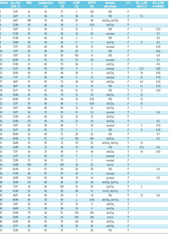
To assess the effect of Akti-1/2 and A-443654 on Akt activity in CLL cells, we examined the phosphorylation status of Akt or Akt substrates which are used to assess the activation status of Akt. Akti-1/2 inhibited Ser473 phosphorylation in a dose-dependent manner (Figure 1A). As previously described for other cell types,18,20 A-443654 induced an increase in Ser473 phosphorylation. In order to confirm that A-443654 was inhibiting Akt, we analyzed the status of the Akt substrates GSK3α/β and FoxO1/FoxO3a. Both inhibitors reduced the phos-phorylation of GSK3α/β and FoxO1/FoxO3a (Figure 1B), demonstrating that they inhibited Akt activity.
Akt inhibitors induce apoptosis in chronic
lymphocytic leukemia cells
To examine the ability of Akt inhibitors to induce apoptosis in CLL cells, we incubated CLL cells with a
range of concentrations of Akti-1/2 (0.5-20 µM) and A-443654 (0.1-1 µM) for 24 h and measured cell viability. Both inhibitors induced apoptosis in a dose-dependent manner (Figure 2A and 2B) and in a time-dependent manner (Online Supplementary Figure S2). The half-max-imal effective concentration (EC50) was 9.85±0.67 µM (range, 5 to 15 µM) for Akti-1/2, and 0.63±0.03 µM (range, 0.40 to 0.85 µM) for A-443654. Similar results were obtained in purified CD19+ CLL cells (Online
Supplementary Figure S3). Akti-1/2 and A-443654
induced apoptosis in all the analyzed samples, inde-pendently of sex, ZAP70 status, CD38 status or genom-ic alterations (data not shown).
Differential effect of Akt inhibitors on B and T cells
from patients with chronic lymphocytic leukemia
and healthy donors
Next, we analyzed the sensitivity of normal B and T cells to Akt inhibitors, in terms of induction of apopto-sis. The number of apoptotic T cells (CD3+cells) was measured in CLL samples and blood samples from healthy donors exposed to several doses of Akti-1/2
Figure 1.Akti-1/2 and A-443654 effects on the phosphorylation of Akt and Akt substrates. CLL cells were incubated with or with-out a range of doses of Akti-1/2 for 2 h. (A) Cells were lysed and whole extracts were analyzed by western blot as described in the Design and Methods section. Results from three patients are shown (n=3). (B) CLL cells were incubated with or without 5 µM
Akti-1/2 and 0.5 µM (patients 2, 4 and 5) or 1 µM (patient 3) A-443654 for 2 h. Cells were lysed and whole extracts were ana-lyzed by western blot as described in the Design and Methods sec-tion. Results from four representative patients are shown (n=9).
Figure 2. Cytotoxic effect of Akt inhibitors on CLL cells and on peripheral blood lymphocytes (PBL) from healthy donors. Cells from CLL patients were incubated for 24 h with or without various doses of Akti-1/2 (A, n=15) and A-443654 (B, n=15). Viability was measured by analysis of phosphatidylserine exposure and PI uptake as described in the Design and Methods section. Cells from CLL patients and healthy donors were incubated for 24 h with or without various doses of Akti-1/2 (C, n=9 and n=3, respec-tively) and A-443654 (D, n=13 and n = 8, respectively). Viability was measured as non-apoptotic CD3+/CD19–T cells from PBL (p)
and CLL (P) or CD3–/CD19+B cells from PBL (m) and CLL (M) as
described in the Design and Methods section. Viability is expressed as the percentage of the viability of untreated cells. Data are shown as the mean value ± SEM. *p<0.005, treated
ver-sus untreated cells (A,B), or B cells versus T cells from patients
with CLL (C,D).
A
A
B
C
D
B
0 0.1 0.3 0.5 1 A-443654 (µM) 0 0.1 0.3 0.5 1 0 0.5 1 5 10 0 0.5 1 5 10 20 100 80 60 40 20 0 100 80 60 40 20 0 100 80 60 40 20 0 100 80 60 40 20 0 A-443654 (µM) Viability (%) Viability (%) Viability (%) Viability (%) Akti-1/2 (µM) Akti-1/2 (µM)Patient 1 Patient 2 Patient 3
ct 0.1 0.5 1 5
Akti-1/2 (µM) Akti-1/2 (µM) Akti-1/2 (µM)
β-ACTIN β-ACTIN β-ACTIN Akt P-Ser473 Akt P-Ser473 Akt P-Ser473 Akt Akt Akt Akti-1/2 A-443654 Akti-1/2 A-443654 P-GSK3α/β P-GSK3α/β P-FoxO3a P-FoxO1 ct 0.1 0.5 1 5 ct 0.1 0.5 1 5 Patient 2 Patient 3 Patient 4 Patient 5
(up to 10 µM) and A-443654 (up to 1 µM) for 24 h. Incubation with 5 µM Akti-1/2 reduced the percentage of viable CLL cells to 64.64±6.12%. In contrast, the percentage of viable T cells from CLL samples was 102.4±3.46%. Interestingly, B and T cells derived from healthy donors were resistant to Akti-1/2-induced apoptosis. Thus, after incubation with 5 µM Akti-1/2 the percentage of viable B cells and T cells were 91.6 ± 2.1% and 100.7±7.2%, respectively (Figure 2C). Similar results were obtained in the treatment of CLL cells and cells from healthy donors with A-443654, but the difference between CLL cells and B or T cells from healthy donors was less pronounced than in the case of Akti-1/2. Incubation with 0.5 µM A-443654 reduced the percentage of viable CLL cells to 64.2±4.9%. In contrast, the percentage of viable T cells was 91.3±3.7%. Furthermore, B and T cells derived from
healthy donors were less sensitive than cells from CLL samples to A-443654-induced apoptosis. Thus, after incubation with 0.5 µM A-443654 the percentages of viable B and T cells were 83.4±5.8% and 87.1±5.9%, respectively (Figure 2D). Similar results were obtained when CLL cells and cells from healthy donors were treated for 48 h with both Akt inhibitors (Online
Supplementary Figure S4). We confirmed this
differen-tial induction of apoptosis between CLL cells and nor-mal lymphocytes by performing an analysis of the DNA content. We observed that CLL cells were more sensitive than T cells and normal B cells to the Akt inhibitor-induced increase in sub-G0 peak (Online
Supplementary Figure S5). These results indicate that B
cells from CLL samples are more sensitive than normal B and T cells to Akt inhibitor-induced apoptosis.
Effect of survival factors in combination
with Akt inhibitors
We studied the effect of two well-known survival factors in CLL cells, IL-426and SDF-1α,27in combina-tion with Akt inhibitors. We used selected CLL sam-ples in which these factors induced a survival effect. Thus, we treated CLL cells with 10 ng/mL IL-4 (Figure
Figure 3. Effect of survival signals on the apoptotic activity of Akt inhibitors. CLL cells were untreated or treated with 10 ng/mL IL-4 (A, n = 10) or 50 ng/mL SDF-1α (B, n = 7) without (white filled bars) or with 10 µM Akti-1/2 (gray filled bars) or 0.5 µM A-443654 (black filled bars) for 48 h. Viability was measured by analysis of phosphatidylserine exposure and propidium iodide uptake as described in the Design and Methods section. Data are shown as the mean value±SEM. **p<0.001 control versus IL-4 and Akt inhibitors treated cells versus control, IL-4 or SDF-1α; *p<0.01 control versus SDF-1α-treated cells.
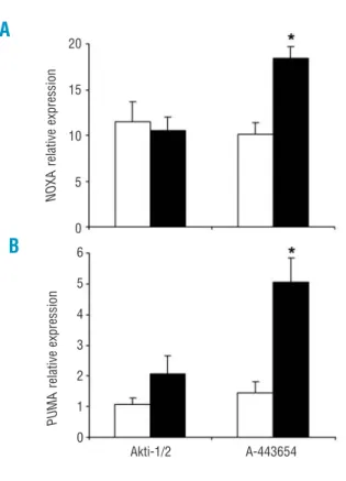
Figure 4. Apoptosis-related gene expression profile induced by Akti-1/2 and A-443654. Cells from CLL patients were untreated (open bars) or treated with 5 µM Akti-1/2 or 0.5 µM A-443654 (black filled bars) for 24 h. Cells were lysed, and NOXA (A) and
PUMA (B) mRNA expression was analyzed by RT-MLPA as
described in the Design and Methods section. The results are shown as the mean value ± SEM of four and eight different exper-iments for Akti-1/2 and A-443654, respectively. *p<0.005 treated
versus untreated cells.
A
B
B
A
Control IL-4
Control SDF-1α Akti-1/2 A-443654
80 70 60 50 40 30 20 10 0 20 15 10 5 0 6 5 4 3 2 1 0 80 70 60 50 40 30 20 10 0 Viability (%) Viability (%) PUMA relative expression NOXA relative expression
M.de Frias et al.
3A) or 50 ng/mL SDF-1α (Figure 3B) and with or with-out 10 µM Akti-1/2 or 0.5 µM A-443654 for 48 h, and then measured cell viability. Neither IL-4 nor SDF-1α was able to inhibit the apoptosis induced by both Akt inhibitors. These results show that Akt inhibitors can induce apoptosis in CLL cells even in the presence of survival signals.
Characterization of Akt inhibitor-induced apoptosis
in chronic lymphocytic leukemia
We investigated the effect of Akt inhibitors on the overall apoptosis mRNA expression profile by perform-ing RT-MLPA.25 Treatment of CLL cells with 5 µM
Akti-1/2 for 24 h did not induce any significant change in the RT-MLPA profile (Figure 4 and Online
Supplementary Figure S6A). The results indicated that
Akt inhibition has minimal effects on the apoptosis mRNA expression profile. Surprisingly, incubation of CLL cells with 0.5 µM A-443654 for 24 h induced an increase in the mRNA levels of NOXA and PUMA and a decrease in the mRNA levels of BMF, BID, one BAX probe, BCLw, NAIP, AIF, APAF and APAFL(Figure 4 and
Online Supplementary Figure S6B). Similar results were
obtained in purified CD19+ CLL cells (Online
Supplementary Figure S7). Thus, A-443654-induced
mRNA changes are likely independent of its activity of inhibiting Akt.
We then investigated the effect of Akt inhibitors on the expression of BCL-2 family proteins. Western blot analysis revealed that Akti-1/2 and A-443654 increased
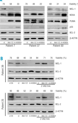
Figure 5. Apoptosis profile induced by Akti-1/2 and A-443654. (A) Apoptosis-related protein expression profile induced by Akti-1/2 and A-443654. Cells were untreated (ct) or treated with 5 µM Akti-1/2 (n = 10) and 0.5 µM A-443654 (n = 13) for 24 h, and MCL-1, NOXA, PUMA, p53 and BCL-2 expression was determined by west-ern blot. Cell viability is expressed at the top of the figure. β-actin was used to standardize protein levels. Results from three repre-sentative patients are shown, two corresponding to Ficoll isolated cells (patients 7 and 37) and one corresponding to purified CD19+
CLL cells (patient 50). (B) Effect of Z-VAD.fmk on Akti-1/2 and A-443654-induced MCL-1 decrease. Cells were pretreated with or without 200 µM Z-VAD.fmk for 30 min and then treated with 5 µM Akti-1/2 and 0.5 µM A-443654 for 24 h (results from two repre-sentative patients are shown, n = 3), and MCL-1 levels were deter-mined by western blot. BCL-2 and β-actin were used to standard-ize protein levels. Cell viability is expressed at the top of the fig-ure.
Figure 6. Apoptosis profile induced by Akti-1/2 and A-443654 in
TP53 deleted/mutated samples. (A) Protein expression profile
and cytotoxic effect of Akti-1/2, A-443654 and Nutlin-3a. Cells were untreated (ct) or treated with 5 µM Akti-1/2, 0.5 µM A-443654 or 5 µM Nutlin-3a for 24 h, and MCL-1, NOXA and PUMA expression was determined by western blot. BCL-2 and β-actin were used to standardize protein levels. Viability was measured by analysis of phosphatidylserine exposure and propidium iodide uptake as described in the Design and Methods section. (B) Apoptosis-related gene expression profile induced by Akti-1/2 and A-443654 in TP53 deleted/mutated CLL samples. Cells were untreated or treated with 5 µM Akti-1/2 (white filled bars) or 0.5 µM A-443654 (gray filled bars) and 5 µM Nutlin-3a (black filled bars) for 24 h. Cells were lysed, and the expression of apoptosis-related genes was analyzed by RT-MLPA as described in the Design and Methods section. The results are shown as fold induc-tion relative to untreated cells.
A
A
B
B
70 58 33 70 48 22 60 31 34 Viability (%) 45 22 15 38 Viability (%) 3 2 1 0 2 1,5 1 0,5 0 1,5 1 0,5 0 41 21 7 40 Viability (%) 72 65 24 78 Viability (%) 76 68 46 34 84 71 76 Viability (%) 90 66 52 31 68 69 58 Viability (%)ct 0h ct Akti-1/2 A-443654 ct Akti-1/2 A-443654
ct 0h ct Akti-1/2 A-443654 ct Akti-1/2 A-443654 ct Akti-1/2 A-443654 Nutlin-3a ct Akti-1/2 A-443654 Nutlin-3a
Akti-1/2 A-443654 Nutlin-3a Akti-1/2 A-443654 Nutlin-3a Akti-1/2 A-443654 Nutlin-3a
ct Akti-1/2 A-443654 Nutlin-3a ct Akti-1/2 A-443654 ct Akti-1/2 A-443654 ct Akti-1/2 A-443654
MCL-1 BCL-2 β-ACTIN MCL-1 MCL-1 NOXA PUMA PUMA NOXA NOXA PUMA NOXA PUMA NOXA PUMA MCL-1 p53 BCL-2 Z-VAD.fmk Z-VAD.fmk BCL-2 BCL-2 β-ACTIN β-ACTIN β-ACTIN PUMA NOXA MCL-1 BCL-2 β-ACTIN PUMA NOXA MCL-1 BCL-2 β-ACTIN
Patient 7 Patient 37 Patient 50
Patient 39 Patient 40 Patient 41 Fold induction Fold induction Fold induction Patient 8 Patient 25
NOXA protein levels and decreased MCL-1 protein levels in all the samples analyzed. PUMA protein lev-els were also increased in 50% of the samples after treatment with Akt inhibitors (Figure 5A). Furthermore, p53 protein was increased by A-443654 but not by Akti-1/2 in all the samples analyzed.
We further analyzed the apoptosis-related protein expression profile induced by Akti-1/2 and A-443654 at different times. We did not observe any change in MCL-1, NOXA and PUMA protein levels after 3 h of treatment with Akt inhibitors (data not shown). NOXA protein was increased by Akt inhibitors at 6 h while PUMA protein levels were increased at 12 h. Moreover, MCL-1 protein was decreased after incuba-tion with A-443654 for 6 h (Online Supplementary Figure
S8). Finally, pretreatment with 200 µM Z-VAD.fmk for
30 min blocked the decrease in MCL-1 induced by Akti-1/2 but not that induced by A-443654 (Figure 5B). These results demonstrate that the decrease in MCL-1 induced by Akti-1/2 is caspase-dependent whereas that induced by A-443654 is caspase-independent and precedes the activation of caspases.
Akti-1/2 and A-443654 induce apoptosis
irrespective of TP53 status in chronic
lymphocytic leukemia cells
To study the role of p53 in Akti-1/2 and A-443654-induced apoptosis we analyzed the effect of these compounds on CLL samples with deleted/mutated
TP53. Patients’ samples with deleted/mutated TP53 or
altered expression have been described previously.28-30 Patient 8 had a 17p deletion in one allele in 43% of peripheral blood lymphocytes, patient 41 had a 17p deletion in one allele in 94% of peripheral blood lym-phocytes, and patient 40 had a frame-shift mutation in one allele (nucleotide deletion in codon 272) and a 17p deletion in the other allele in 86% of peripheral blood lymphocytes. First, we incubated these CLL cells with 5 µM Akti-1/2 or 0.5 µM A-443654 for 24 h. Interestingly, we observed a decrease in viability in two of the three samples with Akti-1/2 treatment and in all three samples with A-443654 treatment (Figure 6A).
Western blot analysis revealed an increase in NOXA levels in the three samples when treated with both inhibitors, while PUMA levels increased in only one sample (patient 8) with 5 µM Akti-1/2 treatment. MCL-1 levels decreased with Akti-1/2 in patients 8 and 41, where Akti-1/2 induced apoptosis. In patient 40, MCL-1 levels increased with Akti-1/2 treatment, and the inhibitor did not induce apoptosis. A-443654 treatment induced a decrease in MCL-1 levels in the three samples (Figure 6A). As a control of TP53 status we used 5 µM Nutlin-3a, which has been described to induce apoptosis and p53 accumulation in cells with wild-type TP53 but not in those with deleted/mutated
TP53.28
Next, we examined the apoptosis mRNA expression profile by performing RT-MLPA. Incubation with 5 µM Akti-1/2 or 0.5 µM A-4436545 induced almost the same mRNA expression profile as that in CLL cells with the wild-type TP53, except that PUMA mRNA
levels did not increase after treatment with A-443654 (Figure 6B).
Discussion
In this study we investigated the effect of two Akt inhibitors on the viability of CLL cells. Both Akt inhibitors induced apoptosis in primary CLL cells. These results are in agreement with a recent report describing that introduction of constitutively active myr.Akt increases the viability of CLL cells.31
The mechanism of action of the two Akt inhibitors is somewhat different (Online Supplementary Figure S9). Surprisingly, Akti-1/2 inhibits Akt but does not induce changes in the RT-MLPA profile. Akt modulates the transcriptional activity of at least one transcription fac-tor, FoxO3a,15 which would induce changes in the mRNA levels of its transcriptional targets BIM32 and
PUMA.33However, RT-MLPA experiments suggest that dephosphorylation of FoxO3a induced by Akti-1/2 is not sufficient to induce the transcription of BIM and
PUMA in CLL cells. As A-443654 is a less specific
Akt-inhibitor,21 the simplest model would be to consider that inhibition of Akt (common to both inhibitors) does not induce changes in the RT-MLPA profile, and that A-443654 has additional targets to explain its effects on the expression of genes. Thus, A-443654 induces an increase in the levels of p53 protein and the induction of PUMA mRNA, a transcriptional target of
TP53 in CLL.28,34 In agreement with these data, the induction of PUMA mRNA by A-443654 was decreased in CLL cells with deleted/mutated TP53. Interestingly, Akti-1/2 induced PUMA and NOXA pro-teins without affecting PUMA and NOXA mRNA. The mechanism for this effect is unknown and could be explained by increased translation or decreased prote-olysis of these proteins.
A common effect of both Akt inhibitors is the mod-ulation of NOXA/MCL-1 balance. It has been reported that in primary CLL cells, the majority of NOXA pro-tein is associated with MCL-1.35
Thus, Akti-1/2 and A-443654 treatment induced an increase in NOXA protein levels and a down-regula-tion of the levels of MCL-1, a critical survival protein in CLL cells. Furthermore, inhibition of caspases pre-vents the down-regulation of MCL-1 induced by Akti-1/2, suggesting that MCL-1 cleavage participates in an amplification loop that increases cytochrome c release and apoptosis in CLL cells, as described for PKC inhibitors.36 In agreement with our results, the intro-duction of constitutively active myr.Akt increases MCL-1 protein, and inhibition of MCL-1 by treatment with siRNA induces apoptosis in CLL.31 The mecha-nism of regulation of MCL-1 protein by Akt in CLL cells is unknown. The Akt substrate GSK-3 has been reported to induce destabilization of MCL-1 protein.16 However, inhibition of GSK-3 does not inhibit the apoptotic effect of PI3K inhibitors.7As GSK-3 is inhib-ited by PKC,37 perhaps the over-expression of active PKC-βII38blocks this pathway in CLL cells.
M.de Frias et al.
sensitive to Akt inhibitors than T cells from CLL sam-ples, and B or T cells from healthy donors. Chemo-therapeutic drugs, including fludarabine, chlorambucil, and doxorubicin induce apoptosis equally in both B and T cells, leading to immunosuppression.39,40 Thus, the differential effect of Akti-1/2 and A-443654 in B and T lymphocytes is of interest. In conclusion, the results presented here suggest that clinically suitable small-molecule inhibitors of Akt alone or in combina-tion with chemotherapeutic drugs might be a new ther-apeutic option for the treatment of CLL.
Authorship and Disclosures
MdeF performed the research and contributed to data analysis and manuscript writing. DIS, AMC, LCM, AFS and DMGG performed the research and contributed with analytical tools. EdelaB contributed with patients’ samples and data. GP designed the research and con-tributed to data analysis. JG designed the research and contributed to data analysis and manuscript writing. The authors reported no potential conflicts of interest.
References
1. Barragan M, Campas C, Bellosillo B, Gil J. Protein kinases in the regula-tion of apoptosis in B-cell chronic lymphocytic leukemia. Leuk Lymphoma 2003;44:1865-70. 2. Barragan M, Bellosillo B, Campas C,
Colomer D, Pons G, Gil J. Involvement of protein kinase C and phosphatidylinositol 3-kinase path-ways in the survival of B-cell chron-ic lymphocytchron-ic leukemia cells. Blood 2002;99:2969-76.
3. Bernal A, Pastore RD, Asgary Z, Keller SA, Cesarman E, Liou HC, et al. Survival of leukemic B cells pro-moted by engagement of the anti-gen receptor. Blood 2001;98:3050-7. 4. Ringshausen I, Schneller F, Bogner C, Hipp S, Duyster J, Peschel C, et al. Constitutively activated phos-phatidylinositol-3 kinase (PI-3K) is involved in the defect of apoptosis in B-CLL: association with protein kinase C∆. Blood 2002;100:3741-8. 5. Jones DT, Ganeshaguru K,
Ander-son RJ, JackAnder-son TR, Bruckdorfer KR, Low SY, et al. Albumin activates the AKT signaling pathway and protects B-chronic lymphocytic leukemia cells from chlorambucil- and radia-tion-induced apoptosis. Blood 2003; 101:3174-80.
6. Cuni S, Aciego P, Perez-Chacon G, Vargas JA, Sanchez A, Martin-Saavedra FM, et al. A sus-tained activation of PI3K/NF-kappaB pathway is critical for the survival of chronic lymphocytic leukemia B cells. Leukemia 2004;18: 1391-400.
7. Plate JM. PI3-kinase regulates sur-vival of chronic lymphocytic leukemia B-cells by preventing cas-pase 8 activation. Leuk Lymphoma 2004;45:1519-29.
8. Nedellec S, Renaudineau Y, Bordron A, Berthou C, Porakishvili N, Lydyard PM, et al. B cell response to surface IgM cross-linking identifies different prognostic groups of B-chronic lymphocytic leukemia patients. J Immunol 2005;174: 3749-56.
9. Hu X, Haney N, Kropp D, Kabore AF, Johnston JB, Gibson SB. Lysophosphatidic acid (LPA)
pro-tects primary chronic lymphocytic leukemia cells from apoptosis through LPA receptor activation of the anti-apoptotic protein AKT/PKB. J Biol Chem 2005;280: 9498-508.
10. Barragan M, de Frias M, Iglesias-Serret D, Campas C, Castano E, Santidrian AF, et al. Regulation of Akt/PKB by phosphatidylinositol 3-kinase-dependent and -independent pathways in B-cell chronic lympho-cytic leukemia cells: role of protein kinase Cβ. J Leukoc Biol 2006;80: 1473-9.
11. Petlickovski A, Laurenti L, Li X, Marietti S, Chiusolo P, Sica S, et al. Sustained signaling through the B-cell receptor induces Mcl-1 and pro-motes survival of chronic lympho-cytic leukemia B cells. Blood 2005; 105:4820-7.
12. Engelman JA, Luo J, Cantley LC. The evolution of phosphatidylinosi-tol 3-kinases as regulators of growth and metabolism. Nat Rev Genet 2006;7:606-19.
13. Leupin N, Cenni B, Novak U, Hugli B, Graber HU, Tobler A, et al. Disparate expression of the PTEN gene: a novel finding in B-cell chron-ic lymphocytchron-ic leukaemia (B-CLL). Br J Haematol 2003;121:97-100. 14. Manning BD, Cantley LC. AKT/PKB
signaling: navigating downstream. Cell 2007;129:1261-74.
15. Brunet A, Bonni A, Zigmond MJ, Lin MZ, Juo P, Hu LS, et al. Akt pro-motes cell survival by phosphorylat-ing and inhibitphosphorylat-ing a Forkhead tran-scription factor. Cell 1999;96:857-68.
16. Maurer U, Charvet C, Wagman AS, Dejardin E, Green DR. Glycogen synthase kinase-3 regulates mito-chondrial outer membrane perme-abilization and apoptosis by desta-bilization of MCL-1. Mol Cell 2006; 21:749-60.
17. Longo PG, Laurenti L, Gobessi S, Petlickovski A, Pelosi M, Chiusolo P, et al. The Akt signaling pathway determines the different prolifera-tive capacity of chronic lymphocytic leukemia B-cells from patients with progressive and stable disease. Leukemia 2007;21:110-20.
18. Luo Y, Shoemaker AR, Liu X, Woods KW, Thomas SA, de Jong R,
et al. Potent and selective inhibitors of Akt kinases slow the progress of tumors in vivo. Mol Cancer Ther 2005;4:977-86.
19 Shi Y, Liu X, Han EK, Guan R, Shoemaker AR, Oleksijew A, et al. Optimal classes of chemotherapeu-tic agents sensitized by specific small-molecule inhibitors of akt in vitro and in vivo. Neoplasia 2005; 7:992-1000.
20. Han EK, Leverson JD, McGonigal T, Shah OJ, Woods KW, Hunter T, et al. Akt inhibitor A-443654 induces rapid Akt Ser-473 phosphorylation independent of mTORC1 inhibi-tion. Oncogene 2007;26:5655-61. 21. Bain J, Plater L, Elliott M, Shpiro N,
Hastie CJ, McLauchlan H, et al. The selectivity of protein kinase inhibitors: a further update. Biochem J 2007;408:297-315. 22. Barnett SF, Defeo-Jones D, Fu S,
Hancock PJ, Haskell KM, Jones RE, et al. Identification and characteriza-tion of pleckstrin-homology-domain-dependent and isoenzyme-specific Akt inhibitors. Biochem J 2005;385:399-408.
23. Logie L, Ruiz-Alcaraz AJ, Keane M, Woods YL, Bain J, Marquez R, et al. Characterization of a protein kinase B inhibitor in vitro and in insulin-treated liver cells. Diabetes 2007;56: 2218-27.
24. Bellosillo B, Pique M, Barragan M, Castano E, Villamor N, Colomer D, et al. Aspirin and salicylate induce apoptosis and activation of caspases in B-cell chronic lymphocytic leukemia cells. Blood 1998; 92:1406-14.
25. Eldering E, Spek CA, Aberson HL, Grummels A, Derks IA, de Vos AF, et al. Expression profiling via novel multiplex assay allows rapid assess-ment of gene regulation in defined signalling pathways. Nucleic Acids Res 2003;31:e153.
26. Dancescu M, Rubio-Trujillo M, Biron G, Bron D, Delespesse G, Sarfati M. Interleukin 4 protects chronic lymphocytic leukemic B cells from death by apoptosis and upregulates Bcl-2 expression. J Exp Med 1992;176:1319-26.
27. Burger JA, Tsukada N, Burger M, Zvaifler NJ, Dell’Aquila M, Kipps TJ. Blood-derived nurse-like cells
pro-tect chronic lymphocytic leukemia B cells from spontaneous apoptosis through stromal cell-derived factor-1. Blood 2000;96:2655-63.
28. Coll-Mulet L, Iglesias-Serret D, Santidrian AF, Cosialls AM, de Frias M, Castano E, et al. MDM2 antago-nists activate p53 and synergize with genotoxic drugs in B-cell chronic lymphocytic leukemia cells. Blood 2006;107:4109-14.
29. Coll-Mulet L, Santidrian AF, Cosialls AM, Iglesias-Serret D, de Frias M, Grau J, et al. Multiplex ligation-dependent probe amplification for detection of genomic alterations in chronic lymphocytic leukaemia. Br J Haematol 2008;142:793-801. 30. Santidrian AF, Cosialls AM,
Coll-Mulet L, Iglesias-Serret D, de Frias M, Gonzalez-Girones DM, et al. The potential anticancer agent PK11195 induces apoptosis irrespec-tive of p53 and ATM status in chron-ic lymphocytchron-ic leukemia cells. Haematologica 2007;92:1631-8. 31. Longo PG, Laurenti L, Gobessi S,
Sica S, Leone G, Efremov DG. The Akt/Mcl-1 pathway plays a
promi-nent role in mediating antiapoptotic signals downstream of the B-cell receptor in chronic lymphocytic leukemia B cells. Blood 2008;111: 846-55.
32. Dijkers PF, Medema RH, Lammers JW, Koenderman L, Coffer PJ. Expression of the pro-apoptotic Bcl-2 family member Bim is regulated by the forkhead transcription factor FKHR-L1. Curr Biol 2000;10:1201-4. 33. You H, Pellegrini M, Tsuchihara K,
Yamamoto K, Hacker G, Erlacher M, et al. FOXO3a-dependent regulation of Puma in response to cytokine/growth factor withdrawal. J Exp Med 2006;203:1657-63. 34. Mackus WJ, Kater AP, Grummels A,
Evers LM, Hooijbrink B, Kramer MH, et al. Chronic lymphocytic leukemia cells display p53-depend-ent drug-induced Puma upregula-tion. Leukemia 2005;19:427-34. 35. Hallaert DY, Spijker R, Jak M, Derks
IA, Alves NL, Wensveen FM, et al. Crosstalk among Bcl-2 family mem-bers in B-CLL: seliciclib acts via the Mcl-1/Noxa axis and gradual exhaustion of Bcl-2 protection. Cell
Death Differ 2007;14:1958-67. 36. Snowden RT, Sun XM, Dyer MJ,
Cohen GM. Bisindolylmaleimide IX is a potent inducer of apoptosis in chronic lymphocytic leukaemic cells and activates cleavage of Mcl-1. Leukemia 2003;17:1981-9. 37. Christian SL, Sims PV, Gold MR.
The B cell antigen receptor regulates the transcriptional activator β-catenin via protein kinase C-mediat-ed inhibition of glycogen synthase kinase-3. J Immunol 2002;169:758-69.
38. Abrams ST, Lakum T, Lin K, Jones GM, Treweeke AT, Farahani M, et al. B-cell receptor signaling in chron-ic lymphocytchron-ic leukemia cells is reg-ulated by overexpressed active pro-tein kinase CβII. Blood 2007;109: 1193-201.
39. Keating MJ. Immunosuppression with purine analogues – the flip side of the gold coin. Ann Oncol 1993; 4:347-8.
40. Hamblin AD, Hamblin TJ. The immunodeficiency of chronic lym-phocytic leukaemia. Br Med Bull 2008;87:49-62.