Tutor:
Prof. Roberto Contestabile Supervisor:
Prof. Martino Luigi Di Salvo
Coordinator: Prof. Stefano Gianni
Ph.D. Student Federico D’Alessio
Ph.D. Course in Biochemistry
Department of Biochemical Sciences “A. Rossi Fanelli” XXXIII Cycle
Regulation of vitamin B
6metabolism in
Escherichia coli
Abstract
The catalytically active form of vitamin B6, pyridoxal 5'-phosphate (PLP), acts as a coenzyme in a variety of different enzymatic reactions. PLP is essential for the normal growth and development of all the organisms, but only plants and microorganisms can synthetise it de novo, while the other organisms must recycle it from nutrients.
The PLP is a very reactive molecule thanks to its aldehyde group and its intracellular concentration must be kept low to avoid undesired toxic reaction in the organism. The regulation of the free PLP concentration in cells occurs through a variety of mechanism, such as its dephosphorylation to PL or, as recently discovered, through the binding to PLP Binding Protein. In
Escherichia coli an important candidate for this role has recently been
discovered: YggS is a non-catalytic protein able to bind PLP. It belongs to the COG0325 family, a class of protein sharing structure homology with PLP-dependent enzymes, such as alanine racemase and some decarboxylases that have the same Fold-type III.
The metabolism of PLP in E. coli has been studied for years. In this work, our group have deepened the regulation of the PLP metabolism, studying and elaborating the state of the art, and crossing the available literature data with those produced in our lab about the regulation of the expression of the genes involved in PLP homeostasis and focusing the analysis on the most important genes. Our studies have also analysed in vivo the phenotypes linked to the genes involved in PLP homeostasis when E. coli is grown in different media and in presence of vitamers, in order to better understand the role of the analysed genes. The study has also considered the expression of the genes involved in PLP homeostasis in presence of vitamin B6 vitamers and during different growth phases.
Finally, given the importance of the gene yggS in E. coli, the attention was focused on the characterization of the protein YggS, whose role has not yet been discovered. This protein is hypothesised to be both a PLP binding protein and a carrier of this important cofactor to the apo PLP-dependent enzymes. Our studies were focused on YggS capacity to bind PLP and, using mutant forms of this protein, we have verified and studied the transfer mechanism of PLP to the apo PLP-dependent enzymes.
I
Index
1. Introduction 1
1.1 Pyridoxal-5’-phosphate: an overview of the biological active form of the
vitamin B6 2
1.2 Vitamin B6 metabolism 3
1.2.1 Vitamin B6 de novo biosynthesis 5
1.2.2 The vitamin B6 salvage pathway 6
1.3 Vitamin B6 in humans 7
1.4 PLP related diseases in humans 11
1.4.1 PLP deficiency related diseases 12
1.4.2 PLP role in cancer 13
1.4.3 PLP role in diabetes 15
1.5 PLP homeostasis: its importance and the involved proteins 18
1.5.1 PLP Binding Protein 21
1.6 Aim of the thesis 27
2. Materials and Methods 28
Materials 29
2.1 Materials, bacterial strains, plasmids and reagents 29
Growth media and supplements 29
Bacterial strains 30
Plasmids 31
Table 2.1 Oligonucleotides for molecular cloning 32
Table 2.2 Oligonucleotides for RT-qPCR 33
Methods 34
2.2 Molecular cloning of WT yggS 34
2.3 Site-directed mutagenesis 34
2.4 RNA analysis through RT-qPCR 35
2.5 Expression and purification of the proteins 36
2.6 Preparation of the apo-forms of the proteins 39
II
2.8 Spectroscopic measurements 41
2.9 Determination of the dissociation constant of PLP from YggS 42
2.10 Kinetic studies 43
2.11 Data analysis 45
2.12 Software used for figures and graphs 46
Results 47
3. Regulation of vitamin B6 metabolism in E. coli 48
3.1 PLP-binding proteins in E. coli 48
3.2 E. coli genes involved in PLP homeostasis 53
3.2.1 Genes involved in PLP biosynthesis 54
3.2.2 Genes of salvage pathway 72
3.2.3 Other PLP- binding proteins 78
3.3. Regulation of PLP metabolism 84
3.3.1 State of the art on transcriptional regulation 84 3.3.2 Transcriptional level analysis of the genes involved in the PLP
metabolism 85
3.3.3 Traslational regulation 95
4. In vivo analysis of genes involved in PLP metabolism 101
4.1 Effect of PL and PN 101
4.2 Effect of 4-deoxypyridoxine 105
5 Characterization of E. coli YggS 119
5.1 Molecular cloning of YggS 119
5.2 Protein expression and purification of YggS 120
5.3 E. coli Serine hydroxy methyltransferase 123
5.4 Docking experiments between YggS and SHMT 124
5.5 Characterization of YggS 126
5.5.1 Size Exclusion Chromatography 126
5.5.2 Second binding site 128
5.5.3 Dissociation constant 129
5.5.5 Circular Dichroism 132
III
6. Conclusions 137
7. Bibliography 141
Collaborations 160
1
2
1.1 Pyridoxal-5’-phosphate: an overview of the biological active form of the vitamin B6
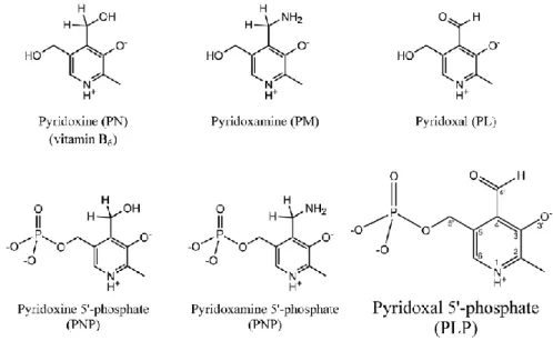
Vitamin B6 is a water-soluble compound represented by six vitamers that are interconvertible thanks to different enzymes. The six vitamers of the vitamin B6 are: pyridoxine (PN), pyridoxamine (PM), pyridoxal (PL) and their phosphorylated forms on their 5' carbon: pyridoxine-5’-phosphate (PNP), pyridoxamine-5’-phosphate (PMP) and the bioactive form pyridoxal-5’-phosphate (PLP) (fig 1.1). The vitamers share the same structure of the 2-methyl-3-hydroxypyridine and differ for the group on the C4 position.
The biologically active vitamer, pyridoxal 5’-phoshate (since here called PLP), acts as a cofactor for more than 150 enzymes (Percudani and Peracchi 2003), and it is essential for normal growth and development of all the organisms. In fact, as enzymatic cofactor, PLP is involved in a lot of essential
Figure 1.1: Structures of the six B6 vitamers. Carbon atom numbering is
3
pathways such as: the synthesis, degradation and transformation of biogenic amines, polyamines and amino acids, transsulfuration, supply of one carbon units, synthesis of tetrapyrrolic molecules, synthesis and degradation of the neurotransmitters (Mills et al. 2005). As a free (not bound to enzyme) molecule, PLP has other important roles, such as chaperon for correct enzymes folding (Cellini et al. 2014), modulator for transcription factors in bacteria (Huq et al. 2007; Tramonti et al. 2018), and as a virulence factor for pathogenic microorganism such as Helicobacter pylori, Mycobacterium
tuberculosis and Actinobacillus pleuropneumoniae (Dick et al. 2010;
Grubman et al. 2010; Xie et al. 2017).
PLP is characterized by an aldehyde group on the C4 carbon and, as a small aldehyde, is a very reactive molecule, so in cells there must be mechanisms for its homeostasis because its free form must be kept in low concentration in order to avoid undesired reaction with other reactive biological molecules (i.e. the amino and thiol groups).
1.2 Vitamin B6 metabolism
Vitamin B6 represented by PLP and its vitamers is essential for all the organisms. Despite the considerable importance of this molecule, only plants and microorganism can synthetize de novo this vitamin. They do this using two different and mutually exclusive biosynthetic pathways: the deoxyxylulose 5-phosphate (DXP)-dependent pathway and the DXP-independent pathway (fig 1.2). All other organisms must assimilate the vitamers from nutrients and interconvert them, depending on their needing, using a so-called salvage pathway (Fitzpatrick, Moccand, and Roux 2010).
4
Fig 1.2: Vitamin B6 biosynthetic pathways: De novo DXP-dependent
pathway (present in some eubacteria): GapB, D-erythrose 4-phosphate dehydrogenase; PdxB, erythronate-4-phosphate dehydrogenase; PdxF/SerC, phosphoserine aminotransferase; PdxA, 4-hydroxythreonine-4-phosphate dehydrogenase; DPXS, 1-deoxy-D-xylulose-5-phosphate synthase; PNP synthase, from PdxJ gene. De novo DXP-independent pathway (present in other eubacteria, fungi, plants and Archaea): PLP synthase complex: synthase domain from Pdx1 gene; glutaminase domain from Pdx2 gene. Salvage pathway (present in all organisms, including mammals): PLK, pyridoxal kinase from PdxK gene and pyridoxal kinase 2 from PdxY gene; PNPOx, pyridoxine 5′-phosphate oxidase from PdxH gene. (Di Salvo et al, 2011)
5 1.2.1 Vitamin B6 de novo biosynthesis
As already mentioned above, the unique living beings able to biosynthesize vitamin B6 are plants and microorganisms. Two different and independent vitamin B6 de novo biosynthetic pathways are known: a DXP-dependent and a DXP-independent pathway, depending on the usage of 1-deoxy-D-xylulose-5-phosphate as a reaction intermediate (Fitzpatrick et al. 2007). The DXP-dependent de novo route was the first to be discovered and studied in Escherichia coli and for a long time was believed to be ubiquitous but, actually, it is used only from some eubacteria (Fitzpatrick et al. 2007; Di Salvo, Contestabile, and Safo 2011). This biosynthetic pathway is composed by two convergent branches (Fig 1.2). The first branch starts with the conversion of D-erythrose 4-phosphate into 4-phosphoerythronate, while the second one starts from the condensation of pyruvate and glyceraldehyde 3-phosphate to produce 1-deoxy-D-xylulose-5-3-phosphate. The two branches converge on the reaction catalysed by pyridoxine 5’-phosphate synthase (PdxJ) to form pyridoxine 5’-phosphate (Laber et al. 1999). The reaction steps used in these two branches are catalysed by five enzymes encoded by the genes gapB, pdxB, pdxF, pdxA, and dxpS. Being PLP the bioactive form of the vitamin B6, PNP generated in the pathway described above must be oxidised to PLP by PNP oxidase (PNPOx) encoded by the gene pdxH (Fitzpatrick et al. 2007).
The so-called DXP-independent de novo route was discovered by chance in Fungi, and after that it was clear that it is more widely diffused than the DXP-dependent route, being found in Archaea, most Eubacteria and plants (Ehrenshaft et al. 1999)(Mittenhuber 2001). This pathway use glutamine, ribose or ribulose 5-phosphate and either glyceraldehyde 3-phosphate or
6
dihydroxyacetone phosphate to produce directly PLP thanks to the catalytic action of the PLP synthase complex encoded by pdx1 and pdx2 genes (Zein et al. 2006, Raschle, Amrhein, and Fitzpatrick 2005).
1.2.2 The vitamin B6 salvage pathway
Some organisms have developed a pathway that bypasses the necessity to produce PLP recycling the vitamers that they absorb from the environment as a nutrient or from the degraded PLP dependent enzymes. This alternative route to PLP production is known as “salvage pathway” and it is made by few enzymes able to interconvert the vitamin B6 vitamers into PLP and vice versa (fig 1.2). The key enzymes of the salvage pathway are: the ATP-dependent pyridoxal kinase (PDXK) that is able to convert each vitamer into their relative phosphorylated form, and the pyridoxine 5’-phosphate oxidase (PNPOx) which oxidases pyridoxine 5’-phosphate into PLP using FMN as a cofactor. These two enzymes are linked also in the regulation of the PLP level in the cell binding this molecule and being regulated themselves by product inhibition. The binding of the PLP on these two proteins contributes to its homeostasis and regulates its production trough different mechanisms that will be discussed in the following paragraphs.
Moreover, in the PLP salvage pathway are present other enzymes such as the phosphorylases that are required to dephosphorylate PLP and other phosphorylated vitamers in order to deliver them through the membranes to the other cells. For example, in human the intestine only absorbs non phosphorylated vitamers (Darin et al. 2016) and therefore, as studied in rats, the vitamers are first dephosphorylated by the intestinal phosphatases (Middleton 1990). Recently PL reductase, which converts PN to PL, has been
7
proposed as an important component of the salvage pathway (Ito and Downs, 2020).
1.3 Vitamin B6 in humans
Vitamin B6 in humans is absorbed from food and is produced from the intestinal microflora. The major nutritional rich foods for vitamin B6 are: fish, beef liver and other organ meats, potatoes and starchy vegetables, and also fruits. In animal-derived food the vitamin B6 is mainly present as PLP associated with glycogen phosphorylase and as PMP, while in vegetables it is present as PN or PN-5’-β-D-glucoside (McCormick 1989).
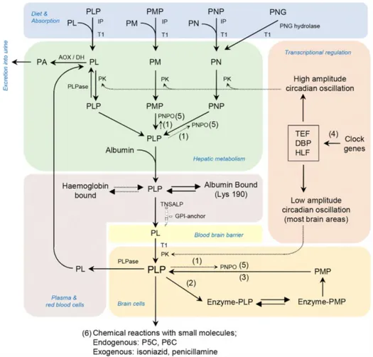
The metabolism of the vitamin B6 in human involves different organs and enzymes as shown in the pathway (Fig 1.3) from dietary assumption, recycle and catabolism of excess PLP (Darin et al. 2016). After their dietary assumption, the phosphorylated vitamers cannot cross the membrane of the intestine (Darin et al. 2016), so some phosphatases such as the tissue-non-specific alkaline phosphatase (TNSALP) hydrolase them in order to allow their transport across the membrane (Said 2004). Once in the intestine PM and PM can be converted before their transport to the liver (Albersen et al. 2013) or, once in the liver in PLP. In the liver PM, PL and PN are first phosphorylated from the pyridoxal kinase and then PMP and PNP are oxidised by the PNPOx to form PLP that, once bound to the albumin (Bohney, Fonda, and Feldhoff 1992), is exported to other cells. The liver is also the organ where the excess PLP is dephosphorylated to PL and then oxidised by an aldehyde dehydrogenase or an aldehyde oxidase (AOX) (Stanulović et al. 1976) to pyridoxic acid to be excreted in urine.
In order to reach the brain, the PLP must dissociate from albumin and be converted in PL by a tissue non-specific alkaline phosphatase. As PL it can
8
pass the blood brain barrier (BBB) probably by facilitated diffusion and it is “trapped” in the brain cells as PLP by the action of the pyridoxal kinase (Spector and Johanson 2007).
9
Fig 1.3: Enzymes and transporters involved in mammalian CNS PLP Synthesis and homeostasis and known human genetic vitamin-B6 dependent epilepsies
Pyridoxal 5’-phosphate (PLP); pyridoxamine 5’-phosphate (PMP); pyridoxal (PL); pyridoxine (PN); pyridoxine 5’-phosphate (PNP); pyridoxine-50-b-D-glucoside (PNG); intestinal phosphatases (IP); transporter (identity unknown; T1); pyridoxal kinase (PK); pyridox(am)ine 5’-phosphate oxidase (PNPO); tissue non-specific alkaline phosphatase (TNSALP); pyridoxal phosphatase (PLPase); aldehyde oxidase (Mo cofactor)/b-NAD dehydrogenase (AOX/DH). (1) PNPO is controlled by feedback inhibition from PLP. (2) PLP functions as a co-factor, forming Schiff bases with the ε-amino group of lysine residues of proteins. (3) PLP can be formed by recycling the cofactor from degraded enzymes (salvage pathway). (4) PLP levels are maintained, in part, by circadian-clockcontrolled transcription factors with PAR bZip transcription factors (DBP, HLF, and TEF) targeting PK. (5) PNPO mutations cause a B6-dependent epilepsy disorder. (6)
Disorders resulting in accumulation of L-pyrroline-5-carboxylic acid (P5C) and D,1-piperideine-6-carboxylic acid (P6C) (hyperprolinaemia type II and pyridoxine-dependent epilepsy due to mutations in ALDH4A1 and ALDH7A1, respectively) cause decreases in bioavailable PLP as do reactions with exogenous small molecules. (Darin et al, 2016)
10
Once PLP is in the cell, its free form must be kept under a certain concentration because of its reactiveness. In order to limit unwanted reactions and toxicity effects given by the free form of PLP, the cells have developed a very efficient protein whose function is not well defined, but that is able to bind PLP. In human the protein that must control the concentration of PLP is the PLP Binding Protein (PLPBP), a protein that does not have any catalytic activity (Darin et al. 2016).
Due to its importance in the homeostasis of the PLP, a deep focus on this protein will be discussed in a dedicated paragraph.
Imbalances in PLP homeostasis can cause severe diseases, PLP deficiency in fact is implicated in several neurological and non-neurological disorders. The vitamin assumed with diet is commonly sufficient since most of the food contains vitamin B6. The most of the PLP imbalance are done by inborn error on the recycle pathway genes or by the assumption of PLP inactivating drugs (Di Salvo et al. 2011). The effect of the PLP concentration imbalance will be discussed in the following paragraphs to better describe the linkage between the low and the high concentration with the most important and studied associated pathologies.
1.4 PLP related diseases in humans
In humans, an insufficient concentration or an excess of PLP can be associated with both mild and severe different pathologies. PLP deficiency due to drug assumption may produce symptoms such as unconsciousness, seizures, sleeplessness, headache, restlessness, agitation, tremors, and hallucination, while the deficiency linked with genetic mutation is implicated in severe pathologies, including neonatal epileptic encephalopathy, seizures, autism, Down syndrome, schizophrenia, autoimmune polyglandular disease,
11
Parkinson's, Alzheimer's, epilepsy, attention deficit hyperactivity disorders and learning disability (Di Salvo et al. 2011).
Some pathologies linked with the low concentration of PLP, such as: symptoms of the premenstrual syndrome (Wyatt et al. 1999), nausea and vomiting during pregnancy (Ebrahimi, Maltepe, and Einarson 2010), carpal tunnel syndrome (Ryan-Harshman and Aldoori 2007) can easily be solved with the assumption of this molecule. More severe pathologies linked with low level of PLP are some neurological diseases and there are also evidences that a low concentration of PLP can contribute to the onset of diabetes and cancer.
Excess of PLP can be toxic too, in fact in humans the effects of large doses (more than 200mg/day) have been observed (Windebank 1985) mostly at the level of peripheral nervous system as sensory and motor neuropathy leading to numbness in hands and feet that usually finish when supplementation is interrupted (Foca 1985). Another symptom of an excess of PLP can be the seizure susceptibility by diminishing GABA levels through several mechanisms (di Salvo, Safo, and Contestabile 2012).
1.4.1 PLP deficiency related diseases
The essential role of PLP in the metabolism of neurotransmitters including GABA, dopamine, epinephrine, serotonin, histamine and D-serine is the main cause of pathological phenotypes when inborn errors lead to a deficiency in the PLP concentration (Hatch et al. 2016).
PLP deficiency occurs through different mechanisms, an example is the PNPOx deficiency due to inborn defect of the enzyme that affects the PLP synthesis and recycle causing a pathology called neonatal epileptic
12
encephalopathy. The patients affected by this pathology present a particular electroencephalogram pattern and some of them can be treated with PLP but not with PN (Wang and Kuo 2007).
Hypophosphatasia is a pathology that affects the PLP concentration in the brain and it is due to a molecular defect on the gene coding for the tissue non-specific alkaline phosphatase (TNSALP). Phosphorylated PLP cannot cross the blood brain barrier and its deficiency in the brain impairs the synthesis of the neurotransmitters causing seizures (Ware et al. 2014).
Symptoms similar to the patients with PNPOx deficiency have been observed also in people with PLP binding protein (PLPBP) deficiency because the intracellular transport of PLP were damaged (Darin et al. 2016).
PLP deficiency is given also by the accumulation of metabolites that inactivate PLP causing pyridoxine dependent epilepsy (PDE). These cases are characterized by deficiency of ALDH7A1 (α-aminoadipic semialdehyde dehydrogenase) or ALDH4A1 (P5C dehydrogenase) (Wilson et al. 2019). All the deficiency described above, when in their most severe cases (neonatal presentations) can have other consequences including anemia, lactic acidosis and hypoglycemia (Wilson et al. 2019).
1.4.2 PLP role in cancer
Despite until the early 1980s the shortage of PLP was considered a strategy against cancer due to the importance of the PLP in the proliferation of the cells (Galluzzi et al. 2013), recent studies have evidenced the opposite relation between vitamin B6 and cancer. In fact, PLP deficiency is linked to an increase of tumor insurgence, in particular in the gastrointestinal tract (Gylling et al. 2017; Kayashima et al. 2011) and lung (Zuo et al. 2019). It is
13
clear that PLP has a multifaced role in cancer being both an antioxidant and an essential micronutrient for cell proliferation (Contestabile et al. 2020). PLP is not only a cofactor for several enzymatic reactions, but is also an efficient ROS scavenger, as the other vitamers, thanks to the presence of both hydroxyl (-OH) and amine (-NH2) substituents on the pyrimidine ring which can directly react with the peroxy radicals (Contestabile et al. 2020; Kannan and Jain 2004).
It has been proposed that in conditions of metabolic stress or in the case of shortage of necessary nutrients, some cellular processes such as DNA acetylation/methylation, synthesis of DNA precursors and ROS production can be altered causing DNA damage, which can drive cells toward cancer (Turgeon, Perry, and Poulogiannis 2018). Although many studies converge towards a protective role of vitamin B6 in cancer, the molecular mechanisms are not completely understood. PLP, as antioxidant molecule, could play an important role in mediating the cross talk between metabolism and DNA damage, in fact PLP acts against the formation of advanced glycation end products (AGEs), that are genotoxic compounds associated with senescence and diabetes (Booth et al. 1997).
PLP does not have only an antioxidant role in cancer, in fact it plays a crucial role as a cofactor of the serine hydroxy methyltransferase (SHMT) whose folate dependent reaction is the main source of one carbon unit in the metabolism, playing an important role in the synthesis of the thymidylate (Fig 1.4)
An eventual PLP deficiency can cause a decrease in activity of the SHMT causing a misincorporation of uracil in the DNA (Giardina et al. 2018; MacFarlane et al. 2011; Paone et al. 2014). SHMT is not the only PLP dependent enzyme whose malfunction reflect its effect on the DNA, in fact also glycine decarboxylase (that also depend on folate) is essential for the
14
synthesis of purines and so, for DNA metabolism (Contestabile et al. 2020; Fleming and Copp 1998).
Given the implications that vitamin B6 have in DNA metabolism, it is not surprising that low levels of this vitamin are associated with cancer.
Fig 1.4:Schematic of B9 metabolism comprising the thymidylate cycle (red
diagram), the methionine cycle (green diagram) and the purine biosynthesis pathway (blue diagram). The enzymes involved are: dihydrofolate reductase (DHFR); thymidylate synthase (TS); serine hydroxymethyltransferase (SHMT); methylenetetrahydrofolate reductase (MTHFR); methionine synthase (MS); methionine adenosyltransferases (MAT); S-adenosylhomocysteinase (SHase); glycine cleavage system (GCS); methylenetetrahydrofolate dehydrogenase (MTHFD); 10-formyltetrahydrofolate dehydrogenase (FDH); formyltetrahydrofolate synthetase (FTHFS). (Contestabile et al, 2020)
15 1.4.3 PLP role in diabetes
This relationship between PLP deficiency and diabetes has been previously described (Rubí 2012), but the molecular and cellular mechanisms underlying this relationship have not yet been completely understood.
PLP deficiency may impact on diabetes in different ways; as reported by Kotake and collaborators (Kotake et al. 1975) the metabolites produced in the altered tryptophan degradation pathway can interfere with insulin activity, causing insulin resistance. Moreover, due to the role of PLP in controlling the expression of genes involved in adipogenesis and in the metabolic pathway of homocysteine, a deficiency of this vitamin could cause insulin resistance (Liu et al. 2016; Moreno-Navarrete et al. 2016).
Studies performed by Rubì and collaborators reported that PLP deficiency may play a role in type I diabetes onset, probably because of the function of this vitamer as a cofactor for two enzymes both present in the pancreatic islet: glutamic acid decarboxylase (GAD65), which synthesizes γ–aminobutyric acid (GABA), that, as part of the GABA cycle it could represent a reserve energy source for the cells based on glutamate; and L-amino acid decarboxylase (L-AADC), a key enzyme in dopamine synthesis (Fig 1.5) (Rubí 2012). Dopamine and serotonin produced by L-AADC could be involved in the regulation of insulin synthesis and secretion, thus, lower levels of dopamine can favour the onset of the type I diabetes (Rorsman et al. 1995). Since GAD65, having the amino acids 250-273 sequence similar to the amino acids 28-50 sequence of the coxsackievirus B protein P2-C, can act as an autoantigen in pancreatic cells (Atkinson et al. 1994). It was proposed that, as a result of this molecular mimicry, cross-reactive T-cell proliferation will occur, leading to autoimmune destruction of the B cells (Rorsman et al. 1995). Also, the key point for the regulation of GAD65 is the change from
16
apo- to holo-enzyme. In fact, PLP binding domain surrounds the auto-antigenic region described above, thus, a PLP-deficiency results in higher levels of apo-GAD65 exposing the antigenic domain and not able to synthesize GABA (Fig 1.5) (Rubí 2012).
Fig 1.5: Hypotetical model for PLP deficiency and the onset of type
I diabetes. B6 vitamers are converted into PLP by pyridoxal kinase,
whose expression is promoted by the transcription factor PAR bZip. Alteration in circadian cycle, alcohol consumption and use of contraceptives decrease PLP synthesis. GAD65 devoid of PLP does not synthesize GABA. Alteration of L-AADC function lowers dopamine levels. GAD65, glutamic acid decarboxylase; PLP, pyridoxal 5'-phosphate; PAR bZip, transcription factor belonging to proline and acidic amino acid-rich basic leucine zipper transcription factor family; L-AADC, L-amino acid decarboxylase; L-DOPA, levodopa. (Rubì, 2012)
17
1.5 PLP homeostasis: its importance and the involved proteins
PLP is a very reactive molecule thanks to the aldehyde group on the C4’ position that can easily forms aldimines with primary and secondary amines (Di Salvo et al. 2011). Its concentration as a free compound must be kept low to avoid undesired reactions and toxic effect on the organisms as observed in human with diseases like sensory neuropathy (Critcher 2015). Also in the microorganisms free PLP levels must be kept low in order to avoid unwanted inhibition as observed in Candida utilis, where PLP can inhibit the enzyme 6-phosphogluconic dehydrogenase binding the lysine residues in the active site through a Schiff base (Rippa, Signorini, and Pontremoli 1967).
Given the dangerousness of the PLP unregulated concentrations, different mechanisms are adopted in the cells to control the level of this molecule and in eukaryotic the concentration is maintained as low as 1µM still providing enough PLP to saturate the PLP-dependent enzymes.
To manage the PLP level to the correct concentration, the most established method is the dephosphorylation to PL by phosphatase. Another mechanism is the conversion of the PL into pyridoxic acid catalysed by the aldehyde oxidase or the NAD-dependent dehydrogenase enzymes (Stanulović et al. 1976). In mammals, the pyridoxal derives from the phosphatase activity of a specific PLP-phosphatase (Gao and Fonda 1994) while in microorganism there should be specific phosphatases, in fact recently it has been discovered a specific PLP-phosphatase in E. coli encoded by the gene ybhA (Sugimoto et al. 2017) that reduces the PLP toxicity.
An important player in PLP homeostasis is the PLPBP and its orthologous that was first identified in E. coli as a member of the COG0325 family (Prunetti et al. 2016).
18
It is interesting that also the PLP producing enzymes (PDXK and PNPOx) can be regulated binding this molecule tightly (Musayev et al. 2003; Yang and Schirch 2000) contributing to its homeostasis. About pyridoxal kinase (PDXK) in E. coli, it was observed that PLP can bind the protein in the active site on the residue Lys229 (fig 1.6), forming an inhibited complex that can be partially reactivated transferring the bound PLP to apo-B6 enzymes, that was suggested to be an important mechanism of activation of the PLP-dependent enzymes (Ghatge et al. 2012).
Fig 1.6: Mechanism of reaction between PLP and the active site K229
A) A scheme showing the structures of the carbinolamine intermediate and the enolimine form of the protonated PLP aldimine.
B) Active site structure of the binary complex of ePL kinase and PL showing the position of K229. (Ghatge et al., 2012)
19
PNPOx is also able to bind PLP on second site different from the catalytic one how is been proved by the experiments of Safo and colleagues (Safo et al. 2001). The PLP binds tightly to the second site, in fact experiments lead by Yang and collaborators resulted in the observation that PLP still bind the second site on PNPOx after gel filtration chromatography (Yang and Schirch 2000). The second binding site has not yet been correctly identified but is surely different from the catalytic site, as demonstrated by the experiments performed by Barile and collaborators using fluorometrics measurements, in which they measured the dissociation constants of PLP for E. coli WT PNPOx and a quadruple catalytic site-mutant (unable to bind PLP at the active site) (Fig 1.7) obtaining comparable values of the dissociation constants. Furthermore, PLP titration experiments on concentrated solutions of the quadruple mutated PNPOx gave a different binding stoichiometry compared to the WT enzyme (Barile et al, 2019).
In presence of high concentration of PLP, human PNPOx activity is reduced but not totally inhibited showing a residual catalytic capacity contrarly to
Fig 1.7: PLP binding equilibrium constant analysis
A) PLP equilibrium curve for WT PNPOx. Kd calculated is 150±0.4 nM;
B) PLP equilibrium curve for the apo form of WT PNPOx (in black) and of the quadruple mutant of PNPOx (in red). Kd calculated are 280±0.1 nM and
20
what has been observed in the E. coli PNPOx that is completely inhibited. The mixed-type inhibition mechanism of the two enzymes is similar, suggesting that the second binding site could be the same on both proteins (Barile et al. 2020).
1.5.1 PLP Binding Protein
Unbalanced levels of free PLP can be toxic in cells and so far, no protein has been assigned specifically to its homeostasis. A good candidate for this important role is the PLP Binding Protein (PLPBP), also called YggS in E.
coli, PROSC in human and PipY in S. elongatus (Darin et al. 2016; Labella et
al. 2017; Prunetti et al. 2016).
YggS was identified in E. coli as a member of the COG0325 family (Eswaramoorthy et al. 2003; Ito et al. 2013). This protein family shares with such decarboxylases and the alanine racemases a protein folding typical of some PLP-dependent enzymes called Fold-type III (Percudani and Peracchi 2003) and characterized by the (α/β)8 barrel structure. The yeast hypothetical protein YBL036C of the family COG0325 have been crystallised in a (α/β)8 barrel structure and, also if it was not predicted to be a PLP binding protein, it was covalently bound to a molecule of PLP (Eswaramoorthy et al. 2003; Percudani and Peracchi 2003). These structures have an α-helical extension of the C-terminal β-strand binding the phosphate of PLP, which could act as a trigger for PLP exchange (Labella et al. 2020) (fig 1.8).
21
This protein family is highly conserved with proteins ranging between a molecular mass 24-30 KDa, present in almost all the kingdom of life including mammals, bacteria, yeast and plants (Fig 1.9). A similar degree of conservation suggests that these proteins play a crucial role for the cellular function (Ito et al. 2013).
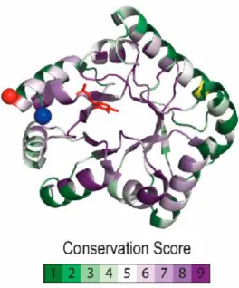
Fig 1.8: Pyridoxal phosphate binding protein (PLPBP) residue conservation and distinctive structural features.
Conservation scores were calculated using CONSURF with COG0325 alignment and a phylogenetic tree from the EGGNOG database as query. The color code illustrates lowest to highest conservation in a green to purple gradient, with yellow for non-informative residues, plotted over the Synpcc7942_2060 (PDB 5NM8) structure. The N-terminal and C-terminal ends are indicated with a red and blue sphere, respectively. The PLP molecule is colored in red. (Labella et al, 2020)
22
Generally, these proteins are monomeric as revealed by size exclusion chromatography analysis of a purified PipY from S. elongatus. Human protein PLPBP expressed in E. coli by Tremino and collaborators has been purified and analysed using the size exclusion chromatography revealing the presence of a second pick corresponding to the dimer. A new preparation of the protein in presence of β-mercaptoethanol reduced the formation of the dimeric fraction, suggesting the presence of an equilibrium where the dimer is formed by the covalent bond between two monomers (Tremiño, Forcada-Nadal, and Rubio 2018).
Proteins present in the COG0325 family binds PLP and are structurally similar to other Fold-type III PLP dependent enzymes, however, they don’t show activity against amino acids and the main substrates of the PLP dependent enzymes (Ito et al. 2013).
A PLP homeostasis role was hypothesized for PLP binding proteins because a knockout of the genes coding for these proteins led to different phenotypes such as: PN toxicity and an imbalance in the amino/ketoacids pools in E. coli,
Fig 1.9: Alignment of short regions of the PLPHP sequence (in single letter amino acid code)
The individual species and the taxonomic group are given to the left. The Clustal 𝜔 alignment has been manually modified to allow the stacking of the same secondary structure elements revealed in crystal. (Tremino et al, 2018)
23
sensitivity to antibiotics that target essential PLP holoenzymes in S.
elongatus and, in human, mutation on the gene coding for PLPBP cause
vitamin B6 dependent epilepsy (Darin et al. 2016; Labella et al. 2020; Plecko et al. 2017; Tremiño et al. 2018).
Deletion of yggS gene in E. coli leads to a pyridoxine sensitiveness as observed by Prunetti and collaborators that can be complemented by an in
trans expression of Zea mays COG0325 family protein (Fig 1.10). The
recovery of the ΔyggS phenotype using a homologous protein was studied independently by Ito and Darin with the respective collaborators. The amino acids relative concentrations in E. coli are modified by yggS knockout and this phenotype can be reverted, along with the phenomenon of the PN toxicity, through complementation with other PLP Binding Protein encoding genes as human (PLPBP), yeast (YBL036C) and Bacillus subtilis (ylmE) variants suggesting a conserved role for these proteins (Darin et al. 2016; Ito et al. 2013). PN toxicity was associated to a toxic accumulation of PNP due to the loss of activity of PNPOx in ΔyggS background, but purified PNPOx from ΔyggS E. coli have shown full activity (Prunetti et al. 2016).
24
Although in several organism PLPBP is not essential for life (Ito et al. 2013; Labella et al. 2017; Prunetti et al. 2016), in humans a court of people has been reported thus far with vitamin B6 dependent epilepsy associated with PLPBP mutation (Darin et al. 2016; Plecko et al. 2017). The mutations found in these patients are both truncating and splicing mutation that lead to PLPBP
Fig 1.10: Toxic effect of PN on the E. coli ΔyggS strain and the universality of YggS function.
(a) DyggS (VDC6594) lawn and drops of PN at concentrations of 5.9 mM (i), 590 mM (ii), 295 mM (iii) or 59 mM (iv);
PN at concentrations of 5.9 mM (i) or 590 mM (ii) was used in experiments (b–f). (b) ΔyggS pBAD18 lawn in presence of 0.2 % arabinose; (c) ΔyggS in pBAD24yggSEc (pBY291.3) lawn in presence of 0.2 % arabinose; (d) ΔyggS transformed with pBAD18 LOC100191932 (pBY298.7) lawn in presence of 0.2 % arabinose; (e) ΔyggS transformed with pBAD18 LOC100191932 in absence of 0.2 % arabinose; (f) ΔyggS transformed with pBEY YBL036C (pBEY329.12) in the presence of 50 ng aTet ml21. The arrows indicate the presence of the ring of toxicity. (Prunetti et al, 2016)
25
loss of function with early in life nervous system pathology as epilepsy with developmental delay if not treated with vitamin B6 supplementation (Tremiño et al. 2018).
Given its diffusion between the living beings and the conservation of PLPBP it is clear that this protein, whose exactly function has not been yet unveiled, has a crucial role in the organisms and given the phenotype when it is mutated, PLPBP is probably directly involved in PLP metabolism.
It was hypothesised the role of PLPBP as a “sponge” for the homeostasis of the PLP in order to have a reservoir, but this role must be confirmed.
Recent studies of Vu and collaborators, have suggested a role of YggS in
Salmonella enterica as an enhancer of the PLP-PMP cycling on PNPOx
26 1.6 Aim of the thesis
Pyridoxal 5'-phosphate (PLP) is the bioactive form of vitamin B6 and is involved in more than 150 enzymatic reactions. This cofactor is involved in several metabolic pathways, such as the synthesis of neurotransmitters, amino acid metabolism, glucose homeostasis, and heme metabolism. In different organisms, PLP has also biological functions other than catalysis, such as those of reactive oxygen species (ROS) scavenger in plants and transcriptional regulator in Eubacteria.
This work aims to deepening our understanding of vitamin B6 metabolism in
Escherichia coli and involves the study of the physiologic effect of deletions
of several genes related to PLP metabolism, and the effect of vitamin B6 vitamers on E. coli growth.
PLP-binding protein encoded by the gene yggS. The effort of these studies has permitted to explore which is its physiological role of YggS in order to assign a specific function to this protein. The hypothesis is that YggS could be a PLP homeostatic protein that acts as a “sponge” to reduce free PLP concentration, and could also act as a carrier protein, able to transfer PLP to the apo-PLP-dependent enzymes that need it as a cofactor. To this aim, we studied the transfer mechanism and the possible interaction of YggS with the PLP-dependent enzyme serine hydroxymethyltranferase.
27
28 Materials
2.1 Materials, bacterial strains, plasmids and reagents
All the chemicals used in this study were purchased from Sigma-Aldrich (Saint Luis, MO, USA) and Carlo Erba (Milan, Italy) except for the chromatographic gels, infact DEAE-sepharose and Phenil-sepharose were purchased from GE Healthcare (Milwaukee, WI, USA) and NiNTA-agarose from Quiagen.
The oligonucleotides used for in situ mutagenesis were synthetized by Metabion international AG (Planegg, Germany) and the DNA sequencing were operated by Microsinth AG (Balgach, Switzerland).
Growth media and supplements
Media utilised for bacterial cultures were previously sterilised using relative temperature and pressure of 120°C and 1 atm for 20 minutes in autoclave. Bacteria were grown both in liquid and solid medium. Selective liquid medium was only added with the appropriate antibiotic when the temperature decreased under 55°C. Selective solid medium was added of 15g/L of agarose before sterilization and then supplemented with the necessary antibiotic when temperature reached 55°C.
Luria Bertani (LB) growth medium is a complex medium containing all the necessary nutrients for an optimal growth of E. coli. LB medium used in our experiments was composed of: 10g/L Tryptone, 5g/L Yeast extract, 5g/L NaCl.
Minimal medium M9 is a defined medium used to grow cells under nutritional control and it was composed of: 0.6% anhydrous Na2HPO4, 0.3%
29
KH2PO4, 0.05% NaCl, 0.1% NH4Cl, 0.4% glucose, 2 mM MgSO4, 0.1 mM CaCl2.
Growth media were supplemented with the following antibiotic doses: 40µg/mL Kanamycin or 100µg/mL Ampicillin.
Bacterial strains
• Escherichia coli DH5α strain was used for transformation and selection of mutants;
• Escherichia coli BL21(DE3) was used for the expression of WT YggS because this gene is already present in the genome and eventual recombination does not affect the correct protein expression. This DE3 strain contain a lysogen of the bacteriophage λDE3 under control of the promoter LacUV5 so a T7 RNA polymerase gene can be expressed on request;
• Escherichia coli HMS174(DE3) is a non-recombinant strain, in this study was used to express all the mutant forms of YggS. As the previous this is a DE3 E. coli strain so, it contains the same T7 RNA polymerase gene;
• Escherichia coli GS1993 is a ΔGlyA strain that in this study was used for the expression of eSHMT both WT and mutants after its transformation with the plasmid pBSglyA;
• Escherichia coli Rosetta (DE3)/pET19hPLPP. • Escherichia coli GC10/pBAD24yggS.
• The following strains of Escherichia coli were bought from the Keio collection and used for the growth curves:
o Escherichia coli BW25113 o Escherichia coli BW25113 ΔyggS
30
o Escherichia coli BW25113 ΔpdxH o Escherichia coli BW25113 ΔpdxJ o Escherichia coli BW25113 ΔpdxK o Escherichia coli BW25113 ΔpdxY
Plasmids
- pET28a(+)/yggS WT and mutant forms; - pBSglyA WT and mutant forms;
- pET19/hPLPP; - pBAD24/yggS WT - pBAD24/yggS K38A - pBAD24/yggS S132A - pBAD24/yggS R229A
31
Table 2.1 Oligonucleotides for molecular cloning
YggS
yggS_NcoI_for GGCCATGGACGATATTGCGCATAACCTG yggS_XhoI_rev GGCTCGAGTTTTTTAGAGTAATCACGCGCACC YggS N-His_up CGGCATATGAACGATATTGCGCATAACCTG YggSN-His_down GCGAATTCTTATTTTTTAGAGTAATCACGCGCAC K36A_for CAGTCAGTgcaACAAAACCTGCGAGCG K36A_rev GGTTTTGTtgcACTGACTGCAAGCAGC K36/38A_for TGCAACAGCGCCTGCGAGCGCCATC K36/38A_rev CTCGCAGGCGCTGTTGCACTGACTG K89A_for CAGTCTAATGCGAGCCGCCTGGTGGCA K89A_rev CAGGCGGCTCGCATTAGACTGCAACGG E134A_for AGTGATgcaAACAGTAAGTCCGG E134A_rev CTTACTGTTtgcATCACTAATGTTAA K137A_for AACAGTgcgTCCGGGATTCAAC K137A_rev CCCGGAcgcACTGTTTTCATC K233/234A_for GCGTGATTACTCTGCAGCACTCGAGCACCACC K233/234A _rev CGCACTAATGAGACGTCGTGAGCTCGTGGTGG ΔK233/234A_for TAAGAATTCGAGCTCCGTCGACAAGCTTGC ΔK233/234A _rev CGGAGCTCGAATTCTTAGAGTAATCACGCGC
eSHMT
D89A_for TCGGCGCTGCCTACGCTAACGTCCAGCC
D89A_rev GTTAGCGTAGGCAGCGCCGAACAGTTC
K354A_for ACGATCCGGCGAGCCCGTTTGTGACCTC
32 Table 2.2 Oligonucleotides for RT-qPCR
QpdxB_for ATTGGTTTTTCCGCTGCACC QpdxB_rev CGCGCTGGACTAACTCATCC QecopdxH_for ATGGTGGTCGCTACCGTGG QecopdxH_rev CTATCACGCGGGCGGCTATG QpdxJ_for TGACCATATCGCTACGCTGC QpdxJ_rev CCAGGCAGCAAAAATGTGGC QpdxK_for TGCCGTGCCTGCTATCAAAC QpdxK_rev AGGATGGTCTTTGCGTAGCG QecopdxY_for AATGGACTGGCTGCGTGATG QecopdxY_rev GCACATGAAACTCTGCGACACC QyggS_for CATACCATCGACCGTTTGCG QyggS_rev GCCATTTGGCGTGCAACTTC rRNA16S_for GACGTTACCCGCAGAAGAAGC rRNA16S_rev GTGGACTACCAGGGTATCTAATCC
33 Methods
2.2 Molecular cloning of WT yggS
The coding sequence of the yggS gene from E. coli was amplified by PCR using primers yggS_NcoI_for and yggS_XhoI_rev (Table 2.1) and pBAD24yggS as template. The amplified 700 bp segment was inserted into a pET28b(+) vector between NcoI and XhoI restriction sites. In this way the
yggS gene contains the HisTAG coding region at the C-terminal terminus.
This new construct, named pET28-YggS, was used to transform E. coli BL21(DE3) competent cells for protein expression. The nucleotide sequence of the insert was determined for both strands and no differences were detected with respect to the sequence reported in data banks. In order to produce an YggS protein form with the HisTAG coding region at the N-terminal terminus, the same steps were performed. Specifically, the yggS gene were amplified using primers YggS N-His_up and YggS N-His_down and the same pBAD24yggS as template. The resulting fragment was inserted into pET28b(+) vector between NdeI and EcoRI restriction sites. Summarizing, two versions of YggS were produced where the His-TAG is present at the N- and C-terminal, respectively.
2.3 Site-directed mutagenesis
YggS
The yggS gene inserted into the pET28-YggS plasmid was used as template in site-directed mutagenesis reactions carried out using two complementary oligonucleotides containing the mutation as primers (K36A_for and K36A_rev, K36/38A_for and K36/38A_rev, K89A_for and K89A_rev, E134A_for and E134A_rev, K137A_for and K137A_rev, K233/234A_for
34
and K233/234A _rev, ΔK233/234A_for and ΔK233/234A _rev). Sequence of the constructs was verified by DNA sequencing. Expression and purification of YggS mutant forms was performed as described for wild-type YggS.
eSHMT
The glyA gene inserted into the pBS/glyA plasmid was used as template in site-directed mutagenesis reactions carried out using two complementary oligonucleotides containing the mutation as primers (D89A_for and D89A_rev, K354A_for and K354A_rev).
2.4 RNA analysis through RT-qPCR
RNA was isolated from three independent cultures (three biological replicates) grown up 1 OD600 in M9 minimum medium added with 0.1 mM PL, 0.1 mM PN and 0.5% casaminoacids (CAA), using the NucleoSpin RNA kit from Macherey and Nagel. RNA concentration and quality were evaluated by measuring the OD at 260 nm and the ratio 260/280 nm, respectively, and by electrophoresis on 1.2% agarose gels. RT-qPCR reactions were performed in two steps. Reverse transcription of DNase-treated RNAs (0.5 g) was carried out using the Maxima® first strand cDNA synthesis kit (Thermo Scientific) with the random primers provided in the kit. Real Time PCR was performed on a Real Time PCR Instrument (Mx3000P QPCR system, Agilent technologies) with a two-step reaction using Maxima® SYBR Green qPCR Master mix (Thermo Scientific) and the oligonucleotides reported in Table 2.2. The relative expression of each target gene was determined by the Pfaffl method (Pfaffl, 2001) using the rRNA 16S gene as normalizer. The fold induction resulting from the different samples was reported as averaged
35
values, and the error (as confidence interval) was measured as standard deviation. The statistical significance of the obtained values was measured using the P-value, calculated using the Student’s t-test.
2.5 Expression and purification of the proteins
YggS
An overnight culture (50 mL) of E. coli BL21(DE3) cells transformed with plasmid pET28-YggS was used to inoculate 4 L of Luria–Bertani broth containing kanamycin (40 mg·L-1). Bacteria were allowed to grow for approximately 5 h at 37 °C (until their OD600 reached to 0.6), then the growing temperature was lowered to 28 °C and the expression of YggS induced with 0.2 mM isopropyl thio-β-D-galactopyranoside (IPTG). Bacteria were harvested after 18 h and suspended in 150 mL of 20 mM potassium-phosphate buffer, pH 7.3, containing 5 mM EDTA, PLP and 2 mg/ml lysozyme. Cell lysis was carried out by sonication on ice (3-min in short 20-s pulses with 20-s intervals). Lysate was centrifuged at 12000 g for 30 min and the pellet was discarded. The supernatant was precipitated by addition of ammonium sulphate to 30% saturation, centrifuged, and the resulted pellet was discarded. Ammonium sulfate was added to the resulting supernatant so to reach the 70% saturation concentration. The sample was centrifuged at 12000 g for 30 min and the pellet suspended in the 20 mM potassium-phosphate buffer, pH 7.3 to dilute the ammonium sulfate to a final concentration less than 10%. The sample was loaded onto a 10 ml Ni-NTA Superflow (QIAGEN) column, previously equilibrated with 20 mM potassium phosphate buffer, pH 7.3 containing 10 mM imidazole. The column was washed with 50 mL of the same buffer, 50 mL of the same
36
buffer containing 20 mM imidazole, and eluted with a linear 20 to 300 mM imidazole gradient (the buffer containing imidazole was adjusted to pH 7.3 with HCl). Fractions containing the expected protein (molecular mass ~26.000 Da) were pooled and dialyzed overnight against 50 mM Na-HEPES buffer pH 7.6, containing 5 mM β-mercaptoethanol. The protein was purified to homogeneity, as judged by SDS-PAGE analysis and the purification yield was about 35 mg YggS per litre of bacterial culture. Recombinant YggS was stored at 4 °C. Protein subunit concentration was calculated using a theoretical extinction coefficient at 280 nm of 16960 M-1 cm-1 (calculated with the Expasy ProtParam tool). PLP content was calculated after the addition of 0.2M NaOH, measuring the absorbance at 388 nm.
eSHMT
An overnight culture (50 mL) of E. coli GS1993 pBSglyA cells was used to inoculate 4 L of Luria–Bertani broth containing ampicillin (100 mg·L-1). Bacteria were allowed to grow for approximately 18h at 37 °C, then they were harvested and suspended in 120 mL of 30 mM TRIS hydrochloride buffer, pH 7.6, containing 20 mM EDTA, PLP and 2 mg/ml lysozyme. Cell lysis was carried out by sonication on ice (3-min in short s pulses with 20-s interval20-s). Ly20-sate wa20-s centrifuged at 12000 g for 30 min and the pellet wa20-s discarded. The supernatant was precipitated by addition of ammonium sulfate to 50% saturation, centrifuged, and the precipitate was discarded. Ammonium sulfate was added to the resulting supernatant so to reach the 75% saturation concentration. The sample was centrifuged at 12000 g for 30 min and the pellet suspended in the 20 mM potassium-phosphate buffer, pH 7.2, containing 100 μM EDTA and 1 mM β-mercaptoethanol. Then the sample was dialyzed overnight against the 20 mM potassium-phosphate buffer, pH 7.2, containing 100 μM EDTA and 1 mM β-mercaptoethanol. The
37
dialyzed sample was loaded onto a DEAE-Sephadex column which had been equilibrated with 20 mM potassium-phosphate, pH 7.2. The column was washed with the equilibrating buffer until the absorbance at 280 nm was below 0.2. The eSHMT enzyme was then eluted with a linear salt gradient (0 to 400 mM sodium chloride). The mixing chamber contained 300 ml 20 mM potassium phosphate, pH 7.2, and the reservoir contained 200 ml of 100 mM potassium phosphate with 400 mM sodium chloride, pH 6.4. The yellow fractions, showing the absorbance bands at 420 nm (corresponding at the PLP internal aldimine) were pooled, and the protein was precipitated by the addition of ammonium sulfate to 75% of saturation. After centrifugation, the precipitate was dissolved in 50 mM potassium-phosphate buffer, pH 7.2, and loaded onto a phenyl-sepharose column which had been equilibrated with the same buffer containing 40% ammonium sulfate. The protein was then eluted with a linear 40% to 0 ammonium sulfate gradient. The yellow fractions were pooled and ammonium sulfate was added to the protein sample to the 75% saturation concentration. Then the precipitate was dissolved in 20 mM potassium-phosphate buffer, pH 7.2, containing 200 μM DTT and 100 μM EDTA and dialysed over night against the same buffer. The purified protein was pure as determined by sodium dodecyl sulfate-polyacrylamide gel electrophoresis. Protein subunit concentration (~45.000 Da M.W.) was calculated using a theoretical extinction coefficient at 280 nm of 44884 M-1 cm-1 and 42790 (calculated with the Expasy ProtParam tool) for the holo and apo forms, respectively. PLP content was calculated after the addition of 0.2M NaOH, measuring the absorbance at 388 nm.
hPLPP
An overnight culture (50 mL) of E. coli Rosetta (DE3)/pET19hPLPP cells was used to inoculate 4 L of Luria–Bertani broth containing ampicillin (100
38
mg·L-1). Bacteria were allowed to grow for approximately 5 h at 37 °C (until their OD600 reached to 0.6), then the growing temperature was lowered to 28 °C and the expression of hPLPP induced with 0.2 mM isopropyl thio-β-D-galactopyranoside (IPTG). Bacteria were harvested after 18 h and suspended in 50 mM sodium-phosphate buffer, pH 8, containing 300 mM sodium chloride and 2 mg/ml lysozyme. Cell lysis was carried out by sonication on ice (3-min in short 20-s pulses with 20-s intervals). Lysate was centrifuged at 12000 g for 30 min and the pellet was discarded. The supernatant was loaded onto a 10 ml Ni-NTA Superflow (QIAGEN) column, previously equilibrated with 50 mM sodium-phosphate buffer, pH 8.0 containing 300 mM sodium chloride and 10 mM imidazole. The column was washed with 50 mL of the same buffer and the protein was eluted with a linear 10 to 300 mM imidazole gradient. Fractions containing the expected protein (molecular mass ~31.000 Da) were pooled and dialyzed overnight against 50 mM Na-HEPES buffer pH 7.2, containing 150 mM sodium chloride. The protein was purified to homogeneity, as judged by SDS-PAGE analysis. Protein subunit concentration was calculated using a theoretical extinction coefficient at 280 nm of 18450 M-1 cm-1 (calculated with the Expasy ProtParam tool).
2.6 Preparation of the apo-forms of the proteins
Both apo-YggS and apo-SHMT were prepared using the same protocol. In order to remove the molecules of PLP bound through a Schiff base to the protein YggS and SHMT, 40 mg of both the proteins have been treated separately with L-Cysteine. Thanks to the reactivity of the PLP with the thiol group of the cysteine, there is the formation of a compound called Thiazolidine (Fig. 2.1) that is more stable than the internal aldimine between Lysine-PLP but less affine so it dissociates from the protein.
39
An amount of 40 mg of purified YggS or SHMT was added to the buffer 20 mM potassium-phosphate (KPi), pH 7.3, containing 5 mM β-mercaptoethanol, 25% ammonium sulphate. The solution was added with 100 mM L-cysteine and kept under mixing until it became clear.
The protein solution was loaded in a chromatographic column containing 7 mL of Phenil Sephadex (GE Healtcare) and washed with the same buffer containing only 50 mM L-cysteine. A second wash of the chromatographic column was done with the same buffer without L-cysteine in order to separate the protein from the thiazolidine. The separation of the thiazolidine were monitored measuring the absorption spectrum at 333 nm, the major absorbance wavelength for thiazolidine.
Elution of the apo-protein happened using the buffer 20mM potassium-phosphate (KPi), pH 7.3, 5 mM β-mercaptoethanol and the fractions containing the protein were selected using spectrophotometric and SDS-PAGE analysis and collected. The obtained proteins were dialyzed overnight against 50 mM NaHEPES buffer pH 7.6 containing 5 mM β-mercaptoethanol.
Figure 2.1Reaction scheme showing the reaction between PLP bound to
40 2.7 Size Exclusion Chromatography
Gel filtration of YggS wild-type and mutant forms was performed on a Superdex 200 10/300 GL column (GE Healthcare, Little Chalfont, UK) at room temperature and at a flow rate of 0.5 ml/min in 50 mM Na-HEPES buffer pH 7.6, containing 150 mM NaCl and 0.5 mM DTT. Elution profiles were obtained from absorbance at 280 nm. Calibration was performed with aldolase (158 kDa) conalbumin (75 kDa) and ovalbumin (44 kDa), carbonic anhydrase (29 kDa), RNase (17.7 kDa). Protein samples (100 µl) were subjected to chromatography at concentration of different concentrations.
2.8 Spectroscopic measurements
Concerning YggS, the spectrophotometric measurements were carried out at 20 °C in 50 mM Na-HEPES buffer pH 7.6. UV-visible spectra were recorded with a Hewlett-Packard 8453 diode-array spectrophotometer. Far-UV (190-250 nm) CD spectra were measured with a Jasco 710 spectropolarimeter equipped with a DP 520 processor using 0.2 and 1 cm path length quartz cuvettes and results were expressed as ellipticity []. Far UV measurements were carried out in 20 mM KPi buffer pH 7.6. Protein samples were heated from 30° C and 80°C, with a heating rate of 1°C per min controlled by a Jasco programmable Peltier element, monitoring the dichroic activity at 220 nm every 0.5 °C. Denaturation curves were fitted with the sigmoidal trace described in the Eqn. 1.
41
2.9 Determination of the dissociation constant of PLP from YggS
PLP binding equilibria were analyzed taking advantage of the protein intrinsic fluorescence quenching observed upon the binding event. The dissociation constant of the binding equilibrium was then calculated from saturation curves obtained measuring the protein fluorescence emission intensity as a function of increasing PLP concentration. The cofactor (from 0 to 2 M and from 0 to 15 M for wt and mutant forms, respectively) was added to apoenzyme samples (50 nM wt and 200nM mutants) at 25 °C in 50 mM Na-HEPES pH 7.6. Preliminary experiments showed that the binding equilibrium was established within the mixing time. Fluorescence emission spectra (300 - 450 nm; 5 nm emission slit) were recorded after mixing PLP and apoenzyme with a FluoroMax-3 Jobin Yvon Horiba spectrofluorimeter, with excitation wavelength set at 280 nm (3 nm excitation slit), with a 1 cm path length quartz cell. For data analysis, we have removed the contribution of PLP at the fluorescence spectrum of the protein samples. Data were analyzed according to a quadratic equation, Eqn. 2.
42 2.10 Kinetic studies
2.10.1 PLP transfer studies
PLP transfer studies were performed with two different methods because the mutation on lysine 354 of the eSHMT cause a loss of activity probably due to the interaction of this residue with the tetrahydrofolate.
The kinetics were studied using a Hewlett-Packard 8453 diode-array spectrophotometer and monitoring in both the assay the variation of absorbance at 340 nm over time.
PLP transfer kinetic assay with L-serine and THF
For this assay 20 µM apo-YggS and 20 µM PLP have been mixed mixed in a vial, then in a different vial have been mixed 70 µM apo-eSHMT and the wanted concentration of hPLPP. All the volumes in the vials were adjusted to have 200 µL of PLP transfer reaction and keep at 37 °C for all the experimental time.
The cuvette has been filled with 598 µM of: 50mM NaHEPES pH 7.6, 25mM L-Serine, 50 µM Tetrahydrofolate (THF), 0,5 µM Methyltetrahydrofolate dehydrogenase (Mtd), 250 µM NADP+.
At different times, the absorbance variation was measured for one minute in order to have a baseline given by the oxidation of the THF; after the minute, 2 µL of the protein reaction mix that was prepared were added into the cuvette and after 3 minutes have been measured the initial velocity of the eSHMT. This enzyme catalyses the reduction of NADP+ to NADPH in order to convert L-Serine in Glycine. The coupled reaction uses Mtd to convert
43
methyltetrahydrofolate and regenerates NADP+ causing an absorbance increase at 340nm.
The initial velocity analysed in a graph over time of measurement, permit to study the kinetic of the reactivation of the apo-eSHMT.
PLP transfer kinetic assay with L-allo-threonine
For this assay 20 µM apo-YggS and 20 µM PLP have been mixed mixed in a vial and in a different vial have been mixed 20 µM apo-eSHMT. All the volumes in the vials were adjusted to have 200 µL of PLP transfer reaction and keep at 37 °C for all the experimental time.
The cuvette has been filled with 558 µL of:
50mM NaHEPES pH 7.6, 10 µL of NADH (8 mg/mL), 10 µL of Alcool dehydrogenase (ADH) (4 mg/mL), 20 µL of L-allo-threonine (0,5M).
At different times, the absorbance variation was measured for one minute in order to have a baseline; after the minute, 2 µL of the protein reaction mix that was prepared were added into the cuvette and after 3 minutes have been measured the initial velocity of the eSHMT. This enzyme has a threonine aldolase activity and through the oxidation of NADH to NAD+ it converts threonine in ethanol and glycine. The coupled reaction uses ADH to convert ethanol in acetaldehyde and regenerates NAD+ to NADH causing an absorbance decrease at 340nm.
The initial velocity analysed in a graph over time of measurement, permit to study the kinetic of the reactivation of the apo-eSHMT.
44 2.11 Data analysis
Eqn.1: 𝜃220 = (∆𝜃 𝑇𝑛
𝑇𝑚𝑛+ 𝑇𝑛) + 𝑐𝑜𝑛𝑠𝑡
θ220: measured ellipticity at 220 nm ∆θ: maximum ellipticity change T: temperature expressed in Celsius
Tm: apparent melting temperature
n: steepness of the sigmoid curve
Eqn.2:
Frel: relative fluorescence measured
F0: fluorescence in the absence of PLP
Finf: fluorescence at infinite PLP concentration
[P]: total protein subunit concentration
[PLP]: residual cofactor concentration bound to the apo-protein
[PLP]x: PLP concentrations used for the titration
Kd: dissociation constant of the equilibrium HOLO APO + PLP
45 2.12 Software used for figures and graphs
All the figures and the graphs reported in this work (except for those taken from the literature and whose reference are reported in the legends) were made using the following software or online services:
• Schemes and enzymatic pathways were realized using Microsoft Office PowerPoint;
• Molecular structures were drawn using Chemdraw 2D;
• The graphs of the reported data were made using GraphPad Prism 7; • The promoter sequence alignment reported in Figure 3.23 was made
46
47
3. Regulation of vitamin B6 metabolism in E. coli
Given the importance of the pyridoxal 5’ phosphate as a cofactor for the most different reactions in E. coli, and given the importance of its homeostasis and regulation due to the toxicity of its free form, in this work we decided to undertake several studies aimed to a better understanding of the mechanisms of regulation of the intracellular concentration of this molecule.
In the following chapters, we studied the different possible mechanism of regulation of PLP. Starting from the identification of E. coli proteins that bind PLP, and elucidating the structures of the genes coding for them. Then, we analyzed the transcriptional levels of the previously identified genes, and hypotesized a possible regulation system that involve the promoter sequences of these genes. As last, we have performed an analysis on the translational level of the genes, and the relative protein abundance in the cell.
3.1 PLP-binding proteins in E. coli
Almost 1.5% of all genes in most prokaryotic genomes encode PLP-dependent enzymes (Percudani and Peracchi 2003). They catalyse many different reactions, such as transamination, decarboxylation, racemization, side chain cleavage, β- and γ-elimination or replacement (John 1995). Using the B6 database (Percudani and Peracchi 2003) (http://bioinformatics.unipr.it/B6db), a tool for the description and classification of vitamin B6-dependent enzymatic activities and of the corresponding protein families, all PLP-binding proteins found in
Escherichia coli MG1655 proteome (57 in total) were retrieved and
48
biosynthesis, while 10 are responsible for the biosynthesis of other molecules, such as coenzymes, polysaccharides, or polyamines. Many are catabolic enzymes, and also a putative PLP carrier protein (YggS, see below) and two PLP-dependent transcriptional regulators (YdcR and YjiR, see below) were reported. Importantly 26% of these proteins are essential for E.
coli life, being the related knock out strains unable to grow in minimal
medium. This indicates how PLP is important for bacterial life.
Protein name
Activity UniProt EC Biological
process Growth in minimal medium IlvE branched-chain-amino-acid aminotransferase P0AB80 2.6.1.42 Branched-chain amino acid biosynthesis No IlvA Threonine deaminase P04968 4.3.1.19 Branched-chain amino acid biosynthesis No
CysK cysteine synthase A
P0ABK5 2.5.1.47 Cys
biosynthesis No
CysM cysteine synthase B P16703 2.5.1.47 Cys biosynthesis Yes TrpB tryptophan synthase, β P0A879 4.2.1.20 Trp biosynthesis No LysA diaminopimelate decarboxylase P00861 4.1.1.20 Lys biosynthesis No
Table 3.1. PLP-binding proteins of E. coli.
Cyan: enzymes involved in amino acid biosynthesis; Yellow: enzymes
involved in the biosynthesis of molecules as cofactors, polysaccharides and polyamines; Orange: different catabolic enzymes; Green: enzymes involved in iron-sulphur cluster assembly; Purple: PLP-dependent transcriptional regulators; Grey: a putative PLP carrier protein; White: putative amino transferases.