Case Report
A Rare Case of Giant Mesenteric Lipoblastoma in a 6-Year-Old
Child and Review of the Literature
Maria Enrica Miscia
,
1,2Gabriele Lisi
,
1,2Giuseppe Lauriti
,
1,2Angela Riccio,
1,2Dacia Di Renzo
,
1Valentina Cascini,
1and Pierluigi Lelli Chiesa
1,21Department of Pediatric Surgery,“Spirito Santo” Hospital, Pescara, Italy
2Department of Medicine and Aging Sciences,“G. d’Annunzio” University, Chieti-Pescara, Italy
Correspondence should be addressed to Giuseppe Lauriti; [email protected] Received 31 January 2020; Accepted 16 July 2020; Published 24 July 2020
Academic Editor: Ragheb Hasan
Copyright © 2020 Maria Enrica Miscia et al. This is an open access article distributed under the Creative Commons Attribution License, which permits unrestricted use, distribution, and reproduction in any medium, provided the original work is properly cited.
Giant mesenteric lipoblastoma is a rare benign tumor arising from the adipocytes. It can mimic malignant tumors, and its diagnosis is difficult before surgery. Imaging studies could lead the diagnosis but not confirm it. Those tumors arising in the abdomen are usually larger and can cause symptoms of compression. Surgical excision is the treatment of choice, and a long-term follow-up is necessary to detect local recurrences. Only a few cases of lipoblastomas arising from the mesentery are reported in literature. We present a case of a rare giant lipoblastoma arising from the mesentery of a 6-year-old girl, with a history of postprandial abdominal pain.
1. Introduction
Adipose tumors are rare in childhood and represent 6% of all
the neoplasms of the soft tissue.
They can be classified into malignant (liposarcoma) and
benign (lipoma, lipoblastoma, lipoblastomatosis) [1, 2].
Lipoblastoma is a rare, encapsulated tumor arising from
the embryonal fat tissue [3].
It is more frequent in children younger than 3 years old,
with a male to female ratio of 3 : 1 [1, 3].
Common localizations are extremities and trunk.
Intra-abdominal lipoblastoma is extremely rare (<7%) and
mesen-teric localization is exceptional [2–4].
We present a case of giant mesenteric lipoblastoma in a
girl, reviewing and discussing pertinent literature.
2. Case Report
A 6-year-old girl came to our attention for a 2-month-lasting
postprandial abdominal pain.
Her medical history was suggestive of constipation for 2
years. A painless, mildly distended, not tender abdomen
was palpable on physical examination.
An abdominal ultrasound showed a hypoechoic
abdominal mass of
9 × 4 cm in size, not vascularized at
the color-Doppler study and well separated from the
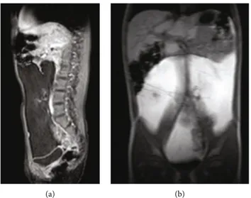
adja-cent organs. In the suspicion of a lipoma, the girl
under-went an abdominal magnetic resonance imaging (MRI),
which con
firmed the presence of an encapsulated
fatty-dense mass (
20 × 4 × 18 cm), occupying the entire
abdom-inal cavity (Figures 1(a) and 1(b)).
Blood exams (complete blood count, C-reactive protein,
and liver function tests) and tumoral markers
(alfa-fetopro-tein, beta-human chorionic gonadotropin, lactic
dehydroge-nase,
carcinoembryonic
antigen,
and
neuron-specific
enolase) were within ranges.
An explorative laparoscopy was then performed. It
showed a huge fatty mass arising from the ileal mesentery
and stretching the ileal loops. The mass was completely
excised through a minilaparotomy, even if a resection of
a tract of intestine involved was necessary. The lesion
Volume 2020, Article ID 3018065, 5 pages https://doi.org/10.1155/2020/3018065
was
21 × 19 × 7 cm and weighted 1,236 g (Figures 2(a) and
2(b)). Bowel continuity was restored through a primary
end-to-end anastomosis.
Histopathological analysis showed adipocytes at di
fferent
stages of maturation with focal myxoid areas and con
firmed
the diagnosis of giant abdominal lipoblastoma.
Postoperative period was uneventful with excellent
esthetic results. No recurrence was noticed at a 1-year
ultra-sonographic follow-up (Figure 3).
3. Review of the Literature
A review of the English literature was performed using a
de
fined search strategy. Scientific databases (PubMed,
Med-line, Cochrane Collaboration, and Scopus) were screened
looking for studies reporting on mesenteric lipoblastoma in
children. MeSH headings and terms used were
“lipoblastoma
AND children”. Reference lists were examined to identify
rel-evant cross-references. Of 408 titles and abstracts, 22 case
(a) (b)
Figure 1: (a, b) MRI ((a) T1 weighted, (b) Thrive sequences) showing the mass occupying the entire abdominal cavity, compressing and displacing the bowel, the inferior vena cava, and common iliac veins.
(a) (b)
Figure 2: (a, b) Intraoperative findings: the mass comes from the mesentery, stretching the involved ileum (a), which was resected together with the mass (b).
Table 1: Review of the English literature. Auth or Year Sex Age Symptoms Size (cm) Position Surgery Follow-up Recurrence Chu ng and Enzinger [9] 1973 F 2 yrs n.a. n.a. n.a. Excision mass+23 cm small bowel n.a. No Friedman et al. [10] 1981 F 13 mo Palpable mass n.a. n.a. n.a. n.a. No Stringel et al. [11] 1982 M 13 mo Abdominal distension, palpable mass 12 × 10 :8× 17 Ileum Excision mass+26 cm small bowel 1 yr No Jimenez [12] 1986 M 11 mo Abdominal distension, palpable mass 12 × 10 × 0: 8 Jejunum Excision mass+part small bowel 3 yrs No Zanetti [13] 1988 F 4 yrs Pain, vom iting 18 n.a. Exci sion mass+part intestine 2 yrs No Denat h [14] 1988 F 2 yrs Abdomina l distension n.a. n.a. n.a. n.a. No Prand o et al. [15] 1990 M 2 yrs Abdominal distension, palpable mass 23 × 19 × 9 n.a. Excision mass n.a. n.a. Schul man et al. [16] 1992 M 2 yrs Abdominal distension, palpable mass n.a. n.a. Excision mass n.a. n.a. Posey et al. [17] 1998 M 10 mo Abdominal distension, palpable mass 10 ×6×1 3 Ileum Excision mass 2 yrs No O ’Do nnell et al. [18] 2000 F 5 mo Abdomina l distension, diarrhea 14 :5 × 11 × 10 :5 Transverse colon Excision mass 6 mo No Mo et al. [19] 2003 F 16 mo Abdominal distension, palpable mass 10 ×9×1 3 Ileum Excision mass+26 cm small bowel n.a. n.a. Al-Salem and Al-Nazer [8] 2003 M 2 yrs Obstruction, midgut volvulus 8×6×5 Ileum Excision mass n.a. n.a. Jung et al. [20] 2005 M 17 mo n.a. 9: 7×7×5 :5 n.a. n.a. 23 mo No Cudnik et al. [5] 2008 M 3 yrs Abdomina l distention, fullness, intermittent pain 15 × 4: 5× 10 Jejunum Excision mass+part small bowel 1 yr No Yu et al. [7] 2009 F 7 yrs Obstruction, midgut volvulus 10 ×8×6 Jejunum Excision mass+part small bowel 1 mo No Ta ng et al. [21] 2009 M 4 yrs Intermittent abdomi nal pain 18 × 15 × 10 Ileocecal Excision mass 4 mo No Jia and Zhang [22] 2009 M 4 yrs n.a. n.a. n.a. n.a. n.a. No Gentimi et al. [4] 2011 M 18 mo Abdominal distension, palpable mass 14 × 11 × 8 Ileum Excision mass+part small bowel 30 mo No Capasso et al. [23] 2014 M 3 yrs Abdominal pain and vomiting n.a. n.a. Excision mass n.a. n.a. Gho sh et al. [24] 2015 F 14 mo Palpable abdominal mass, intestinal obstruction n.a. n.a. Excision mass n.a. n.a. Yang et al. [6] 2016 n.a. n.a. n.a. n.a. n.a. n.a. n.a. No Pres ent case 2019 F 6 yrs Postprandial abdominal pain 21 × 19 × 7 Ileum Excision mass+part small bowel 1 yr No n.a.: not available .
reports were included, reporting data on 22 cases from 1973
to date (Table 1) [4–24]. We found a male to female ratio of
1.3 : 1 (12 M, 9 F; 1 not available). Mean age at diagnosis was
29 ± 21 months, and preferred localization, when reported,
was the ileal mesentery (6/11).
Three out of 18 patients had an acute onset, with volvulus
[7
–9]. None of the patients underwent a laparoscopic
exci-sion of the mass. A resection of small bowel has been
reported in 9/17 cases.
Mean time of follow-up was
16 ± 12 months. No
recur-rences have been reported, when mentioned.
4. Discussion
Lipoblastoma is a rare benign, encapsulated tumor of
adipo-cytes, accounting for 5-30% of all the adipose tumors [3].
When di
ffuse and infiltrative, it is called lipoblastomatosis.
It was
firstly described by Jaffe in 1926 [4].
It arises from the abnormal proliferation of embryonic
fat cells but its etiopathogenesis is not completely
under-stood [1, 3, 25].
It is more frequent in the
first decade of life, with a peak
of incidence in males younger than 3 years (about 90% of all
cases) [1, 3].
These tumors are usually asymptomatic; however, they
can cause a mass e
ffect when reaching considerable
dimen-sions [3, 25].
Lipoblastoma is commonly located at the level of the
trunk and extremities. Intra-abdominal localizations are rare;
nonetheless, intraperitoneal tumors usually reach superior
dimensions [2, 3].
Typical symptoms of abdominal lipoblastomas are
abdominal pain, constipation, and vomiting, secondary to
organ compression. Acute manifestations, secondary to
volvu-lus or intussusception, are also reported, although rare [7
–9].
Imaging studies, especially MRI, are useful to de
fine the
adipose nature, the extent, and tissue involvement of
neopla-sia. However, this study is unable to di
fferentiate among
lipoma, lipoblastoma, and liposarcoma [3, 26, 27]. In fact,
the proportion of the myxoid stroma determines the imaging
appearance of the lesion: a well-defined predominantly fatty
lesion is likely to be a lipoblastoma, while an infiltrative lesion
with a less-represented fatty component could be a
lipoblas-tomatosis [27]. The
first imaging study performed is the
ultrasound; however, an MRI or a computed tomography
scan is usually required to better de
fine the adipose nature
and the relationship with the surrounding organs. De
finitive
diagnosis is secondary to histopathological examination, and
surgical excision is the treatment of choice.
Microscopic features consist of lobular architecture and
myxoid areas with spindle cells and lipoblasts at various
stages of differentiation [28, 29].
Rearrangements of chromosome 8q11 codifying for the
oncogene PLAG1 are found in more than 70% of cases [2–
4, 28–30]. The different molecular alteration found in
adipo-cytic tumors could help in di
fferentiating between lipoma,
lipoblastoma, and liposarcoma [31]. This information could
be useful to get before surgery, especially if the resection of
the tumor requires a long length of intestinal resection.
Recurrence rate is reported to range between 9 and
46%, and it is usually secondary to an incomplete surgical
excision [3, 6, 30].
A problem related to mesenteric lipoblastomas is the
massive size of the mass and its close contiguity to the
mesen-teric vessels and intestinal loops, which makes a purely
lapa-roscopic excision virtually impossible to perform. In
children, laparotomy incision should be as small as possible,
to limit the extent of the scar, as we did in our case.
5. Conclusions
Mesenteric lipoblastoma is a rare benign tumor.
It can be completely asymptomatic or can cause
symp-toms related to a mass e
ffect. Imaging studies are unable to
reach the de
finitive diagnosis; therefore, surgical excision is
the treatment of choice and leads to a de
finitive diagnosis
through the histopathological examination of the specimen.
We stress the importance of a surgery that should be as
mini-invasive as possible, considering the localization and
the size of the mass. A long-term ultrasonographic
follow-up is necessary to detect local recurrences.
Consent
A written informed consent from parents has been achieved.
Disclosure
The current study has been presented as conference abstract
and oral communication at the Joined Meeting of the
Sec-tion of Oncology and the SecSec-tion of Pediatric Urology of
the Italian Society of Pediatric Surgery (17
thMay 2019,
Bologna, Italy).
Conflicts of Interest
The authors show no con
flict of interest.
Authors’ Contributions
Each author gave a substantial contribution for the
prepara-tion of the manuscript.
References
[1] H. Susam-Sen, B. Yalcin, T. Kutluk et al., “Lipoblastoma in children: review of 12 cases,” Pediatrics International, vol. 59, no. 5, pp. 545–550, 2017.
[2] V. Cascini, G. Lisi, G. Lauriti, G. Sindici, and P. Lelli Chiesa, “Giant abdomino-pelvic adipose tumors of childhood,” Pedi-atric Surgery International, vol. 28, no. 1, pp. 89–93, 2012. [3] E. Séguier-Lipszyc, A. Baazov, S. Fichman, S. Ash, and
E. Freud,“Current management of lipoblastoma,” European Journal of Pediatrics, vol. 177, no. 2, pp. 237–241, 2018. [4] F. Gentimi, D. Antoniou, E. Papandreou, A. A. Tzovaras, and
M. Moschovi, “A giant mesenteric lipoblastoma in an 18-month old infant: a case report and review of the literature,” African Journal of Paediatric Surgery, vol. 8, no. 3, pp. 320– 323, 2011.
[5] R. Cudnik, P. A. Efron, M. K. Chen, J. D. Reith, and E. A. Beierle,“Mesenteric lipoblastoma: a rare location in children,” Journal of Pediatric Surgery, vol. 43, no. 12, pp. E5–E7, 2008. [6] C. Yang, S. Wang, J. Zhang, X. R. Kong, Z. Zhao, and C. C. Li,
“An unusual cause of paediatric abdominal pain: mesenteric masses accompanied with volvulus,” The Turkish Journal of Gastroenterology, vol. 27, no. 4, pp. 325–329, 2016.
[7] D. C. Yu, P. J. Javid, K. R. Chikwava et al.,“Mesenteric lipo-blastoma presenting as a segmental volvulus,” Journal of Pedi-atric Surgery, vol. 44, no. 2, pp. E25–E28, 2009.
[8] A. H. Al-Salem and M. Al-Nazer,“Mesenteric lipoblastoma in a 2-year-old child,” Pediatric Surgery International, vol. 19, no. 1-2, pp. 115–117, 2003.
[9] E. B. Chung and F. M. Enzinger,“Benign lipoblastomatosis. An analysis of 35 cases,” Cancer, vol. 32, no. 2, pp. 482–492, 1973.
[10] A. C. Friedman, D. S. Hartman, J. Sherman, E. M. Lautin, and M. Goldman, “Computed tomography of abdominal fatty masses,” Radiology, vol. 139, no. 2, pp. 415–429, 1981. [11] G. Stringel, B. Shandling, K. Mancer, and S. H. Ein,
“Lipoblas-toma in infants and children,” Journal of Pediatric Surgery, vol. 17, no. 3, pp. 277–280, 1982.
[12] J. F. Jimenez,“Lipoblastoma in infancy and childhood,” Jour-nal of Surgical Oncology, vol. 32, no. 4, pp. 238–244, 1986. [13] G. Zanetti,“Benign lipoblastoma: first case report of a
mesen-teric origin,” Tumori, vol. 74, pp. 495–498, 2018.
[14] F. M. Denath,“Case of the season,” Seminars in Roentgenology, vol. 23, no. 4, pp. 241-242, 1988.
[15] A. Prando, S. Wallace, J. L. C. Marins, R. M. Pereira, E. R. de Oliveira, and M. Alvarenga,“Sonographic features of benign intraperitoneal lipomatous tumors in children: report of 4 cases,” Pediatric Radiology, vol. 20, no. 8, pp. 571–574, 1990. [16] H. Schulman, Y. Barki, and Y. Hertzanu,“Case report:
mesen-teric lipoblastoma,” Clinical Radiology, vol. 46, no. 1, pp. 57-58, 1992.
[17] Y. Posey, E. Valdivia, D. L. Persons et al.,“Lipoblastoma pre-senting as a mesenteric mass in an infant,” Journal of Pediatric Hematology/Oncology, vol. 20, no. 6, pp. 580–582, 1998. [18] K. A. O'Donnell, M. G. Caty, J. E. Allen, and J. E. Fisher,
“Lipo-blastoma: better termed infantile lipoma?,” Pediatric Surgery International, vol. 16, no. 5-6, pp. 458–461, 2000.
[19] Y. H. Mo, S. S. Peng, Y. W. Li, and C. T. Shun,“Mesenteric lipoblastoma: case report,” Pediatric Radiology, vol. 33, no. 1, pp. 37–40, 2003.
[20] S.-M. Jung, P.-Y. Chang, C.-C. Luo, C.-S. Huang, J.-Y. Lai, and C. Hsueh, “Lipoblastoma/lipoblastomatosis: a clinicopatho-logic study of 16 cases in Taiwan,” Pediatric Surgery Interna-tional, vol. 21, no. 10, pp. 809–812, 2005.
[21] X. B. Tang, T. Zhang, Y. Z. Bai, and W. L. Wang,“Giant mes-enteric lipoblastoma in a 4-year-old child,” Journal of Pediatric Surgery, vol. 44, no. 4, pp. 859–861, 2009.
[22] H. M. Jia and K. R. Zhang,“Mesenteric lipoblastoma in a 4-year-old,” Pediatric Radiology, vol. 39, no. 10, p. 1126, 2009. [23] R. Capasso, E. Rossi, L. Castelli et al.,“Mesenteric lipoblastoma
and cervical lipoblastomatosis: ultrasound, elastosonography, and computed tomography findings in two children,” Case Reports in Radiology, vol. 2014, Article ID 478252, 4 pages, 2014.
[24] P. Ghosh, R. N. Das, R. Ghosh, C. Datta, and P. K. Mishra, “Lipoblastoma and lipoblastomatosis: a clinicopathological
study of six cases,” Journal of Cancer Research and Therapeu-tics, vol. 11, no. 4, p. 1040, 2015.
[25] J. W. Han, H. Kim, J. K. Youn et al.,“Analysis of clinical fea-tures of lipoblastoma in children,” Pediatric Hematology and Oncology, vol. 34, no. 4, pp. 212–220, 2017.
[26] C. W. Chen, W. C. Chang, H. S. Lee, K. H. Ko, C. C. Chang, and G. S. Huang,“MRI features of lipoblastoma: differentiat-ing from other palpable lipomatous tumor in pediatric patients,” Clinical Imaging, vol. 34, no. 6, pp. 453–457, 2010. [27] S. Moholkar, N. J. Sebire, and D. J. Roebuck, “Radiological–
pathological correlation in lipoblastoma and lipoblastomato-sis,” Pediatric Radiology, vol. 36, no. 8, pp. 851–856, 2006. [28] M. A. Brundler, K. C. Kurek, K. Patel, and I. Jester,
“Submuco-sal colonic lipoblastoma presenting with colo-colonic intus-susception in an infant,” Pediatric and Developmental Pathology, vol. 21, pp. 401–405, 2017.
[29] J. Ferreira, G. Esteves, R. Fonseca, C. Martins, S. André, and M. M. Lemos,“Fine-needle aspiration of lipoblastoma: cyto-logical, molecular, and clinical features,” Cancer Cytopathol-ogy, vol. 125, no. 12, pp. 934–939, 2017.
[30] J. Abdul-Ghafar, Z. Ahmad, M. U. Tariq, N. Kayani, and N. Uddin, “Lipoblastoma: a clinicopathologic review of 23 cases from a major tertiary care center plus detailed review of literature,” BMC Research Notes, vol. 11, no. 1, p. 42, 2018. [31] B. Dadone, S. Refae, C. Lemarié-Delaunay, L. Bianchini, and
F. Pedeutour,“Molecular cytogenetics of pediatric adipocytic tumors,” Cancer Genetics, vol. 208, no. 10, pp. 469–481, 2015.