Università degli Studi del Piemonte Orientale “Amedeo Avogadro”
Department of Pharmaceutical Sciences Ph.D. in Chemistry and Biology
XXXII cycle 2016-2019
Involvement of ionic channels in chemically-induced neurotoxicity: examples of different molecular mechanisms
SSD Bio 09
DOTTORANDA MARIANNA DIONISI
TUTOR PROF. CARLA DISTASI COORDINATORE DEL DOTTORATO PROF. LUIGI PANZA
2
Contents
Chapter 1 ... 5
Introduction ... 5
1.1 Nanoparticles and potential neurotoxicity ... 5
SiO2 NPs ... 7
1.2 Chemotherapy-induced peripheral neuropathy (CIPN) ... 10
Oxaliplatin-induced peripheral neuropathy ... 12
1.3 Features facilitating neurotoxicity ... 15
Metabolism and Neuronal Structure ... 15
Ion Channels and Neurotrasmission ... 17
The BBB ... 19
1.4 Mechanisms of Neurotoxicity ... 21
Formation of ROS ... 21
Ca2+ Signalling ... 24
pH Homeostasis ... 28
1.5 Ionic channels involved ... 33
TRP channels ... 33
Large conductance channels: Connexins and Pannexins ... 36
Two-pore domains (K2P) channels ... 39
1.6 Neurotoxicity screening ... 41
Electrophysiological screens ... 42
Outline of the Project ... 44
Chapter 2 ... 58
SiO2 nanoparticles modulate the electrical activity of neuroendocrine cells without exerting genomic effects ... 58
Abstract ... 58
3
2.2 Materials and Methods ... 61
2.3 Results ... 68
SiO2 NPs interact with the cell membrane and are incorporated in endocytic compartments ... 68
SiO2 NPs elicit depolarizing responses in GT1-7 cells and adult mouse sensory neurons ... 70
SiO2 NPs transiently increase the electrical activity of GT1-7 cells in long lasting recordings ... 73
Incubation with NPs up to 24 hours does not affect gene expression ... 78
2.4 Discussion ... 79
Chapter 3 ... 88
The interaction of SiO2 nanoparticles with the neuronal cell membrane: activation of ionic channels and calcium influx ... 88
Abstract ... 88
3.1 Introduction ... 89
3.2 Materials and methods... 91
3.3 Results ... 96
Silica particles ... 96
SiO2 NPs elicit long lasting inward currents in GT1–7 cells ... 97
Effects of channel blockers ... 101
Biophysical characterization of NPs-activated channels: TRPV4 ... 106
Large conductance nonselective channels ... 110
SiO2 NPs induce lipid peroxidation in GT1-7 cells ... 114
Preincubation with NAC affects the calcium increases induced by SiO2 NPs ... 115
3.4 Discussion ... 117
Conclusions ... 120
4
Oxaliplatin induces pH acidification in dorsal root ganglia neurons ... 131
Abstract ... 131
4.1 Introduction ... 132
4.2 Materials and Methods ... 134
4.3 Results ... 141
Toxicity of OHP in dorsal root ganglia neuronal cultures. ... 141
OHP modifies responses to capsaicin and icilin ... 142
Icilin responses are mediated by TRPA1 and not by TRPM8 ... 144
Sodium Oxalate or cisplatin do not mimic the effect of OHP ... 145
OHP does not modify mRNA levels for TRPA1 or TRPV1 ... 147
pH changes occur upon OHP treatment in vitro and in vivo ... 149
Restoration of physiological pH suppresses TRPA1 hyper-sensitivity ... 153
4.4 Discussion ... 156
Chapter 5 ... 164
Contribution of hyperpolarizing ion channels to acute cold hypersensitivity induced by oxaliplatin administration ... 164
Abstract ... 164
5.1 Introduction ... 165
5.2 Material and methods ... 167
5.3 Results ... 172
OHP effect on expression of TREK and TRAAK mRNA in DRG neurons ... 172
OHP effect on electrical activity in DRG neurons ... 174
OHP effect on TREK-2 channel ... 178
5.4 Discussion ... 182
Chapter 6 ... 194
5
Chapter 1
Introduction
Neurotoxicity is defined as “an adverse change in the structure or function of the nervous system that results from exposure to a chemical, biological or physical agent”1. This is a very general term that includes the wide variety of adverse effects elicited by an external agent acting directly on the central nervous system (brain, spinal cord, optic nerves), and/or on the peripheral nervous system (motor, sensory and/or autonomic components and end organs), and/or indirectly via peripheral organs1. The objective of the research project I pursued during my PhD training was the study of possible molecular targets and mechanisms involved in two specific cases of neurotoxicity, widely documented but not completely understood: 1) the potential neurotoxicity induced by Silica Nanoparticles (SiO2) and 2) the Oxaliplatin
(OHP)-induced neurotoxicity.
1.1 Nanoparticles and potential neurotoxicity
Nanomaterials (NMs) are defined as materials having at least one dimension in the range of 1–100 nm. Based on their structure, nanomaterials can be categorized into: 1) zero-dimensional (nanoparticles and quantum dots); 2) one-dimensional
6
(nanofibers, nanotubes, and nanowires); 3) two-dimensional (graphene and graphene oxide); and 4) three-dimensional nanomaterials, generally known as bulk nanomaterials, which consist of equiaxed nanometer sized grains, and characterized by three arbitrary dimensions which are not confined to the nanoscale. In this context, it should be noted that these categories are not so restrictive and some materials can be positioned on the margin of two of these categories2.
The International Organization for Standardization (ISO) has defined Nanoparticles (NPs) as nano-objects with all three external dimensions in the nanoscale (https://www.iso.org/standard/51240.html). These NPs can differ in sizes and shapes (such as spheres, cylinders, cubes, triangles, rings, or disks with various dimensions) and for nature (organic, inorganic and carbon based).
At the nanoscale, the inorganic matter present chemical and physical properties that are not present when the same materials are analyzed at a micro-macroscopic scale. The increased surface area, optical properties (such as quantum effects and increased absorption efficiency), uniformity, quantum confinement (a phenomenon that causes spontaneous properties of semi-conductivity) and increased reactivity are just some example of the most important properties acquired2.
These findings are the reason of the increasing applications of engineered NPs. NPs have been successfully applied not only to traditional manufacturing (i.e. catalysis, energy, electronics, cosmetics and personal care products), but also to emerging pharmaceutical and biomedical fields (I.e. imaging, diagnosis, drug delivery, and
cancer immunology)3.
However, the expansion of the use of NPs has raised great public attention because of potential toxicity concerns in animals and humans. Being their dimensions
7
comparable to those of common subcellular and molecular structures from biological material, this fact allows NPs to interact with tissues and cells and to
induce unknown and/or not always well understood alterations of their functions4.
The main cellular entrance routes are endocytosis based, and usually these require the activation of a trigger according to the cellular machinery involved in the
internalization processes 5. However, recent reports are available regarding a direct
permeation of NPs through plasma membrane6,7.
The plasma membrane represent the first site of interaction between nanoobjects and the cell before any internalization process is initiated. Therefore, the highly organized lipids and proteins of which it is composed are likely targets. In neurons (and glial cells), the main modulators of neuronal activity and of its changes in responses to external stimuli are located in the plasma membrane. It is therefore to be expected that in these tightly regulated and highly responsive cells, NPs can induce significant and potentially damaging effects by interfering with transporters
and channels, receptors for neurotransmitters and neurohormones 4.
While a significantly expanded literature, especially in the last decade, on potential toxicity induced by NPs on living beings is available, the amount of data about
harmful effects on central nervous system (CNS) still remains less abundant than on
other tissues4.
SiO2 NPs
Amorphous silica powders, conversely to crystalline forms, are considered biocompatible and thus they are widely used in a large range of industrial applications, from food to catalysis. Depending on which kind of application is
8
considered, the size range of the powder particles can vary and, in some cases, may also include a fraction of nanometric particles8.
Stober was the pioneer in developing a system of chemical reactions for the synthesis of spherical monodisperse micron size silica particles. From then on, the method is known as “Stober synthesis”. Many modifications have constantly been added to the Stober synthesis to yield monodisperse, ordered, nanosized silica particles. Manipulating the reaction parameters resulted in particles with different shapes and sizes, produced ad hoc to be used in various fields, including nanomedical applications ranging from drug delivery to cell tracking and subcellular imaging. On the basis of the performances required, they can be used as either produced or functionalized at the surface of the object8,9.
The control of Ca2+ homeostasis is a key point for neuronal physiological and
pathological processes. Any single step in neuronal function is linked to a tight control of intracellular Ca2+ levels and all the mechanisms involved in the regulation
of this parameter. For several kinds of nanomaterials, such as Ag, ZnO, TiO2,
hydroxyapatite, quantum dots (QDs), Au and PM10, alterations of intracellular Ca2+
concentration [Ca2+]
i have been reported9.
Although the data for SiO2 NPs are still more scarce9, oscillatory and reversible
changes in [Ca2+]
i have been observed following perfusion of neuronal cells with the
silica NPs10. In the same work, even if the NPs could be internalized, and so the
intracellular organelles could represent potential targets, the Ca2+ imaging data
supported the hypothesis of the existence of an influx from the extracellular
medium as, apparently, the exclusive Ca2+ pathway (Fig. 1). This work suggests that
target(s) that mediate the interaction with neurons should also be searched on the plasma membrane, as primary gate for all cellular interactions with the external milieu10.
9 Figure 1. Upper panel. Time course of incorporation of cyanine-doped fluorescent silica NPs into GT1–7 cells. 3D reconstructions of XYZ confocal stacks. Cells were stained with a FITC conjugated with an anti-β-Tubulin III antibody. Lower panel. A) Perfusion with an extracellular Ca2+-free solution completely and reversibly abolished the NPs-induced Ca2+ signals B)
10 1.2 Chemotherapy-induced peripheral neuropathy (CIPN)
Cancer presents an enormous global disease burden. However, the availability of sensitive tests and diagnostic methods to detect cancer at an early stage and the use of increasingly effective treatments (including chemotherapeutic agents), are improving the cancer survival rates11.
The antineoplastics commonly used for the treatment of many types of cancers, alone or in combination with surgery and radiation therapy, belong to platinum-based antineoplastics (particularly oxaliplatin (OHP) and cisplatin), vinca alkaloids (particularly vincristine and vinblastine), epothilones (ixabepilone), taxanes
(paclitaxel, docetaxel), proteasome inhibitors (bortezomib) and
immunomodulatory drugs (thalidomide) (Tab. 1) . However, despite their efficacy, these agents are associated with significant, debilitating and sometimes even devastating side effects. The reduction of chemotherapy doses or even their cessation may be necessary thereby limiting its effectiveness11,12.
One of the most relevant side effects caused by platinum-based antineoplastic agents is a condition known as chemotherapy-induced peripheral neuropathy (CIPN). Predominant CIPN is characterized by sensory symptoms, which usually develop first, characterized by paresthesias, dysesthesias, tingling and numbness especially in the hands and feet. Pain and sensory abnormalities may persist after the cessation of chemotherapy, in some cases even for years. Frequently, then sensory symptoms, motor and autonomic symptoms (including weakness, cramps, gait dysfunction, sweating abnormalities, constipation, and light-headedness) can also be encountered. The life quality of cancer patients developing CIPN can be profoundly affected. In the most severe cases, CIPN can lead to paresis, complete patient immobilization and severe disability 11.
11 Table 1. Cancer therapy, mechanism of action and side effects of antineoplastic drugs that induce CIPN, as reviewed in 11,13,14.
Antineoplastics Cancer therapy Mechanism of action Side effects
platinum-based (cisplatin, carboplatin and oxaliplatin) solid tumors (see Tab. 2) mitochondrial damage, altered expression and function of ion channels (Na+, K+, TRP), activation of microglia and astrocytes
neuroinflammation, altered excitability of peripheral neurons vinca alkaloids (vincristine, vinblastine, vinorelbine and vindesine)
Hodgkin and non– Hodgkin lymphoma, testicular cancer and non–small cell lung cancer
microtubule disruption, induction of changes of large axons and DRG neurons, attraction and activation of immune cells
neuroinflammation, altered excitability of peripheral neurons taxanes (paclitaxel, docetaxel and cabazitaxel) ovarian cancer, breast cancer, non-small cell lung cancer and prostate cancer
microtubule disruption, mitochondrial damage, altered expression and function of ion channels (Na+, K+, TRP), activation of microglia and astrocytes
neuroinflammation, altered excitability of peripheral neurons proteasome inhibitors (bortezomib and carfilzomib) myeloma and lymphoma
attraction and activation of T-lymphocytes and monocytes, mitochondrial damage, increase in sphingolipid metabolism within astrocytes neuroinflammation, altered excitability of peripheral neurons immunomodulatory drugs (Thalidomide)
multiple myeloma downregulation of TNF-α, inhibition of NF-kB, antiangiogenic effects (inhibition of b-FGF and VEGF) altered excitability of peripheral neurons
12 Oxaliplatin-induced peripheral neuropathy
Platinum-based chemotherapeutics are third generation agents widely used for the treatment of several types of solid tumors (Tab. 2) . Among these, OHP is indicated for the treatment of the digestive tract tumor (advanced colorectal, esophageal, stomach, liver and pancreatic cancers)11.
Essentially, OHP toxicity is associated to the ability of the drug to interact with DNA and to induce alterations able to stop DNA replication and cell cycle, as well as to inhibit the mechanisms of DNA repair and, finally, to induce cell death through apoptosis. In particular, the PlatinumII atom center, released by the drug hydrolysis together with other metabolites (i.e. oxalate), is able to form DNA adducts, intra/inter-strand DNA cross-links and also DNA-protein cross-links. Thus, the DNA double helix structure is completely distorted and the replication is prevented causing an inhibition of proliferation15.
It is necessary remember that the metal-based molecules, such as OHP and, in general, all platinum-based drugs, are biologically active molecules and, for this reason, they can bind, not only nucleic acids, but also to other important biological entities such as proteins, enzymes and hormones. Thus, they can interfere with the functionality of normal intracellular organelles (particularly mitochondria); influence signalling and neurotransmission by altering intracellular homeostasis and affecting the activity of membrane receptors and ion channels; or cause neuroinflammation by inducing the release of pro-inflammatory cytokines and activating apoptosis11,15.
Despite OHP shares the typical chronic syndrome with others platinum compounds, during or immediately after its administration, the onset of an acute syndrome has
13
been observed in the majority of patients (85%–95%). This acute peripheral neurotoxicity, which appears to be unique for OHP treatments, is characterized by an altered cold sensitivity and by a neuronal hyperexcitability that can be resolved
in hours or days after the onset12,16. Since the acute neurotoxicity rarely causes an
early discontinuation of the treatment, mechanistic studies are usually directed to elucidate the chronic form of neurotoxicity. Despite no solid proof of a relationship between the two kinds of neurotoxicity has been given yet, it has been reported that patients with more complicated acute symptoms usually show a more severe type of chronic neurotoxicity15.
The pathomechanism by which OHP damages the nervous system structures seems to be multifactorial, but the hypothesis concerning its effect on ion channels and Ca2+ signalling remains mainly related to the acute form of OHP neurotoxicity. A
dysregulation of voltage-gated Na+ channels and the alteration of Ca2+ signalling
seems to be the most accepted explanation. However, the mechanisms at the basis
of ion channel dysfunction are not sufficiently clarified15. The understanding of the
pathogenesis of the acute form OHP neuropathy can not be longer considered a second aim.
14 Table 2. Cancer therapy, side effects and main mechanisms of neurotoxicity of Platinum-based antineoplastics, as reviewed in 11,12,15.
Platinum-based antineoplastics
Cancer therapy Side effects Main Mechanisms of neurotoxicity
Cisplatin testicular, ovarian, bladder, lung (small-cell and non-small-cell) cancer, sarcomas, solid tumors of head and neck
neurotoxicity (paresthesias and/or numbness in a stocking and glove distribution, sensory ataxia in the most severe cases), nephrotoxicity, ototoxicity, severe nausea and vomiting, mild hematologic toxicity
nuclear DNA damage, mitochondrial DNA damage and oxidative stress
Carboplatin ovarian, lung, and breast cancer
hematologic toxicity (thrombocytopenia and anemia), neurotoxicity (less frequent and less severe)
nuclear DNA damage, mitochondrial DNA damage and oxidative stress
Oxaliplatin advanced and metastatic colorectal cancer (in combination with 5-fluorouracil/leucovorin),eso phageal, stomach, liver and pancreatic cancers
peripheral
neurotoxicity (acute and chronic form), hematologic toxicity (neutropenia, thrombocytopenia), nausea/vomiting, and diarrhea
nuclear DNA damage, mitochondrial DNA damage and oxidative stress, altered calcium homeostasis
15 1.3 Features facilitating neurotoxicity
The nervous system is organized in networks in which different cell types of many anatomic locations can communicate via electric and chemical signals. The sheer complexity of the system makes it highly vulnerable.
Despite the nervous system is armed with compensating mechanisms, a widespread lesion induced by a given toxicant may correspond to a leak of functional capacity. Especially during development when the basic structures of the brain are forming, a chemical disruption of this tightly regulated process can have
profound structural and functional (including behavioral) consequences for life17.
For this reason, the nervous system is protected by the blood–brain barriers (BBBs). These barriers act effectively to retard the transfer of charged and large molecular weight compounds from circulation to nervous tissue, but do not provide protection to the BBB against lipid soluble agents or toxins that may damage and render porous. Furthermore, there are some regions that are not completely protected by BBB, including areas involved in the neuroendocrine activity (i.e. hypothalamus, pineal), as well as motor and sensory nerve terminals, and thus may represent potential points of entry for toxins18.
Metabolism and Neuronal Structure
To maintain this extraordinary complexity, the highly differentiated cells of the CNS need a large amount of energy. More in detail, the high metabolic rate required includes the maintaining of the synapses, the restoring of ion gradients across the cell membranes for the generation of action potentials, the synthesis and packaging
16
of neurotransmitters in vesicles, the axonal transport, and, of course, all the “normal” cell requirements19.
In comparison to other cell types, neurons have an exceptional cellular morphology with long projections called axons. Whereas most cells are measured in micrometers, neurons can extend their axons and dendrites for millimeters, centimeters, and, in the case of human peripheral nerves up to a meter (their length can exceed the diameter of the cell body by a factor of >100,000)20,21.
Reflecting the functional heterogeneity presented in neuronal segments, each part of a neuron (dendrites, soma, axon, presynaptic segment) might have quite different energy demands. For this reason, mitochondria, that are thought to arise from the cell soma, are distributed along the cellular body in a more orderly way than in other cells, adapting to the local requirement of energy.
As for others organelles, anterograde and retrograde mitochondrial trafficking is supported by cytoskeletal transport mechanisms primarily by microtubule-based
motors—kinesins and dynein20. This process, together with the maintaining of
neuronal excitability, covers about the 60–80% of the ATP consumption in the CNS. The remaining 10–20% is invested in the neurotransmission, which includes synthesis and packaging of neurotransmitters in vesicles and the transport to
presynaptic endings19. The high dependence of neurons on oxidative
phosphorylation, as the major source of energy, makes them extremely vulnerable
to substances that are able to interfere with the mitochondrial respiratory chain19.
The capacity of some platinum-based chemotherapeutic, such as OHP and
cisplatin15,22, to bind mitochondrial DNA (mDNA) and form mDNA adducts may
affect the replication and transcription of mDNA, resulting in the synthesis of abnormal proteins and the following damage on the mitochondrial activity.
17
Otherwise, the interruption of the respiratory chain function can also derived in an abnormal accumulation of substances inside the mitochondria, as in the case of NPs
after the interaction with cells23–25. In any case, the impairment of the
mitochondrial physiological function is followed by a decrease in the cellular metabolism and, thus, an increase in the production of ROS (reactive oxygen species) that, most likely, reaches the oxidative stress condition, often accompanied by the degeneration of the organelle itself and the inevitable cell death4,11,26.
Furthermore, neurons are susceptible to any, direct or indirect, impairment to the normal cytoskeleton structure. The stress on the axonal transport system is so high that non-critical perturbations for other cell types can cause degeneration in neurons. It has been reported the role of several kinds of NPs in perturbing the cytoskeletal architecture in cells of the CNS, as observed, for example, for Ag NPs, which affect key cytoskeletal proteins β-tubulin and F-actin in rat cortical neurons
27.
Ion Channels and Neurotrasmission
The proper functioning of ion channels in the membrane is necessary for preserving the neuronal excitability. An elevated active transport maintains unequally distributed the ions on each side of the axonal membrane. In this way, the axonal membrane separates areas of unequal electrochemical potential, generating a resting potential. In this condition, the internal compartment results negative because the majority of Na+ selective channels are closed and the K+ selective
18
in the presence of a stimulation, during which voltage-dependent Na+ selective
channels start to open making the membrane permeable to mainly Na+ ions and
thus the internal compartment of the cells more positive (depolarization). Right after, Na+ selective channels start to inactivate and voltage-dependent K+ selective
channels to open, bringing the membrane back to resting levels, ready to restart the generation of another electrical signal. In order to avoid the accumulation of Na+ (but also Ca2+ and Cl-) in the neuronal cytosol, and of K+ to the extracellular
space, the combination of ATPase (Na+-K+ ATPases and Ca2+ ATPases) activity and
K+ buffering of astrocytes acts to restore and maintain neuronal excitability. When
a nerve impulse reaches a terminal branch of an axon, it depolarizes the synaptic boutons. Here, a second type of transmission by means of the cellular release of special messenger chemicals (neurotransmitters) initiates. The amount of secretion is related to the degree of depolarization, which, in turn, is a function of the number of nerve impulses that reach a synaptic bouton17. Thus, any disturbance in the
neuronal and glial functioning affects the membrane excitability and, as consequence, the release of neurotransmitters.
Before any internalization process is initiated, the plasma membrane represents the first site of interaction between nanoobjects and cell. Thus, receptors for neurotransmitters and neurohormones, transporters, channels and lipids that composed the plasma membrane, may represent possible targets of this
interaction4. Large amounts of data support the hypothesis that ionic channels can
be modulated in different ways by several kinds of NPs. Among these, there are Na+, K+, Ca2+ voltage operated channels 28–30, but also members of the highly
heterogeneous family of Transient Receptors Potentials channels (TRP)10, as well as
members of large-conductance non-selective channels (i.e. Connexin and Pannexin)31.
19
In a similar way, the hyperexcitability observed following a single administration of
OHP may be partially induced by the altered action of Na+ channels (Na
V)32,33, TRP
channels34,35 and members of the two-pore domain potassium channels K
2P
subfamily36,37, as main players in the transduction of the thermal, chemical and
noxious stimuli in the peripheral nervous system.
The BBB
The physical BBB has evolved for preventing the entrance of various toxic compounds in the brain and thus preserving its homeostatic microenvironment. The BBB comprises three parts: 1) the arachnoid barrier, composed by three layers of connective tissue that are collectively termed the meninges (dura mater, arachnoid mater and pia mater) and are present in the brain as well as in the spinal cord; 2) the blood–cerebrospinal fluid barrier (BCSFB), which comprises the epithelial cells that surround the specialized vascular tissue (choroid plexus) present in each lateral ventricles of the brain, where the cerebrospinal fluid (CSF) originates; and 3) the BBB itself, which is the closest to the neurons. The BBB is formed by brain microvessel endothelial cells (BMECs), which separate the blood in the capillaries from the interstitial fluid (ISF) in the brain compartment. Tight junctions, adherent junctions, apicobasal polarity, and a luminal surface-bound glycocalyx provide to BBB with unique properties that make it a physically imposing gateway that control the penetration of any molecule to brain. Different types of cells (i.e. neurons, BMECs, vascular smooth muscle cells, astrocytes, microglia, pericytes, oligodendrocytes, mast cells), constituting all together the
20
neurovascular unit (NVU), are in constant cross-talk to each other. They create a very complex interdependence that dynamically regulates the permeability of the
BBB and, thus, preserves the optimal functioning of the CNS38.
However, it is not completely impermeable for all substances since there is directed transport by diffusion of lipophilic substances through cell membranes, and
paracellular passage of water-soluble substances across tight junctions21.
Furthermore, not all the neurons in the body are protected. In the peripheral nervous system the lack of the BBB, that is supplied by capillaries with fenestrated walls, allows the free passage of molecules between the bloodstream and the extracellular fluid. This explains, in part, why sensory neurons located in the dorsal and trigeminal root ganglion (DRG and TG) may represent the main target of chemotherapeutic agents like platinum-based drugs. Moreover, many studies have shown a new neuropsychological syndrome associated with cancer treatment/chemotherapy-induced cognitive impairment.
A direct action of chemotherapeutics on BBB endothelial cells (EC) has not been demonstrated. However, in vitro administration of OHP induces significant changes in the junctional and cytoskeletal apparatus of endothelial cells, and these alterations in the BBB may be associated with higher concentrations of OHP in the
brain, what probably contributes to the development of a chronic form of pain11 .
A consistent set of data, both in vivo and in vitro assays, confirms that many types of NPs can reach the CNS by two main pathways: 1) by crossing the BBB when either orally administered or injected (transcellular diffusion, paracellular diffusion, receptor-mediated transcytosis, adsorptive-mediated transcytosis), 2) or through
the nasal epithelium to the olfactory bulb and the brain, when inhaled3. The initial
uncertainty about the accessibility of NPs to the central CNS is probably because of the lower amount of data on potential NP-induced neurotoxicity, respect to
21
literature dedicated to describe the interaction between NMs on other tissues and cells4.
1.4 Mechanisms of Neurotoxicity
Among the possible mechanisms by which external agents can trigger a neuronal
damage, the oxidative stress, the dysregulation of Ca2+ and pH homeostasis have a
high impact on the functional modulation of the proteins that govern the resting membrane potentials, that set the threshold for firing action potential, that influence the duration and/or shape of an action potential and that synchronize neuronal activity. Even small perturbations in these regulatory pathways can lead to serious pathological events, including apoptosis and cell death. Even more, all
these mechanisms appear to be mutually interconnected to each other39.
Formation of ROS
The term ROS is used to describe a variety of molecules and free radicals derived from molecular oxygen. The main sources of intracellular ROS are mitochondria, endoplasmic reticulum (ER), peroxisomes, microsomes, and a family of cell membrane enzymes namely nicotinamide adenine dinucleotide phosphate (NADPH) oxidases (NOX).
MITOCHONDRIAL ROS SOURCES Mitochondrial electron-transport system
represent the main intrinsic source of ROS. During the generation of ATP and water, the individual complexes of the respiratory chain mediate the transfer of the electrons from the reducing equivalents NAD(P)H and FADH2
22
(Flavin adenine dinucleotide) to molecular oxygen. Since mitochondria are
not thermodynamically perfect machines, some of these electrons inevitably escape the physiological default pathway40,41. The one-electron
reduction of molecular oxygen operated by complex I (NADH ubiquinone oxidoreductase) and complex III (co-enzyme Q, bc1complex, and uniquinone/cytochrome c reductase) activity in the mitochondrial matrix and intermembrane space, respectively, generate the first ROS element: the superoxide anion (O2−). Once in the intermembrane space, O2− can be
converted, spontaneously or by the catalysis of an enzyme family known as SOD (superoxide dismutase), into a more stable form, hydrogen peroxide
(H2O2). However, further reactions may lead to the formation of hydroxyl
radicals (OH), through the Fenton or Haber-Weiss reactions42,43.
NON-MITOCHONDRIAL ROS SOURCES In addition to the mitochondrial
electron transport, other cellular systems are able to generate ROS, including NADPH oxidases, xanthine oxidase, nitric oxide synthases, cyclooxygenases and lipoxygenases. Among them, NADPH oxidases, originally discovered in neutrophils, which play a key role in the microbicidal activity, are a distinct ROS producing family of enzymes. They are multi-subunit membrane-bound enzymes, expressed at the cell surface and intracellular organelles, that catalyze the reduction of oxygen into superoxide (O2-) by using NADPH as an electron donor and oxygen as an
electron acceptor. Thus, whereas the other oxidoreductases, mentioned above, only produce ROS as by-products along with their specific catalytic pathways, NADPH oxidases can be considered as “professional” ROS producers. Recently, seven different homologues have been identified in
23
is not constitutive (i.e. neurons), but Nox-dependent ROS generation can be stimulated by a large collection of chemical, physical, environmental, and biological factors. These findings suggest that NADPH oxidase is an important component of the cellular stress signal transduction network, a sort of cellular “alarm system”, that can alert and prime the cells either to
be adapted to the stress or to undergo apoptosis40.
For most organ systems, including the CNS, only uncontrolled levels of ROS are deleterious. Regulated ROS generation has key functions in physiology and intracellular signaling pathways, such as inhibition of protein tyrosine phosphatases, activation of certain redox-sensitive transcription factors, and modulation of the functions of some ion channels45. Therefore, to maintain the
concentration of these oxidants at non-toxic levels, many enzymatic mechanisms, as well as nonenzymatic components with antioxidant capacity that derive directly from the dietary (i.e. β-carotene, ascorbate (vitamin C), α-tocopherol (vitamin E)), are present 42.
SOD enzyme removes superoxide ions to form hydrogen peroxide. The cytosolic form of this enzyme contains Cu+ and Zn+ (CuZn–SOD), while the mitochondrial
form contains Mn (Mn–SOD). H2O2 produced this way can be removed by
glutathione peroxidase or catalase, both of which are haem-containing enzymes42.
Due to the close proximity to the electron transport chain, the major locus for free radical production, and the lack of protective histones, the mitochondrial DNA (mtDNA) is, therefore, the principal cellular target of oxidative stress. Although the presence of DNA-repairing enzymes, ROS can impair the mtDNA that, in turn, may induce a decrease of cellular metabolism through the disruption of electron transport (many electron carriers are synthetized from mitochondrial
24
chromosome), mitochondrial membrane potential, ATP generation and an
increased production of ROS46.
The stimulation of lipid peroxidation is another important factor that may contribute to the suppression of mitochondrial metabolism. The uncontrolled formation of lipid peroxides exerts its toxic effect through two general mechanisms: first, affecting the composition, structure and dynamics of cellular membranes, since they are important for the maintaining of the membranes integrity; second, being highly reactive compounds that may react with proteins and DNA,
contributing, in this way, to the propagation and generation of new ROS47.
Mitochondria are one of the internal organelles involved in the regulation of Ca2+
homeostasis. A tight correlation between the oxidative stress and calcium signal
disruption is markedly evidenced by the Ca2+-dependent mitochondrial
permeability transition (MPT). When intracellular Ca2+ rise and the retention
capacity of the mitochondria is exceeded, the protein channels, commonly known
as the permeability transition pores (PTP), open. The efflux of Ca2+ causes drastic
changes in mitochondrial ultrastructure and functional activity. The result is a mitochondrial failure, which can lead to necrosis due to ATP depletion, or, if MPT induction occurs in a subpopulation of mitochondria, to caspase-mediated apoptosis46.
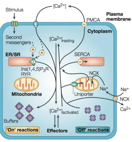
Ca2+ Signalling
Ca2+ regulates many divergent cellular processes, including, but not limited to,
muscle contraction, exocytosis and gene transcription. Signals that have a widely different spatial and temporal profiles, from microseconds, for Ca2+ triggering
25
exocytosis at the synaptic junction, to hours, for gene transcription and cell proliferation.
This versatility emerges from the use of an heterogenous molecular array of
components that constitute the complex toolkit of Ca2+ signalling. Pumps, channels,
exchangers and binding proteins tightly control changes in free cytosolic Ca2+
concentration [Ca2+]
c, that in resting conditions is maintained around the value of
10-100 nM (Fig. 2). While internal and external sources of Ca2+ are used by the cells
to generate Ca2+ signals 48,49.
Ca2+-entry mechanisms When the permeability of the plasma membrane to
Ca2+ changes, the ions enter the cell from the outside, where the [Ca2+] is
around 2 mM. The large electrochemical gradient across the plasma
membrane is guaranteed by the activity of the Ca2+-ATPase (PMCA) and of
the Na+/Ca2+ exchanger (NCX), that removes Ca2+ from the cytoplasm in
order to restore the resting state50.
There are various entry channels, each of them characterized by widely
different properties. Voltage-operated Ca2+ channels (VOCCs), found mainly
in excitable cells, generate rapid Ca2+ fluxes that mediate fast cellular
responses such as muscle contraction or exocytosis at the synaptic endings. Furthermore, there are also channels modulated by ligands, that can be external ligands, as in the case of NMDA (N-methyl-D-aspartate) receptors (NMDARs) that respond to glutamate and, for this reason, belonging to the class of Receptor-operated channels (ROCs); or they can be internal ligands, as in the case of channels gated by cyclic-nucleotides, found mainly in sensory systems, that belong to the class of second-messenger-operated channels (SMOCs).
26
Moreover, other types of stimuli are translated by different channels. Many of them belong to the superfamily of TRP channels, composed by 28 members, encoded by up to 28 different genes and one pseudogene generally characterized by a low conductance that allow them to operate
over much longer time scales without swamping the cell with too much Ca2+
48.
Ca2+ release from internal stores Other important sources for Ca2+ signalling
are represented by the internal stores. The principal organelle responsible
to rapid rise of Ca2+ is the ER, or its muscle equivalent, the sarcoplasmatic
reticulum (SR). The release from such store is controlled principally by Ca2+
itself, but also, by several other messengers, such as inositol-1,4,5-trisphosphate (Ins(1,4,5)P3), cyclic ADP ribose (cADPR), nicotinic acid adenine dinucleotide phosphate (NAADP) and sphingosine-1-phosphate
(S1P)48. Since little is known about the channels opened by NAADP and S1P,
in this section, the focus will be on Ryanodine (RYR) and Inositol (1,4,5) trisphosphate (Ins (1,4,5) P3R) receptors. These two channels are often
mediators of Ca2+ signal amplification. In general, at low concentrations of
Ins (1,4,5) P3, they have a bell-shaped Ca2+ dependence. Therefore, at low
concentrations of cytosolic Ca2+ (100-300 nM) they take part to the so-called
Ca2+-induced Ca2+ release (CICR) process, but, when the concentration of
27
Figure 2. Overview of the calcium homeostasis and signaling mechanisms. Ca2+ signalling can be divided into “on” and “off” reactions. During the “on” reactions, the [Ca2+]i increases as a consequence of Ca2+ entry from the external environment or through the release from intracellular stores. Most of this Ca2+ (shown as red circles) is bound to buffers and effectors. During the “off” reactions, Ca2+ leaves the effectors and buffers and is removed from the cell by various exchangers and pumps. The Na+/Ca2+ exchanger (NCX) and the plasma-membrane Ca2+ -ATPase (PMCA) extrude Ca2+ to the outside, whereas the sarco(endo)plasmic reticulum Ca2+ -ATPase (SERCA) pumps Ca2+ into the ER. Mitochondria also have an active function: they rapidly sequester Ca2+ through a uniporter, and then slowly release it back into the cytosol (adapted from Berridge, Bootman, et al. 2003).
A unique array of channels can be found in each cell type, in order to create a
perfect “toolkit” able to generate a Ca2+ signalling systems that differ in terms of
28
The cytosolic buffers proteins are essential for shaping the amplitude, as well as the
duration of Ca2+ signals. They are molecules that serve as Ca2+ ion chelators that
present appropriately spaced acidic side-chain residues (i.e. glutamate, aspartate) and/or backbone carbonyl groups, in which a Ca2+ ion may fit in. This evolutionarily
well-conserved protein families differ in the way the Ca2+ ions are bound52.
In the neurons, for example, high concentration of buffers like parvalbumin and calbindin, belonging to the EF-hand cytosolic protein family, limits the spatial
spreading of local Ca2+ signals. This is particularly important to maintain confined
the signal in the synapses in which different types of channels are strategically located51.
pH Homeostasis
The intracellular pH represents another fundamental parameter that can affect a wide range of biological processes, including membrane excitability, intercellular communication, signal transduction and vesicle trafficking. The ionization state and, therefore, the structure and function of virtually all proteins of the cells depend on pH. Not surprisingly, dynamic and sustained mechanisms have been
evolved to maintain long-term pH homeostasis53.
Under physiological conditions, the extracellular pH is slightly alkaline (~7.3–7.4), while the cytosolic pH is slightly more acidic (7.2). The tendency of cells to acidify can be explained by 1) the electrical potential across the membrane and 2) the acid equivalents produced during metabolic reactions. Being more negative inside, the cells promote the uptake of the positively charged protons and, at the same time, the efflux through specific pathway of the negatively charged bases. Furthermore, the continuous generation of acid equivalents during metabolic reactions, like ATP
29
production by glycolysis and oxidative phosphorylation in the cytoplasm and in the
mitochondria, respectively, can boost the gradual accumulation of free H+54.
Although the cells have a high buffer capacity, characterized by various intracellular weak acids and bases that protect them from rapid and localized pH changes, active processes are needed to promote the protons extrusion from the cytosol across the plasma membrane in sustained stress situations. A steady state condition is maintained when a balance between the rate of acid loaders (JL, i.e., rate of acid influx/generation or alkali efflux/consumption) and the rate of acid extrusion (JE, i.e., rate of acid efflux/consumption or alkali influx/generation) is achieved55,56 (Fig.
3).
ACID LOADERS Mammalian cells are continuously exposed to CO2, which
being uncharged, can cross the biological membrane and enter into the cells. Once inside, CO2 can combine with H2O to form H2CO3, most of which
dissociates very slowly to form HCO3- and H+. Most cells have a specific
enzyme, carbonic anhydrase, that catalyzes the conversion of CO2+H2O to
H2CO3, accelerating the acidification process of the cell. Although HCO3- is an
effective proton buffer, it is negatively charged and, as well as OH-, can
passively exit to the cell and then contribute to the instauration of a chronic intracellular acid load53.
The main acid loader that is involved in the JL is the Cl-/HCO3- exchanger
(Anion Exchanger AE, encoded by the SLC4A gene). AE3 is the most expressed exchanger in the CNS and, as the other members of the family,
mediates the electroneutral exchange of one Cl− for one HCO
3-. Generally,
Cl− concentration inside the cells is lower than extracellularly, so the inward
gradient of Cl− provides the driving force for a net efflux of HCO
3-54. Thus,
30
if the gradient driving transport reverses, the acid-base transporters can switch the direction of net transport53.
Finally, the electrogenic Na+/HCO
3- cotransporter will be explained in the
next paragraph in detail since this same protein can either operate as acid loader and acid extruder.
ACID EXTRUDERS The extrusion of acid equivalents is an active process that
tends to raise the intracellular pH an to restore it after an acid loading, for example, after an intense neuronal activity55. The membrane associated
vacuolar H+-ATPase (V-ATPase), firstly found in association with endosomal
membranes, but also distributed in the plasma membrane of specialized cell
types (i.e. phagocytes and osteoclasts54), is a pump that uses the energy of
ATP hydrolysis to extrude cytosolic H+ into the extracellular space.
The net extrusion of H+ or the influx of HCO
3- can be also accomplished by a
secondary active transport. In this context, the main acid extruders are the Na+-H+ antiporters or exchangers (NHE) from the SLC9 family of solute
carriers and the Na+-coupled HCO
3- transporters (NCBTs) in the SLC4 family.
NHEs use, in an electro-neutral manner, the inwardly directed electrochemical Na+ gradient generated by Na+/K+-ATPases to export H+.
Until now, nine different isoforms of NHE exchangers have been identified and all of them are broadly expressed in the CNS.
Among the acid-extruding HCO3- transporters, the six mammalian NBCTs are
the predominant. They translocate Na+ and HCO
3– both in the same
direction, but the coupling stoichiometry varies among members of this family. In particular, several Na+-driven HCO
3- (or CO32-) operate as acid
extruders (NBCn1 and NCBn2), with a Na+: HCO
31
others, as acid loaders (NCBe1 and NCBe2), if Na+:HCO
3- stoichiometry is
1:353,54.
NBCn1 and NCBn2 are electroneutral isoforms, what means that they are unaffected by the transmembrane potential, and perhaps the magnitude and directionality of transport is dependent solely of the combined chemical
gradients of Na+ and HCO
3–.
Conversely, for the electrogenic NBCe1 and NBCe2, the driving force depend of the electrical potential, as well as the concentration of HCO3- and Na+ on
either side of the membrane. Therefore, for example in the case of renal proximal tubule, in which the electrical component outstrips the driving force of the concentration gradients, the net direction of transport is outward, resulting in a cytosolic acidification.
Finally, the Na+-driven Cl-/HCO
3- exchanger that mediates the electroneutral
exchange of one Na+ plus two HCO
3− equivalents for one Cl−. This was the
first transporter ever to be studied as a pHi regulator53.
A straight correlation between the excitability of the neurons and pH changes exists. Membrane proteins, such as channels, transporters, receptors and even ATPase pumps have intracellular and extracellular moieties that result in pH sensitivity. The direction of the responses evoked by pH changes depends on the individual components that are responsible for dictating overall excitability in each neuron. For this reason, there are neurons that exhibit hyperexcitability following an acidification of the cytosol (i.e. chemosensitive neurons), while others could exhibit a decrease in the excitability55.
32
Figure 3. pH regulation in the CNS. (A) pHi dependence on the balance between the rate of acid extrusion (JE) and the rate of acid loading (JL). Steady-state of pHi is achieved when JE = JL (intersection of dark-blue and dark-red lines: point A). A shift to a more alkaline value (intersection of light-blue and dark-red lines: point B) follows an JE increase (J’E). Conversely, a shift to a more acidic value (intersection of dark-blue and light-red lines: point C) follows an JL increase (J’L). If the rise in JE is matched by an equal increase in JL (J’E = J’L) there will be no net change in pHi (red circle). (B) Acid loaders (red) and acid extruders (dark blue) in neurons, astrocytes and oligodendrocytes (Ruffin, et el. 2014).
33 1.5 Ionic channels involved
TRP channels
The TRP superfamily is a very heterogenous class of ion channels displaying a great diversity of ion selectivity, modes of activation, and physiological functions57.
Despite this variability, all the members of this family share a typical protein structure consisting of six putative transmembrane segments (S1 to S6) that assemble in tetramers (homo- or heterotetramers) to form the functional channel. Each subunit contributes to selectivity filter and to ion-conducting pore with a reentrant loop located between S5 and S6. They also have an intracellular amino and carboxyl termini that result variable in length and in domains.
From protein homology, TRP channels can be divided into seven subfamilies: TRPA, TRPC, TRPM, TRPML, TRPN, TRPP and TRPV58. They are expressed in all cellular
membranes, with the exception of the nuclear envelope and mitochondria, of virtually all cell types. The majority are located in the plasma membrane, where
they mediate the transport of ions59. Although they are defined as non selective
Ca2+-permeable cation channels, the variety of homo/heteromultimerizations that
can be achieved, makes the overall picture of TRP channel ion selectivity more complex. Most TRPs show little preference for Ca2+ over Na+, some others (i.e.
TRPC1, TRPM1 and TRPP2) are completely non-selective cation channels, and both
Ca2+-selective (i.e. TRPV5 and TRPV6) and Na+-selective (i.e. TRPM4 and TRPM5) are
present in the superfamily57.
Regarding the regulation of their activity, a lot of mechanisms have been described. The mechanism(s) through which a given TRP is activated and regulated cannot be
predicted reliably on the basis of its subfamily assignment.Members of this family
34
number of exogenous and endogenous ligands. The heat-sensitive TRPV1 can be also activated by capsaicin (the pungent extract of hot peppers), as well as the cold receptor TRPM8 by menthol (derived from the mint plant Mentha piperita), and TRPV4 directly by bisandrographolide (derived from the plant Andrographis
paniculata)59. The recurring topic is that most TRP channels are activated through a
diversity of mechanisms60.
Initially considered as rather insensitive to voltage, it is well documented now, that the mid-point activation of TRPs, evoked for example by temperature or ligands,
can undergo to a voltage-dependent shift of even several millivolts magnitude61.
This intrinsic voltage dependence regards mainly TRPs involved in sensory
perception59. However, in some cases there are similarities among subfamily
members, as all TRPC channels that are sensitive to membrane phospholipids, like phosphatidylinositol (4,5) bisphosphate (PtdIns (4,5) P2).
The modulation by phospholipids can be direct or indirect. The great number of
membrane-associated proteins sensitive to the levels of PtdIns (4,5) P2 can, in turn,
affect the activity of TRP channels62. The hydrolysis of PtdIns (4,5) P
2 operated by
phospholipases C (PLCs) can be activated by two different pathways: 1) G protein– coupled receptors (GPCRs) and 2) tyrosine kinases receptors. Once the hydrolysis
of PtdIns (4,5) P2 is obtained, two important signaling molecules are produced: 1)
diacylglycerol (DAG) and 2) inositol (1,4,5) trisphosphate (IP3)58,59. DAG can affect
the TRP channel opening directly or indirectly, by producing several metabolites of (i.e.). While IP3, which activates IP3 receptors (IP3R) on the ER membrane, evokes a
release of Ca2+ from intracellular stores responsible of the activity modulation of
TRP (store-operated Ca2+ entry (SOCE) or capacitative Ca2+ entry (CCE))57. Additional
activation mechanisms include phosphorylation by PKA, PKC and PKG, that sensitize them by other stimuli; changes in extracellular and intracellular pH; ROS
35
Detecting and responding to this wide variety of external environment stimuli make their contribution essential for several physiological processes, ranging from pure sensory (chemosensation, thermosensation, nociception, mechanosensation and
photoreception) and homeostatic (such as reabsorption and osmoregulation of Ca2+
and Mg2+) functions to many other motile functions, such as muscle contraction and
vasomotor control. Thus, an increasing number of evidences suggest the implication of several TRP channels in a wide range of diseases in humans, following either mutations in the encoding gene or an acquired mechanism.
Genetically and pharmacological evidences have confirmed the involvement of these channels in the generation and transduction of pain (TRPV1,TRPA1,TRPM8); in the activation of central and peripheral reflex responses, including cough, mucus secretion and bronchospasm, in the mammalian respiratory tract following the interaction with irritant and/or inflammatory stimuli (TRPV1, TRPA1, TRPM8, TRPV4, TRPC6); in the skin, where they control a variety of biological processes such as itch, regulation of barrier function, keratinocyte differentiation, hair growth, inflammation and wound healing, being expressed both on neuronal and non-neuronal cells (TRV1, TRPV3, TRPA1); in CNS disorders, like learning and memory formation through regulation of synaptic plasticity (TRPC6), or neurodegenerative disorders, Guamanian amyotrophic lateral sclerosis (ALS-G) and Parkinsonism dementia (TRPM2, TRPM7), or in axonal and neuronal degeneration in Multiple Sclerosis (MS, TRPM4) and in cardiac hypertrophy associated with arrythmias,
36 Large conductance channels: Connexins and Pannexins
Gap junctions (GJs) were primarily known as mediators of direct intracellular communication. When two hemichannels (HCs), belonging of two neighboring cells, interact together, a GJ is formed. These junction put directly in contact the cytosolic compartments of adjacent cells allowing the flux of several small molecules, such as ATP, glutamate and prostaglandins, as well as ions, including Ca2+. Each
hemichannel is composed of 6 connexin (Cx) proteins, all of which consist of 4 transmembrane domains, 2 extracellular loops (EL), 1 cytoplasmic loop (CL), 1 cytoplasmic C-terminal tail (CT) and 1 cytoplasmic N-terminal tail. Now, it is clear that Cx HCs are not only structural precursors of GJs but themselves, alone, can provide a circuit for communication, albeit between the cytosol and the
extracellular environment69. Although Cx- based GJC and hemichannels share
similar structure, they differ in their localization (inside or outside the contacting zone with others cells), opening and closing regulation and their roles in cellular
processes70. Usually, while the GJs are open in physiological conditions, Cx HCs are
typically closed, because of their low open probability, which would be enough to participate in several cellular processes. For example, Cx46 and Cx50 hemichannels openings seem to have a crucial role in the lens, an avascular tissue in which the microcirculatory homeostasis depends on nutrient, electrolyte and water flow through these plasmalemmal channels. In human CNS, where 11 out 21 Cx isoforms have been identified, are differently distributed between astrocytes, oligodendrocytes, microglia and neurons. Here, hemichannels activity has been correlated to release of precursor or signaling molecules such as Nicotinamide
adenine dinucleotide (NAD+), ATP, adenosine, IP
3 and glutamate. The opening of
Cx43 hemichannels, which represents also the most abundantly expressed connexin in the human body, and the following release of ATP have been observed
37
in astrocytes in which it seems to be involved in the Ca2+ wave propagation
mediated by an extracellular paracrine signaling component 71.
However, in the presence of several type of stimuli such as increases in membrane depolarization, an increase in intracellular Ca2+ concentration or a decrease in
extracellular Ca2+ concentration, mechanical stimulation, changes in the Cx
phosphorylation status, oxidative stress, pH, ischemia/reperfusion insults, fatty acid overload and in the inflammatory conditions the Cx- hemichannels open probability increases. When the hemichannel activity becomes too high and uncontrolled, cell viability diminishes. In this cases, they form “leaky hemichannels” and/or their number at the plasma membrane increase dramatically up to enhance
cell membrane permeabilization, enough to cause an accelerated cell death70. In
addition of a possible abnormal connexin hemichannel activity that could be considered as a gain-of-function mutation, many connexins undergo to loss of gap junction function owing to trafficking deficiencies (i.e. gap junction plaque formation), channel malformation (non-functional or communication-deficient channels) or the genesis of aberrant hemichannel activity. At least the half part of the total human connexin genes identified can be linked to inherited human diseases including neuropathies, deafness, skin diseases, oculodentodigital dysplasia (ODDD) and cardiac arrhythmias 69,70,72.
In 2000, a novel class of Cx-like proteins, with similar membrane topology to the connexins but without forming GJs, has been identified. The Panx family consists of three members, namely Panx1, Panx2, Panx3. Panx1 is the most widely represented one in diverse tissues and cell types, while the other two are mainly expressed respectively in the CNS and, in skin and skeletal tissues. Similar to Cx hemichannels, Panx ones are implicated in a wide variety of physiological and pathological contexts. Although Panx channels are opened at positive potential, a wide number
38
of triggers that promote their opening at membrane resting potential, has been
identified69,73. Large amount of data refers their activation following the
intracellular Ca2+ increments, the phosphorylation mediated by Src family kinase or
by other types of kinases (i.e. c-Jun NH2-terminal kinase (JNK); protein kinase G), the ATP binding to purinergic P2 receptors (such as P2Y receptors), the oxygen deprivation (in erythrocytes and neurons), the elevation of extracellular potassium or a mechanical stress (in various cell types)74.
Both reversible (i.e. G-protein-coupled receptor) and irreversible (caspase- mediated cleavage) forms of channel activation induce a progressive channel opening, characterized by a robust macroscopic currents across the plasma membrane of individual cells (ion- and large-molecule transport) that presents at least five open substates (corresponding to 5, 25, 30, and 90% of the fully open unitary conductance, ranging from ∼300 pS to >500 pS)74.
The involvement of pannexins in the induction of inflammation has been reported in multiple publications. There are many evidences that support the idea that Panx1 and its associated P2X receptors are essential for upstream regulation of inflammasomes and proteolytic activation of Casp1 and Casp11. This has been reported in many cell types, however a strong demonstration has been only
39 Two-pore domains (K2P) channels
The two- pore domain are the third major family of K+ channels, along with
voltage-gated (KV) and inwardly rectifying (Kir). They are structurally different to the others
K+ channels. While voltage-gated K
V channels and inwardly rectifying Kir channels
assemble as tetramers and each subunit symmetrical contribute to the ion permeation pathway through a single copy of P-domain, the K2P have two pore
domains (P1 and P2) in tandem per subunit and four transmembrane domains (TM1–TM4). A large extracellular cap-domain linked to the P1 from TM1, stabilized by an apical disulphide bridge, can be considered as the most prominent structural
motif of K2P. The steric hindrance created by the specific position of the cap-domain
seems to modulate the gating of the K2P channels, rendering them less sensitive to
classical toxin peptides and pore blocking molecules like tetraethyl-ammonium ions that, instead, act on other classes of K+ selective channels.
In mammals, K2P derive from 15 genes and can be classified in six subfamilies: TWIK
(weak inward rectifiers), TREK (lipid and mechanosensitive channels), TASK (acidification-inhibited), TALK (alkalinization-activated), THIK (inhibited by halo- thane), and TRESK (spinal cord channels)76. They are called leak or background
channels for their primary function, which is to maintain hyperpolarized resting the membrane potential of virtually every excitable and non-excitable eukaryotic cell. Increasing evidence suggests that the activity of these ion channels can be also modulated by a great variety of stimuli, including changes in membrane tension, voltage, temperature, oxygen-tension, extracellular and intracellular pH, phospholipids, and other signaling molecules arising from G-protein–coupled
receptor activation76,77. Although they do not have a conventional voltage-sensor,
40
recent work provides a possible explanation of the voltage-dependence of K2P78.
The intriguing hypothesis relies crucially on the movement of the permeant ion, along the selectivity filter (SF), and therefore on its electrochemical potential difference. Only a depolarization would push ions into the SF, otherwise empty. Once fully occupied, it become permeant in outward direction. High occupancy would not be reached by forcing ion movement in the inward direction; under these conditions, the SF would become inactive. Many stimuli including intracellular
acidification , lipids, membrane tension can affect the voltage- dependence78.
K2P channels are expressed throughout the body and each type of channels have a
different expression profile. It is well established that several members of this family are highly expressed in DRG and TG neurons77. Therefore, an increasing
number of evidences is revealing their central role in the transduction of noxious stimuli by modulating the somatosensory excitability. The painful stimuli activate various classes of cationic ion channels like ASIC, TRPV1 and TRPA1 in the peripheral nerve terminals, in which they induce a depolarization of the membrane potential.
Voltage-dependent Na+ channels generate an action potential when the threshold
is reached. The leak K+ channels are able to prevent the generation of an action
potential hyperpolarizing the membrane potential. Thus, these channels operate a counteraction by decreasing conduction fidelity across the axon or by limiting the
release of neurotransmitter79. Further evidences are provided, for example, by less
amplitude current required to elicit an action potential in cells derived by TRESK-1 knockout mice; or the mechanical and thermal hypersensitivity showed by both TREK-1- and TRAAK-deficient mice in response to a chronic inflammation. On the contrary, the overexpression of TRESK channels reduces mechanical and thermal hypersensitivity in response to nerve injury80.
41
The implication of these channels in the regulation of multiple types of pain, such as inflammatory, neuropathic, mechanical and thermal, migraine and cancer pain, make them possible molecular targets for the development of pain therapeutic strategies. However, the pharmaceutical design is restrained by 1) the broad range of transcript processing and post-translational modification mechanisms (phosphorylation, SUMOylation, glycosylation) and 2) the high capacity of heterodimerization of specific pairs of subunits. The result of these mechanisms is a high rate of diversification of channels, with completely distinct physiological and pharmacological properties by combining the functional properties of single constituent. However, the lack of structural data remains the first most prominent bottleneck in the development of drugs that act at K2P channels77,80,81.
1.6 Neurotoxicity screening
Traditionally, the paradigms for hazard identification and risk assessment requires an animal-based experimental approach. In the last years, a change towards new approaches using integrate in silico and in vitro testing to identify the mechanisms of toxicity is occurring. Many practical and scientific considerations support this evolution, first of all the increase of safety testing reliability by reducing the need for animals and by lowering the costs with the implementation of high-throughput methods82.
The endpoints commonly used for in vitro neurotoxicity testing can be divided into three categories: 1) viability read-outs, 2) morphological read-outs and 3) functional read-outs. The first step is, essentially, to identify which are the compounds that cause toxicity in one or more cell types and to individuate which is


![Table 2 Particles Extra/Intra- Cellular Solutions N° Cell s N° Responsive Cells Current Density (pA pF-1 ) I-V V rev (mV)Media n IQR Median IQR NP (20 µg mL -1 ) Tyrode/KAsp 34 34 -7.8 [-14.8; -4.4] Outward rectification -1.7 [-12](https://thumb-eu.123doks.com/thumbv2/123dokorg/4807008.49645/101.773.28.723.505.977/particles-cellular-solutions-responsive-current-density-outward-rectification.webp)
