XXVII ciclo A.A. 2011-2014
STUDY OF STRATEGIES FOR INCREASING
BIOCOMPATIBILITY AND EFFECTIVENESS OF
POLYETHYLENIMINE CARRIERS FOR GENE
THERAPY
XXVII ciclo A.A. 2011-2014
STUDY OF STRATEGIES FOR INCREASING
BIOCOMPATIBILITY AND EFFECTIVENESS OF
POLYETHYLENIMINE CARRIER FOR GENE
THERAPY
Elena Nicolì
Supervised by Prof. Michela Bosetti
Prof. V. Prasad Shastri
Contents
1 Introduction ... 1
1.1 Nanomedicine ... 1
1.1.1 Definition and challenges ... 1
1.1.2 Nanomedicine for therapeutic application ... 2
1.1.3 Active and passive targeting ... 4
1.1.4 Nanomedicine in Tissue Engineering ... 6
1.2 Drug delivery strategies ... 9
1.2.1 Impact of physical properties of nanoparticles ... 10
1.3 Gene therapy ... 12
1.3.1 RNAi mechanism. ... 12
1.4 Gene delivery strategies ... 15
1.4.1 Viral delivery ... 15
1.4.2 Non-viral delivery ... 18
1.5 Non-viral delivery materials for gene therapy ... 20
1.5.1 Physical methods ... 20
1.5.2 Chemical methods ... 22
1.5.3 Challenges of non-viral gene delivery. ... 25
1.5.4 Endocytosis mechanisms ... 27
1.6 Polyethylenimine and toxicity issues... 29
1.7 Active targeting: the potential of albumin. ... 31
1.8 References ... 33
2 Outline of the Thesis ... 40
3 The genotoxicity of PEI-based nanoparticles is reduced by acetylation of PEI amines in human primary cells ... 44
3.1 Summary ... 44
3.2 Introduction... 45
3.3 Materials and Methods ... 47
3.4 Results... 55
3.5 Discussion and conclusion ... 71
3.6 References ... 75
4 Enhanced gene silencing through human serum albumin-mediated delivery of polyethylenimine-siRNA polyplexes ... 79
4.1 Summary ... 79
4.2 Introduction... 80
4.3 Materials and Methods ... 82
4.4 Results and discussion ... 88
4.5 Conclusion ... 105
4.6 References ... 106
5 Biocompatible cationic nanoparticles for gene delivery in bone tissue engineering: uptake trafficking, localization and activity in human primary osteoblasts... 110
5.1 Summary ... 110
5.2 Introduction... 111
5.3 Materials and methods ... 113
5.4 Results... 122 5.5 References ... 137 6 Conclusions ... 140 List of publications ... 147 Acknowledgements ... 150
1
1 Introduction
1.1 Nanomedicine
1.1.1 Definition and challenges
Nanotechnology is the branch of science that investigates the potential of materials in nanometre range of size. The specific definition was given in 2000 by the National Nanotechnology Initiative: ―Nanotechnology is concerned with materials and systems whose structures and components exhibit novel and significantly improved physical, chemical and biological properties, phenomena and processes due to their nanoscale size‖ [1].
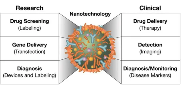
Nanotechnology applied for medical purposes takes the name of ―Nanomedicine‖. Nano-scale devices for treatment, diagnosis, monitoring and control of biological system have been referred as ―Nanomedicine‖ by the National Institute of Health [2]. Nanomedicine aspires to develop nanoscale systems, from a few atoms to sub-cellular size (1-1000 nm), that can reach high level of cell targeting, with minimal impact on cell viability [1]. Applications of Nanomedicine are summarized in Figure 1.
2
Figure 1.Research and clinical approaches of Nanomedicine.
1.1.2 Nanomedicine for therapeutic application
The successes of Nanomedicine are reported, for example, in the treatment of cancer. Cancer mortality counts more than 10 million new cases every year and in the past two years has decreased, owing to better understanding of tumor biology and improved diagnostic devices and treatments [3]. Ordinary cancer treatments include surgical intervention, radiation and chemotherapeutic drugs, and the higher limitations are due to low tumor specificity, low solubility and short circulation time of the drug. The concept of nanomedicine in therapeutic application is, thereby, closely related with drug-delivery. The design of an optimal nanocarrier, which ensures drug encapsulation, efficient cell targeting and intracellular drug release, has revolutionized the pharmaceutical field.
3
In the recent years a lot of efforts were employed to develop the perfect delivery carrier for individual therapeutic agents. Careful screening of the drug, interaction with the carrier, binding/dissociation properties and cell uptake/cytotoxicity are important issues to consider for obtaining a therapeutic effect. Furthermore, blood stability, tolerance from the immune system and extravasation to reach a target cell type are included in the multitask properties that a nanocarrier, for in vivo therapy, should possess (Figure 2).
Rational design of nanocarrier for cancer therapy
What a nanocarrier should do What a nanocarrier should be
Protect the drug from premature degradation
Be made from a material that is biocompatible, well characterized, and easily functionalized Prevent drugs from prematurely interacting
with the biological environment
Exhibit high differential uptake efficiency in the target cells over normal cells (or tissue) Enhance absorption of the drugs into a
selected tissue (for example, solid tumor)
Be either soluble or colloidal under aqueous conditions for increased effectiveness Control the pharmacokinetic and drug
tissue distribution profile Have an extended circulating half-life Improve intracellular penetration Have a low rate of aggregation and a long
shelf life
4
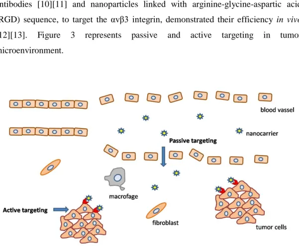
1.1.3 Active and passive targeting
In cancer delivery, the specific drug accumulation is enhanced by the disorganized endothelial structure and increased permeability of the endothelial barrier, besides of the reduced lymphatic drainage. This passive tumor targeting has been defined as ―Enhanced Permeability and Retention (EPR) effect‖, discovered by Matsumura and Maeda [4]. Endothelial pores are reported to be in sizes varying from 10 to 1000 nm, depending on the type of tumor [5] and efficient vascular extravasation occurs with nanoparticles size profile lower than 400 nm [6].
Active targeting consists, instead, in conjugating the nanoparticles with a specific molecule able to target a specific tissue and cell type. Proteins, peptides, monoclonal antibodies, small molecules, aptamers can be used to target a specific cell type [7]. Most of tumors up-regulates the expression of specific receptors to cope with the higher proliferation of malignant cells. Nowadays, the more studied over-expressed surface molecules are transferrin receptor, folate receptor, specific glycoproteins and the epidermal growth factor receptor (EGFR). Transferrin transports iron to the cells and folic acid is important for nucleotides synthesis. Several lectins have been found to specifically interact with glycoproteins expressed on cancer cells, and EGF is involved in proliferation, angiogenesis and metastasis [6]. Targeting tumor vasculature, through specific endothelial receptors, it is also possible to inhibit tumor-induced angiogenesis. The vascular endothelial growth factor receptors VEGFR-1 and VEGFR-2 are over-expressed on tumor endothelium, like αvβ3 integrin is highly expressed on neo-vascular endothelial cells [6][8].
Therefore, the possibility to target tumors based on the different pattern/expression of specific molecules on the cell surface is becoming extremely promising. PLGA nanoparticles, modified with isopropyl myristate moieties, showed increased delivery of chemotherapeutic Paclitaxel to the lung and superior in vitro effectiveness [9].
5
Furthermore, the development of nanoparticles carrying transferrin and/or transferrin antibodies [10][11] and nanoparticles linked with arginine-glycine-aspartic acid (RGD) sequence, to target the αvβ3 integrin, demonstrated their efficiency in vivo [12][13]. Figure 3 represents passive and active targeting in tumor microenvironment.
Figure 3. Representative chart of the role of endothelial fenestration in enhancing passive transportation in the tumor microenvironment and the role of tumor targeting molecules in developing selective nanocarriers.
6
1.1.4 Nanomedicine in Tissue Engineering
Nowadays, Nanomedicine is not only limited for therapeutic application, but offers also opportunities in regenerative medicine. The increased understanding of cell response to extracellular signals and the intracellular pathways involved in cell survival, proliferation and differentiation, inspired regenerative medicine strategies, with the aim to replicate biological instructions expressed during embryogenesis. Growth factors are the signaling molecules that instruct cells during tissue development, which can be studied to achieve tissue regeneration in adults. Encapsulation and controlled delivery of these factors may potentially activate specific proliferation/differentiation pathways to regenerate tissues [14][15].
Considering in details bone regeneration, traditional treatment methods for promoting bone healing include bone grafts or synthetic materials, to fill the defect and provide structural support. Bone autografts are considered the gold standard for treating bone defects, owing to the low risk of an adverse immune response, osteoinductive, osteoconductive, and osteogenic properties [16].
Recently, several growth factors have been discovered to play a role in bone regeneration, directly acting on osteoblasts, which produce mineralized bone matrix. Growth factors delivery by nanocarriers is promising for overcoming the actual disadvantages with conventional scaffold implantation, like immunogenicity, low growth factors expression and low cell proliferation. Furthermore, a nano-delivery system offer major control and longer-term release of the growth factor, compared to the direct adsorption on the surface of implanted scaffolds [17] [14].
Osteoblast commitment from pluripotent mesenchymal stem cells is mainly regulated by the growth factor family of TGF-β, with the demonstrated activity in vitro and in
7
guiding the differentiation process [18][19]. Besides, a number of other growth factors are being investigated for their potential to regenerate bone, including platelet-rich plasma (PRP), platelet-derived growth factor (PDGF), vascular endothelial growth factor (VEGF), and fibroblast growth factor (FGF) [17].
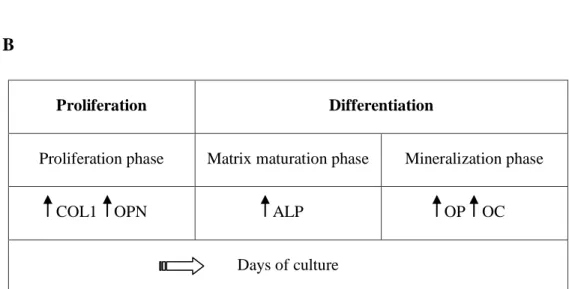
Osteoblast commitment involves the expression of phase-specific genes, which are important to evaluate the response of an ―osteogenic treatment‖. Collagen type I (COL1) is expressed in the early stage of osteoblast differentiation and is the main structural component of bone matrix. Osteopontin (OPN), a non-collagenous matrix protein, and alkaline phosphatase (ALP) are important in stabilizing the matrix in formation, and are considered early markers of osteoblasts maturation. In the late stage of differentiation, osteocalcin (OC) expression is up-regulated [16][20].
Bone formation is a physiological balance between bone synthesis and bone resorption. Differentiated osteoblasts produce and release the cytokine RANKL, member of the Tumor Necrosis Factor (TNF) superfamily, that induce osteoclast differentiation and fusion of pre-osteoclasts [21] [22]. Therefore the activation of RANKL, in response to delivery agents, should be considered in order to induce effective osteogenesis.
The differentiation process of osteoblasts can be divided in four stages (Figure 4):
Proliferation phase: In this phase, genes required for proliferation (c-fos) and
progression of the cell cycle (cyclins, histones) are expressed, together with genes associated with the biosynthesis of the extracellular matrix (COL1, TGFβ) and cell adhesion proteins (fibronectin).
Matrix-maturation phase: In this phase the expression of genes involved in the
maturation and organization of the bone extracellular matrix (alkaline phosphatase) are up-regulated. The role of alkaline phosphatase (ALP) is still uncertain, but since
8
this enzyme cleaves phosphate groups from monophosphate-ester substrates, it is assumed that the enzyme is involved in bone mineralization.
Mineralization phase. In this phase, the expression of genes associated with the
organized deposition of hydroxyapatite (osteopontin and osteocalcin) is up-regulated.
Apoptosis phase. In this phase were observed an increased COL1 and collagenase
gene expression, apoptotic cell death and compensatory proliferative activity [20].
9
B
Proliferation Differentiation
Proliferation phase Matrix maturation phase Mineralization phase COL1 OPN ALP OP OC
Days of culture
Figure 4. Bone regeneration. A) Proliferation of mesenchymal stem cell (MSC) and differentiation to osteoblast lineage by the use of specific growth factors. B) Progressive stages of osteoblast differentiation with the increase in the expression or activity of phase specific markers.
1.2 Drug delivery strategies
Nowadays, the most used drug delivery strategies are lipids, inorganic materials, proteins and polymers, which can be strategically combined to obtain an optimal delivery agent.
Lipids, due to their dynamic nature are easily modifiable for cell targeting, because
their dynamic nature allows clustering of peptides or other ligands. However, their dynamic nature reduces the stability of the system.
Inorganic materials (gold, carbon, and iron oxide) provide the advantage of stability;
but the strength causes retention in the body and limited clinical application.
Polymers offer enhanced biocompatibility and major control of drug release.
10
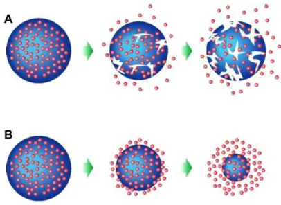
(PLL-A), poly(ε-caprolactone) (PCL), chitosan, gradually release the therapeutic agent, with the degradation of the carrier, ensuring safe clearance of degraded products [12][23] (Figure 5).
Figure 5. Degradation mechanisms of biodegradable polymeric nanoparticles: A) bulk erosion, B) surface erosion.
1.2.1 Impact of physical properties of nanoparticles
It is already known that the physics of the nanocarrier influences the delivery efficiency, and determinant parameters, independently from the specific material, are grouped under ―physical targeting‖:
Size - The size of the nanoparticles regulates uptake, clearance from the kidney and
immune system activation [24]. Small particles inferior of 10 nm are easily excreted from the urinary system, while big particles or aggregation can cause vascular occlusion and immune response [25].
11
The size limitation for tumor targeting depends on the cut-off limit of the EPR effect on the endothelial barrier. Main pore size ranges between 380 and 780 nm in diameter, but change according with the type, location and cause of the tumor [26][27]. For tumor infiltration, usually small particles, around 20 nm or smaller, are facilitated in crossing the dense extracellular matrix [28].
Shape - The shape of the nanocarrier can be spherical, disk-like, rod-like, flexible
[29] and can influence the rate of cellular uptake and body distribution [30].
Stiffness - A clear example of how stiffness can regulate efficiency of drug delivery,
despite of the size of nanoparticles, is given by red blood cell physiology. Red blood cells (RBCs), biconcave microstructures of around 8 µm, are able to deform and pass through small blood vessels and have inspired the development of soft polymeric microparticles, with the aim to simulate the same deformability. Although only few studies have been done in this direction, stiffness seems to regulate uptake efficiency in different cell lines, for example, has been reported that cervical cancer Hela cells can take up soft particles better than hard ones [31].
Charge - The surface charge is another important factor to be considered in drug
delivery. Positive charges improve non-specific cell uptake by electrostatic interaction with the cellular membrane, although hydrophobic nanoparticles can benefit of negative charges in promoting hydrophobic contacts [32]. Positive charges have the disadvantage to cause in vivo aggregation with serum proteins and erythrocytes and being rapidly excreted through the urinary system [33].
12
1.3 Gene therapy
Gene therapy is a scientific discipline that has been developed with the aim of correcting genetic diseases. Genes are the functional units of heredity and encode instructions to make proteins, which regulate most of the life functions. When the gene sequence is altered, proteins are unable to carry their normal function and result in genetic disorders. Genetic medicine is applicable in the treatment of monogenic hereditary disorders, cancer and viral infections, with the possibility to replace a defective gene or introducing a new gene to confer new properties to the cell [34][35]. In tissue regeneration growth factors gene delivery resulted being more effective than protein delivery, for the possibility to overcome the unstable biological activity, short half-life and low tissue penetration of the growth factors [17]. The strategies of gene therapy include gene encapsulation in inactivated viral carrier or alternatively into non-viral delivery, with the incorporation of the gene sequence in a bacterial plasmid. Recently the discovery of RNA-modification therapy opened the possibility to modify the gene expression through the activation of an endogenous pathway, RNA interference (RNAi), which specifically blocks the translation of a target mRNA, through antisense oligonucleotides [36].
1.3.1 RNAi mechanism.
Since 1998, when the researchers Fire, Mello and colleagues discovered the ability of double strand antisense RNA to silence the expression of a target protein, a new approach for gene therapy gained international attention. Synthetic double strand RNA sequences opened the possibility to specifically regulate the expression of a protein in cells and potentially reach any part of the genome. We can distinguish two
13
main pathways of the RNAi mechanism (Figure 6). The first mechanism is physiologically activated as anti-viral response, where long double strand RNA sequences (dsRNA), are interpreted as pathogens by cells and degraded in the cytoplasm. The enzyme Dicer processes dsRNA into small interfering RNAs (siRNAs), which are then recognized and incorporated into the RNA-induced-silencing complex (RISC). Argonaute2, a multifunctional protein part of the RISC complex, unwinds the siRNA, and mediates the degradation of the sense strand. The activated RISC works binding the antisense strand, using this sequence as a template to perfectly match the sequence of a target messenger RNA (mRNA) in the cell cytoplasm. RISC mediates cleavage and degradation of the target, inhibiting protein translation.
The synthetic sequences, to mimic this process, are formulated to be around 21-25 bp, skipping the Dicer functional cleavage, in order to reduce the inflammatory response mediated by interferon-γ (IFN-γ) [37] [36].
The second pathway is mediated by endogenous microRNAs (miRNAs), codified by the cell genome. They have been discovered to finely regulate the gene expression in cells, especially during embryogenesis and development [38]. miRNAs often reside into introns or polycistronic units in the genome and are processed in the cytoplasm with nearly the same mechanism of siRNAs. Different Argonaute proteins are involved (1-4) and the main difference in the process is the imperfect match with the target mRNA in sites within the 3’untranslated regions (UTRs), steric inhibition of mRNA translation or destabilization of mRNA by deadenylation of the poly-A tail [39].
For therapeutic application the use of siRNA is more practical and specific than miRNA, since miRNA can regulate the expression of multiple genes and one gene can be regulated by multiple miRNA.
14
The gene silencing through endogenous siRNAs ensure its therapeutic effect for 3-7 days in high proliferating cells and for several weeks in non-dividing cells [40]. Considering the limitations of siRNA therapeutics, siRNAs, like pDNA, do not cross the cellular membrane spontaneously, because of their high negative charge. The development of a delivery strategy is also required in order to reduce the high extracellular degradation and to counteract the poor targeting and high clearance by the urinary system [37].
Figure 6. RNAi endogenous mechanisms for protein silencing: similarity and differences in siRNA and miRNA mechanism [36].
15
1.4 Gene delivery strategies
Nowadays, a lot of efforts are directed to develop an efficient carrier for nucleic acids, to be applicable for gene and siRNA therapeutics. The two main strategies involve 1) engineered viruses, deprived of the genes responsible for pathogenesis, carrying the gene of interest and the proteins important for the transfection; 2) Non-viral carriers, that create Wan der Walls or electrostatic interactions with pDNA or siRNA sequences. Viral systems ensure high transfection efficiency, but they have the disadvantage to provoke immune response, toxicity and mutagenesis [41]. Non-viral carriers are preferred for the possibility to increase the safety of the gene delivery system for in vivo application, the possibility to insert targeting molecules, simple preparation and low production costs [42].
1.4.1 Viral delivery
Viral-mediated gene delivery consists in the utilization of viruses deprived of the capacity to replicate, but engineered to deliver exogenous genes. The most common viral systems involve functional modification of adenoviruses, adeno-associated virus vectors, retroviruses and lentiviruses (Figure 7). These systems provide high delivery efficiency and a constant expression of the therapeutic gene. The major limitations that restrict the in vivo utilization are immunogenicity, toxicity, mutagenesis and limited size of DNA encapsulation [42][35].
16
Figure 7. Representative T4 Phage assembly for gene delivery.
Adenovirus vectors - Adenoviruses are double strand DNA viruses, they do not
contain lipid or membrane, and the genetic material is encapsulated in icosahedral particles, containing a complex combination of structural proteins. Adenovirus vectors can contain a relatively large amount of DNA, up of 36 kb; they cause receptor mediated endocytosis and do not integrate in the host genome, remaining as episomes in the infected cells. Therefore, the expression of the exogenous gene is transient. The main disadvantage of this system is the strong immune and inflammatory response [43].
Adeno-associated virus vectors - Adeno-associated viruses are small single strand
DNA viruses, with a simple protein coat. The absence of a complex envelope structure induces a less problematic immunogenicity compared to adenoviruses. They can infect cells with good efficiency and they can be engineered to persist in
17
cells in an extra-chromosomal state, without integration in the host genome. The major limitation of these vectors is the low packaging capacity (4.5 kb)[43] [35].
Retroviral vectors - Retroviruses are circular-enveloped RNA viruses and they have
the capacity to be integrated in the host genome. The infection of retroviruses is not cell specific and it is mediated by glycoproteins on the viral surface. Viral RNA is injected into cells.
Retroviral vectors require three important genes that should be conserved in the engineered viral vector: ENV, POL and GAG. ENV codifies for the viral envelop,
GAG for the viral matrix, capsid and glycoproteins and POL for the reverse
transcriptase (RT) and the integrase enzyme (IN). RT is a viral DNA polymerase that converts the viral RNA in single strand DNA in the cell cytoplasm, which in the nucleus will be transformed in double strand DNA and integrated in the host genome by the integrase. The casual insertion in the genome, mediated by LTR sequences, causes a high risk of mutagenesis. Retroviral vectors show high transfection efficiency, with the capacity to deliver DNA up to 8 kb, although the transgene expression ceases in a range of days or weeks. The reason of the silencing is not well known, although the methylation of DNA into condensed chromatin seems to be involved [43][35][42].
Lentiviruses - Lentiviruses belong to the family of Retroviridae. Lentivirus vectors
are mostly based on components of HIV1. HIV is the lentivirus responsible to cause chronic immune deficiency, known as acquired immune disorder syndrome (AIDS). Lentiviruses have a good capacity to transfect non dividing cells, as monocyte/macrophage lineage and neurons, with potential use for gene delivery in the central nervous system (CNS). Compared to retroviruses, they have a more complex structure, with additional regulatory genes like TAT, REV, VIF, VPR, NEF
18
1.4.2 Non-viral delivery
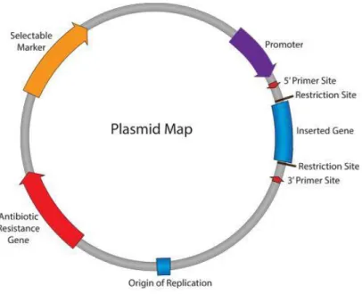
The development of non-viral gene delivery carriers aims to minimize immunogenicity, toxicity and mutagenesis of the viral delivery system. The potential of non-viral delivery systems, beyond the lower toxicity for in vivo applications, is the possibility to link antibodies, proteins, aptamers and ligands that can confer specificity for a target cell type [41]. In this case the genetic material is not compacted into the viral nucleus, but it is incorporated in bacterial plasmids, commonly isolated from the bacteria Escherichia Coli. In nature, plasmids are small molecules that codify for some additional survival characteristics, like resistance to antibiotics. They consist in extra-chromosomal DNA and they can be transmitted from one bacteria to another one. The plasmid can replicate independently in the nucleus of a suitable host, exploiting the replication and transcription machinery of the cell. Important features, for constructing a plasmid able to replicate in bacterial/eukaryotic cells and express the gene of interest, are: the origin of
replication (ORI), an antibiotic resistance gene, which allows the selection of the
transfected cell population, a promoter and a multiple cloning site, a short region rich in several commonly used restriction sites, to insert the foreign gene (Figure 8).
19
Figure 8. Representation of key characteristics of plasmids used for gene delivery.
Cell delivery of genetic material, as pDNA or siRNAs, requires a non-viral delivery system able to promote gene transfection. Native nucleic acids are large molecules with negative charge, that result in poor cellular uptake and immune stimulation through Toll-like receptors activation [36]. Therefore, nucleic acids need to be compacted in small particles, which reduce the electrostatic repulsion with the cellular membrane, promoting cell internalization and masking the immunogenic nature. In the recent years, a lot of efforts have been made to improve non-viral gene delivery. Non-viral delivery mainly involves the formulation of cationic lipids and cationic polymers. The electrostatic-based condensation allows the formation of lipoplexes or polyplexes, in nano-scale, to be easily internalized by non-specific endocytosis [44].
20
1.5 Non-viral delivery materials for gene therapy
In order to efficiently describe the current strategies of non-viral gene therapy, the delivery methods have been divided in physical and chemical systems.
Physical methods based their efficiency in creating transitory permeability in the cellular membrane. The applicability in vivo is limited for the lack of specificity, toxicity, for tissue damage and in most of the cases low transfection efficiency [41][42].
Chemical methods group the non-viral carriers for gene delivery, commonly cationic lipids (Liposomes) or cationic polymers. Electrostatic interactions with pDNA/siRNA regulate the condensation in lipoplexes and polyplexes and the cell internalization exploits the physiological cell uptake mechanisms. Chemical methods have higher prospective for in vivo application, focusing the research efforts in reducing the cytotoxicity and improving selective targeting.
1.5.1 Physical methods
Microinjection - consists in the use of a micropipette to inject the genetic material
through the cell membrane of a single cell and if necessary, through the nuclear envelop. This technique limits the transfection to few cells, with manipulation of each single cell, although allows to transfect large amount of genetic material without toxicity.
Gene Gun - The DNA is encapsulated in heavy metal particles, which are accelerated
with pressurized gas at high speed, to penetrate the cell membrane. Usually gold, tungsten and silver have been used. This method allows the penetration of few
21
millimetres into the tissue, but tissue damage and activation of the immune response are the main disadvantages.
Electroporation - uses high voltage to create transitory nanometric pores into the
cellular membrane. This method, if optimized can reach high transfection efficiency that is comparable with viral systems. The major disadvantage for in vivo application is the large area of tissue that is subjected to the high voltage and the necessity of surgical operation to position the electrodes.
Sonoporation - exploits ultrasound waves to facilitate passive gene transfection. A
contrast agent, often air-filled microbubbles stabilized with polymers or phospholipids, is used to increase the permeability of the membrane of surrounding cells. The advantage, compared to electroporation is the safety of the method that does not require surgical procedures, but the transfection efficiency is low.
Needle injection - applies the direct injection of naked DNA to tissues, organs and
bloodstream. The method could reach high efficiency when the genetic material is complexed with a chemical non-viral vector, to reduce the level of extracellular degradation and increase the transfection efficiency.
Jet injection: high-speed delivery of DNA, through the use of pressurized gas,
facilitates cell penetration into a target tissue, compared to needle-injection. The high-speed contact with the cell membrane creates pores that increase the transfection capability.
Hydrodynamic gene transfer: hydrodynamic pressure is the driving force for
DNA/RNA delivery into target organs. The volume of the injected solution is the main problem that limits the applicability in vivo, since humans cannot tolerate solutions equivalent to 8% of the body weight.
22
1.5.2 Chemical methods
Recently chemical non-viral vectors have been extensively studied. Condensation with various formulations of cationic lipids or cationic polymers has been tested, with particular regard to pDNA/siRNA protection, transfection efficiency, in vivo application and toxicity issues.
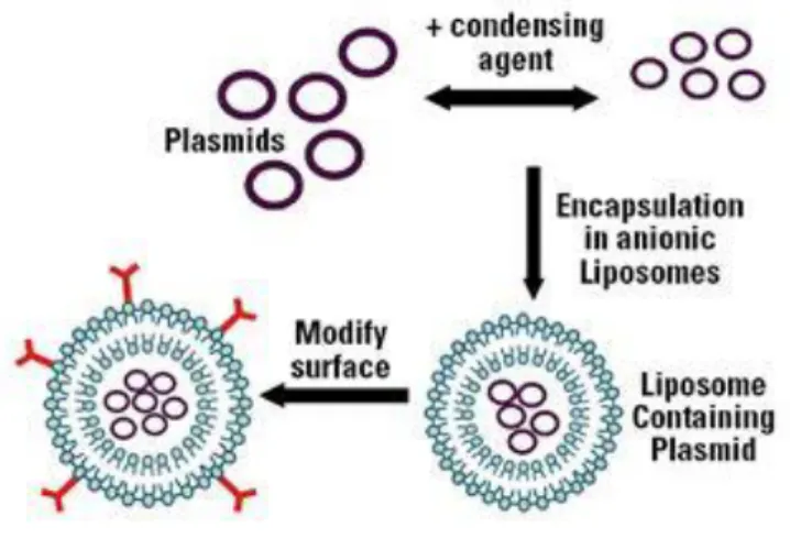
Liposomes - are spherical vesicles composed of a single lipid bilayer, where the
hydrophilic head groups are in contact with the aqueous environment and the hydrophobic tails face each other. The association with highly hydrophilic molecules, as pDNA and siRNA, increases with the utilization of cationic lipids, which contain one or more amines in the head group (Figure 9). The positive charge of the amino groups head group is not only necessary to incorporate the genetic material, but also for efficient binding to the cellular membrane.
Figure 9. Schematic incorporation of plasmids in liposomes, forming lipoplexes that can be modified with surface molecules for specific targeting.
23
Typical reagents for cationic lipid transfection are N-[1-(2,3-dioleyloxy)propyl]-N,N,N-trimethyl ammonium chloride (DOTMA), [1,2-bis(oleoyloxy)-3-(trimethylammonio)-propane] (DOTAP), 3β[N-(N’, N’-dimethylaminoethane)-carbamoyl] cholesterol (DC-Chol), and dioctadecylamidoglycylspermine (DOGS) [45]. Dioleoylphosphatidylethanolamine (DOPE) is often used, as neutral lipid, to improve the transfection efficiency, probably due to the conformational shift to an inverted hexagonal structure [45][41]. Besides, the use of cholesterol has been shown to increase the stability of liposomes, decreasing the probability of gene release by exposure to serum proteins [46]. However, the clinical application of liposomes is hindered by toxic-side effects, large size of the complexes and high surface charge, which lead to rapid renal clearance [46]. PEGylated-liposomes, with absorption or covalent attachment of polyethylene glycol (PEG), improve gene transfection in presence of serum, in vivo half-life of lipoplexes and reduced macrophage phagocytosis [45][46]. The property of phospholipids to fuse with the cellular membrane is one possibility of cell internalization, although endocytosis seems the major mechanism involved [41].
Cationic polymers - can be divided in natural and synthetic and biodegradable and
non-biodegradable [42]. Natural and biodegradable polymers like polyamides, polypeptides as poly-lysine (PLL), dextran and chitosan showed comparatively low transfection efficiency than synthetic polymers, like branched and linear polyethylenimine (PEI), and PAMAM dendrimers [47][41][48]. The biodegradability, if in one hand ensures the safety of the delivery system, can on the other hand undergo through degradation in biological environment, before reaching the target tissue.
24
Cationic polymers have the capacity to interact with nucleic acids for the high density of amino groups, which are protonable at neutral pH, but the high transfection efficiency mostly correlates with high cytotoxicity [48][49]. PEI is one of the most efficient cationic polymers in pDNA/siRNA delivery, but the high molecular weight and the high nitrogen/phosphate ratio (N/P) necessary for efficient transfection, cause cytotoxicity with limited application in vivo. PAMAM dendrimers have also shown good interaction with siRNA, with efficient ability to escape from endosomes, due to the internal tertiary amino groups [50]. The higher efficiency of PEI, compared with the other polymers, has been attributed to the higher capacity to escape from endosomes [41][48]. At intracellular level the ability to release pDNA/siRNA in the cytoplasm is of critical importance to avoid lysosomal degradation and has been found that endosomal escape is proportional to the protonable amines of the delivery agent. PLL has been demonstrated to have, at physiological pH, almost all the N-atoms protonated and the low transfection efficiency is attributed to the low capacity of activating the proton-sponge effect [47]. The proton-sponge effect assumes that amino group protonation delays the acidification that occurs during the maturation of endosomes. The ATPase proton pumps continue to actively translocate protons into the endosomes, which resist to acidification. The endosomal accumulation of protons is followed by passive entry of chloride ions and the ionic concentration leads to water influx. The osmotic pressure cause endosome swelling and wall rupture, causing pDNA/siRNA release in the cytoplasm [51][47].
PEI is partially protonated at physiological pH and the remaining amino-groups can be protonated in the acidic environment of endosomes, resulting more efficient in activating the proton-sponge effect. The importance of the phenomenon is demonstrated by the fact that most of the polyplexes showing low transfection
25
efficiency increase their delivery capacity after co-transfection with chloroquine, a lysomotropic compound [47].
For in vivo application the main concern about cationic polymers is the cytotoxicity and the poor circulation time, due to serum proteins and erythrocytes aggregation [44]. Davis et al. have recently developed the first polymer-based nanoparticle formulation for siRNA delivery entered in clinical trial and systemically administered to humans [52]. The formulation CALAA-01 is a four component system for siRNA therapeutics to be administered in cancer patients: a cyclic oligosaccharide (β-ciclodextrin) was incorporated into the backbone of linear polycation chains to condense the siRNA, amantane polyethylene glycol (AD-PEG) was used to stabilize the nanoparticles in biological fluids and human transferrin was adsorbed to the surface to target tumors [52][48].
1.5.3 Challenges of non-viral gene delivery.
Biological barriers and physical/chemical properties of the delivery system need to be considered for a therapeutic application. First, whether we consider, in vitro or in
vivo application, the genetic material needs to be protected by extracellular
nucleases. This point is particularly critical for siRNA delivery, because RNA is highly susceptible to RNAse degradation [41].
Secondly, the material should not aggregate with serum proteins in the bloodstream, it should possess a high circulation time, avoid the activation of the immune system, and finally cross the endothelial barrier. As discussed before, this operation is facilitated in case of tumor treatment, by EPR effect, but crossing the endothelium in presence of a healthy intima structure requires a specific uptake mechanism. The molecules known to be transported through endothelial cells are mainly plasma
26
proteins with bloodstream transport function, like low density lipoproteins (LDL), very low density lipoproteins (VLDL), albumin, insulin and the iron transporter transferrin [53].
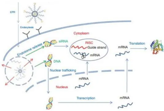
Once the gene delivery system reaches the target tissue it should be able to penetrate into the target cell and efficiently disassemble. Important difference to be considered between pDNA and siRNA delivery is the intracellular site where the mechanism takes place: RNAi pathway entirely plays in the cytoplasm, while pDNA needs to further penetrate the nuclear membrane to be expressed (Figure 10). Considering that endocytosis is the major uptake mechanism involved in the internalization of nanoparticles, the delivery system requires the ability to activate endosome escape and intracellular release of free pDNA/siRNA molecules [54].
Figure 10. Intracellular trafficking of nanoparticles for gene therapy. Endosome escape allows nuclear trafficking of DNA for gene expression and siRNA incorporation in the RISC complex for gene silencing.
27
1.5.4 Endocytosis mechanisms
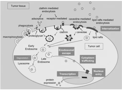
Cell membrane is impermeable to most of the macromolecules and particles and non-viral delivery systems have to follow specific pathways to be up-taken by cells. The knowledge of the uptake mechanisms and how they vary between the cell types is recognised, but not yet completely understood [49][55]. A part from certain types of liposomes, which have the capacity to fuse with the cellular membrane for their hydrophobic nature, most of the nanoparticles require a specific energy-dependent internalization mechanism [41]. Physical properties (nanoparticles size, charge, stiffness) and active targeting, through molecules that activate a receptor-mediated endocytosis, mainly regulate the uptake direction.
Phagocytosis is an uptake mechanism mostly activated by phagocytes, like monocytes and macrophages, which, being part of the immune system, are specialized in the internalization of viruses and bacteria [49]. The other mechanisms are grouped under the term of pinocytosis and are distinguished in: macropinocytosis, clathrin-mediated pathway, caveolae-mediated pathway and lipid raft-mediated pathway (Figure 11). The size of the nanoparticles firstly give an idea of the possible internalization mechanism, although the rigid classification into size parameters is under discussion [47]. Clathrin-coated pits are generally large 100-150 nm, caveolin-1 flask-shaped invagination around 50-80 nm, while macropinocytosis can cover big sizes of 500-2000 nm [48]. The abundance of caveolae, despite clathrin pits, depends on the cell type in consideration [56]. Recent studies have shown the ability of ―caveosomes‖, derived from multiple association of caveolae, to internalize big particles [57]. Microspheres with a diameter superior of 500 nm were selectively inhibited by cholesterol depletors, which block the internalization mediated by lipid rafts. Serum albumin, viruses like cholera toxin and bacteria are known to be internalized by caveolae-mediated endocytosis [58][59][60]. The knowledge about
28
uptake mechanisms is nowadays limited and other pathways might be involved in regulating the uptake of macromolecules. Besides, the uptake mechanism regulates the intracellular trafficking of nanoparticles: clathrin-based pits are transported through early and late endosomes directly to lysosome to be degraded, while caveolae-mediated endocytosis seems to transport molecules to the Golgi apparatus, convenient route for pathogens to avoid lysosomal degradation [60].
The capacity of the gene delivery carrier to escape form endosomes is particularly important to ensure the release of the therapeutic agent in the cytoplasm.
Figure 11. Schematic representation of the known intracellular trafficking pathways involved in the uptake mediated by endocytosis.
29
1.6 Polyethylenimine and toxicity issues.
PEI, in linear and branched form, resulted, as described before, one of the best pDNA/siRNA delivery carrier, but the efficiency is critically correlated with high molecular weight (25kDa) and high N/P ratio required for efficient transfection, with toxicity issues for systemic administration [41]. PEI has been demonstrated to induce cell membrane damage (necrosis post-treatment) and mitochondrial-mediated apoptosis in a later stage, 24 hours post-treatment. Furthermore, the high positive charge of the polyplexes lead to nanoparticles aggregation with serum proteins and erythrocytes in vivo [48].
Linear and branched PEI showed different ability to complex pDNA or siRNAs. In particular, linear polyethylenimine (lPEI) resulted more efficient to complex pDNA than siRNA; contrarily, branched polyethylenimine (bPEI) showed good ability to transport siRNA, probably due to the three-dimensional structure that allows multiple folding options for the siRNA [51].
Research efforts are, nowadays, focused in reducing PEI cytotoxicity, improving at the same time pDNA/siRNA delivery: modifications of PEI backbone and conjugation with neutral charged polymers or proteins are the most considered options for improving biocompatibility, bloodstream circulation and cell targeting [50]. Modification of the PEI backbone with poly-ethylene glycol (PEG) has been already demonstrated to reduce non-specific localization in liver, spleen and lung, compared to the PEI polyplexes. However, the uptake efficiency by cells resulted low for the dramatic reduction of the surface charge [61].
A lot of efforts are nowadays directed to functionalize PEI structure: amino groups acetylation, succinylation, alkylation are proposed for improving siRNA-therapeutics, reducing toxic side effects [50] (Figure 12). Moreover, the oxidation of
30
the amino groups of PEI with hydrogen peroxide (H2O2) showed reduced
cytotoxicity of PEI-DNA polyplexes, without compromising, although increasing green fluorescent protein (GFP) expression [62].
Figure 12. Functionalization of bPEI by acetylation, succinylation, PEGylation and modification with hydrophobic choresterol, long alkyl chains and PBLG chains for decreasing the cytotoxicity, increasing the stability of polycation complexes and prolonged blood circulation time [50].
Besides the structural modifications, the conjugation of PEI with ―cell-targeting ligands‖, as serum proteins or macromolecules, have been tested. The conjugation with cholesterol and long alkyl groups, for example, can increase the possibility to target the liver by interacting in vivo with the high-density lipoprotein (HDL) and low-density lipoprotein (LDL) receptors [37]. More recently, the conjugation of PEI
31
with galactose has been investigated to increase a receptor-mediated endocytosis in hepatocytes [48].
PEI conjugation with transferrin (Tf), epithelial growth factor (EGF) and folic acid was studied for the possibility to target cancer, considering that tumor cells over-express their receptors. Ogris et al., have demonstrated that PEI conjugated with transferrin, improves gene transfection in human erythromyeloid leukemia cells, compared to unmodified PEI polyplexes [63].
1.7 Active targeting: the potential of albumin.
Serum proteins have been recently discovered to serve as endogenous targeting ligands and for increasing nanoparticles circulation, after injection [44][53]. In Kim
et al., have reported that apolipoprotein A-I, a component of the high density
lipoprotein (HDL), can be assembled with liposomes to address siRNA delivery to the liver, through specific receptor-mediated internalization in hepatocytes [64]. Transferrin, the iron transporter in the bloodstream, in the recent years has been largely employed for non-viral gene therapy, as the cited CALAA-01 siRNA-therapeutic agent and PEI-Tf-DNA complexes. The future direction of genetic medicine is, therefore, direct the therapeutic agent to the target cells, ensuring a good transfection efficiency, which translates in high therapeutic effect.
Nowadays, plasma proteins are gaining attention for the possibility to increase the half-life of a therapeutic agent and the possibility to target tumors, due to the EPR effect. Serum proteins, as transferrin, albumin, LDL are promising as anti-neoplastic agents [53].
Human serum albumin (HSA) is the most abundant protein in human plasma and it transports a broad range of molecules in the bloodstream: fatty acids, therapeutic
32
drugs (aspirin, penicillins, benzodiazepines), metabolites and metal ions (copper II, nickel II, calcium II, zinc II) [53].
HSA has been discovered to accumulate in malignant tumors and inflamed tissues and the therapeutic application as drug delivery carrier has been validate with the commercialized agent Abraxane (ABI-007) [65]. Abraxane consists in paclitaxel albumin-loaded nanoparticles, which improves the anticancer therapeutic efficiency of paclitaxel in the treatment of metastatic breast cancer. Other types of malignant tumors have been tested for the therapeutic efficiency of Abraxane, for example head and neck cancer [66].
A recent study has demonstrated the role of albumin in preventing non-specific protein absorption to nanoparticles in presence of serum, with potential to enhance nanoparticles circulation time [67]. Moreover, it is known that albumin, further being transported to tumors by EPR effect, possesses the ability to actively cross the endothelial membrane by gp60 receptor- mediated transcytosis [56][68][69]. The internalization of HSA has been demonstrated to involve caveolae-mediated endocytosis in endothelial cells [56].
In the intra-tumor environment the secreted protein acidic and rich in cysteine (SPARC) has been shown to have a role in binding albumin, facilitating the transportation in tumor cells, to probably being used as source of amino-acids and energy [70]. SPARC and gp60 share a region of structural homology, which is associated with the common albumin binding activity [71].The over-expression of SPARC and caveolae in many malignant tumors have been correlated with high tumor invasion and metastasis [66][72].
Therefore, many advantages could derive from the use of albumin as gene delivery carrier, including high circulation time in the bloodstream, tolerance from the immune system and tumor targeting.
33
1.8 References
[1] K. Riehemann, S. W. Schneider, T. a Luger, B. Godin, M. Ferrari, and H. Fuchs, ―Nanomedicine--challenge and perspectives.,‖ Angew. Chem. Int. Ed. Engl., vol. 48, no. 5, pp. 872–97, Jan. 2009.
[2] S. M. Moghimi, a C. Hunter, and J. C. Murray, ―Nanomedicine: current status and future prospects.,‖ FASEB J., vol. 19, no. 3, pp. 311–30, Mar. 2005.
[3] D. Peer, J. M. Karp, S. Hong, O. C. Farokhzad, R. Margalit, and R. Langer, ―Nanocarriers as an emerging platform for cancer therapy.,‖ Nat. Nanotechnol., vol. 2, no. 12, pp. 751–60, Dec. 2007.
[4] H. Maeda, J. Wu, T. Sawa, Y. Matsumura, and K. Hori, ―Tumor vascular permeability and the EPR effect in macromolecular therapeutics: a review,‖ J. Control. Release, vol. 65, no. 1–2, pp. 271–284, Mar. 2000.
[5] V. P. Torchilin, ―Drug targeting,‖ Eur. J. Pharm. Sci., vol. 11, pp. S81–S91, Oct. 2000. [6] F. Danhier, O. Feron, and V. Préat, ―To exploit the tumor microenvironment: Passive and
active tumor targeting of nanocarriers for anti-cancer drug delivery.,‖ J. Control. Release, vol. 148, no. 2, pp. 135–46, Dec. 2010.
[7] S. Nie, ―Understanding and overcoming major barriers in cancer nanomedicine.,‖ Nanomedicine (Lond)., vol. 5, no. 4, pp. 523–8, Jun. 2010.
[8] J. S. Desgrosellier and D. a Cheresh, ―Integrins in cancer: biological implications and therapeutic opportunities.,‖ Nat. Rev. Cancer, vol. 10, no. 1, pp. 9–22, Jan. 2010.
[9] Y. Mo and L.-Y. Lim, ―Preparation and in vitro anticancer activity of wheat germ agglutinin (WGA)-conjugated PLGA nanoparticles loaded with paclitaxel and isopropyl myristate.,‖ J. Control. Release, vol. 107, no. 1, pp. 30–42, Sep. 2005.
[10] J. D. Byrne, T. Betancourt, and L. Brannon-Peppas, ―Active targeting schemes for nanoparticle systems in cancer therapeutics.,‖ Adv. Drug Deliv. Rev., vol. 60, no. 15, pp. 1615–26, Dec. 2008.
[11] J. Y. Yhee, S. J. Lee, S. Lee, S. Song, H. S. Min, S.-W. Kang, S. Son, S. Y. Jeong, I. C. Kwon, S. H. Kim, and K. Kim, ―Tumor-targeting transferrin nanoparticles for systemic polymerized siRNA delivery in tumor-bearing mice.,‖ Bioconjug. Chem., vol. 24, no. 11, pp. 1850–60, Nov. 2013.
34
[12] J. M. Morachis, E. a Mahmoud, and A. Almutairi, ―Physical and chemical strategies for therapeutic delivery by using polymeric nanoparticles.,‖ Pharmacol. Rev., vol. 64, no. 3, pp. 505–19, Jul. 2012.
[13] R.-Y. Lin, K. Dayananda, T.-J. Chen, C.-Y. Chen, G.-C. Liu, K.-L. Lin, and Y.-M. Wang, ―Targeted RGD nanoparticles for highly sensitive in vivo integrin receptor imaging.,‖ Contrast Media Mol. Imaging, vol. 7, no. 1, pp. 7–18, 2012.
[14] K. Lee, E. a Silva, and D. J. Mooney, ―Growth factor delivery-based tissue engineering: general approaches and a review of recent developments.,‖ J. R. Soc. Interface, vol. 8, no. 55, pp. 153–70, Feb. 2011.
[15] E. Engel, A. Michiardi, M. Navarro, D. Lacroix, and J. a Planell, ―Nanotechnology in regenerative medicine: the materials side.,‖ Trends Biotechnol., vol. 26, no. 1, pp. 39–47, Jan. 2008.
[16] B. Munksgaard, ―Regeneration of vascularized bone,‖ vol. 41, pp. 109–122, 2006.
[17] K. Kim and J. P. Fisher, ―Nanoparticle technology in bone tissue engineering.,‖ J. Drug Target., vol. 15, no. 4, pp. 241–52, May 2007.
[18] H. G. Schmoekel, F. E. Weber, J. C. Schense, K. W. Grätz, P. Schawalder, and J. a Hubbell, ―Bone repair with a form of BMP-2 engineered for incorporation into fibrin cell ingrowth matrices.,‖ Biotechnol. Bioeng., vol. 89, no. 3, pp. 253–62, Feb. 2005.
[19] T. N. Vo, F. K. Kasper, and A. G. Mikos, ―Strategies for controlled delivery of growth factors and cells for bone regeneration.,‖ Adv. Drug Deliv. Rev., vol. 64, no. 12, pp. 1292–309, Sep. 2012.
[20] H. C. S. Der, L. Kurz, W. E. G. M, and B. Lorenz, ―Polyphosphate in Bone,‖ vol. 65, no. 3, pp. 296–303, 2000.
[21] J. Caetano-lopes, H. Canhão, and F. J. Eurico, ―Osteoblasts and bone formation,‖ pp. 103– 110.
[22] Y. L. C.S. Soltanoff, W.Chen, S. Yang, ―Signaling Networks that Control the Lineage Commitment and Differentiation of Bone Cells,‖ Crit Rev Eukaryot Gene Expr, vol. 19, no. 1, pp. 1–46, 2009.
[23] R. Dinarvand, N. Sepehri, S. Manoochehri, H. Rouhani, and F. Atyabi, ―Polylactide-co-glycolide nanoparticles for controlled delivery of anticancer agents.,‖ Int. J. Nanomedicine, vol. 6, pp. 877–95, Jan. 2011.
35
[24] H. S. Choi, W. Liu, P. Misra, E. Tanaka, J. P. Zimmer, B. Itty Ipe, M. G. Bawendi, and J. V Frangioni, ―Renal clearance of quantum dots.,‖ Nat. Biotechnol., vol. 25, no. 10, pp. 1165–70, Oct. 2007.
[25] C. Wong, T. Stylianopoulos, J. Cui, J. Martin, V. P. Chauhan, W. Jiang, Z. Popovic, R. K. Jain, M. G. Bawendi, and D. Fukumura, ―Multistage nanoparticle delivery system for deep penetration into tumor tissue.,‖ Proc. Natl. Acad. Sci. U. S. A., vol. 108, no. 6, pp. 2426–31, Mar. 2011.
[26] R. K. Jain, ―Understanding Barriers to Drug Delivery : High Resolution in Vivo Imaging Is Key Understanding Barriers to Drug Delivery : High Resolution in Vivo Imaging Is Key,‖ pp. 1605–1606, 1999.
[27] H. Hashizume, P. Baluk, S. Morikawa, J. W. McLean, G. Thurston, S. Roberge, R. K. Jain, and D. M. McDonald, ―Openings between defective endothelial cells explain tumor vessel leakiness.,‖ Am. J. Pathol., vol. 156, no. 4, pp. 1363–80, Apr. 2000.
[28] a Pluen, Y. Boucher, S. Ramanujan, T. D. McKee, T. Gohongi, E. di Tomaso, E. B. Brown, Y. Izumi, R. B. Campbell, D. a Berk, and R. K. Jain, ―Role of tumor-host interactions in interstitial diffusion of macromolecules: cranial vs. subcutaneous tumors.,‖ Proc. Natl. Acad. Sci. U. S. A., vol. 98, no. 8, pp. 4628–33, Apr. 2001.
[29] R. a Petros and J. M. DeSimone, ―Strategies in the design of nanoparticles for therapeutic applications.,‖ Nat. Rev. Drug Discov., vol. 9, no. 8, pp. 615–27, Aug. 2010.
[30] H.-C. Huang, S. Barua, G. Sharma, S. K. Dey, and K. Rege, ―Inorganic nanoparticles for cancer imaging and therapy.,‖ J. Control. Release, vol. 155, no. 3, pp. 344–57, Nov. 2011. [31] J.-O. You and D. T. Auguste, ―Nanocarrier cross-linking density and pH sensitivity regulate
intracellular gene transfer.,‖ Nano Lett., vol. 9, no. 12, pp. 4467–73, Dec. 2009.
[32] J. Voigt, J. Christensen, and V. P. Shastri, ―Differential uptake of nanoparticles by endothelial cells through polyelectrolytes with affinity for caveolae.,‖ Proc. Natl. Acad. Sci. U. S. A., vol. 111, no. 8, pp. 2942–7, Feb. 2014.
[33] M. Lundqvist, J. Stigler, G. Elia, I. Lynch, T. Cedervall, and K. a Dawson, ―Nanoparticle size and surface properties determine the protein corona with possible implications for biological impacts.,‖ Proc. Natl. Acad. Sci. U. S. A., vol. 105, no. 38, pp. 14265–70, Sep. 2008.
[34] S. Misra, ―Human gene therapy: a brief overview of the genetic revolution.,‖ J. Assoc. Physicians India, vol. 61, no. 2, pp. 127–33, Feb. 2013.
[35] T. P. O’Connor and R. G. Crystal, ―Genetic medicines: treatment strategies for hereditary disorders.,‖ Nat. Rev. Genet., vol. 7, no. 4, pp. 261–76, Apr. 2006.
36
[36] R. L. Kanasty, K. a Whitehead, A. J. Vegas, and D. G. Anderson, ―Action and reaction: the biological response to siRNA and its delivery vehicles.,‖ Mol. Ther., vol. 20, no. 3, pp. 513– 24, Mar. 2012.
[37] K. a Whitehead, R. Langer, and D. G. Anderson, ―Knocking down barriers: advances in siRNA delivery.,‖ Nat. Rev. Drug Discov., vol. 8, no. 2, pp. 129–38, Feb. 2009.
[38] A. Pauli, J. L. Rinn, and A. F. Schier, ―Non-coding RNAs as regulators of embryogenesis.,‖ Nat. Rev. Genet., vol. 12, no. 2, pp. 136–49, Feb. 2011.
[39] T. G. McDaneld, ―MicroRNA: mechanism of gene regulation and application to livestock.,‖ J. Anim. Sci., vol. 87, no. 14 Suppl, pp. E21–8, Apr. 2009.
[40] D. W. Bartlett and M. E. Davis, ―Insights into the kinetics of siRNA-mediated gene silencing from live-cell and live-animal bioluminescent imaging.,‖ Nucleic Acids Res., vol. 34, no. 1, pp. 322–33, Jan. 2006.
[41] W. Wang, W. Li, N. Ma, and G. Steinhoff, ―Non-Viral Gene Delivery Methods,‖ Curr. Pharm. Biotechnol., vol. 14, no. 1, pp. 46–60, Jan. 2013.
[42] E. Cevher, A. D. Sezer, and E. Ş. Çağlar, ―Gene Delivery Systems : Recent Progress in Viral and Non-Viral Therapy,‖ 2012.
[43] L. S. Young, P. F. Searle, D. Onion, and V. Mautner, ―Viral gene therapy strategies: from basic science to clinical application.,‖ J. Pathol., vol. 208, no. 2, pp. 299–318, Jan. 2006. [44] R. Kanasty, J. R. Dorkin, A. Vegas, and D. Anderson, ―Delivery materials for siRNA
therapeutics.,‖ Nat. Mater., vol. 12, no. 11, pp. 967–77, Nov. 2013.
[45] D. a Balazs and W. Godbey, ―Liposomes for use in gene delivery.,‖ J. Drug Deliv., vol. 2011, p. 326497, Jan. 2011.
[46] T. M. Allen and P. R. Cullis, ―Liposomal drug delivery systems: from concept to clinical applications.,‖ Adv. Drug Deliv. Rev., vol. 65, no. 1, pp. 36–48, Jan. 2013.
[47] S. C. De Smedt, J. Demeester, and W. E. Hennink, ―Cationic Polymer Based Gene Delivery Systems,‖ vol. 17, no. 2, 2000.
[48] Y. Yue and C. Wu, ―Progress and perspectives in developing polymeric vectors for in vitro gene delivery,‖ Biomater. Sci., vol. 1, no. 2, p. 152, 2013.
[49] L. Parhamifar, A. K. Larsen, a. C. Hunter, T. L. Andresen, and S. M. Moghimi, ―Polycation cytotoxicity: a delicate matter for nucleic acid therapy—focus on polyethylenimine,‖ Soft Matter, vol. 6, no. 17, p. 4001, 2010.
37
[50] A. Tamura and Y. Nagasaki, ―Smart siRNA delivery systems based on polymeric nanoassemblies and nanoparticles.,‖ Nanomedicine (Lond)., vol. 5, no. 7, pp. 1089–102, Sep. 2010.
[51] W. Liang and J. K. W. Lam, ―Endosomal Escape Pathways for Non-Viral Nucleic Acid Delivery Systems,‖ 2012.
[52] J. E. Zuckerman, I. Gritli, a. Tolcher, J. D. Heidel, D. Lim, R. Morgan, B. Chmielowski, a. Ribas, M. E. Davis, and Y. Yen, ―Correlating animal and human phase Ia/Ib clinical data with CALAA-01, a targeted, polymer-based nanoparticle containing siRNA,‖ Proc. Natl. Acad. Sci., vol. 111, no. 31, Jul. 2014.
[53] F. Kratz and U. Beyer, ―Serum Proteins as Drug Carriers of Anticancer Agents : A Review,‖ Inf. Heathcare, vol. 5, no. 4, pp. 281–299, 1998.
[54] C. Scholz and E. Wagner, ―Therapeutic plasmid DNA versus siRNA delivery: common and different tasks for synthetic carriers.,‖ J. Control. Release, vol. 161, no. 2, pp. 554–65, Jul. 2012.
[55] M. Morille, C. Passirani, A. Vonarbourg, A. Clavreul, and J.-P. Benoit, ―Progress in developing cationic vectors for non-viral systemic gene therapy against cancer.,‖ Biomaterials, vol. 29, no. 24–25, pp. 3477–96, 2008.
[56] R. D. Minshall, C. Tiruppathi, S. M. Vogel, W. D. Niles, a Gilchrist, H. E. Hamm, and a B. Malik, ―Endothelial cell-surface gp60 activates vesicle formation and trafficking via G(i)-coupled Src kinase signaling pathway.,‖ J. Cell Biol., vol. 150, no. 5, pp. 1057–70, Sep. 2000. [57] A. Manuscript and L. E. Cells, ―NIH Public Access,‖ vol. 3, no. 12, pp. 4110–4116, 2013. [58] J. Rejman, A. Bragonzi, and M. Conese, ―Role of clathrin- and caveolae-mediated endocytosis
in gene transfer mediated by lipo- and polyplexes.,‖ Mol. Ther., vol. 12, no. 3, pp. 468–74, Oct. 2005.
[59] J. Rejman, V. Oberle, I. S. Zuhorn, and D. Hoekstra, ―Size-dependent internalization of particles via the pathways of clathrin- and caveolae-mediated endocytosis.,‖ Biochem. J., vol. 377, no. Pt 1, pp. 159–69, Jan. 2004.
[60] I. A. Khalil, K. Kogure, H. Akita, and H. Harashima, ―Uptake Pathways and Subsequent Intracellular Trafficking in Nonviral Gene Delivery,‖ vol. 58, no. 1, pp. 32–45, 2006.
[61] M. Neu, D. Fischer, and T. Kissel, ―Recent advances in rational gene transfer vector design based on poly(ethylene imine) and its derivatives.,‖ J. Gene Med., vol. 7, no. 8, pp. 992–1009, Aug. 2005.
38
[62] W. Y. Seow, K. Liang, M. Kurisawa, and C. a E. Hauser, ―Oxidation as a facile strategy to reduce the surface charge and toxicity of polyethyleneimine gene carriers.,‖ Biomacromolecules, vol. 14, no. 7, pp. 2340–6, Jul. 2013.
[63] M. Ogris, P. Steinlein, S. Carotta, S. Brunner, and E. Wagner, ―DNA/polyethylenimine transfection particles: influence of ligands, polymer size, and PEGylation on internalization and gene expression.,‖ AAPS PharmSci, vol. 3, no. 3, p. E21, Jan. 2001.
[64] S. I. Kim, D. Shin, H. Lee, B.-Y. Ahn, Y. Yoon, and M. Kim, ―Targeted delivery of siRNA against hepatitis C virus by apolipoprotein A-I-bound cationic liposomes.,‖ J. Hepatol., vol. 50, no. 3, pp. 479–88, Mar. 2009.
[65] F. Kratz, ―Albumin as a drug carrier: design of prodrugs, drug conjugates and nanoparticles.,‖ J. Control. Release, vol. 132, no. 3, pp. 171–83, Dec. 2008.
[66] N. Desai, V. Trieu, B. Damascelli, and P. Soon-shiong, ―SPARC Expression Correlates with Tumor Response to Albumin-Bound Paclitaxel in Head and Neck Cancer Patients,‖ Transl. Oncol., vol. 2, no. 2, pp. 59–64, 2009.
[67] C. M. Kummitha, A. S. Malamas, and Z.-R. Lu, ―Albumin pre-coating enhances intracellular siRNA delivery of multifunctional amphiphile/siRNA nanoparticles.,‖ Int. J. Nanomedicine, vol. 7, pp. 5205–14, Jan. 2012.
[68] C. Tiruppathi, ―Gp60 Activation Mediates Albumin Transcytosis in Endothelial Cells by Tyrosine Kinase-dependent Pathway,‖ J. Biol. Chem., vol. 272, no. 41, pp. 25968–25975, Oct. 1997.
[69] W. Schubert, P. G. Frank, B. Razani, D. S. Park, C. W. Chow, and M. P. Lisanti, ―Caveolae-deficient endothelial cells show defects in the uptake and transport of albumin in vivo.,‖ J. Biol. Chem., vol. 276, no. 52, pp. 48619–22, Dec. 2001.
[70] E. Frei, ―Albumin binding ligands and albumin conjugate uptake by cancer cells.,‖ Diabetol. Metab. Syndr., vol. 3, no. 1, p. 11, Jan. 2011.
[71] J. A. N. E. Schnitzer, ―Antibodies to SPARC inhibit albumin binding endothelium to SPARC , gp60 , and microvascular,‖ 1992.
[72] Y. Lavie, G. Fiucci, and M. Liscovitch, ―Upregulation of caveolin in multidrug resistant cancer cells: functional implications.,‖ Adv. Drug Deliv. Rev., vol. 49, no. 3, pp. 317–23, Jul. 2001.
40
2 Outline of the Thesis
The overall aim of this thesis is to improve branched polyethylenimine (bPEI) delivery system for gene therapy application. As previously discussed in the introduction, despite the good efficiency in condensing nucleic acids and promoting cell transfection, the major limitation of PEI for in vivo application is the high cytotoxicity, due to the high positive charged amino groups density.
The effect of amino groups acetylation on bPEI backbone and the incorporation of human serum albumin (HSA) in bPEI-siRNA polyplexes have been investigated to improve cell transfection efficiency and reduce the toxicity that accompanies the delivery efficiency.
Firstly, my project was focused in investigating the effect of partial acetylation of primary amino groups, along PEI backbone, in the capacity to form nanoparticles (NPs) with poly(lactic-co-glycolic acid) (PLGA), the influence of acetylated PEI (AcPEI) in nanoparticles size and surface charge and the ability of NPs to deliver genetic material. Particular attention was paid in testing cell cytotoxicity, genotoxicity and immunotoxicity, following transfection of both PEI-PLGA (PEI-NPs) and AcPEI NPs, with the aim to increase the biocompatibility of the system for in vivo applications. Nanotoxicity is often underestimated, but it is of fundamental importance for therapeutic use. Considering the application of NPs as medical devices, collateral effects as inflammation, immunoreaction, or cancer, can emerge due to NPs toxicity. Free oxygen radicals production, following cell stress and immune system activation, can contribute in the risk of DNA mutation and cancer. Therefore we concentrated our work in studying PEI-NPs capability of promoting
41
DNA damage and intracellular ROS release, investigating the potential of AcPEI-NPs, with 50% of acetylated amino groups, in reducing the toxicity of PEI, without compromising the transfection efficiency.
After having demonstrated the effectiveness of AcPEI gene delivery system in human umbelical vein endothelial cells (HUVEC), we focused our study on the possibility to use acetylated PEI nanoparticles as gene delivery carrier in tissue engineering. In this study, the internalization efficiency, the mechanism of uptake involved and the endosome escape efficiency were investigated in human primary osteoblasts (hOBs), isolated from human trabecular bone. The cytotoxicity was evaluated in terms of membrane damage (necrosis) or activation of apoptosis, while the effect of the nanoparticles on hOB activity and differentiation was studied quantifying alkaline phosphatase (ALP) activity, collagen type I production (COL1), mineralization and gene expression of markers of differentiated osteoblasts. The results will be used for future application of these nanoparticles in gene delivery to promote osteogenesis and in siRNA delivery to inhibit bone formation in blood vessel walls.
The project, developed at the University of Freiburg, involved the incorporation of human serum albumin (HSA) into pre-formed bPEI-siRNA polyplexes, with the aim to improve transfection efficiency in the tumor microenvironment, since HSA has been demonstrated to accumulate in inflamed tissues and tumors [65]. The ternary complex, formed by electrostatic interactions, was characterized in terms of size and surface charge. The intracellular delivery efficiency, mechanism of uptake and silencing efficiency were evaluated with in vitro studies, investigating a direct role of HSA in promoting cell uptake and gene silencing, compared to bPEI-siRNA complexes. Further studies will be focused in investigating a possible HSA receptor
42
mediated endocytosis of the complexes, for HSA utilization as a tumor targeting molecule.
![Figure 2. Physical and chemical properties for the design of an optimal drug-delivery agent [3]](https://thumb-eu.123doks.com/thumbv2/123dokorg/4809869.49803/12.893.183.808.562.1021/figure-physical-chemical-properties-design-optimal-delivery-agent.webp)
![Figure 6. RNAi endogenous mechanisms for protein silencing: similarity and differences in siRNA and miRNA mechanism [36]](https://thumb-eu.123doks.com/thumbv2/123dokorg/4809869.49803/23.893.213.803.538.997/figure-endogenous-mechanisms-protein-silencing-similarity-differences-mechanism.webp)