1 Gut Month 2017 Vol 0 No 0
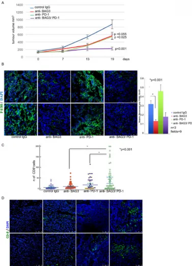
Figure 1 Combined effect of anti-Bcl-2-Associated athanoGene 3 (anti-BAG3) and anti-PD-1
antibodies in inhibiting pancreatic ductal adenocarcinomas growth and increasing the number of CD8+cells in tumours. (A) Tumour growth of syngeneic grafted tumours treated as indicated.
(B) Analysis of macrophage infiltrate in tumours by F480 staining. (C) Count of CD8+ infiltrating
lymphocytes in tumours. (D) Representative images of CD8 positivity in excised tumours of the four different groups. For more details see online supplementary information.
Letter
Combined effect of anti-BAG3
and anti-PD-1 treatment on
macrophage infiltrate, CD8
+T cell number and tumour
growth in pancreatic cancer
We read with great interest the article by Zhang et al1 showing that CD8+ cellinfiltration in pancreatic tumours can be enhanced by depletion of myeloid cells (CD11b+ macrophages and
myeloid-de-rived suppressor cells) and that the depletion of CD11b+ cells resulted in
decreased PD-L1 expression on cancer cells thus impairing the triggering of the inhibitory receptor PD-1 on T cells.1
Recruitment and activation of CD8+
lymphocytes in tumours are suppressed by mechanisms only partially understood and
rescuing CD8+ cell infiltrate in tumours
is one of the objectives of immunothera-pies.1 2 Tumour-associated macrophages
(TAMs) play a crucial role in the relation between tumour cells and their environ-ment.3 Here, we confirm the interplay
between macrophages and CD8+ cells in
pancreatic cancer and identify a poten-tial way to exploit this enhancing effect of anti-PD-1 treatment. Indeed, we show that reduction of macrophage infiltrate, through treatment with an anti-Bcl-2-As-sociated athanoGene 3 (BAG3) antibody,4
results in increased number of CD8+
cells in pancreatic tumours in a murine model. BAG3 is a co-chaperone of the heat shock protein 70 whose expression is induced in response to stress but has been shown to be constitutive in cancers including pancreatic ductal adenocar-cinomas (PDAC). We recently showed that BAG3 is secreted by PDAC cells and binds to TAMs, inducing their activation and the release of factors that support
tumour growth and metastatic spreading. Treatment of PDAC-carrying mice with an anti-BAG3 mAb that interacts with a portion of BAG3 protein (from aa 385 to aa 399) not overlapping the BAG domain resulted in reduced tumour growth and metastatic spreading; analysis of tumour biopsies from anti-BAG3-treated mice showed a marked reduction of TAMs and a decrease in macrophage-released cyto-kines.4 5
Now we used the anti-BAG3 mAb in combination with an anti-PD-1 antibody to treat mice PDAC allografts. To this end, murine Kras-driven pancreatic cancer cells (mt4-2D)6 were subcutaneously grafted in
syngeneic immunocompetent (C57BL6) mice4 and when tumours size reached
about 100 mm3 mice were treated for 19
days with an BAG3 mAb, an anti-PD-1 antibody or a combination of the two. Histochemical staining showed that tumours appeared as low differentiated pancreatic adenocarcinoma; as expected for tumours derived from K-Ras-mutant cells, all tumour showed high expression of phospho-ERK (online supplementary figure 1) Treatment with either anti-BAG3 mAb or anti-PD-1 antibody resulted in a significant reduction of tumour growth that was even more impressive when the two antibodies were used in combina-tion, suggesting that the block of BAG3 activity was additive with the block of PD-1 (figure 1A) as demonstrated by the not significant interaction term in a two-way analysis of variance (online supplementary figure 2). As previously reported, treatment with anti-BAG3 mAb resulted in reduced macrophage infil-trate4 that on the contrary appeared to
be increased in anti-PD-1-treated animals (figure 1B). Interestingly, the macrophage infiltrate was also reduced in the animals treated with the combination of the two antibodies. Analysis of tumour sections showed that CD8+ cells were hardly
detectable in control tumours, while their number was increased following treatment with anti-BAG3 or anti-PD-1 antibody and a higher effect was observed in mice treated with the two in combina-tion (figure 1C, D) .
In conclusion, blocking BAG3 activity results in an increased number of CD8+
cells, with potential antitumour effects. Such increase is likely to be due, at least in part, to the decrease of TAMs-derived factors4 that suppress CD8+ lymphocytes
influx or subsistence in tumour tissues. Whether BAG3 also impacts on other regulatory circuits requires further inves-tigation, nevertheless, our observations disclose a BAG3-mediated mechanism
PostScript
Gut Online First, published on August 11, 2017 as 10.1136/gutjnl-2017-314225
Copyright Article author (or their employer) 2017. Produced by BMJ Publishing Group Ltd (& BSG) under licence.
group.bmj.com
on October 9, 2017 - Published by
http://gut.bmj.com/
2 Gut Month 2017 Vol 0 No 0
PostScript
that suppresses CD8+ cell recruitment.
Furthermore, our findings indicate the potential effectiveness of anti-BAG3-di-rected and anti-PD-1-dianti-BAG3-di-rected strategies in fighting pancreatic cancer.
Vittoria Iorio,1,2 Alessandra Rosati,1,2
Raffaella D'Auria,1,2 Margot De Marco,1,2
Liberato Marzullo,1,2 Anna Basile,1,2
Michelina Festa,1,3 Maria Pascale,1,3
Paolo Remondelli,2 Mario Capunzo,2
Gianluca Sala,4 Verena Damiani,4
Giuseppina Amodio,2 Marta Di Nicola,4
Rossano Lattanzio,4 Maria Caterina Turco,1,2
Vincenzo De Laurenzi1,4
1Biouniversa srl, c/o University of Salerno, Montoro, Avellino, Italy
2Department of Medicine,Surgery and Dentistry, Schola Medica Salernitana, University of Salerno, Baronissi, Salerno, Italy
3Department of Pharmacy, Division of Biomedicine ‘A Leone’, University of Salerno, Fisciano, Salerno, Italy 4Department of Medicine and Biotechnology, University G d'Annunzio and CeSI-Met, Chieti, Italy
Correspondence to Professor Maria Caterina turco, Department of Medicine and Surgery Schola Medica Salernitana, University of Salerno, Baronissi, Salerno 84081, Italy; mcturco@ unisa. it
Acknowledgements this work was supported in part by Associazione Italiana per la ricerca sul Cancro (AIrC IG-14701) to MCt (AIrC IG-15196), VDL and (AIrC IG −18467) GS and by Ministero dell’ Istruzione, dell’Università e della ricerca (PrIN 2012CK5rPF_003). We acknowledge Professor Dave tuveson for kindly providing murine Kras-driven pancreatic cancer cell line (mt4-2D).
Contributors VI, Ar, MCt and VDL were involved in the study concept and design and drafting and revision of the manuscript. VI, Ar, rDA, MDM, LM, AB, MF, MP, MC, GS, MDN, rL and VD were involved in acquisition of data, analysis, interpretation of data and statistical analysis. Pr and GA provided technical support. Competing interests Ar, MDM, LM, AB, MF, MP, VDL and MCt are shareholders of the Academic Spin-off BIOUNIVerSA that provided anti-BAG3 antibodies. the remaining authors declare no competing financial interests.
Provenance and peer review Not commissioned; externally peer reviewed.
Open Access this is an Open Access article distributed in accordance with the Creative Commons Attribution Non Commercial (CC BY-NC 4.0) license, which permits others to distribute, remix, adapt, build upon this work non-commercially, and license their derivative works on different terms, provided the original work is properly cited and the use is non-commercial. See: http:// creativecommons. org/ licenses/ by- nc/ 4. 0/
© Article author(s) (or their employer(s) unless otherwise stated in the text of the article) 2017. All rights reserved. No commercial use is permitted unless otherwise expressly granted.
► Additional material is published online only. to view please visit the journal online (http:// dx. doi. org/ 10. 1136/ gutjnl- 2017- 314225)
VI and Ar contributed equally.
To cite Iorio V, rosati A, D'Auria r, et al. Gut Published Online First: [please include Day Month Year]. doi:10.1136/gutjnl-2017-314225 Received 28 March 2017 revised 26 July 2017 Accepted 29 July 2017 Gut 2017;0:1–2. doi:10.1136/gutjnl-2017-314225 RefeRences
1 Zhang Y, Velez-Delgado A, Mathew e, et al. Myeloid cells are required for PD-1/PD-L1 checkpoint activation and the establishment of an
immunosuppressive environment in pancreatic cancer.
Gut 2017;66:124–36.
2 Anderson KG, Stromnes IM, Greenberg PD. Obstacles posed by the tumor microenvironment to t cell activity: a case for synergistic therapies. Cancer Cell
2017;31:311–25.
3 Mantovani A, Marchesi F, Malesci A, et al. tumour-associated macrophages as treatment targets in oncology. Nat Rev Clin Oncol 2017;14:399–416. 4 rosati A, Basile A, D'Auria r, et al. BAG3 promotes
pancreatic ductal adenocarcinoma growth by activating stromal macrophages. Nat Commun 2015;6:8695. 5 ray K. Pancreatic cancer: new insights into PDAC
growth promotion via a BAG3-mediated paracrine loop. Nat Rev Gastroenterol Hepatol 2015;12:669. 6 Boj SF, Hwang CI, Baker LA, et al. Organoid models
of human and mouse ductal pancreatic cancer. Cell
2015;160:324–38.
group.bmj.com
on October 9, 2017 - Published by
http://gut.bmj.com/