Università degli Studi di Urbino “Carlo Bo”
Dipartimento di Scienze Biomolecolari
Dottorato di Ricerca in
Metodologie Biochimiche e Farmacologiche
Ciclo XXVIII
Development of an innovative strategy
based on engineered autologous erythrocytes
as “enzyme replacement therapy” for the
treatment of Phenylketonuria
SSD BIO/10
RELATRICE
DOTTORANDA
Chiar.ma Prof.ssa
Dott.ssa
LUIGIA ROSSI
CLAUDIA GABUCCI
CO-RELATORE
Dott. GIOVANNI MAMBRINI
Alla mia famiglia presente e futura,
perché nonostante tutto c’è
Index
INTRODUCTION
... 1
1. PHENYLKETONURIA: THE EPITOME OF HUMAN BIOCHEMICAL DISORDERS ... 2
2. THE DISEASE ... 3
2.1. Clinical manifestations and pathogenic mechanism ... 4
2.2. The role of BH4 cofactor in PKU pathogenesis ... 9
2.3. Other clinical manifestations ... 12
3. MATERNAL PKU ... 12
4. PHENYLALANINE HYDROXYLASE ... 14
4.1. Enzyme and gene ... 14
4.2. PAH regulation ... 16
4.3. PAH mutations ... 18
5. CLASSIFICATION ... 19
5.1. Classification according to blood L-Phe ... 20
5.2. Classification according to tolerance to dietary L-Phe intake ... 20
5.3. Classification according to clinical course ... 21
6. DIAGNOSIS... 21
6.1. Differential diagnosis ... 23
6.2. The BH4 loading test ... 24
7. THERAPEUTIC APPROACHES TO PKU ... 25
7.1. L-Phe restricted diet... 26
7.2. Glycomacropeptide (GMP) ... 27
7.3. Large neutral amino acids (LNAAs) ... 27
7.4. Supplementation with BH4 ... 28
7.5. Gene therapy ... 29
7.6. Cell therapy ... 29
7.7. Enzyme replacement or substitution therapy ... 30
7.7.1. Phenylalanine Ammonia Lyase ... 30
8. DRUG DELIVERY SYSTEMS: ERYTHROCYTES AS THE BEST CHOICE ... 33
8.1. Carrier erythrocytes in enzyme substitution therapies ... 35
AIM OF THE WORK ... 37
MATERIALS AND METHODS ... 39
1. Enzymes... 40
1.1. Recombinant AvPAL ... 40 1.2. Hexokinase (E.C. 2.7.1.1) ... 402. Human blood ... 40
3. Animals... 41
4. In vitro studies ... 42
4.2. L-Phenylalanine in vitro metabolism by murine rAvPAL-RBCs ... 42
5. Phenylalanine Ammonia Lyase activity assay ... 43
6. In vivo studies ... 43
6.1. Loading procedure and in vivo efficacy of rAvPAL-RBCs: dose finding study ... 43
6.2. Loading procedure and in vivo efficacy of rAvPAL-RBCs: repeated administration study .. 44
7. Tandem mass spectrometry ... 45
8. Evaluation of plasma anti-rAvPAL IgG titers ... 45
9. Annexin V staining ... 46
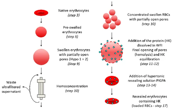
10. Loading of HK with Red Cell Loader® ... 46
10.1. The Red Cell Drug Loading System ... 46
10.2. Processing solutions... 49
10.3. The EryDex System applied to proteins ... 49
10.4. Optimization of the loading procedure and RBC membrane stability test in different buffers ... 51
11. Hexokinase activity assay ... 52
12. Statistical Analysis ... 53
12.1. Dose finding study ... 53
12.2. Repeated administration study ... 53
12.3. Stability of human erythrocytes loaded by RCL® ... 53
RESULTS ... 54
1. In vitro studies ... 55
1.1. Development of murine rAvPAL-RBCs ... 55
1.2. In vitro L-Phe metabolism by murine rAvPAL-RBCs ... 55
2. Preclinical studies ... 56
2.1. Efficacy of rAvPAL-RBC treatment: dose finding study ... 56
2.2. Preliminary evaluation of anti-rAvPAL IgG concentrations ... 59
2.3. Efficacy of rAvPAL-RBC treatment: repeated administration study ... 60
2.3.1. Step 1 – rAvPAL-RBC infusions at 18-19 day intervals ... 61
2.3.2. Step 1 – anti-rAvPAL IgG production ... 63
2.3.3. Step 2 – rAvPAL-RBC infusions at 9-10 day intervals ... 63
2.3.4. Step 2 – anti-rAvPAL IgG production ... 65
3. Loading of hexokinase in human RBCs with the Red Cell Loader®... 66
3.1. Optimization of the loading procedure ... 66
3.2. Loaded RBC membrane stability in different storage buffers ... 67
3.3. Loading of HK in standard conditions ... 68
DISCUSSION ... 72
CONCLUSION AND FUTURE PERSPECTIVES ... 81
BIBLIOGRAPHY ... 83
1. PHENYLKETONURIA: THE EPITOME OF HUMAN BIOCHEMICAL DISORDERS
When in 1934 the Norwegian endocrinologist Dr. Asbjørn Følling first diagnosed the presence of a high concentration of an unknown substance (which he called phenylpyruvic acid) in the urine of two siblings with mental retardation, probably he was not aware of the importance his discovery would have for thousands of people from that moment on. The credit for the discovery was also due to that caring and stubborn mother, who could not resign herself to the mental retardation of her children without having found a reason [http://pkuworld.org/home/history.asp].
By means of a traditional assay of classical chemistry for the detection of ketones, consisting in the addition of ferric chloride to the urine of diabetic patients, Dr. Følling observed the appearance of a deep green colour, which he had never seen before. Further chemical analyses and steps of purification on many other urine samples from patients sharing the same neurocognitive and developmental delays with the first ones, led to the identification of a chemical substance whose empirical formula was C9H8O3. The physical and chemical characterization of this molecule revealed that it was acidic, with a molar mass of 164; under slight oxidizing conditions it produced a benzaldehyde-like odour whereas a strong oxidation gave origin to benzoic acid and oxalic acid. All these features permitted to identify the compound as phenylpyruvic acid. What was still to be discovered was the causal relation to the mental retardation. From the analysis of the urine from another 430 mentally impaired subjects, Dr. Følling identified eight patients excreting the same substance and for the first time he understood the correlation between mental impairment and excretion of phenylpyruvic acid, a condition he named “oligophrenia phenypirouvica” (also named after him as Følling’s disease) [1]. Two years later the condition was renamed “phenylketonuria” to link the disease to the metabolic phenotype [2]. When L-Phe is not metabolized by the specific enzyme phenylalanine hydroxylase (PAH), it enters an alternative pathway of transamination and decarboxylation, which leads to the formation of phenylpyruvate, phenyllactate, and o-hydroxyphenylacetate, the metabolites whose excretion in the urine confers to it the typical colour and odour [3]. Further studies of family relationships highlighted an autosomal recessive mechanism of transmission [4]. To explain the causes of the phenylpyruvic acid excretion, he hypothesized some kind of defect in phenylalanine metabolism, with phenylalanine present at high concentration in the blood of such patients if the hypothesis was verified; all this was later on confirmed [5] through a microbiological test developed by Dr. Robert Guthrie which exploited the reversal of growth inhibition observed in Bacillus subtilis ATCC 6051 in the presence of a high level of phenylalanine [6].
Phenylketonuria is the first example of impaired cognitive development which was recognized to have a chemical etiology, i.e. hyperphenylalaninemia [2]. Today PKU is considered “the epitome of metabolic disorders” and is often employed as a model to describe and understand many other inborn errors of amino acid metabolism [7, 8].
2. THE DISEASE
Phenylketonuria (OMIM# 261600) is an inborn error of amino acid metabolism, caused by a deficiency of the enzyme phenylalanine hydroxylase (PAH, EC 1.14.16.1) that catalyzes the conversion via para-hydroxylation of the amino acid L-phenylalanine (L-Phe) into L-tyrosine (L-Tyr) [9]. The lack of PAH activity results in phenylalanine accumulation in body fluids (including brain and liquor), reaching neurotoxic levels and thus causing a progressive severe and irreversible neurological and intellectual disability, due to the lack of neurotransmitters for which Tyr is a precursor [10, 11]. PKU is inherited as an autosomal recessive trait [4] with a prevalence varying in a wide range according to geographic region and ethnicity; nevertheless, its rate of 1 out of 10,000 live births in Europe makes the pathology one of the most widespread genetic diseases among Caucasians [10, 12], with peak values in those populations with a high rate of consanguinity [13].
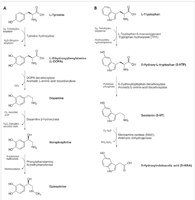
The disease is mainly caused by a mutation in a gene located in chromosome 12 (region 12q22-q24.2, GenBank U49897) encoding the cytosolic hepatic enzyme phenylalanine hydroxylase (PAH, E.C. 1.14.16.1) which catalyzes the irreversible conversion of the amino acid phenylalanine into tyrosine, a limiting step for the complete oxidation of L-Phe to CO2 and H2O. To work properly, PAH needs the presence of a pterin cofactor, terahydrobiopterin (BH4), as well as molecular oxygen and iron (Figure 1). The final concentration of phenylalanine in the body is in fact the result of a finely regulated balance: Phe input amount, coming from diet and the endogenous recycling of amino acids, and L-Phe output amount, represented by that fraction integrated in newly synthesized proteins and the one oxidized to L-Tyr through the PAH-mediated reaction [9].
When PAH does not carry out its own activity, phenylalanine accumulates in body fluids, including liquor, giving origin to hyperphenylalaninemia (HPA) and the related wide spectrum of mental disturbances typical of this condition [9, 14].
Figure 1. Metabolic pathways involving phenylalanine. a) Phenylalanine metabolisms in normal conditions; b) the conversion
catalyzed by Phenylalanine Hydroxylase.
2.1. Clinical manifestations and pathogenic mechanism
The main organic symptom of the disease is hyperphenylalaninemia; L-Phe reaches plasma concentration of up to and over 1200 µM, depending on the severity of the mutation. The high concentration L-Phe is the main agent of the other clinical manifestations, because it acts on different aspects of the brain functioning, as described below.
Patients with PKU in its most severe (untreated) form present with impaired growth and brain development. Symptoms include microcephaly, epilepsy, seizures, motor deficits, severe intellectual disability and behavioral disturbances, including psychotic, autistic, and aggressive disorders [15, 16]. Mental retardation is thought to arise from a decreased myelination that confers a reduced protection from proteolytic degradation [17] in combination with a reduced myelin production [18]. Many studies
have identified problems related to the altered development of the brain architecture due to the exposure to excessive L-Phe, as extensively reviewed by several Authors [16, 18, 19]; these modifications include abnormal myelination, cortical plate width and altered dendritic arborization with a reduced number of synaptic spines. The exposure to high concentration of L-Phe makes the already formed myelin unstable, whereas demyelinated axons undergo a reverse maturation, with consequent neuronal dysfunction [20]. Even in early and well-treated PKU patients dysmyelination still occurs, which may cause some of the above mentioned clinical manifestations [21]. In vitro studies have also demonstrated a depressed glutamaergic synaptic transmission in the presence of high L-Phe levels [22], data confirmed by a subsequent in vivo study [23]. The reported impaired activity of the glutamate receptor may explain, at least partially, the reduced dendritic arborization and number of spines experienced by untreated PKU patients; this effect was not observed for the transamination products of L-Phe, i.e. phenylpyruvic, phenylactic and phenylacetic acid, further indicating L-Phe as the neurotoxic agent in PKU [24]. The accumulation of potentially toxic L-Phe metabolites in the blood does not reach indeed a level sufficiently high to cause brain injury [18].
A significant inverse correlation has been found between L-Phe levels and IQ score, especially during the critical developmental period (age 0-12 years), even in early treated children: each 100 µM rise in L-Phe concentration corresponded to a 1.3-3.1 IQ point decline [25]. Later, a meta-analysis study identified a larger IQ decline, ranging from 1.9 to 4.1 points per each 100 µM mean rise in L-Phe in children treated since the neonatal age; a similar correlation was also found between lifetime L-Phe levels and IQ scores in early-treated individuals [26].
Moreover, Anastasoaie and colleagues [27] demonstrated that a better explanation of the cognitive outcome be provided by blood L-Phe variability, rather than the mean lifetime blood value as itself in early and continuously treated children, thus suggesting the importance of maintaining an L-Phe concentration as stable as possible within the recommended range of 120–360 µM over time, in order to prevent the deterioration of the cognitive performance.
All the neurological alterations encountered in PKU patients ultimately account for deficits commonly belonging to the field of the executive functioning, including also response speed, academic abilities, language-related tasks (including reading and arithmetic), problem solving ability, attention, interhemispheric transfer of information, and visuo-spatial and visual-motor abilities, as observed by Scriver et al. [14] and then extensively reviewed in the works by Bone et al. [16] and by Huijbregts et al. [28]. In a meta-analysis study, processes such as planning, working memory, inhibition, processing speed, and cognitive flexibility resulted to be impaired in early diet-treated patients, compared to controls [29].
The psychological and psychiatric problems documented in adolescent patients concern the area of social life, with negative findings in terms of autonomy, self-esteem, frustration threshold, school
achievements, attention, mood disturbances, depression and anxiety [30], even in early treated children [31]. Discontinuation of the dietary therapy as well as high levels of L-Phe are of course associated with a higher incidence of behavioral problems. Untreated individuals show more severe symptoms such as autism, hyperactivity, aggression, social withdrawal, anxiety, depression, psychosis, and profound intellectual disability [31], whereas adult patients early treated in childhood present with generalized depressed and anxious mood, lack of autonomy, low self-esteem and a tendency to social isolation. Phobias are also typical, and the most common one is agoraphobia [31-33].
L-Phe accumulation in blood results in its increased accumulation in the brain. This is due to the fact that phenylalanine belongs to the group of the Large Neutral Amino Acids (LNAAs), to which valine, leucine, isoleucine, threonine, histidine, tryptophan, methionine and tyrosine also belong. All these amino acids share a common carrier, the L-amino acid transporter-1 (LAT-1), to cross the blood-brain barrier (BBB) and enter the brain [34]; when there is an excessive circulating amount of L-Phe, this one is thought to compete with the other LNAAs for the available LAT-1 transporter, saturating it and thus resulting in a L-Phe overload in the brain and in a corresponding decreased amount of the other LNAAs, particularly L-Tyr and L-Trp [18, 34]. Moreover, across species LAT-1 appeared to have a higher affinity for L-Phe than the other LNAAs, and this is particularly marked for the human species, making it more susceptible to the negative effects of HPA [35].
Besides being important for protein synthesis, the LNAAs L-Tyr and L-Trp are also precursors for neurotransmitters, namely dopamine (DA, and consequently norepinephrine (NE) and epinephrine) and serotonin (5-hydroxytryptamine, 5-HT) respectively (Figure 2) [18]. Dopamine plays an important role in motor and cognitive functioning; norepinephrine is involved in learning and memorization processes, in the arousal of attention, fear and anxiety, and in the development of the maternal behaviour in females; serotonin is important for neuronal proliferation, synaptogenesis and morphogenesis [36, 37].
Indeed, there are two possible mechanisms by which L-Phe alters brain functioning: if on the one hand the increased L-Phe presence in brain results in a decreased level of the other LNAAs including L-Tyr and L-Trp, as described above, on the other hand it acts as a competitive inhibitor of the other two amino acid hydroxylases, TyrOH and TrpOH [18, 38-41], thus generating a lack of their products. In fact, high brain L-Phe was reported to negatively affect the activity of the other hydroxylases, notwithstanding the increased availability of BH4 (whose production is stimulated by L-Phe itself [42]) [18].
Before acting as neurotransmitters, biogenic amines also represent fundamental signals for the correct early brain development [43], as demonstrated in experiments in mice whose genes encoding the biosynthetic enzymes TyrOH and dopamine β-hydroxylase (DBH) were deleted [44, 45].
Figure 2. Biosynthetic pathways of neurotransmitters lacking in the brain of PKU patients. (A) Synthesis of catecholamines
(dopamine, DA, norepinephrine, NE, and epinephrine); (B) synthesis of serotonin (5-hydroxytryptamine, 5-HT) (adapted from http://www.hdri-usa.com/).
The development of the cerebral cortex occurs following a precise sequence of events, well defined in time and space, also spanning over the post-natal period especially as regards the synapses and dendrites in the prefrontal cortex. These time-windows are characterized by a different availability of brain amines, with peak increases of catecholamines and serotonin during the critical developmental period followed by reduction to adult levels, and are the most susceptible to L-Phe-induced damages, as extensively demonstrated by studies on animal models [36, 37, 41, 44-53]. Low levels of biogenic amines have been found in the cerebrospinal fluid of hyperphenylalaninemic patients [54].
The healthy prefrontal cortex is particularly rich in dopamine, which is fundamental for the executive functions, and PKU children have been found to have an impaired dopaminergic innervation, with subsequent attentional deficits [55, 56]. A work by Burlina et al. [57] confirmed a reduced aminergic
synthesis in the brain of adult PKU patients even if compliant with a free L-Phe diet. Some cognitive and behavioral alterations, significant though subtle, have also been reported in patients with a good control of blood L-Phe levels [29, 58], which could be explained as a consequence of small permanent cortical dendritic changes, secondary to NE depletion in the perinatal period [49].
In a series of in vivo studies, Pascucci et al. [41, 51, 59] demonstrated in mice with the BTBR-Pahenu2 genotype, the genetic model of human PKU, a severe lack of whole brain serotonin during critical post-natal periods (PND14-21) and deficits in the level of its immediate and limiting precursor 5-hydroxytryptophan (5-HTP), not corresponded by a reduction in its initial amino acidic precursor L-Tryptophan; these evidence support the hypothesis of TrpOH activity inhibition exerted by L-Phe excess, rather than a hampered access of L-Trp across the blood brain barrier (BBB) [40, 51], thus confirming the minor involvement of tryptophan in the L-Phe induced alterations [59]. In addition, Pascucci and colleagues detected a 50% reduction in 5-HT, followed by a 40% decrease in NE levels and a 30% decrease in DA, thus confirming previous observations [47, 51].
As reported, among the considered biogenic amines dopamine and its precursor L-3,4-dihydroxyphenylalanine (L-DOPA) are the less affected by HPA, owing to the fact that when L-Tyr levels are abnormally low and L-Phe is extremely high, the latter can serve as substrate for TyrOH for the production of L-DOPA [60].
All the described pathophysiologic mechanisms involve L-Phe as a single molecule. In the recent few years, a new amyloidosis-like etiology has been proposed: it has been observed that when L-Phe is present at extremely high concentrations, such as in the brain of PKU patients, it self-assembles to form fibrils with amyloid-like morphology and cytotoxicity [61-63]. Such deposits have been found in the hippocampus of model mice and in the parietal cortex tissue obtained from phenylketonuric individuals [61] and their toxicity seems to derive from the fibril arrangement in a hydrophilic core and hydrophobic exterior made up of aromatic side chains of the L-Phe monomers. Therefore the lipophilic character of the fibril outer surface would promote insertion into cell membranes, while the hydrophilic core interacts with ions; the result is ion leakage and a consequent cellular damage [63]. Interestingly, Singh and coworkers demonstrated inhibition of L-Phe fibril formation by D-Phe enantiomer when added at ≥ 8% of L-Phe concentration in solution. The DL-Phe solution formed instead aggregates with a completely different morphology of flat flakes with irregular edges, which proved to be more stable than fibers and, most importantly, unable to propagate further [62]. Supplementation with D-Phe could therefore be a possible strategy to prevent HPA-associated brain alterations both by preventing accumulation of the amyloid-like L-Phe fibers, and by competing with the L-enantiomer for the LAT1 transporter to cross the blood-brain barrier [64].
Another recent hypothesis is that excessive L-Phe impacts on DNA methylation patterns, like other stressors. To verify this idea Dobrowolsky et al. [65] studied the DNA from the frontal cortex of two
PKU patients, and then compared the methylation patterns and subsequent gene expression to those observed in the DNA from leukocytes (chronically immersed in a hyperphenylalaninemic environment) of other PKU patients on diet, with both well-controlled and poorly-controlled blood L-Phe. The Authors found epigenetic alterations both in PKU brains, suggesting neurological involvement, and in patients’ leukocytes, with a strikingly higher methylation rate in regulatory miRNA genes in the non diet-compliant group. A subsequent inversely proportional altered expression (down-regulation) was also identified in the miRNA-targeted genes, which, in most cases, code for structural proteins or proteins involved in synapse formation and functioning and in myelination process. These findings confirm and extend to human PKU what had already been observed in a preclinical study on mouse fetuses affected by maternal PKU [66] and suggest DNA methylation pattern in leukocytes as a possible biomarker for historic L-Phe exposure, as well as epigenome as a candidate target for PKU treatment [65].
2.2. The role of BH4 cofactor in PKU pathogenesis
Hyperphenylalaninemia (HPA) can be caused indeed not only by defects in the PAH gene, but also in those genes encoding enzymes involved in the pathways of de novo synthesis or recycling of the PAH cofactor tetrahydrobiopterin. This condition was initially referred to as “malignant phenylketonuria” [67]. The biosynthetic enzymes involved are GTP cyclohydrolase I (GTPCH), 6-pyruvoyl-tetrahydropterin synthase (PTPS) and sepiapterin reductase (SR) which catalyze the de novo synthesis of BH4 starting from guanosine triphosphate; on the other hand, the recycling enzymes are pterin-4a-carbinolamine dehydratase (PCD) and dihydropteridine reductase (DHPR) which catalyze the reduction of the oxidized cofactor quinonoid dihydrobiopterin (qBH2) once Phe conversion to L-Tyr has occurred [68]. Deficiencies in the BH4 synthesis or recycling enzymes are inherited similarly to the PAH mutations as autosomal recessive traits, and account for approximately 2% of HPAs detected in babies by newborn screening [69]. In this case the metabolic block can be overcome by supplementing a biologically active synthetic form of the cofactor, i.e. sapropterin dihydrochloride ((6R)-2-amino-6-[(1R,2S)-1,2-dihydroxypropyl]-5,6,7,8-tetrahydro-4(3H)-pteridinone dihydrochloride), investigated for the first time by BioMarin Pharmaceutical Inc. (Novato, CA) and commercialized since 2007 both in the U.S.A. and Europe with the name of Kuvan® [https://www.bmrn.com/about-us/history.php#2008]. To date, this is the only drug approved by FDA with the “orphan” designation for the treatment of PKU. In Figure 3 are represented the metabolic pathways through which the cofactor is produced in the organism.
Figure 3. BH4 metabolism. The different colours identify the three metabolic pathways, with the one in blue being the main
anabolism. The “green” enzymes belong to an alternative pathway operating in parallel with the main biosynthetic route (Adapted from Werner et al. 2011 [70]).
The pterinic cofactor BH4 has a very important role in PKU symptom determination. The lack of tetrahydrobiopterin is one of the causative agents of a group of pediatric disorders named monoamine deficiencies and also known as “atypical PKU” [71, 72]. Such alterations take origin from defects in the neurotransmitter biosynthetic enzymes, as well as in BH4 availability, which serves as cofactor in the reactions [73]. In fact, apart from Phenylalanine Hydroxylase, tetrahydrobiopterin is a common cofactor for the other two hydroxylating enzymes, TyrOH and TrpOH, meaning that individuals with
enzymatic defects in the BH4 biosynthetic or recycling pathways suffer not only from the lack of neurotransmitter precursors, but in addition are not even able to synthesize catecholamines and serotonin from tyrosine and tryptophan introduced with the diet [74].
Among the other options, the regulation of BH4 production relies also on BH4 itself and blood L-Phe; both molecules act on the first enzyme, GTPCH, via the GTPCH feedback regulatory protein (GFRP), the first one (BH4) inhibiting it and the second one (L-Phe) activating it. As a consequence, in HPA there is a high plasma concentration of pterin cofactor which, however, cannot be used due to the lack of PAH [75]. On the other hand, Phenyalanine Hydroxylase is not inhibited by the product of BH4 oxidation, i.e. BH4-4a-carbinolamine, but rather by primapterin, an isomer of biopterin carrying the dihydroxypropyl chain at position 7 instead of 6 of the pteridine ring. When the genetic defect occurs in the enzyme pterin-4a-carbinolamine dehydratase (PCD), the carbinolamine cofactor can also be non-enzimatically dehydrated to quinonoid dihydrobiopterin, but this reaction is not efficient enough to supply the reduced form of the cofactor in the required amounts [76]. Therefore, a lack of PCD mainly induces BH4-4a-carbinolamine to chemically rearrange in two steps to dihydroprimapterin (that is, 7-substituted dihydrobiopterin) which is then further converted into primapterin (7-biopterin) and in this form excreted into the urine, thus defining a condition named “primapterinuria” [71, 72, 77-80]. The 7-substituted form of biopterin (7-BH4) was reported not to be a good cofactor for PAH, due to the uncoupling of the reaction to the production of L-Tyr [81]; moreover, it exerts a potent inhibition on the enzyme already at 1 µM concentration in the presence of physiological concentration of BH4 and saturating L-Phe, while its oxidation generates hydrogen peroxide, which further inhibits PAH activity [79]. Therefore, the hyperphenylalaninemia observed in patients with normal PAH activity is probably due to the inhibition exerted by the 7-sustituted cofactor generated by an altered BH4 metabolism [79]; on the other hand, PAH inhibition raises even more L-Phe concentration which, in turn, stimulates the activity of the BH4-synthesising enzyme GTPCH I through its regulatory protein GFRP [42, 82], thus activating a loop of mutual feedback regulations and making primapterinuria actually dependent on plasma L-Phe levels and liver BH4 availability, with the possibility to modulate their amounts by means of an L-Phe-restricted diet [80].
When the genetically altered protein is dihydropteridin reductase (DHPR), the quinonoid form resulting from the first step of the recycling pathway is readdressed to a non-enzymatic rearrangement, yielding 7,8-dihydrobiopterin, a substrate for dihydrofolate reductase (DHFR) instead of DHPR. However, this alternative regeneration pathway is not sufficiently effective in restoring the proper concentration of tetrahydrobiopterin, as demonstrated by severe clinical neurological manifestations arising in DHPR-deficient patients [83]. One of the proposed explanation is the accumulation of 7,8-dihydrobiopterin, a recognized strong inhibitor of all aromatic amino acid
hydroxylases, which would further contribute to the diminished synthesis of both L-DOPA and 5-OH-tryptophan [84].
2.3. Other clinical manifestations
The reduced availability of L-DOPA is the factor underlying another hallmark of phenylketonuria, i.e. fair colour of patient hair and skin. It has been demonstrated that tyrosinase (E.C. 1.14.18.1), the Cu2+ -dependent oxidoreductase representing the first enzyme in the melanogenesis pathway, works in concert with TyrOH, the latter providing L-DOPA necessary to activate and promote tyrosinase activity and, as a consequence, skin and hair pigmentation [85].
Moreover, a causative relation was discovered between L-Phe and the inhibition of brain and liver -hydroxy--methylglutaryl-coenzyme A reductase (E.C. 1.1.1.88), the rate-limiting enzyme in cholesterol biosynthetic pathway, together with a reduced production of mevalonic acid [86]. The resulting hypocholesterolemia is hypothesized to have a protective effect against cardiovascular diseases in adults [3]. The impairment of the mevalonate pathway also implies a decreased synthesis of coenzyme Q10 (ubiquinone-10; CoQ10), actually observed in both serum and lymphocytes of PKU patients, thus supporting a role of excessive L-Phe as an indirect pro-oxidant factor [87, 88].
3. MATERNAL PKU
An adequate control of blood L-Phe levels is particularly important in women willing to become pregnant, since the exposure to elevated L-Phe concentrations is teratogenic for the fetus, causing the so-called “maternal phenylketonuria syndrome” or maternal PKU [34, 89]. Pregnant patients with poor or absent control of HPA or PKU have a high probability to experience pregnancy complications, the most frequent being spontaneous abortion, intrauterine fetal death (IUFD) or growth retardation, and preterm delivery [89-91]. Affected newborns present with low birth weight (and are defined as small for gestational age, SGA), microcephaly, congenital heart disease (CHD), intellectual or developmental disabilities (IDDs), and facial dysmorphism (FD) [90-92]. The incidence of such pregnancy complications and/or neonatal sequelae is strongly correlated with maternal blood L-Phe levels, particularly during the first weeks of embryogenesis, i.e. when organogenesis occurs; this is especially the case for FD [89].
The study of Prick and colleagues [89] clearly demonstrates a significant correlation between the increase in mean L-Phe concentration per trimester in maternal blood and the occurrence of SGA, microcephaly and IDDs, and underlines an increase in CHD as L-Phe per trimester doubled.
Hence, it becomes very important for women with a desire to become pregnant to accurately plan the pregnancy itself and to comply with a strict L-Phe restricted diet also enriched in multivitamin complexes, vitamin B12 and folic acid, which had better to be initiated before conception and should
be continued throughout pregnancy, in order to prevent as much as possible fetal damages [89, 90, 93, 94]. During pregnancy maternal L-Phe tolerance increases to some extent thanks to fetal PAH activity, resulting in lower blood levels and in a better cognitive performance; this should permit a relaxation of the strict diet regimen during the second trimester, thus avoiding the nutritional problems that could arise from the adherence to the diet itself [89].
The Maternal PKU Collaborative Study (MPKUCS), which lasted from 1984 to 2002, represented an international effort aimed at assessing the effects of such diet on the offspring of PKU mothers, also in relation to the timing of the treatment [95, 96]. From this study emerged that the main causative agent is the maternal PKU phenotype, determining maternal blood L-Phe concentration and therefore fetal exposure; this could also represent a problem in case of mild HPA, since during pregnancy there is a placental gradient of L-Phe favoring the fetus [97] that can be dangerous if not kept under control [98]. Moreover, the socio-economic status of such women could influence their level of education and subsequently the cognitive outcome [99]. On the other hand, the maternal phenotype becomes determinant only when dietary treatment is discontinued during childhood and adolescence, leading to impaired maternal intelligence and, as a consequence, to the delayed onset of treatment and a poor quality of care both in anticipation of and during pregnancy and the post-natal period [100, 101]. Women should reach a blood L-Phe concentration within the recommended range of at least 120-360 µM (2-6 mg/dl) – better 60-250 µM (1-4 mg/dl) [102] because of the positive gradient across placenta – already before conception or, in any case, not later than the 8th week of pregnancy, and should manage to keep it in this range throughout pregnancy, in order to bear a baby with normal birth parameters (as regards CHD, microcephaly and SGA [103, 104]) and cognitive performance [91, 93, 95, 96, 98]. These reference values origin from the fact that a concentration higher than 120 µM is necessary to avoid the damages produced by hypophenylalaninemia [96, 104], whereas the upper limit comes from the identification of a critical threshold value of 330-360 µM beyond which damages to the fetus occur [105].
PKU mothers bearing non-PKU babies (i.e. healthy carriers of a single mutated allele) are encouraged to breastfeed their children without restriction, since the single non mutated copy of the PAH gene is sufficient to metabolize the amount of L-Phe introduced with breast milk [106], also because breast milk contains only 43 mg L-Phe/dl compared to 59-73 mg/dl of infant formulas and 164 mg/dl of cow’s milk [107].
Another important finding of the MPKUCS was that L-Phe fluctuations, even within the safe range of concentrations, has a negative effect on offspring cognitive outcome, which on the contrary was not evident for L-Phe mean levels [94]. In fact, Maillot et al. [94] demonstrated a strong negative correlation between the standard deviation (describing maternal blood L-Phe variations within the recommended interval) and offspring IQ measured at the age of 4, 8 and 14 years. Blood L-Tyr does
not seem to have effect on the developing fetus [96]. The additional treatment with BH4 supplementation was reported to help the development of fetal PAH activity with a protective effect from maternal HPA [99, 108].
In conclusion, the solution resides in the achievement of the proper degree of L-Phe control already before conception; this might prove to be very difficult, though, especially for women with the most severe forms of the disease, who used to have themselves poor L-Phe control throughout their lives (and consequently a higher probability of mental impairment): it has been demonstrated that a maternal IQ <85 is associated with a later achievement of the target L-Phe range and a more negative influence on the fetus [101, 109]. Hence, it is important to provide these and all other patients with special support and education on the importance of diet compliance, together with more frequent monitoring throughout pregnancy [90, 94, 110]. Adherence to such recommendations makes it possible for PKU mothers to have children with the same expectancy of cognitive development as non-PKU people [90].
4. PHENYLALANINE HYDROXYLASE
4.1. Enzyme and gene
Phenylalanine hydroxylase (also named phenylalanine-4-monooxygenase, symbol PAH or PheOH, EC 1.14.16.1) is part of the enzymatic family of pterin-dependent aromatic amino acid hydroxylases. This family also includes two more monooxygenases, i.e. tryptophan hydroxylase (tryptophan-5-monooxygenase, symbol TPH or TrpOH, EC 1.14.16.4) and tyrosine hydroxylase (tyrosine-3-monooxygenase, symbol TH or TyrOH, EC 1.14.16.2); all these proteins share the use of BH4, molecular oxygen and reduced iron (Fe2+) to carry out their own activity [111, 112].
The human PAH enzyme is encoded by a gene mapped to the long arm of chromosome 12 (locus PAH 12q22-q24.2) and expressed in liver and kidney [113-115]; the cDNA of the gene was first obtained and cloned in the 1980s [116-118] (GenBank NM_000277 for PAH mRNA; U49897.1 for PAH cDNA) whereas the full-length genomic sequence of the gene was obtained later (GenBank AF404777) [119]. PAH gene is composed of 13 exons and 12 large introns to reach a total length of 121,526 Kbp in the minus strand of the DNA [http://www.genecards.org/cgi-bin/carddisp.pl?gc_id=PAH]. The gene coding sequence (cds, nt 473-1,831) is flanked by a 5’-UTR region (nt 1-472) and a 3’-UTR region (nt 1,832-2,680); it is transcribed into a mature mRNA of approximately 2.6 Kb (2,680 bp), which is in turn translated into a 452 amino acid monomer [http://www.ncbi.nlm.nih.gov/nuccore/U49897.1, last update 1997]. Each polypeptide weighs 51.8 KDa and consists of three domains: an N-terminal regulatory domain (residues Glu19-Leu142, also identified as ACT domain), a catalytic domain (residues Asp143-Phe410), and a short C-terminal tetramerization domain (residues Ser411-Lys452) [3, 120, 121; GenBank
AAC51772.1]. Figure 4 shows the nucleotide cds and the corresponding amino acidic sequence of the
PAH gene.
Figure 4. Nucleotide sequence of the coding region (cds, nt 473-1831) of the PAH gene (line above) and corresponding amino
acidic sequence (line below). Underlined in red is the regulatory domain, in green the catalytic domain and in blue the tetramerization domain. In bold and red are the residues forming the autoregulatory sequence (ARS, Gly19-Gly33); in bold and green the putative L-Phe binding site (residues Gly46-Leu49 and Glu66-Pro69); in bold and blue the amino acids forming the cofactor binding site (Gly247, Leu249, Phe254, Ala322, Tyr325); in bold and purple the metal ion binding site (His285, His290, Glu330); in bold and black the Ser16 residue, the phosphorylation site important for activity regulation.
Despite the great difficulty to obtain the crystallographic structure of the whole enzyme, the availability of the structures of many truncated forms of PAH, containing the regulatory and catalytic domains [122] or the catalytic and tetramerization domains [123, 124], allowed the construction of a full-length crystal model by superimposing the catalytic domains (Figure 5).
Figure 5. (A) Full-length structure of human phenylalanine hydroxylase monomer obtained by superimposing the catalytic
domains of the truncated forms. The red sphere represents iron. (B, C) Two perpendicular views of the full-length PAH model structure. The iron is shown as a gray sphere in all four monomers (adapted from Erlandsen and Stevens 1999 [120]).
Full-length PAH has been described to exist in solution both as a functional homodimer and homotetramer, in a pH- and phenylalanine-dependent equilibrium, with a marked shift towards the tetrameric form as pH decreases or L-Phe concentration increases [17, 125, 126]. However both oligomeric forms are functional, as demonstrated by studies on truncated forms of both PAH and TyrOH including only the tetramerization and catalytic domains, which still maintain the enzymatic activity though losing substrate specificity [124], but with the peculiarity that the tetramer formed by PAH is asymmetrical: this is due to the fact that, actually, this tetramer is a “dimer of dimers” [120], where a mechanism of domain swapping between monomers takes place such that secondary elements switch their mutual position in order to promote a stable oligomerization, together with the formation of an antiparallel coiled-coil structure with the other monomers [124, 127].
4.2. PAH regulation
Given the negative effects of an excessive circulating phenylalanine concentration, it is very important to maintain phenylalanine homeostasis in vivo; therefore, PAH activity is strictly regulated through different mechanisms, i.e. substrate activation, cofactor inhibition and reversible phosphorylation [19, 126, 128-131; for review see Refs. 132, 133]. The autoregulatory sequence located at the N-terminus of the protein includes a residue of Ser16 which has been demonstrated to be the site of phosphorylation by the cAMP-dependent protein kinase A (cAMP-PKA) [130]. This sequence is named autoregulatory because it sterically limits the access of the substrate to the catalytic site of the enzyme [133]. When the first 30 N-terminal residues are removed, PAH shows a higher affinity and a consequent higher rate of L-Phe conversion [134].
L-Phe acts on PAH as a homotropic allosteric activator with a highly cooperative action which can be exerted only on the tetrameric form with extensive structural changes occurring at the tertiary/quaternary level [17, 131]. In fact, the substrate-mediated mechanism of activation involves all the four monomers, inducing modifications in the monomeric structures such that a stronger interaction at the dimer interface is promoted, whereas the interactions between dimers in a tetramer
are weakened. As a result, the dimer/tetramer equilibrium is shifted towards the tetrameric form of the enzyme upon binding of L-Phe [126], the volume of the tetramer increases and a competent catalytic site is exposed [17]. L-Phe binds in a specific allosteric site located in the regulatory domain [135], different from the active site of the catalytic domain, and the binding on one site do not automatically excludes the binding on the other one, although the affinity for the allosteric site is seven-fold higher [131, 136]. In fact, in each monomer the N-terminal tail stretches over the active site, thus preventing access for the substrate, unless L-Phe binds on the regulatory sequence and causes its displacement [124].
Besides being L-Phe-dependent, the rate of PAH activation by its substrate seems to be also pH-dependent, with less L-Phe necessary for activation at basic values of pH; at low L-Phe concentration the enzyme shows a low activity, in accordance to its allosteric regulatory mechanism [17].
If L-Phe is an allosteric activator of the enzyme, on the other hand its cofactor BH4 acts as a classic allosteric inhibitor, which binds to the N-terminal autoregulatory sequence, thus preventing the conformational changes necessary for enzyme activation [137] (whereas the alterations induced upon L-Phe binding prevent BH4 from exerting its negative effect on activity [138]) and moreover closing the access to the active site [139]. Nevertheless, BH4 is necessary for the primary reduction of the Fe3+ ion to Fe2+, an inevitable prerequisite for enzyme activation [136].
Another regulatory element is represented by the unique post-translational modification undergone by the protein, i.e. phosphorylation, which occurs on the Ser16 by cAMP-dependent PKA. Phosphorylation promotes the enzymatic transition from the steady state towards the active state, facilitating L-Phe access to the active site [126]; it is a reciprocal mechanism in that the substrate makes PAH more susceptible to phosphorylation by cAMP-PKA, which in turns sensitizes the enzyme towards allosteric activation by L-Phe by lowering the concentration of substrate needed for activation, but without changing its positive cooperativity [128-130].
Moreover, the phosphoric moiety disrupts the binding site of BH4, therefore interfering with its inhibitory action [19]; on the other hand, BH4 is a potent inhibitor of the enzyme in that it prevents the access to Ser16 for phosphorylation. This effect is compensated by L-Phe when L-Phe and BH4 are simultaneously present [130]. Notwithstanding the main regulatory role exerted by both L-Phe and BH4 and the more acute response to L-Phe in the presence of BH4 [140], phosphorylation can also represent another way to modulate the hydroxylase activity. Shiman [141] demonstrated that both phosphorylated and unphosphorylated forms of the enzyme require L-Phe for activation; hence, phosphorylation does not equal allosteric activation, but lowers the energy needed for it to occur. This happens by means of two mechanisms: by promoting the transition to the active state of the protein, and by reducing BH4 affinity for its inhibitory binding site. These aspects explain why in vivo phosphoPAH has a higher affinity for the substrate L-Phe, a higher activation rate and a lower
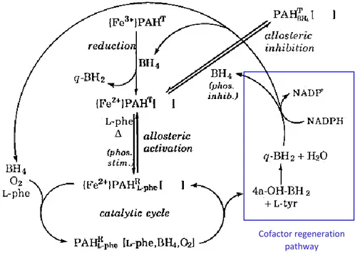
sensitivity to BH4-mediated inhibition [19]. In Figure 6 is a schematic representation of the regulatory pathways in which PAH is involved.
Figure 6. Regulatory pathways of PAH (from Kappock and Caradonna 1996). PAHT = PAH in the steady state; PAHR = PAH in
the active state; the empty square brackets represent a substrate-binding pocket located in the active site.
4.3. PAH mutations
After Følling’s discovery of the disease, many other studies followed and allowed the identification of the underlying metabolic block [142, 143]. The first PAH mutations could be detected after the gene cloning and mapping [116], thus paving the way for structure and functional analyses in vitro. The phenylalanine hydroxylase gene is characterized by great allelic diversity, as reported in the PAH variation database which currently contains 890 records of gene variants [PAHvdb, Blau N and Yue W, and Perez B, http://www.biopku.org/pah/]. To date, 567 mutations both pathologic and polymorphic have been discovered and are collected in a database named PAHdb created in 1996 (http://www.pahdb.mcgill.ca) [11, 144]. Of these, the most common are missense mutations (60.1%), followed by deletions (13.4%), splice alterations (10.9%) and silent or non-sense mutations (5.6% and 4.9% respectively) (http://www.pahdb.mcgill.ca/cgi-bin/pahdb/mutation_statistics-1.cgi); the final result is a total or partial lack of enzymatic activity that can result in different phenotypic severity. In fact, most patients are not homozygous for a single mutation; they are instead compound heterozygous for two different allelic alterations. Some patients, who are compound heterozygous, are phenotypically functional hemizygous, due to a combination of a severe mutation (such as a null one) with an allele that still allows the production of enzyme, even if only partially functioning: in those cases, the less severe mutation determines the PKU metabolic phenotype [145]. Together with the polymorphic nature of the phenylalanine hydroxylase gene, this is the main reason underlying the
Cofactor regeneration pathway
great phenotypic diversity associated with the disease, and which, in addition, makes PKU very widespread in spite of its recessive inheritance pattern [146, 147].
While many of the most severe disease-causing mutations map to exon 7 which encodes part of the catalytic core of the enzyme [17], the most common (missense) alterations occur in the junction between the catalytic and tetramerization domains [124], making PKU a loss-of-function misfolding disease. By means of in vitro expression and site-directed mutagenesis, Gersting and colleagues [148] demonstrated how mutations located not only in the oligomerization domain but also in the catalytic and regulatory portions alter protein folding and assembly, with severe effects on allosteric regulation and proteolytic stability. Interestingly, the majority of these mutations did not reduce the catalytic activity below 50% of the wild type protein, strongly indicating that the loss of function is due to a mechanism of increased degradation. To this purpose, Pey et al. [149] proposed an effective model to analyze the destabilizing effect of missense mutations on the native protein’s conformation, in order to predict the phenotypic outcome, and those results were then confirmed in the work by Gersting et al. [148].
The identification of the etiologic agent permitted the development of a treatment aimed at limiting the phenotypic effects of the disease: in fact, phenylketonuric patients show a different tolerance with respect to the daily amount of phenylalanine intake, and, on these basis, a dietary therapy has been proposed in the 1950s, with first positive results published in 1953 [150-152].
Indeed, phenylketonuria has often been defined as a disease born from the discordance between nature and nurture [10, 11], where the nurture component is the essential amino acid L-Phe and the nature is represented by the mutation in the PAH gene. The result of the discordance is hyperphenylalaninemia, the PKU metabolic phenotype, which leads and is associated with the clinical phenotype of impaired cognitive development and function. The possibility to act externally on the metabolic manifestation of the pathology makes PKU the first genetic disease to have a pharmacological treatment, thus smoothing the negative effects of gene alterations [10].
5. CLASSIFICATION
Given the multiple factors influencing the disease outcome, a classification of the phenotype can be made considering different aspects, first of which is the type and position of the PAH mutation, which determines the rate of enzymatic activity and consequently the level of hyperphenylalaninemia [34]. Hence, the classification is primarily made on the basis of the severity of HPA, considering that the normal L-Phe concentration in the blood of healthy individuals ranges from 50 to 110 µM [34]. Other criteria, i.e. tolerance to dietary phenylalanine intake, clinical course of the disease and BH4 responsiveness, are also employed to describe the phenotype [34, 153, 154].
5.1. Classification according to blood L-Phe
This classification was introduced in 1980 by Güttler et al. [155] and defines the following phenotypes according to pre-treatment L-Phe levels:
Classical PKU: pre-treatment L-Phe > 1200 µM
Variant PKU: pre-treatment L-Phe between 600 and 1200 µM
Mild HPA or non-PKU HPA: pre-treatment L-Phe between 120 and 600 µM
The class named “variant PKU” was later divided into two subcategories [145, 156], resulting in:
Classical PKU: L-Phe > 1200 µM
Moderate PKU: L-Phe between 900 and 1200 µM
Mild PKU: L-Phe between 600 and 900 µM
Mild HPA: L-Phe above 110 µM but < 600 µM
The amino acidic levels are currently used by approximately 80% of the PKU centers to identify the patients’ phenotype, but this approach can lead to some mistaken evaluations, since blood L-Phe depends on some variables, such as the diet followed before L-Phe assessment, the timing of the analysis and the effect of the neonatal catabolism, which can result in false positive or false negative outcomes [153]. For example, the current practice of screening blood L-Phe in newborns within the third day of life can result in a negative conclusion, due to the fact that the amino acid might not have had time to reach its maximal concentration [34].
Recently, a new term was proposed to describe the intermediate range of values between the critical threshold of 360 µM and 600 µM (6-10 mg/dl); currently, there is still uncertainty about whether L-Phe concentrations comprised in such range may have a negative influence on cognitive and executive functioning, thus requiring treatment [157, 158]: this is the so-called “Mild-HyperPhe-gray zone” [159].
5.2. Classification according to tolerance to dietary L-Phe intake
L-Phe tolerance is defined as the amount of daily L-Phe a patient can introduce with a normal diet without experiencing an increase in blood amino acid levels above the upper limit of the recommended range (360 µM) [153]. This parameter has proven to be stable and reliable to the purpose of PKU phenotyping. The first classification proposed by Güttler et al. [155] included three classes:
Classic PKU: L-Phe tolerance < 20 mg/kg body weight/day
Variant PKU: L-Phe tolerance between 20 and 50 mg/kg body weight/day Mild HPA: L-Phe tolerance > 50 mg/kg body weight/day
Classic PKU: L-Phe tolerance < 20 mg/kg/day, corresponding to 250-300 mg L-Phe/day Moderate PKU: L-Phe tolerance of 20-25 mg/kg/day (350-400 mg/day)
Mild PKU: L-Phe tolerance of 25-50 mg/kg/day (400-600 mg/day) Mild HPA: patients not requiring dietary restriction
According to the paper by Camp et al. [159], between mild HPA and mild PKU is the Mild-HyperPhe-gray zone, corresponding to a L-Phe tolerance > 50 mg/kg body weight/day.
The assessment of L-Phe tolerance must be performed under standardized conditions so as to obtain a reliable indication of the phenotype [153]. It is generally performed at the age of 5 years in the majority of the medical centres [145], but van Spronsen et al. [160] have demonstrated that reliable determinations can be made already at the age of 2 years and that L-Phe tolerance at 2, 3 and 5 years correlates well with that at the age of 10 years [160]. On the contrary, L-Phe tolerance must be reassessed in adulthood in relation to body weight in order to satisfy as much as possible the criterion of 9.1 mg L-Phe/kg ideal body weight/day [161]. This kind of classification is currently used by 70% of medical centers [162].
An additional classification based on BH4-responsiveness has been proposed by Blau and Muntau [163] and consists in BH4-non-responsive HPA and BH4-responsive HPA, the latter being further divided into BH4-responsive PAH deficiency and HPA due to defects in the BH4 pathway.
5.3. Classification according to clinical course
The parameters to evaluate the clinical course include the intellectual outcome, in terms of patient education and IQ, the maximum L-Phe concentration reached in particular conditions or periods of life (such as non compliance to the restricted diet or the occurrence of infectious diseases) and, most importantly, the variations in blood L-Phe levels and the phenylalanine-to-tyrosine (L-Phe/L-Tyr) ratio [27, 164, 165]. The latter two parameters have been demonstrated to influence the cognitive outcome to a large extent, larger than blood L-Phe level itself, and therefore their determination may allow a simplified classification of the phenotypes according to the need of treatment:
PKU: patients who need a strict dietary control of L-Phe levels
Non-PKU HPA: patient who do not need any dietary treatment to keep L-Phe under control BH4-responsive PKU: patients who may take advantage from BH4 supplementation.
6. DIAGNOSIS
The identification of PKU or HPA should be performed as early as possible, so as to introduce an opportune treatment and thus limit, if not completely avoid, all the neurological and metabolic consequences of a too high uncontrolled hyperphenylalaninemia [34, 153]. For this reason, now all
newborns are routinely tested for PKU/HPA soon after birth according to national screening programs [166]. Blood samples are drawn for analysis between the 2nd and the 5th day of life in most centers [167]. Today blood sampling is considered suitable for analysis within 24h-48h from birth when L-Phe/L-Tyr ratio is employed for the diagnosis [168].
Usually, affected babies born from healthy mothers do not display physical alterations, except for a tendency to a reduced body weight and head circumference [169]. Therefore, diagnosis mainly consists in the biochemical assessment of blood L-Phe and L-Tyr, biopterin and neopterin content in blood or urine, and the measurement of specific enzyme activities [153]. All forms of the disease reveal upon neonatal screening a common pattern of blood Phe higher than 120 µM, normal or reduced L-Tyr concentrations (with a L-Phe/L-L-Tyr ratio > 2) and normal values for the remaining amino acids [34]. The analytical methods employed to assess blood L-Phe include:
- Guthrie’s test or bacteriological inhibition assay (BIA): first proposed in 1963 by Robert Guthrie [6], it is based on the inhibition of Bacillus subtilis, which requires L-Phe for its growth. A small amount of peripheral blood is drawn from the patient (usually from the heel) and collected on a standardized filter paper, the so-called “Guthrie’s card”. The dried blood spot (DBS) obtained is then submitted to the analysis. This test was introduced for mass screening and nowadays is being increasingly replaced by more modern techniques (e.g. tandem mass spectrometry) characterized by improved precision, sensitivity, practicability, and faster time of analysis; - Fluorimetric assay: a simplified and automated method yielding a lower rate of false positive
results compared to the Guthrie’s test [170, 171];
- Analysis by reverse-phase liquid cromatography [172, 173];
- Enzymatic colorimetric assay: based on the reaction catalyzed by phenylalanine dehydrogenase, it was employed to detect L-Phe in plasma samples [174, 175];
- Tandem mass spectrometry (TMS): this procedure is able to simultaneously identify even small amounts of amino acids (L-Phe and L-Tyr) in dried blood spots collected on Guthrie’s cards, providing the L-Phe/L-Tyr ratios and yielding a low rate of false positive results [176-179]. - PAH locus sequencing: the genetic characterization allows the detection of all the subjects
bearing a mutation in one or both PAH alleles with a high degree of certainty, also revealing the number and nature of the alterations and thus permitting to evaluate the potential residual enzymatic activity. This approach is particularly useful during prenatal screenings to identify healthy carriers and to recognize those genotypes resulting in a milder phenotype, also presenting a higher probability of BH4-responsiveness [153].
The early determination of blood L-Phe levels if on the one hand allows the early introduction of the opportune treatment, on the other hand may present some problems regarding the certainty of
patient identification. In newborns, the liver enzymes involved in amino acid metabolism might not have reached complete maturity, especially in pre-term babies, who therefore display a transient hyperphenylalaninemia that is detected as pathological and represents a false positive outcome. Moreover, babies fed cow’s milk experience a protein overload that can result in positive tests; false positives may also origin from an improper preparation of the sample or a too thick blood spot, or a combination of two or more of these factors [15], whereas false negatives may origin from analyses performed in extraordinary conditions, such as sickness, parenteral nutrition or blood transfusions [153]. Temporarily higher levels of L-Phe may be due to possible heterozygosity for PAH deficiency [180], as well as to maternal PKU or other non-PKU disorders; for all these reasons, the test on dried blood spot must be repeated a second time in order to confirm the first result [3]. Analysis of L-Phe metabolites (mainly pterin compounds) in urine is not accepted as a diagnostic tool on its own, since their levels vary considerably between blood and urine, and excretion greatly depends upon transaminase activity, which might be reduced in newborns [181].
6.1. Differential diagnosis
Once hyperphenylalaninemia has been detected, it is necessary to distinguish, among the different forms, those originating from disorders of BH4 metabolism, which are responsible for about 2-3% of the reported cases of HPA [9, 153, 182]. The differential diagnosis involves the measurement of urinary neopterin and biopterin, as well as the activity of the enzymes of BH4 metabolism in blood, with particular attention to DHPR [9, 15, 153, 183, 184]. Quantification of folates and neurotransmitter metabolites 5-hydroxyindoleacetic acid and homovanillic acid (deriving from serotonin and dopamine respectively) in the cerebrospinal fluid, together with a BH4 loading test, provide further information
on the disease, thus enabling a correct differentiation among the various severity forms of PAH or BH4 deficiency [183-185]. All the determinations requiring blood samples can be performed on a single dried blood spot by means of tandem mass spectrometry [179, 183].
Nowadays, the so-called BH4 loading test is integral part of the neonatal screening tests. It was initially
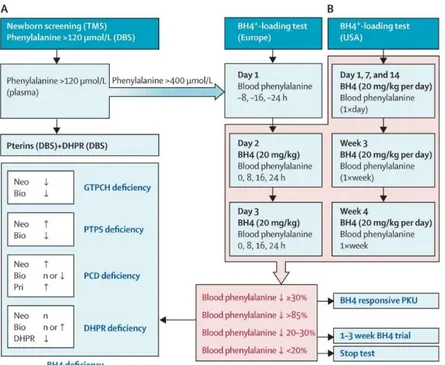
performed to distinguish the cases of HPA due to BH4 deficiency from those caused by PAH defects, but now it is also employed to identify patients affected by PKU variants responsive to BH4 supplementation [186, 187]. In Figure 7 is reported the flow-chart commonly followed to perform the differential diagnosis of PKU or BH4 deficiencies, once HPA has been detected.
Figure 7. A) Diagnostic flow-chart employed to distinguish the different disorders of PAH and BH4 metabolism that can result
in hyperphenylalaninemia. B) BH4 loading test protocols in Europe [according to Blau et al. 2009] and USA [according to Levy
et al. 2007]. DBS = Dried blood spot; n = normal; Neo = neopterin; Bio = biopterin; Pri = primapterin.*BH4 can be either
tetrahydrobiopterin or sapropterin (Kuvan®) (From Blau et al. 2010 [34]).
6.2. The BH4 loading test
Many of the PAH mutations catalogued in the BIOPKU database, are responsible for mild PKU or mild HPA phenotypes: this means that the protein originating from those altered sequences still retains partial hydroxylating activity that can be stimulated by BH4 supplementation, with a consequent reduction in measured L-Phe levels. The first observations of such response date back to 1999 [154] and patients with up to 800 µM L-Phe are most likely to respond to the treatment [190, 191].
There is lack of consensus as regards the definition of BH4 responsiveness, since various criteria can be considered for definition. A dose of 10 mg BH4/kg body weight (BW) has been used for the analysis [192], otherwise 20 mg BH4/kg BW [193] or a combination of 20 mg BH4/kg BW and 100 mg L-Phe/kg BW has also been employed to challenge patients over a period of 24 hours [194, 195]. Sometimes variations exceeding normal individual variability in L-Phe levels have also been considered as a criterion [193] as well as a 1- or 2-fold increase in patient tolerance to daily L-Phe intake [34].
The most widely accepted definition of BH4 responsiveness consists in a blood L-Phe reduction of at least 30% of the pre-treatment value upon a single administration of 20 mg BH4/kg body weight [163, 190, 191]. A reduction ≥ 20% may also be considered of clinical relevance [189].
Test procedures for BH4 responsiveness are different between Europe and USA [34] and there are many protocols, differing from one another as for timing and dose administration and total duration of the analysis, with longer procedures being more common in the USA [189]. The “24 h protocol” commonly employed in European centers [163] consists in the administration per os of 20 mg BH4/kg
body weight and in the subsequent blood L-Phe and pterin measurement at 0, 4, 8 and 24 hours after treatment. Since the response is dose- and time-dependent, some patients may not be identified by this test, if it takes them a longer time to show a sufficient effect on L-Phe; therefore an extended version of the test has been proposed, the “48 h loading test”, with a second administration of 20 mg BH4/kg BW 24 hours after the first one, followed by additional L-Phe assessment at 32 and 48 h [196, 188]. When a protocol involving L-Phe loading is selected, administration of 13C-L-Phe enables subsequent evaluations to be performed non-invasively on patient breath (Phenylalanine breath test); in this case, the amount of exhaled 13CO
2 is measured, obtaining indications on PAH activity and on the overall condition of the L-Phe catabolic pathway of the organism (L-Phe oxidation capacity), which is also expected to reflect the clinical phenotype [194, 195, 197].
On the basis of the percentage reduction in blood L-Phe, patients can be divided as follows:
Rapid responders: patients experiencing a decrease in L-Phe ≥ 30% after 8 h and ≥ 50% by 24 h from BH4 administration;
Moderate responders: patients showing a decrease rate ≥ 20% after 8 h, ≥ 30% after 24 h and ≥ 50% after 48 h;
Slow responders: patients whose plasma L-Phe levels decrease of ≥ 20% by 24 h and ≥ 30% after 48 h.
When L-Phe reduction overcomes 85% upon BH4 treatment, the condition is identified as BH4 deficiency [34].The BH4 loading test should be performed early after birth and before the introduction
of the low L-Phe diet, so as blood L-Phe variations upon BH4 treatment are more evident. Blood L-Phe must be over 400 µM, otherwise older patient who are already on dietary regimen must increase the protein intake before and during the testing period, or are submitted to a concomitant phenylalanine load, consisting in a single administration of 100 mg L-Phe/kg BW [34, 198]. Although the test is effective at all ages, its sensitivity in newborns has been questioned due to liver immaturity and to the fact that only 24 h protocols can be employed at this age [199]. Performing the analysis early allows the early introduction of the restricted diet in non-responders, but at the same time, implies the possibility to miss slow responders (who are mistakenly considered negative to the test). Therefore, it is advised to repeat the analysis according to longer protocols after 3 months of life, that is when the liver has reached complete maturity and longer testing protocols can be applied [199].
7. THERAPEUTIC APPROACHES TO PKU
PKU is the first genetic disease to have an effective treatment, though not decisive. The main aim of all available therapeutic approaches is to restore at least near-physiological levels of circulating L-Phe and L-Tyr, so as to remove as much as possible the biochemical and neurological consequences
resulting from their imbalance. There is great lack of consensus among medical centers worldwide as regards the threshold value beyond which treatment must be introduced. The most common concentrations employed to this purpose are 360 µM, 400 µM and 600 µM [34] and the range of L-Phe concentrations considered safe is between 120 and 360 µM, at least until 12 years of age [106, 200], with the upper limits rising up to 900 µM after the 12th year of age [106]. However, there is great inconsistency about the target range to be reached in adolescence and adulthood, resulting in a wide spectrum of disease management and outcomes [201, 202].
7.1. L-Phe restricted diet
The chief treatment for the disease is a low-phenylalanine diet, introduced for the first time in the early 1950s [151, 152] and still remaining the mainstay of the available approaches. Dietary treatment must be adopted as soon as possible after birth [34]. Since genotype is not always a reliable predictor of the patient phenotype, diet should be individually tailored in order to reach the target concentration, with particular attention to the specific needs of every age and condition [15].
Patients must exclude foods rich in protein [15, 34] and pay particular attention to those containing the artificial sweetener aspartame (L-aspartyl-L-Phe methyl esther) which releases L-Phe, L-aspartic acid and methanol when metabolized [3]. Low-Phe containing natural foods such as potatoes and most vegetables can be consumed only in small amounts, whereas low-protein versions of some foods (i.e. bread and pasta) also exist [34]. The foods allowed in this approach make the diet appear as a medically prescribed vegetarian or vegan diet, but it implies even more food restrictions [203]. This regimen alone does not enable the achievement of the target L-Phe level, as well as sufficient protein and energy intake; hence, it must be integrated with commercially available phenylalanine-free medical foods and protein substitutes [15, 34]. Moreover, the diet must be carefully monitored, so as to adapt it to individual L-Phe tolerance, age and growth requirement, illnesses, physical activity and pregnancy in females [15, 109]. Breastfeeding is encouraged in infants in combination with the medical formula [34]; the strict dependence (approximately 85% [204]) of the required protein intake on semi-synthetic formulas is likely to introduce many nutritional deficiencies, involving also L-Phe and L-Tyr [15]. Even too low concentrations of L-Phe (< 30 µM) must be avoided in order not to impair development [205, 206], but levels between 60 and 120 µM should not be regarded as too low especially in patients with more relaxed adherence to diet [109]. Two systematic reviews by Demirkol [203] and Singh [204] report that the most frequent nutrient deficiencies experienced by PKU patients on-diet concern essential long-chain polyunsaturated fatty acids (particularly arachidonic acid and docosahexanoic acid, DHA) and micronutrients, such as minerals (zinc, copper, manganese, selenium, calcium, iron) and vitamins (A, C, E, B2, B6, B12, D), as well as other metabolically important compounds (i.e. CoQ10,