Research Article
Red Cell Distribution Width and Platelet Count as
Biomarkers of Pulmonary Arterial Hypertension in Patients with
Connective Tissue Disorders
Mattia Bellan
,
1,2,3,4Ailia Giubertoni,
5Cristina Piccinino,
5Arnaldo Dimagli,
1,5Federico Grimoldi,
1,2Maurizio Sguazzotti,
1,2Michela Emma Burlone,
1Carlo Smirne
,
1Daniele Sola,
1,2,4Paolo Marino
,
1,5Mario Pirisi
,
1,2,4and Pier Paolo Sainaghi
2,4 1Department of Translational Medicine, Università del Piemonte Orientale (UPO), Via Solaroli 17, 28100 Novara, Italy2Division of Internal Medicine, Immunorheumatology Unit,“Maggiore della Carità” Hospital, Corso Mazzini 18, 28100 Novara, Italy 3Division of Internal Medicine,“Sant’Andrea Hospital”, Corso Abbiate 21, 13100 Vercelli, Italy
4IRCAD (Interdisciplinary Research Center of Autoimmune Diseases), Novara, Italy
5Division of Cardiology,“Maggiore della Carità” Hospital, Corso Mazzini 18, 28100 Novara, Italy Correspondence should be addressed to Mattia Bellan; [email protected]
Received 5 November 2018; Revised 17 April 2019; Accepted 30 April 2019; Published 2 June 2019 Academic Editor: Ralf Lichtinghagen
Copyright © 2019 Mattia Bellan et al. This is an open access article distributed under the Creative Commons Attribution License, which permits unrestricted use, distribution, and reproduction in any medium, provided the original work is properly cited. Introduction/Objective. In the present paper, we aimed to test the value of the red cell distribution width (RDW) coefficient of variation as a candidate biomarker for pulmonary arterial hypertension (PAH) in patients with connective tissue disorders (CTD), correlating it with the degree of cardiopulmonary impairment in these patients. Methods. The study population included N = 141 patients with CTD and N = 59 patients affected by pulmonary hypertension of other etiologies, all referred to the Pulmonary Hypertension Clinic of the Cardiology Division of an Academic Hospital in Northern Italy for evaluation (including right catheterization). Clinical, instrumental, and laboratory data were collected and related to RDW and other full blood count indexes. Results. Twenty out of 141 CTD patients (14%) received a diagnosis of PAH. In comparison to those without PAH, CTD patients with PAH displayed a larger RDW (14.9% (13.5-17.2) vs. 13.8% (13.1-15.0);p = 0 02) and a lower platelet count (205 177‐240 × 109/l vs. 244 197 5‐304 2 × 10⁹/l; p = 0 005). Moreover, with respect to CTD patients without PAH, RDW
was significantly larger also in PH of other etiologies. In contrast, the platelet count was significantly lower only in CTD-related PAH, with a value > 276 × 109/l being 100% sensitive in ruling out PAH. Finally, RDW, but not the platelet count, was related
directly to systolic pulmonary arterial pressure (r = 0 381; p = 0 0008) and right ventricle diameter (r = 0 283; p = 0 015) and inversely to diffusing capacity of the lung for carbon monoxide (r = −0 325; p = 0 014). Conclusion. RDW is a promising candidate biomarker for the screening and the prognostic stratification of PAH in CTD patients.
1. Introduction
Pulmonary hypertension (PH) is a clinical entity that com-prises multiple conditions of different etiologies and patho-physiology, defined by the presence of a mean pulmonary arterial pressure (mPAP) equal to or greater than 25 mmHg, measured during invasive right heart catheterization (RHC) at rest [1]. Although reliable estimates of prevalence are lacking, PH as defined above is not uncommon. Group 1
PH (i.e., pulmonary arterial hypertension (PAH), character-ized by a pulmonary artery wedge pressure≤ 15 mmHg), on the other hand, is a rare disease that may complicate connec-tive tissue diseases (CTD) [2]. The estimated prevalence and incidence of PAH are around 15-60 cases and 5-10 cases per million adult population, respectively [3]; roughly, half of these cases occur in association with CTD, with a prevalence of 21-29% in mixed connective tissue disease (MCTD), up to 14% systemic lupus erythematosus (SLE) [4], and close to Volume 2019, Article ID 4981982, 7 pages
20% in systemic sclerosis (SSc) [5]. Lower rates have been reported in Sjogren’s syndrome (SS) [4] and in dermatomyo-sitis/polymyositis (PM/DM) [6].
Consensus exists that these high-risk patients should be screened for PAH, since CTD-related PAH has a severe prog-nosis, even poorer than the idiopathic form [7]; in fact, PAH is the leading cause of death in systemic sclerosis [8]. More-over, early treatment of PAH patients leads to improved out-comes, whereas treatment delay is associated with clinical worsening [9]. The screening strategy most used today is based on the application of a two-step algorithm (DETECT) on systemic sclerosis patients, which is 97% sensitive and 35% specific for the diagnosis of PAH [10]. Despite the rela-tively good performance of the DETECT algorithm, no uni-versally agreed approach by which to screen these patients exists, and novel biomarkers of PAH are actively pursued.
The red cell distribution width (RDW) coefficient of var-iation, a measure of the variability in size of circulating eryth-rocytes, is routinely reported as a component of the complete blood count and has been used in the differential diagnosis of anemia [11]. Recently, RDW has been shown to stratify prog-nosis among patients with idiopathic and thromboembolic PAH [12, 13]. Moreover, RDW has been proposed as a pre-dictor of PAH in SSc and in SS [14, 15].
In the present paper, we aimed to evaluate RDW as a can-didate biomarker for PAH in CTD patients and to correlate it with the severity of cardiopulmonary impairment.
2. Methods
2.1. Patients. We performed a cross-sectional observational study. From October 1st, 2016, to April 20th, 2018, we recruited 141 consecutive patients, affected by CTD and referred to the Pulmonary Hypertension Outpatient Clinic of the Cardiology Department, A.O.U.“Maggiore della Carità,” Novara, Italy.
We applied the following exclusion criteria: (a) Age < 18 years
(b) Refusal to participate in the study
(c) Impossibility to undergo the required cardiopulmo-nary assessment
We also recruited 59 patients affected by PH of other eti-ologies, applying the same exclusion criteria, to check whether the predictive factors identified in CTD-related PAH were valid in other clinical settings.
2.2. Main Outcome Variable. Following echocardiography estimation of systolic pulmonary artery pressure (sPAP), right heart catheterization (RHC) was performed to con-firm PH, when appropriate. PAH was defined by mean pulmonary artery pressure mPAP ≥ 25 mmHg, pulmonary capillary wedge pressure ≤ 15 mmHg, and pulmonary vascular resistance > 3 Wood Units. Whenever contraindi-cations to RHC occurred, PH was diagnosed based on echocardiography-estimated sPAP≥ 35 mmHg and addi-tional high-probability criteria, according to 2015 ESC/ESR guidelines. In the control group, PH etiology was carefully
searched for, and patients were classified according to the Nice 2013 criteria [1].
2.3. Procedures. All patients underwent a comprehensive medical history and a thorough physical examination, aimed at assessing the presence and severity of signs and symptoms compatible with PH. Cardiovascular risk factors and other comorbidities were investigated. Past medical his-tory and drug hishis-tory were recorded. Rheumatologic disease history was recalled through an extensive review of the medical records.
Complete blood count (CBC), serum creatinine, C-reactive protein (CRP), and brain natriuretic peptide (BNP) were measured. The serum autoantibody profile was exam-ined. Further assessment of these patients consisted of
(i) 12-lead electrocardiogram with 6 limb and 6 precor-dial leads with paper speed set at the standard rate of 25 mm/s
(ii) six-minute walking distance (6MWD), which mea-sures the distance that the patient could quickly walk over a total of six minutes on a hard,flat surface (iii) posteroanterior and lateral chest X-rays and, when
necessary, high-resolution computed tomography (HRCT) (to rule out interstitial lung disease) (iv) pulmonary function tests (PFTs), performed on
the same day the patient attended the PH clinic. The spirometry was performed using standardized equipment and technique with a spirometer, which measured the amount of air the subject exhaled and the rate at which it was exhaled. The device was connected to a computer employing“Medisoft ExpAir 1.28.20” to convert the signals into numeri-cal values and graphics. The following standardized measurements were evaluated: forced vital capacity (FVC), forced expiratory volume in the 1st second (FEV1), FEV1/FVC (%) (also known as the Tiffe-neau index)
(v) the diffusing capacity of the lung for carbon monox-ide (DLCO), measured with the single-breath Jones-Meade protocol. Once the mouthpiece and the nose clip were in place, the subject made a maximal expi-ration and then, with a maximal inspiexpi-ration, inhaled a gas blend containing carbon monoxide (0.3%) and other inert gases (0.3% CH4). Then, the patient was asked to hold his/her breath for about 10 seconds and exhale afterwards. During the expiration, alveo-lar air was analyzed: the ratio between carbon mon-oxide in inspired gas and carbon monmon-oxide in exhaled air determined the diffusion of carbon monoxide. Predicted DLCO corrected for haemo-globin and alveolar volume was assessed
(vi) transthoracic echocardiography, performed using the Vivid 7 or E9 cardiovascular ultrasound machine by GE Medical Systems (Horten, Norway) with a 1.7/3.4 MHz tissue harmonic transducer. All data
were obtained in standardized patient positions, according to the guidelines of the American Echo-cardiography Society. The exam was performed by an echocardiography expert in PH. The following parameters were studied [16]: sPAP, right atrium area (RAA), right ventricle diameter (RVD), and left ventricular ejection fraction (LVEF). LVEF has been evaluated with the modified Simpson method; sys-tolic pulmonary artery pressure (sPAP) was esti-mated from the maximal velocity of tricuspid regurgitation (TR) and RAP (right atrial pressure) using Bernoulli’s equation (sPAP = 4TR2+ RAP).
RAP was estimated with the respiratory motion and the size of the inferior vena cava (IVC) from the subcostal view. TR jet was graded according to recommendations for the quantification of native valvular regurgitation
(vii) right ventricle systolic function was evaluated by estimating the Tricuspid Annular Plane Systolic Excursion (TAPSE)
The study protocol was approved by the institutional eth-ical committee and conducted in strict accordance with the principles of the Declaration of Helsinki. Informed consent was obtained from all individual participants included in the study.
2.4. Statistical Analysis. Anthropometric, clinical, and bio-chemical data were recorded in a database and analyzed by the statistical software package MedCalc v.18.10.2 (MedCalc Software, Broekstraat 52, 9030, Mariakerke, Belgium). The measures of centrality and dispersion of data chosen were medians and interquartile range (IQR). Continuous vari-ables were compared between groups by the Mann–Whitney and Kruskal-Wallis (K-W) tests. Exact Fischer’s test and
Pearson’s χ2 test were used, as appropriate, to explore the
associations of categorical variables. To test the diagnostic performance of RDW and platelet count, receiver operating characteristic curves were built, with calculation of the respective areas under the curve (AUC). The level of signif-icance chosen for all statistical tests was 0.05 (two-tailed).
3. Results
Among the 141 patients with CTD, the following diagnoses were recorded: SSc (N = 102, 72.3%), SLE (N = 3, 2.1%), SS
(N = 1, 0.7%), PM/DM (N = 13, 9.3%), MCTD (N = 10,
7.1%), and undifferentiated CTD (UCTD; N = 12, 8.5%). Out of 141, N = 20 received a diagnosis of PAH (14%): in
N = 10 patients, the diagnosis was based on RHC data.
In Table 1, we report the main features of these patients, as well as the comparison between patients with and without PAH. As shown in the table, the patients with PAH were older and characterized by a significantly higher BNP and sPAP; moreover, the creatinine clearance and the DLCO were significantly lower in the case of PAH. Interestingly, PAH was also associated with a larger RDW and a lower platelet count. We therefore analyzed the diagnostic accuracy of RDW and PLTs; in Figure 1, we report the respective ROC curves. An RDW value≥ 16% was 40.0% sensitive and 88.3% specific for the diagnosis of PAH, with an AUC 0.666 (CI95%: 0.581-0.783; p = 0 015). A platelet count ≤ 276 × 109/l was 100.0% sensitive and 36.3% specific for the
diag-nosis of PAH, with an AUC 0.697 (CI95%: 0.614-0.771;
p = 0 0001). 5/20 patients were affected by chronic
respira-tory failure requiring continuous oxygen supplementation. This subset of patients had a trend, though not statistically significant, towards a larger RDW (17.7% (15.4-18.2) vs. 14.2% (13.4-16.0)) when compared to those with PAH and no chronic respiratory failure. Platelet count was not differ-ent between groups (data not shown).
Table 1: Main features of the study population and differences related to the presence of PAH. We report the main features of the whole study population; moreover, we compared CTD alone and CTD complicated by PAH. Categorical variables are shown as frequencies (%); continuous variables are shown as medians (IQR). Mann–Whitney and Fisher’s exact tests were used as appropriate.
Variables Entire CTD population (N = 141) CTD without PAH (N = 121) CTD with PAH (N = 20) p
Age (years) 68 (57-76) 62 (52-72) 74 (69-79) 0.0001 Gender (F/M) 127 (90.1)/14 (9.9) 110 (90.9)/11 (9.1) 17 (85.0)/3 (15.0) 0.42 Hb (g/dl) 12.9 (11.8-14.0) 12.9 (11.7-13.8) 12.0 (11.6-13.8) 0.35 RDW (%) 14.0 (13.2-15.5) 13.8 (13.1-15.0) 14.9 (13.5-17.2) 0.02 PLTs (×109/l) 228 (190-295) 244 (197.5-304.2) 205 (177-240) 0.005 BNP (pg/ml) 78.7 (35.3-182.1) 52.1 (29.7-102.2) 204.9 (81.0-465.5) <0.0001
Creatinine clearance (ml/min) 82.5 (60.0-97.5) 89.0 (71.5-102.0) 65 (49.0-78.0) 0.0005 DLCO (ml/min/mmHg) 72.5 (52.0-87.0) 76.0 (64.0-87.2) 45.0 (40.2-54.7) <0.0001
FEV1 (%) 96.0 (78.0-113.0) 101.0 (84.7-115.0) 103.0 (87.5-115.0) 0.98
FEV1/FVC (%) 107.0 (101.0-114.0) 109.0 (102.5-115.5) 107.0 (102.0-111.0) 0.41
sPAP (mmHg) 33 (25-45) 27 (23-31) 46 (42-57) <0.0001
LVEF (%) 61.5 (57-66) 63.0 (59.0-67.0) 61.5 (55.5-65.5) 0.15
N: number; CTD: connective tissue diseases; PAH: pulmonary arterial hypertension; Hb: haemoglobin; RDW: red cell distribution width; PLTs: platelets; BNP: brain natriuretic peptide; DLCO: diffusion lung capacity for carbon monoxide; FEV1: forced expiratory volume in the 1st second; FVC: forced vital capacity; sPAP: systolic pulmonary artery pressure; LVEF: left ventricular ejection fraction.
We further tried to evaluate whether the increase in RDW and the decrease in platelet count were limited to CTD-related PAH or generalizable to other PH patients. In Table 2, we report the main features of the PH control group (Figure 2). With respect to CTD not complicated by PAH, RDW was significantly higher in both CTD-related PAH (14.9% (13.5-17.2)) and PH of other etiologies (14.8% (13.2-17.0); K-W 13.6;p < 0 001). On the contrary, the plate-let count was significantly lower only in CTD-related PAH (205 177‐240 × 109/l; K-W 7.30; p = 0 02), being similar
between PH of other etiologies (222 184‐266 × 109/l) and
CTD not complicated by PAH (242 194‐302 × 109/l) (see
also Figure 2).
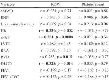
Finally, we evaluated the correlations between RDW and platelet count measured in patients with PH and di ffer-ent clinical and instrumffer-ental parameters. In Table 3, we report the results of this analysis. Platelet count was not related to any of the functional and echocardiographic markers analyzed. On the contrary, RDW was inversely asso-ciated with DLCO and directly related to sPAP and right ven-tricle diameter.
4. Discussion
The results of the present study indicate that RDW, a simple parameter routinely obtained with the automated full blood count, may be of help in the diagnostic evaluation and prog-nostic stratification of CTD patients, with a specific focus on PAH. These data will be discussed at the light of the existing literature on the topic.
The rationale of the present study stems from previous reports, suggesting that red blood cells and platelets are pos-sibly involved in the pathogenesis of several inflammatory diseases and may serve as diagnostic and prognostic bio-markers in this context [17]. Growing attention has been paid recently towards the prognostic implication of an increased RDW: in fact, this parameter has been used as an indicator of ineffective red cell production or haemolysis but has recently been identified as a predictor of poor
prognosis in different cardiovascular and noncardiovascular diseases [18, 19]. With specific regard to PH, a high RDW predicts a poor prognosis in chronic thromboembolic PH and in PAH [12, 13] and may be a factor in CTD-related PAH, at least in Asian patients [14, 15]. Furthermore, in SSc patients, a higher RDW has been observed in those subjects with at least one clinical vascular involvement (acral ulcers, PAH, or renal crisis) [20]. Our data confirm and extend the abovementioned observations, suggesting
100 100 80 80 60 60 100 − specificity Sensitivity 40 40 20 20 0 0 (a) 100 100 80 80 60 60 100 − specificity Sensitivity 40 40 20 20 0 0 (b)
Figure 1: Receiver operating characteristic (ROC) curves for red cell distribution width (RDW) coefficient of variation and platelet count. In (a), we report the ROC curve of RDW for the diagnosis of PAH; in (b), we report the ROC curve of platelet count for the diagnosis of PAH.
Table 2: Main features of the PH control group. We report the main features of the control group with PH of other etiologies. Categorical variables are shown as frequencies (%); continuous variables are shown as medians (IQR).
Variables N = 59 PH class (1/2/3/4/5) 17 (28.8)/10 (16.9)/7 (12.0)/10 (16.9)/15 (25.4) Age (years) 73 (64-79) Gender (F/M) 33 (56)/26 (44) Hb (g/dl) 12.9 (11.9-14.8) RDW (%) 14.8 (13.2-17.0) PLTs (×109/l) 222 (184-266) BNP (pg/ml) 182.5 (77.7-299.5)
Creatinine clearance (ml/min) 70.0 (51.0-87.0) DLCO (ml/min/mmHg) 53.0 (42.0-80.0)
FEV1 (%) 70.0 (57.0-89.5)
FEV1/FVC (%) 102.0 (93.2-111.0)
sPAP (mmHg) 53 (44-69)
LVEF (%) 60 (55-66)
N: number; class 1: pulmonary arterial hypertension; class 2: PH related to left heart disease; class 3: PH related to pulmonary disease; class 4: thromboembolic PH; class 5: others; Hb: haemoglobin; RDW: red cell distribution width; PLTs: platelets; BNP: brain natriuretic peptide; DLCO: diffusion lung capacity for carbon monoxide; FEV1: forced expiratory volume in the 1st second; FVC: forced vital capacity; sPAP: systolic pulmonary artery pressure; LVEF: left ventricular ejection fraction.
that RDW may predict the presence of PAH in CTD patients; moreover, RDW has a potential prognostic implication, given its association with sPAP, right ventricular size, and DLCO, which are well-known prognostic markers in SSc-related PAH [21, 22]. Wefinally showed that RDW is elevated in PH belonging to classes different from class 1, therefore marking more generally PH rather than PAH in CTD. The reason why RDW is elevated under these circumstances is still unknown, although it might reflect the microvascular damage or the chronic inflammatory state, since elevation of RDW levels was shown to reflect the circulating levels of
proinflammatory cytokines, such as tumor necrosis factor
α, interleukin- (IL-) 1, and IL-6 [20].
We also reported a significantly lower platelet count in CTD patients with PAH; this observation might be explained by an intravascular thrombosis due to endothelial damage, leading to the local consumption of platelets [23]. However, a low platelet count seems to be more specifically associated with CTD-related PAH, since it was not observed in patients with other forms of PH and in those with uncomplicated CTD. Given that the platelet count was not associated with other important prognostic markers of PAH but has high sensitivity, its role may consist in the possibility of ruling out PAH, when above the threshold indicated.
It might be argued that the increased RDW and reduced platelet count belong to the development of hypoxia, leading to increased erythropoiesis at the expense of megakaryopoi-esis. We have no data about blood gas analysis of our patients; however, those on oxygen supplementation because of chronic respiratory failure were not characterized by a dif-ferent platelet count. However, a trend towards a larger RDW was observed. Hypoxia is, therefore, an intriguing potential explanation of these associations, deserving consideration in further studies.
A main limitation of the present study is related to the small sample size of our cohort which, being representative of a relatively rare complication of a rare disease, allowed us to enrol only 20 patients with CTD-related PAH. This significantly limits our possibility to draw definitive conclu-sions, particularly with regard to the identification of diag-nostic thresholds. Therefore, the ROC curves presented have the value of an exploratory analysis, requiring con firma-tion in larger cohorts.
Finally, for the present study, the diagnosis of PAH was based on the 2015 ESC/ESR guidelines which defined pul-monary arterial hypertension in the presence of a mPAP≥ 25 mmHg; this cut-off has been recently revised during the
30 28 26 24 RDW (%) 22 20 18 16 14 12 CTDs CTDs with
PAH PAH of otheretiology
(a) 800 700 600 500 400 300 200 100 0 PLTS (10 9/l) CTDs CTDs with
PAH PAH of otheretiology
(b)
Figure 2: RDW and platelet values according to underlying diagnosis. The box and whiskers of RDW (a) and platelet (b) values according to the underlying diagnosis. RDW: red cell distribution width; PLTs: platelets; CTD: connective tissue diseases; PAH: pulmonary arterial hypertension.
Table 3: Correlation between functional parameters and RDW and platelet count in patients with PAH.
Variable RDW Platelet count
6MWD r = ‐0 051; p = 0 71 r = 0 021; p = 0 88 BNP r = 0 045; p = 0 69 r = 0 006; p = 0 96 Creatinine clearance r = ‐0 009; p = 0 94 r = 0 213; p = 0 06 Hb r = ‐0 331; p = 0 002 r = ‐0 031; p = 0 79 sPAP r = 0 381; p = 0 0008 r = 0 071; p = 0 54 LVEF r = 0 089; p = 0 45 r = 0 182; p = 0 12 RAA r = 0 199; p = 0 10 r = ‐0 081; p = 0 50 RVD r = 0 283; p = 0 015 r = ‐0 038; p = 0 75 DLCO r = ‐0 325; p = 0 014 r = 0 037; p = 0 79 FEV1 r = ‐0 179; p = 0 17 r = ‐0 169; p = 0 20 FEV1/FVC r = ‐0 152; p = 0 25 r = ‐0 188; p = 0 15
RDW: red cell distribution width; 6MWD: 6-minute walking distance; BNP: brain natriuretic peptide; Hb: haemoglobin; sPAP: systolic pulmonary artery pressure; LVEF: left ventricular ejection fraction; RAA: right atrium area; RVD: right ventricle diameter; DLCO: diffusion lung capacity for carbon monoxide; FEV1: forced expiratory volume in the 1st second; FVC: forced vital capacity.
2018 PH World Symposium. The new proposed definition of PAH is based on the detection of a mPAP≥ 20 mmHg with a pulmonary vascular resistance≥ 3 WU at RHC [24]. Our findings do not automatically extend to those patients with a mPAP 21-24 mmHg and, therefore, need to be confirmed using this new diagnostic threshold.
5. Conclusions
In conclusion, RDW is a promising tool for the screening and the prognostic stratification of PAH in CTD patients; its role in complementing and refining the approach by which we currently screen for PAH on these high-risk patients is worth to be established in future studies.
Data Availability
The data used to support thefindings of this study are avail-able from the corresponding author upon request.
Conflicts of Interest
The authors declare that there is no conflict of interest regarding the publication of this article.
Acknowledgments
This research is original and had afinancial support of the Università degli Studi del Piemonte Orientale.
References
[1] N. Galiè, M. Humbert, J.-L. Vachiery et al., “2015 ESC/ERS Guidelines for the diagnosis and treatment of pulmonary hypertension,” European Heart Journal, vol. 37, no. 1, pp. 67–119, 2016.
[2] V. Thakkar and E. M. T. Lau, “Connective tissue disease-related pulmonary arterial hypertension,” Best Practice & Research Clinical Rheumatology, vol. 30, no. 1, pp. 22–38, 2016.
[3] A. J. Peacock, N. F. Murphy, J. J. V. McMurray, L. Caballero, and S. Stewart, “An epidemiological study of pulmonary arterial hypertension,” European Respiratory Journal, vol. 30, no. 1, pp. 104–109, 2007.
[4] V. Cottin, “Pulmonary arterial hypertension in connective tissue disease,” Revue des Maladies Respiratoires, vol. 23, Supplement 4, pp. 13S61–13S72, 2006.
[5] J. Avouac, P. Airò, C. Meune et al.,“Prevalence of pulmonary hypertension in systemic sclerosis in European Caucasians and metaanalysis of 5 studies,” The Journal of Rheumatology, vol. 37, no. 11, pp. 2290–2298, 2010.
[6] L. Cavagna, E. Prisco, C. Montecucco, and R. Caporali, “Pulmonary arterial hypertension in antisynthetase syndrome: comment on the article by Chatterjee and Farver,” Arthritis Care & Research, vol. 63, no. 4, pp. 633-634, 2011.
[7] M. J. Ruiz-Cano, P. Escribano, R. Alonso et al.,“Comparison of baseline characteristics and survival between patients with idiopathic and connective tissue disease-related pulmonary arterial hypertension,” The Journal of Heart and Lung Trans-plantation, vol. 28, no. 6, pp. 621–627, 2009.
[8] N. F. Chaisson and P. M. Hassoun, “Systemic sclerosis-associated pulmonary arterial hypertension,” Chest, vol. 144, no. 4, pp. 1346–1356, 2013.
[9] N. Galiè, L. Rubin, M. Hoeper et al.,“Treatment of patients with mildly symptomatic pulmonary arterial hypertension with bosentan (EARLY study): a double-blind, randomised controlled trial,” The Lancet, vol. 371, no. 9630, pp. 2093– 2100, 2008.
[10] J. G. Coghlan, C. P. Denton, E. Grünig et al.,“Evidence-based detection of pulmonary arterial hypertension in systemic scle-rosis: the DETECT study,” Annals of the Rheumatic Diseases, vol. 73, no. 7, pp. 1340–1349, 2014.
[11] T. C. Evans and D. Jehle, “The red blood cell distribution width,” The Journal of Emergency Medicine, vol. 9, pp. 71–74, 1991.
[12] C. J. Rhodes, J. Wharton, L. S. Howard, J. S. R. Gibbs, and M. R. Wilkins, “Red cell distribution width outperforms other potential circulating biomarkers in predicting survival in idio-pathic pulmonary arterial hypertension,” Heart, vol. 97, no. 13, pp. 1054–1060, 2011.
[13] A. Smukowska-Gorynia, I. Tomaszewska, K. Malaczynska-Rajpold et al.,“Red blood cells distribution width as a potential prognostic biomarker in patients with pulmonary arterial hypertension and chronic thromboembolic pulmonary hyper-tension,” Heart, Lung and Circulation, vol. 27, no. 7, pp. 842– 848, 2018.
[14] J. Zhao, H. Mo, X. Guo et al., “Red blood cell distribution width as a related factor of pulmonary arterial hypertension in patients with systemic sclerosis,” Clinical Rheumatology, vol. 37, no. 4, pp. 979–985, 2018.
[15] M. Hui, J. Zhao, Z. Tian et al.,“Red blood cell distribution width as a potential predictor of survival of pulmonary arterial hypertension associated with primary Sjogren’s syndrome: a retrospective cohort study,” Clinical Rheumatology, vol. 38, no. 2, pp. 477–485, 2019.
[16] L. G. Rudski, W. W. Lai, J. Afilalo et al., “Guidelines for the echocardiographic assessment of the right heart in adults: a report from the American Society of Echocardiography. Endorsed by the European Association of Echocardiography, a registered branch of the European Society of Cardiology, and the Canadian Society of Echocardiography,” Journal of the American Society of Echocardiography, vol. 23, no. 7, pp. 685–713, 2010.
[17] O. O. Olumuyiwa-Akeredolu and E. Pretorius, “Platelet and red blood cell interactions and their role in rheumatoid arthri-tis,” Rheumatology International, vol. 35, no. 12, pp. 1955– 1964, 2015.
[18] L. L. Abrahan IV, J. D. A. Ramos, E. L. Cunanan, M. D. A. Tiongson, and F. E. R. Punzalan,“Red cell distribution width and mortality in patients with acute coronary syndrome: a meta-analysis on prognosis,” Cardiology Research, vol. 9, no. 3, pp. 144–152, 2018.
[19] B. D. Horne, J. B. Muhlestein, S. T. Bennett et al., “Extreme erythrocyte macrocytic and microcytic percentages are highly predictive of morbidity and mortality,” JCI Insight, vol. 3, no. 14, 2018.
[20] N. Farkas, A. Szabó, V. Lóránd et al.,“Clinical usefulness of measuring red blood cell distribution width in patients with systemic sclerosis,” Rheumatology, vol. 53, no. 8, pp. 1439– 1445, 2014.
[21] S. R. Johnson, J. R. Swiston, and J. T. Granton,“Prognostic fac-tors for survival in scleroderma associated pulmonary arterial
hypertension,” The Journal of Rheumatology, vol. 35, no. 8, pp. 1584–1590, 2008.
[22] M. I. Acosta Colmán, G. Avila Pedretti, M. E. Acosta, C. P. Simeón Aznar, V. Fonollosa Plá, and M. Villardel Torrés, “Can we predict the severity of pulmonary hypertension in patients with scleroderma?,” Reumatología Clínica, vol. 8, no. 5, pp. 259–262, 2012.
[23] P. Herve, M. Humbert, O. Sitbon et al.,“Pathobiology of pul-monary hypertension. The role of platelets and thrombosis,” Clinics in Chest Medicine, vol. 22, no. 3, pp. 451–458, 2001. [24] N. Galiè, V. V. McLaughlin, L. J. Rubin, and G. Simonneau,
“An overview of the 6th World Symposium on Pulmonary Hypertension,” European Respiratory Journal, vol. 53, no. 1, article 1802148, 2019.
Stem Cells
International
Hindawi www.hindawi.com Volume 2018 Hindawi www.hindawi.com Volume 2018 INFLAMMATIONEndocrinology
International Journal ofHindawi www.hindawi.com Volume 2018 Hindawi www.hindawi.com Volume 2018
Disease Markers
Hindawi www.hindawi.com Volume 2018 BioMed Research InternationalOncology
Journal of Hindawi www.hindawi.com Volume 2013 Hindawi www.hindawi.com Volume 2018Oxidative Medicine and Cellular Longevity
Hindawi
www.hindawi.com Volume 2018
PPAR Research
Hindawi Publishing Corporation
http://www.hindawi.com Volume 2013 Hindawi www.hindawi.com
The Scientific
World Journal
Volume 2018 Immunology Research Hindawi www.hindawi.com Volume 2018 Journal ofObesity
Journal of Hindawi www.hindawi.com Volume 2018 Hindawi www.hindawi.com Volume 2018 Computational and Mathematical Methods in Medicine Hindawi www.hindawi.com Volume 2018Behavioural
Neurology
Ophthalmology
Journal of Hindawi www.hindawi.com Volume 2018Diabetes Research
Journal of Hindawiwww.hindawi.com Volume 2018
Hindawi
www.hindawi.com Volume 2018 Research and Treatment
AIDS
Hindawiwww.hindawi.com Volume 2018
Gastroenterology Research and Practice
Hindawi www.hindawi.com Volume 2018