Contents lists available atScienceDirect
International Journal of Surgery Case Reports
j o u r n a l h o m e p a g e :w w w . c a s e r e p o r t s . c o mPHACES syndrome: Diode laser photocoagulation of intraoral
hemangiomas in six young patients
Gianfranco Favia
a, Luisa Limongelli
a,∗, Angela Tempesta
a, Matteo Favia
b,
Eugenio Maiorano
caDepartment of Interdisciplinary Medicine, Complex Operating Unit of Odontostomatology, Aldo Moro University, 70124 Bari, Italy bMedical School, Aldo Moro University, 70124 Bari, Italy
cDepartment of Emergency and Organ Transplantation, Pathological Anatomy, Aldo Moro University, 70124 Bari, Italy
a r t i c l e i n f o
Article history:Received 12 December 2014
Received in revised form 23 March 2015 Accepted 24 March 2015
Available online 27 March 2015 Keywords:
Laser treatment Rare disease Vascular tumor
a b s t r a c t
INTRODUCTION: The acronym PHACES describes the association of posterior fossa malformations, facial hemangiomas, arterial anomalies (cardiovascular or cerebrovascular), coarctation of the aorta and car-diac defects, eye abnormalities, and sternal or ventral defects. In this study we report on 6 patients affected by the PHACES syndrome and showing 34 intraoral hemangiomas (IH), treated by diode laser photocoagulation (DLP).
CASE PRESENTATION: IH appeared as red-bluish soft masses, smooth or lobulated, from a few millimetre to several centimetres in size, covered by intact mucosa and blanching on pressure. IHs were treated by DLP with 320m fibres at a wavelength of 800 ± 10 nm. The diode laser techniques applied were: Transmucosal DLP (DLTP), a no-contact technique in which laser energy is delivered by a flexible optic quartz fiber, which is kept 2–3 mm apart from the lesion, and Intralesional DLP (DLIP), in which the fibre is introduced into the lesion through a transmucosal access. DLTP was used for 20 flat, superficial IHs and, after a variable number of laser sessions (average = 3) depending on the size of the lesion, 65% completely regressed, while in the remaining 35% shrinkage of the lesion was achieved with minor and few complications.
The remaining 14 deep/multi-lobulated IHs were treated by DLIP, resulting in complete regression of 79% of them.
CONCLUSIONS: DLP techniques are an effective and minimally invasive procedure for IH in patients with PHACES, in consideration of the multiple lesions to treat, of the necessity of multiple interventions and the higher compliance of the patients.
© 2015 Published by Elsevier Ltd. on behalf of Surgical Associates Ltd. This is an open access article under the CC BY-NC-ND license (http://creativecommons.org/licenses/by-nc-nd/4.0/).
1. Introduction
PHACES is an acronym which refers to a rare syndrome char-acterized by Posterior fossa defects, facial hemangiomas, arterial lesions, cardiac abnormalities, eye anomalies and sternal cleft[1,2]. Firstly described by Frieden in 1996[1], this syndrome shows predilection for the female gender (M:F = 9:1)[3,4]; there is no evidence of familial tendency[5].
∗ Corresponding author at: Department of Interdisciplinary Medicine, Odontostomatology, Aldo Moro University, P.zza G. Cesare, 11, 70124, Bari, Italy. Tel.: +39 3398790106/0805218784; fax: +39 0805218784.
E-mail addresses: [email protected] (G. Favia), [email protected](L. Limongelli),[email protected] (A. Tempesta),[email protected](M. Favia),[email protected] (E. Maiorano).
Single/multiple facial hemangiomas are the most impor-tant clinical manifestation of PHACES. Hemangiomas manifest during the first weeks of life as teleangiectasias or erythema-tous plaques; subsequently, they increase in size (>5 cm), and converge together, occupying one or more distinct segments: fronto-temporal, maxillary, mandibular or fronto-nasal[6]. In 80% of the cases, hemangiomas in the mandibular region are associ-ated with sternal clefts[7]and cardiovascular abnormalities[2,7,8]. Hemangiomas could completely or partially regress during the first decade, thus acquiring a port-wine like aspect.
The most common extracutaneous features are: cerebrovascu-lar, structural brain and cardiovascular abnormalities[9].
Cerebrovascular anomalies consist in abnormalities of major cerebral arteries and are divided into four categories: dysplasia, narrowing, aberrant course/origin and persistence of embryonic anastomoses[8].
A spectrum of congenital structural brain abnormalities have been described in the PHACES syndrome, the most common of which involving the cranial posterior fossa[8].
http://dx.doi.org/10.1016/j.ijscr.2015.03.045
2210-2612/© 2015 Published by Elsevier Ltd. on behalf of Surgical Associates Ltd. This is an open access article under the CC BY-NC-ND license (http://creativecommons.org/licenses/by-nc-nd/4.0/).
Table 1
Diagnostic criteria for PHACES syndrome.
Organ system Major criteria Minor criteria
Cerebrovascular Anomalies of major cerebral arteries: Persistent embryonic artery other than trigeminal artery: Dysplasiaaof the large cerebral arteriesb Proatlantal intersegmental artery (types 1 and 2)
Arterial stenosis or occlusion with or without moyamoya collaterals Primitive hypoglossal artery Primitive otic artery Absence or moderate to severe hypoplasia of the large cerebral arteries
Aberrant origin or course of the large cerebral arteries Persistent trigeminal artery
Saccular aneurysms of any cerebral arteries Structural
brain
Posterior fossa anomaly Enhancing extra-axial lesion with features consistent with intracranial hemangioma
Dandy-Walker complex Midline anomalyc
Unilateral/bilateral cerebellar hypoplasia/dysplasia Neuronal migration disorderd
Cardiovascular Aortic arch anomalies: Ventricular septal defect
Coarctation of aorta Right aortic arch (double aortic arch) Dysplasiaa
Aneurysm
Aberrant origin of the subclavian artery with or without a vascular ring
Ocular Posterior segment abnormalities: Anterior segment abnormalities: Persistent fetal vasculature (persistent hyperplastic primary vitreous) Sclerocornea
Cataract Coloboma
Retinal vascular anomalies Microphtalmia
Morning Glory disc anomaly Optic nerve hypoplasia Peripapillary staphyloma Coloboma
Ventral or midline Sternal defect Hypopituitarism
Sternal cleft Ectopic thyroid
Supraumbilicam raphe Sterna defects
aIncludes kinking, looping, tortuosity, and/or dolichoectasia.
bInternal carotid artery, middle cerebral artery, posterior cerebral artery, or vertebrobasial system. c Callosal agenesis or dysgenesis, septum pellucidum agenesis, pituitary malformation, or pituitary ectopia. d Polymicrogyria, cortical dysplasia, or gray matter hetrotopia.
Fig. 1. IH on the right cheek before (1a) and after Diode Laser Transmucosal Photocoagulation (1b).
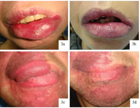
Fig. 3. (a) and (b). Case 1: haemangioma of lower lip before and after Diode Laser Intralesional Photocoagulation. (c) and (d). Case 2: haemangioma of upper lip before and
after Diode Laser Intralesional Photocoagulation.
The most important cardiovascular anomaly associated with PHACES is coarctation of the aorta, which occurs in 14.5% of the affected patients[8]. Abnormalities of the eye are rare and most frequently consist in microphthalmia and exophtalmo[9,10].
Ventral development defects consist of sternal clefting and supraumbilical abdominal raphe[10].
Other extracutaneous manifestation of PHACES syndrome is intraoral haemangioma (IH), which has been only occasionally reported in the English literature.
According to the consensus statement on diagnostic criteria for PHACES syndrome, the diagnosis requires the presence of segmen-tal haemangioma of the head, greater than 5 cm, plus one major criterion or 2 minor criteria, as illustrated inTable 1 [10].
The PHACES syndrome may show variable clinical presentation, according to a major and a minor phenotype, the former being char-acterized by several life-threatening abnormalities, which could kill the patient within the first years of life, the latter manifesting less serious anomalies, which allow patients to reach adulthood.
This work was aimed at describing the clinical features of the vascular lesions occurring in 6 young patients affected by the PHACES syndrome, and to illustrate the efficacy of diode laser pho-tocoagulation for the treatment of IH.
2. Case presentation
This study was carried out in accordance with the code of ethics of the world medical association (Declaration of Helsinki). The patients released informed consent on diagnostic and therapeu-tic procedures and for the possible use of biological samples for research purposes.
In this study we report the cases of 6 PHACES patients (aver-age (aver-age: 17,5 years), 2 males and 4 females, who collectively were affected by 34 Intraoral Hemangiomas.
IHs appeared as red-bluish soft masses, smooth or lobulated, from few millimetres to several centimetres, covered by intact mucosa and blanching on pressure. As such lesions produced bleed-ing, functional and aesthetic/psychological problems, they were treated with diode laser (GaAlAs - A2G Laser “Surgery 35”) with a flexible optic quartz fibre of 320m, at a 800 ± 10 nm wave-length by either diode laser transmucosal photocoagulation (DLTP) or diode laser intralesional photocoagulation (DLIP).
Patients were treated under local anaesthesia without adrenaline in multiple sessions. The laser was used in pulsed mode (t-on 190 ms/t-off 250 ms) at the power of 9 W. With the DLTP technique, the laser source was kept 2–3 mm apart from the lesion, and moved slowly over the lesion.
With the DLIP technique the fibre was introduced into the lesion and a radial pattern of laser energy was delivered, staying at least 5 mm below the mucosal surface.
Before, during and after the laser sessions, cool packs were used to avoid tissue thermal damage. Each session ended with the appli-cation of a gel made of hyaluronic acid and amino acids.
Overall, 34 IHs were detected, 20 of which, located on the fornix and palate (5), tongue (4), floor of the mouth (2), cheek (3) and gin-giva (1) appeared as flat, superficial/reddish lesions; the remaining 14 IHs (lower lip: 4, upper lip: 4, fornix: 1, gingiva: 2 and cheek: 3) appeared as deep, multi-lobulated bluish lesions. 3 IHs of the lower lip and 2 of the tongue also showed ulceration and bleeding.
The first 20 lesions treated by DLTP, showed slight post-operatory pain, bleeding and oedema (15%) with low incidence (5%) of complications (ulceration). After a variable number of laser sessions (average = 3) depending on the size of the lesions, 65% of such lesions (gingiva: 1, tongue: 3, cheek: 2, fornix: 3 and palate: 4) completely regressed, while the remaining 35% showed consistent shrinkage with minor/absent complications (Fig. 1).
Post-operatory pain, bleeding and oedema were noticed in patients affected by 6 of the 14 deep/multi-lobulated bluish IHs
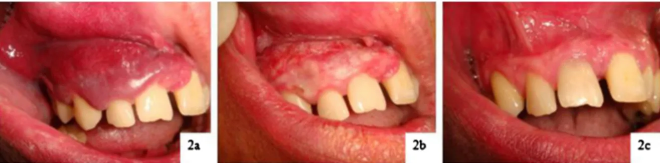
treated by DLIP. After a variable number of DLIP sessions (aver-age = 4), 79% (lower lip: 4, upper lip: 3, 1 on fornix, gingiva: 1 and cheek: 2) completely regressed, the remaining 21% having presented shrinkage of the lesions and reduced complications (Figs. 2 and 3). The follow-up period after the treatment was 18–72 months (average: 45 months) with no evidence of recurrences of the healed lesions.
3. Discussion
Facial hemangiomas are the most common PHACES syndrome features. One of the most common extracutaneous localizations of hemangiomas is the oral cavity, although rarely reported in the English literature. PHACES IHs, like those in non-syndromic patients, appear as red-bluish soft masses, smooth or lobulated, often covered by intact mucosa[12]. Those located on lower lip are at greater risk of complications (ulceration)[11]. In the current report, 8 lesions occurred on the lips (4 lower and 4 upper), 6 on the cheek and fornix, 5 on the palate, 4 on the tongue, 3 on the gingiva and 2 on the floor of the mouth.
In the past years, a lot of surgical and non-surgical treatments were tested for the management of hemangiomas. In view of the haemorrhagic risk and high invasiveness related to surgical excision, attention was paid to the possible alternative use of phar-macological and/or minimally invasive interventions. Several drugs have been tested, such as: propranolol[13,14], steroids[15], vin-cristine[16]and bleomycin[17], which were proved effective on hemangiomas in their proliferative phase, and recommended in paediatric patients affected by PHACES syndrome. Laser photoco-agulation has been considered an effective and non-invasive option for the treatment of adult PHACES patients with IHs without rele-vant side effects and toxicity[18,19].
In view of hemangiomas containing high amounts of haemoglobin absorbing in the spectral range of about 400–1100 nm, Diode Laser with a wavelength of 800± 10 nm was chosen because of its affinity to oxyhaemoglobin, producing photothermolysis, erythrocytes microagglutination and vessel obliteration.
The diode laser therapeutic approach to vascular lesions is based on different techniques, such as transmucosal photocoagulation (DLTP) or intralesional photocoagulation (DLIP). DLTP, preferred for flat superficial/reddish IH, being a no contact technique, resulted a minimally invasive treatment, with high rate of complete regres-sion (65%), low incidence of post-operatory pain, bleeding and oedema (15%). The DLIP technique, though at higher risk for post-operatory pain, bleeding and oedema, in comparison with DLTP (42,85%), was chosen for deeper lesions and resulted in complete regression of 79% of the lesions.
The additional use of the gel compound of hyaluronic acid and amino acids reset to zero post-operatory complications and allowed faster healing of the lesions.
4. Conclusions
Diode laser photocoagulation techniques have been proved a very effective and minimally invasive treatment for PHACES IHs, considering the presence of several concomitant lesions and the necessity of numerous interventions.
Conflict of interest
All authors disclose any financial and personal relationships with other people or organizations that could inappropriately influ-ence (bias) their work.
Sources of funding
All Authors declare no sources of funding or any study sponsors.
Ethical approval
This study was carried out in accordance with the code of ethics of the world medical association (Declaration of Helsinki) and approved by our institution ethical committee (study nr 4576 – Prot. 1443/C.E.). The patients released informed consent on diag-nostic and therapeutic procedures and possible use of biologic samples for research purposes.
Consent
The patients released informed consent on diagnostic and thera-peutic proceduresand possible use of biologic samples for research purposes.
Authors contribution
Gianfranco Favia: study concept or design, data collection, data analysis or interpretation, writing the paper, final revision.
Luisa Limongelli: study concept or design, writing the paper. Angela Tempesta: study concept or design, writing the paper. Matteo Favia: study concept or design, writing the paper. Eugenio Maiorano: study concept or design, data collection, data analysis or interpretation, writing the paper, final revision.
Guarantor
The corresponding author is the guarantor of submission.
Ethical standards
We hereby declare that this paper has not been previously pub-lished or submitted elsewhere,
the authors have read and approved its content and all authors do not have competing interest to declare.
Also,
- The work compiles with the ethical policies of the journal. - It has been conducted under internationally accepted ethical standards.
- All diagnostic and therapeutic procedures were carried out according to “standard good clinical practice”.
- No experimental procedures were carried out that would require approval by an ethical committee.
- Data from all patients included in this study were treated anon-imously and the patients released informed consent for the use of such data for scientific publication.
References
[1] I.J. Frieden, V. Reese, D. Cohen, PHACE syndrome: the association of posterior fossa brain malformations, hemangiomas, arterial anomalies, coarctation of the aorta and cardiac defects, and eye abnormalities, Arch. Dermatol. 132 (1996) 307–311.
[2] D.W. Metry, C.F. Dowd, A.J. Barkovich, I.J. Frieden, The many faces of PHACE syndrome, J. Pediatr. 139 (2001) 117–123.
[3] D.W. Metry, D.H. Siegel, R.M. Cordisco, E. Pope, J. Prendiville, B.A. Drolet, et al., A comparison of disease severity among affected male versus female patient with PHACES Syndrome, J. Am. Acad. Dermatol. 58 (2008) 81–87.
[4] S.S. Arora, B.M. Plato, R.J. Sattenberg, R.K. Downs, K.S. Remmel, J.O.
Heidenreich, Adult presentation of PHACES syndrome, Interv. Neuroradiol. 17 (2011) 137–146.
[5] J.H. Levin, S.G. Kaler, Non-random maternal X-chromosome inactivation associated with PHACES, Clin. Genet. 72 (2007) 345–350.
[6] A.N. Haggstrom, E.J. Lammer, R.A. Schneider, R. Marcucio, I.J. Frieden, Pattern of infantile hemangiomas: new clues to hemangioma pathogenesis and embryonic facial development, Pediatrics 117 (2006) 698–703.
[7] D.W. Metry, A.N. Haggstrom, B.A. Drolet, E. Baselga, S. Chamlin, M. Garzon, et al., A prospective study of PHACE syndrome in infantile hemangiomas: demographic features, clinical findings, and complication, Am. J. Med. Genet. 140A (2006) 975–986.
[8] D. Metry, G. Heyer, C. Hess, M. Garzon, A.N. Haggstrom, P. Frommelt, et al., Consensus statement on diagnostic criteria for PHACE syndrome, Pediatrics 124 (2009) 1447–1456.
[9] K. Brandon, P. Burrows, C. Hess, D. Metry, Arteriovenous malformation a rare manifestation of PHACE syndrome, Pediatr. Derm. 28 (2001) 180–184. [10] D.A. Hartemink, Y.E. Chiu, B. Drolet, J.E. Kerschner, PHACES syndrome: a
review, Int. J. Pediatr. Otorhinolaryngol. 73 (2009) 181–187.
[11] S.L. Chamlin, A.N. Haggstrom, B. Drolet, E. Baselga, I.J. Frieden, M. Garzon, et al., Multicenter prospective study of ulcerated hemangiomas, Pediatrics 151 (2007) 684–689.
[12] J.S. Gill, S. Gill, A. Bhardawaj, H.S. Grover, Oral haemangioma, Case Rep. Med. (2012) 347939.
[13] A.L. Marqueling, V. Oza, I.J. Frieden, K.B. Puttgen, Propranolol and infantile hemangiomasfour years later: a systematic review, Pediatr. Dermatol. 30 (2013) 182–191.
[14] T. Solomon, J. Ninnis, D. Deming, T.A. Merritt, A. Hopper, Use of propranolol for treatment of hemangiomas in PHACE syndrome, J. Perinatol. 31 (2011) 739–741.
[15] A. Izadpanah, A. Izadpanah, J. Kanevsky, E. Belzile, K. Schwarz, Propranolol versuscorticosteroids in the treatment of infantile hemangioma: a systematic review and meta-analysis, Plast. Reconstr. Surg. 131 (2013)
601–613.
[16] R.S. Glade, K. Vinson, D. Becton, S. Bhutta, L.M. Buckmiller, Management of complicated hemangiomas with vincristine/vinblastine: quantitative response to therapy using MRI, Int. J. Pediatr. Otorhinolaryngol. 74 (2013) 1221–1225.
[17] Q.F. Luo, F.Y. Zhao, The effects of Bleomycin A5 oninfantile maxillofacial haemangioma, Head Face Med. 7 (2011) 11.
[18] A. Vesnaver, D.A. Dovsak, Treatment of vascular lesions in the head and neckusing Nd:YAG laser, J. Cranio-Maxillo-Fac. Surg. 34 (2006) 17–24.
[19] U. Romeo, A. Del Vecchio, C. Russo, G. Palaia, G. Gaimari, J.
Arnabat-Dominquez, et al., Laser treatment of 13 benign oral vascular lesionsby three different surgical techniques, Med. Oral Patol. Oral Cir. Bucal. 18 (2013) 279–284.
Open Access
This article is published Open Access atsciencedirect.com. It is distributed under theIJSCR Supplemental terms and conditions, which permits unrestricted non commercial use, distribution, and reproduction in any medium, provided the original authors and source are credited.