UPPER TRIASSIC CALCAREOUS ALGAE FROM THE PANTHALASSA OCEAN
IOAN I. BUCUR1*, SYLVAIN RIGAUD2, NICOLÒ DEL PIERO3, ANDREA FUCELLI3,
ERIC HEERWAGEN3, CAMILLE PEYBERNES3, GIOVAN PEYROTTY3, CHRISTIAN VERARD3,
JÉRÔME CHABLAIS4 & ROSSANA MARTINI3
1*Corresponding author. Babeş-Bolyai University, Department of geology and Center for Integrated Geological Stuides, M. Kpgălniceanu str.,
1, 400084 Cluj-Napoca, Romania. E-mail: [email protected]
2Asian School of the Environment, 62 Nanyang Drive, 637459 Singapore
3University of Geneva, Department of Earth Sciences, Rue des Maraîchers 13, CH-1205 Genève, Switzerland
4Hydro-Géo Environnement, Chemin Fief-de-Chapitre 7, 1213 Petit-Lancy, Switzerland
To cite this article: Bucur I.I., Rigaud S., Del Piero N., Fucelli A., Heerwagen E., Peybernes C., Peyrotty G., Verard C., Chablais J. & Martini R. (2020) - Upper Triassic calcareous algae from the Panthalassa Ocean. Riv. It. Paleontol. Strat., 126(2): 499-540.
Abstract. Upper Triassic calcareous algae, abundant and well-diversified in Tethyan deposits, have rarely been
described in rocks of Panthalassan origin. Over the past ten years, several studies were performed on Upper Triassic carbonate deposits of Panthalassan affinity in North America, Japan and Far East Russia, revealing unexpectedly rich and diversified assemblages. The samples were collected from nine localities situated on both sides of the Pacific Oce-an. The identified algal assemblage consists of green and red algae, including fourteen dasycladaleans, rare bryopsida-leans, and several rhodophyceans. This paper describes the main algal taxa, including six new species: Holosporella?
ros-sanae Bucur & Del Piero n. sp., Holosporella magna Bucur & Fucelli n. sp., Griphoporella minuta Bucur & Peybernes n. sp., Patruliuspora pacifica Bucur, Del Piero & Peyrotty n. sp., Patruliuspora oregonica Bucur & Rigaud n. sp. and Collarecodium? nezpercae Bucur & Rigaud n. sp. Rivulariacean-like cyanobacteria and thaumatoporellacean algae are also present. The
whole Panthalassan algal assemblage comprises both unknown (?endemic) and common taxa of the Tethyan domain. To explain the cosmopolitan distribution of various Upper Triassic benthic organisms (scleractinian corals, calcified sponges, foraminifera), a close connection with the Tethys Ocean was hypothesized by different authors. During the Late Triassic, the Tethys was open to the east on the Western Panthalassa but not to the west, suggesting that Triassic calcareous algae were able to efficiently colonize environments that are estimated to be more than 10’000 km apart. An adventitious transport of calcareous algae and/or their spores is proposed to explain this long-range algal dispersal.
Received: February 25, 2020; accepted: May 29, 2020
Keywords: Calcareous algae; Dasycladales; Bryopsidales; Rhodophyta; Upper Triassic; Panthalassa.
I
ntroductIonThe Late Triassic represents a time-period with a relatively high abundance of calcareous al-gae (Flügel 1985, 1991; Barattolo 1991; Bucur 1999; Granier & Grgasović 2000; Nose et al. 2018). This seems related to the re-emergence and increasing development of reefal and associated environments from the Middle to the Upper Triassic (Flügel 2002).
Most known species of calcareous algae in the Up-per Triassic (including dasycladaleans, udoteaceans, and “solenoporaceans”) have been identified in limestone rocks from Europe, mainly in the Alps and Carpathians, but also in central and southern Italy, Greece and the Dinarides. They were first de-scribed at the end of the nineteenth century (e.g., Gümbel 1872; Salomon 1895) and then widely studied in the twentieth century and over the last twenty years (Pia 1912, 1920; Bistricky 1964; Ott 1967, 1968; Flügel 1961, 1975; Senowbary-Daryan 1980; Di Stefano 1981; Barattolo et al. 1993, 2008;
Parente & Climaco 1999; Senowbari-Daryan & Zamparelli 2005; Grgasovič 2007). Ouside of Eu-rope, Upper Triassic calcareous algae are known from Indonesia (Timor and Moluccas) (Vinassa de Regny 1915; Pia 1924), Eastern Tibet (Flügel & Mu 1982), Turkey (Senowbari-Daryan & Link 2005; Se-nowbari-Daryan et al. 2006), Iran (Senowbari-Dary-an & Hamadani 2000), and Oman (Berneker 1996, 2005). In contrast with this relative abundance, only few manuscripts dealing with Triassic limestone de-posits from North and South America mentioned or described calcareous algae. A report of phylloid-like algae from Lime Peak, Yukon was published by Reid (1986), and later the species was re-described by Torres (2003) as Ivanovia triassica Torres, 2003. In her PhD Thesis, Reid (1985), also illustrated cal-careous algae from Lime Peak assigned to Cyano-phyceae (Garwoodia or Cayeuxia), “Solenoporaceae” (Parachaetets sp., and Parachaetetes triassicus (sic)(Vi-nassa de Regny, 1915), and dasycladales (Clypeina). These Clypeina specimens were interpreted by Flü-gel et al. (1989) as bryozoans although their struc-ture is typical of dasycladales with laterals arranged around a central cavity and sparitic calcification of the lateral’s wall. The dasycladalean Diplopora
ore-gonensis Flügel, Senowbari-Daryan & Stanley, 1989
was described from the Excelsior Gulch Conglom-erate (Hurwal Formation, Wallowa Mountains, Ore-gon) (Flügel et al. 1989). The Triassic reef limestone from Summit Point (Oregon), and Mina (Nevada) studied by Martindale et al. (2012, 2015) contain “solenoporacean” red algae (the authors mention seven species: Solenopora cf. alcicornis Ott, 1966, S. cf.
undata Senowbari-Daryan & Link, 2005, S. cf. endoi
Flügel, 1975, S. cf. simionescui Dragastan, 1969,
Para-chaetetes cf. tauricus Senowbari-Daryan & Link, 2005,
P. cf. cassianus Flügel, 1961, Tauristorea cf. discursa
Senowbari-Daryan, Link & Isintek, 2006) as well as unspecified Dasycladacean green algae.
For the past ten years, in the frame of the REEFCADE Project (to R.M.), several studies made on Upper Triassic deposits of North Amer-ica, as well as far East Russia and Japan (Chablais et al. 2010a, b, c, 2011; Heerwagen & Martini 2018, 2020; Khalil et al. 2018; Onoue et al. 2009; Pey-bernes et al. 2015, 2016a, b; Peyrotty et al. 2020; Rigaud 2012; Rigaud et al. 2012, 2013a, b, 2015a, b, 2016; Sano et al. 2012; Senowbari-Daryan et al. 2010) revealed the existence of distinct calcareous algae in many of the studied sections, but none of
these publication discussed in detail the algal as-semblages or gave a systematic description of the observed algae. The aim of our study is to describe and illustrate Panthalassic algae found by all the above-mentioned authors and examine their pal-aeobiogeographical distribution. The whole asso-ciation is rich and consists of algae known already from the Tethys as well as of new species, possibly endemic to Panthalassa.
G
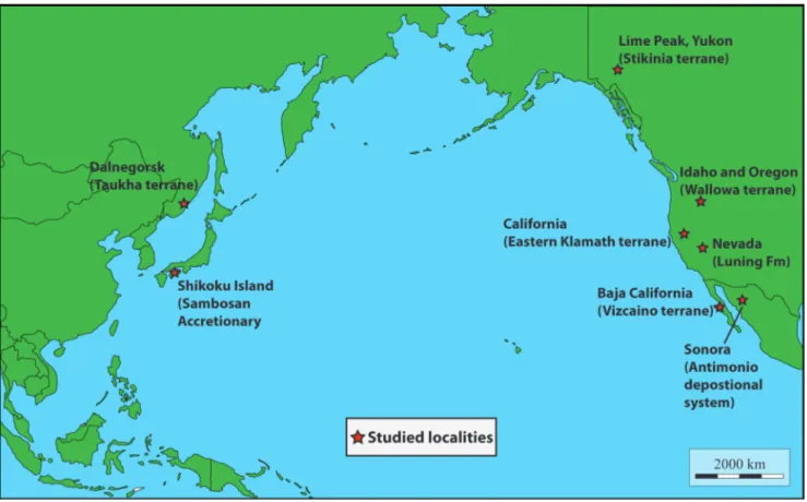
eoloGIcalframeworkThe studied samples containing algae were collected from nine localities situated on the Cir-cum-Pacific region: seven from North America, and two from Eastern Asia (Fig. 1 and Tab. 1).
North America
Yukon, Oregon and Idaho
The material from the northwestern part of North America comes from three distinct Upper Triassic localities: (1) the Black Marble Quarry in Oregon, USA (lower-middle parts), (2) the Mission Creek Quarry in Idaho, USA (base of the Quarry), and (3) Lime Peak in Yukon, Canada (West side of the Peak) (Tab. 1). All these localities are part of allochthonous terranes that originated in the Panthalassan Ocean and accreted onto the Ameri-can continental margin in the Mesozoic (Coney et al. 1980). Lime Peak is part of Stikinia (Bordet 2016a, 2016b) whereas the Black Marble Quarry and, with less certainty, the Mission Creek Quarry (both tectonically isolated blocks) are regarded as part of the Wallowa terrane (Hoover 1991; Stan-ley et al. 2008; Rigaud 2012), possible southern prolongation of Stikinia (Mortimer 1986; Oldow et al. 1989; Yancey & Stanley 1999). The samples used in this study come from bedded, fossiliferous shallow-water limestones of lagoonal facies that are notably rich in molluscs, foraminifers, ostracodes, echinoderms, and possibly associated with small coral and/or sponge thickets. The preservation of originally aragonitic components (e.g., gastropods, scleractinian corals, involutinid foraminifers, green algae) is quite atypical: although fully recrystallized, specimens are often pervasively impregnated by hy-drocarbons (now metamorphosed into graphite) so that their original structure is still visible (see Rigaud
et al. 2015a for details). The Black Marble Quarry (BMQ) is well-known for its foraminiferal content, particularly rich in involutinids (Kristan-Tollmann & Tollmann 1983; Rigaud et al. 2010, 2012, 2013a-b, 2015a-2013a-b, 2016) and for its giant wallowaconchid bivalves (Stanley 1979; Yancey & Stanley 1999). It presents a foraminiferal assemblage typical of the Norian (see Koehn-Zaninetti 1969; Blau & Schmidt 1990), including the Norian marker Aulosina
ober-hauseri (Koehn-Zaninetti & Brönnimann 1968). The
dark grey to black, restricted lagoon limestone beds in which algae have been discovered are found in the first fifty meters of the quarry, below the transition to a more open marine facies (Scotch Creek Mem-ber) of the Martin Bridge Formation (see Rigaud 2012 for details). Age-diagnostic fossils (ammo-noids) found by Nolf (1966, p. 54) would position such levels within the Lower Norian, Kerri Zone.
The Mission Creek Quarry or Lapwai Quarry (LQ) is particularly famous for its fossil-rich silici-fied beds that contain a rich and diversisilici-fied inver-tebrate lower upper Norian (Sevatian: age based on the ammonite Gnomohalorites cordilleranus Tozer, 1979, see Nützel & Erwin 2004) fauna, including bivalves (Newton, unpublished data), brachiopods
(Cooper 1942; Hoover 1991), corals (Squires 1956; Stanley 1979, 1986), and gastropods (Haas 1953; Nützel & Erwin 2001, 2004). Only few authors have investigated the quarry and most studies are based on the collection of J. S. Williams, J. Reed, and Norman D. Newell Newell (see Squires 1956 for details). These silicified beds are found south of the quarry, near a zig-zagging gravel road, few meters below the transition to a Hurwal-type (red/ brown argillite) lithology (see Rigaud 2012 for de-tails). The here described algae/porostromate-rich wackestone-floatstone/rudstone levels were recov-ered from other, slightly older sedimentary strata (in a stratigraphic position estimated to be 55-65 meters below the silicified beds). The preservation of these older levels is uneven and marbleized lime-stone rocks are majoritarily observed. However, in some levels, usually darker (dark gray) or yellowish, fossils such as corals (Stanley 1986) and foraminifers (Rigaud et al. 2015a) are preserved and may abound. The here reported algae come from yellowish levels found at the foot of the quarry.
Lime Peak is one of the Upper Triassic lime-stone bodies which form a northeast trending belt along the East side of Lake Laberge. These massive
to stratified carbonates, are part of the Hancock Member of the Aksala Formation (Bostock & Lees 1938; Hart 1997; Tozer 1958). The Lime Peak area is the place where Hancock Member carbonates are better exposed and preserved (Tab. 1). The samples were collected on the north-west side of the peak: there, a 20-m-thick outcrop is formed by ca 12 m massive limestones which are underlain by ca 8 m of medium to thick-bedded (20-40 cm) algae-bear-ing strata. Based on conodont assemblages the age of the deposits is late Norian (Lei et al. 2019).
California
Hosselkus Limestone material comes from a series of outcrops located north of the city of Redding in Shasta County, USA (Tab. 1). These outcrops are part of the Eastern Klamath terrane, a sequence of Palaeozoic and Mesozoic island arc deposits that were built upon oceanic crust (Irwin 1977) and accreted onto the American continental margin during the Mesozoic (Coney et al. 1980). It is worth to notice that the best-preserved limestone units crop out in the Shasta Lake area and not at the type locality (Taylorsville, Plumas County). In the Shasta area, the lower part of the Hosselkus Lime-stone shows a great variety of open basin facies (e.g., deep environment with radiolarians, condensed lev-els of halobid-like bivalves, crinoids packstone, cal-citurbidites with ammonoids). After a progressive transition toward shallower basinal facies, the whole succession is covered by several meters of breccia composed of centimetric to decimetric limestone clasts within a radiolarian-rich fine matrix. The lime-stone clasts derive from an Upper Triassic carbon-ate platform that no longer exists. Facies mostly re-flect platform top to foreslope environments, with mud-dominated and grain-dominated elements associated with shallow-water organisms; the last are observed in millimetric to decametric reworked elements, and notably comprise the here-described algae and coral-rich facies. Conodonts recovered in all three types of facies indicate a late Carnian age.
Nevada
The Luning Formation crops out in a wide area of Western Nevada and is part of the Luning Allochthon, a series of nappes composed of Trias-sic and JurasTrias-sic sedimentary rocks and Permian to Triassic volcanic rocks (Oldow 1981). The thi-ckest succession of the Luning Formation is
loca-ted north of the Pilot Mountain (Tab. 1) where it shows a total thickness exceeding 2000 m (Sandy & Stanley 1993), divided in three members: Lower, Middle and Upper. The Lower Member is an alter-nation of shales and coral and sponge biostromes, while the middle member is fully terrigenous, show-ing alternation of shales, sandstones and conglome-rates. The Upper (algae-bearing) Member shows a regular alternation of lagoonal-peritidal facies, with abundant bivalves, gastropods and megalodonts, alternated with decimeter-thick coral horizons and nodular marls. Muller & Ferguson (1936) had alrea-dy evidenced the abundance of fossils in the Upper Member of the Luning Formation. Preliminary co-nodont results indicate a middle Norian age for this portion of the formation.
The presence of a thick terrigenous member, around 600 m (Sandy & Stanley 1993) plus the oc-currence of marls with variable percentage of clay minerals, might suggest that the Luning Formation was probably deposited close to the American con-tinental margin and not in the middle of Pantha-lassa.
Mexico
Outcrops of Upper Triassic carbonates of shallow-marine facies are rare in Mexico. They are limited to two areas in Sonora and in Baja California Sur (Tab. 1).
In Sonora, the Antimonio depositional system (ADS) encompasses the El Antimonio Group, ge-nerally considered as part of the northwest-southe-ast trending tectonostratigraphic Antimonio terrane (González-León et al. 2009 and references therein). The shallow-marine strata that yielded the studied algae belong to the Upper Triassic Rio Asunción Formation. Outcrops in northern Sonora are loca-ted near the cities of Caborca and Hermosillo (He-erwagen & Martini 2018). The main localities are Sierra del Álamo and Barra los Tanques (Tab. 1). At Sierra del Álamo, the Rio Asunción Formation (upper part of the El Antimonio group succession, ca. 109 m), consists of shallow-marine limestone, siltstones to fine grained sandstones, some inter-calated volcanics, and shales. A distinct carbonate interval, dated by ammonoid and bivalve biostrati-graphy as Norian in age (Damborenea & González-León 1997 and references therein; González-González-León 1997), contains sponge-coral biostromes with algae. At Barra los Tanques, a precise biostratigraphic
fra-mework has not been established yet. The shortly exposed section (ca 29 m) is predominantly com-posed of limestones with terrigenous material in-cluding sponge-coral biostromes and patch-reefs with algae, comparable to those at Sierra del Álamo. Intercalated in the succession are lumachelle beds,
and carbonate-cemented siltstones to fine sandsto-nes (Heerwagen & Martini 2018).
In Baja California Sur, the studied algae come from the San Hipólito Formation on the Vizcaíno terrane. The Vizcaíno terrane is a northwest-sou-theast trending tectonostratigraphic unit, which
G e olog ic a l c on te x t G P S P oin ts Lit h olog y / F a cie s Ag e S a m pl e s 1 D a ln eg o rsk (F ar E as t R u ss ia) T au k h a t e rr an e 44° 23' 47. 86" N 135° 46' 45. 04" E Lag o o n al lim e st o n e (I n v o lu tin id -M e g alo d o n tid W a ck e st on e t o R u d st on e ) lo w e r-m id d le No rian G P -152; G P -153; G P -157; G P -206C ; G P -206C -2; G P -206C -3; G P -206C -4 2 S h ik ok u Is la n d (J a p a n ) S a m b os a n A ccr e ti on a ry Co m p le x (S A C) Sam bo san t ype lo cal it y: 33 ° 3 4. 45 2′ N 13 3° 4 2. 73 3′ E W a jik i lo ca lit y: 33° 52' 04. 1" N 34° 29' 50. 7" E Tak ay am a l o cal it y: 33° 18' 17. 84" N 132° 24' 55. 23" E R e e fal lim e st o n e ( sp o n g e -alg ae B o u n d st o n e a n d G ra in st o n e ) Lag o o n al lim e st o n e ( In v o lu tin id -alg ae W ac k e st o n e ) O p e n l a g oon ( p e loi d -b iocl a st P a ck st on e t o Gr a in st on e ) U p p e r T rias sic C P -259A ; C P -259B ; C P -261 (S am bo san T ype lo cal it y) C P -94A ; C P -94B (W a jik i lo ca lit y) JC 231a (T ak ay am a l o cal it y) 3 Y u ko n (C an ad a) S tik in ia t e rr an e 61° 3' 57. 35" N 134° 54' 57. 95" W Lag o o n al lim e st o n e W a ck e st on e t o r u d st on e lat e No rian W H -227a ; W H -227b ; W H -232a ; W H -232b ; W H -233a ; W H -233b ; W H -234; W H -235a ; W H -235b ; W H -236d ; W H -259; W H -260; W H -261 (L im e P ea k) 4 Ida ho (U SA ) ? W allo w a t e rr an e 46° 17' 34. 37" N 116° 42' 02. 69" W Lag o o n al lim e st o n e (D as y clad ac e an -P o ro st ro m at e W a ck e st on e -P a ck st on e a n d F lo a ts to n e -R u d ts o n e ) No rian ( ? S e v at ian ) LQ -5; L Q -6; L Q -23; L Q -24; L Q -27; L Q -39A ; LQ -39B ; L Q -39C ; L Q -39D ; L Q -39F ; L Q -39G ; L Q -39I (M issi o n C re ek Q uar ry ) 5 O re g o n (U SA ) W allo w a t e rr an e 45° 22' 23. 27" N 117° 21' 16. 40" W O rg an ic -r ic h lim e st o n e , r e st ric te d lag o o n al fac ie s (I n v o lu ti n id -r ic h M u d st o n e t o P a ck st o n e ) lo w e r No rian ( K e rr i Z o n e ) B M Q -33R 4; B M Q -36-9; B M Q -48; B M Q -48R 2; M Q -7D ; M Q -16; M Q -16D ; M Q -18R -2; M Q -19-1; M Q -33R 3; M Q -34b -17; M Q -34B ; M Q -34-9; M Q -36; M Q -38R ; M Q -41; M Q -41-10; M Q -41D ; M Q -48-2; M Q -48-3; M Q -48-4; M Q -48D ; M Q -48R -2; M Q -49R ; M Q -77; M Q A C -17b ; M Q V IC -41 (B lac k M ar bl e Q uar ry ) 6 C a lif or n ia (U SA ) E as te rn K lam at h t e rr an e R ee f fac ie s l o cal it y: 40° 46' 45. 24" N 122° 0' 9. 62" W D e b ris d o m in at e d f o re slo p e S h allo w o rg an is m s r e w o rk e d in o p e n b as in e n v ir o n m e n t as c e n tim e tr ic b re cc ia; m at rix in d ic at e a d e e p e n v ir o n m e n t w it h rad io lar ian s u p p e r C a rn ia n (b a se d o n C o n o d o n ts ) F A 48b ; F A -50-2A ; F A -129; F A -210; F A -217 (S hast a C o unt y) 7 B a ja C a lif or n ia S u r (M e xi co) V iz caín o t e rr an e Sa n H ip ó lit o lo ca lit y: 26° 59' 36. 04" N 113° 59' 12. 19" W P at ch r e e f m ar g in t o b ac k r e e f ( e ch in o id -lit h o clas t-p e lo id G rain st o n e t o R u d st o n e a n d W a ck e st o n e ) lat e No rian E H -509 (S an H ipo lít o F o rm at io n) 8 N e vad a (U SA ) A tt a ch e d , or r e a ll y cl os e t o A m e ric an C rat o n H o lo spo re lla m agna be d: 38° 29' 12. 96" N 118° 13' 1. 89" W Lag o o n al t o t id al e n v ir o n m e n t (m os tl y W a ck e st on e a n d P a ck st on e ) m id d le No rian (b a se d o n C o n o d o n ts ) F A -81a ; F A -81c (no rt h-east f lank o f G ar fi el d H ill s) 9 S on or a (M e xi co) A n tim o n io d e p o sit io n al syst e m ( A D S ) B ar ra l o s T anque s l o cal it y: 30° 29' 41. 91' 'N 112° 50' 48. 24" W Sie rr a d el Á la m o ( SA ) lo ca lit y: 30° 43' 16. 80" N 112° 33' 9. 30" W Sie rr a d el Á la m o ( EA ) lo ca lit y: 30° 44' 1. 40" N 112° 35' 7. 00" W S a n d s h e e ts a n d s a n d s h o a ls (s ilic ic las tic o st rac o d – g as tr o p o d – li th ocl a st Gr a in st on e t o R u d st on e ) S an d s h e e ts an d s an d s h o als , ag it at e d w at e r ( lit h o clas tic e ch in o id – g a st ro p o d – sp o n g e – p e lo id Gr a in st on e t o R u d st on e ) S an d s h e e ts an d s an d s h o als , ag it at e d w at e r ( silic ic las tic lit h o clas tic os tr a cod /r ich W a ck e st on e t o Gr a in st on e ) lat e No rian E H -118A (B ar ra l o s T anque s) E H -212A ; E H -216A ; E H -216b (S ie rr a d el Á la m o S A ) E H -306A ; E H -311A ; E H -319A ( Si er ra d el Á la m o EA ) R e p os it or y : A ll t h e s a mp le s a n d t h in s e ct io n s a re s to re d i n t h e M u se u m d ’H is to ir e N a tu re ll e d e G e n è v e , S wi tz e rl a n d ( M H N G -G E P I- 2020-0001 t o M H N G G E P I- 2020-0082) . AS IA A RIC ME H A NORT Loc a lit y
Tab. 1 - Coordinates of the investigated localities, together with their geological context, lithologies and facies content, ages, and collected samples.
constitutes the entire Vizcaíno Peninsula. However, its classification as a terrane is somewhat unclear (Heerwagen & Martini 2020 and references therein). The San Hipólito Formation (ca 2400 m thick mari-ne succession) is named after the mari-nearby village and represents an arc-ophiolite assemblage (Busby 2004; Morán Zenteno et al. 1994) with basinal to slope fa-cies spanning from the Norian to the Pliensbachian (Orchard et al. 2007). The formation comprises a limestone breccia (ca 105 to 260 m thick), which in-cludes re-worked upper Norian shallow-water lime-stones. According to Heerwagen & Martini (2020), the breccia itself consists of volcaniclastic sandsto-nes containing limestone clasts in a poorly sorted, massive to weakly bedded, fine to medium-coarse litharenite matrix. The size of the clasts ranges from several decimeters up to few meters in diameter. Li-thologically, the limestone clasts are composed of light gray to gray, sometimes with a pinkish hue, micritic limestone. The macrofossil assemblage is dominated by brownish-weathered coral colonies; subordinate are fragments of thalamid sponges and fossil debris including echinoids, gastropods, bival-ves, brachiopods, ostracodes, and foraminifers. Only in some samples red algae and dasycladalean algae, serpulids, bryozoans, coprolites, and fragments of calcimicrobe colonies were found. Less commonly found were blocks of micritic limestone that contain megalodontid bivalves.
Asia
On the Asian part of the Circum-Pacific re-gion, the algae described in this paper have been found in in Far East Russia in the Taukha terrane and in Japan in the Sambosan Accretionary Com-plex (SAC) (Tab. 1).
Far East Russia
The Russian outcrops are located in the sou-thern part of the Sikhote-Alin orogenic belt (Pri-morsky Kray), defined as an upper Tithonian-Hau-terivian accretionary prism (Kemkin et al. 1997). The Tetyukha suite (Kojima 1989; Khanchuk et al. 2016), situated in the northern part of the Taukha terra-ne (Dalterra-negorsk area) is made of Norian atoll-type carbonates from the Panthalassa ocean (Peyrotty et al. 2020). The sedimentology and biostratigraphy of the limestone have been studied and described in detail by Peyrotty et al. (2020). The algae pre-sented in this paper have benn observed in
involu-tinids-megalodontids wackestone to rudstone facies interpreted as lagoonal limestones. Algae are associa-ted with megalodontids, involutinids (dominaassocia-ted by
Parvalamella friedli (Kristan-Tollmann, 1962)),
gastro-pods and undetermined calcimicrobes. The age of this facies is defined as early-middle Norian on the basis of foraminifers. According to Kojima (1989), the Taukha terrane represents the northern exten-sion of the SAC but the here-described algae differ, possibly suggesting a different origin or depositional environment of the Upper Triassic shallow-water li-mestone from Japan and Russia.
Japan
The material from South West Japan comes from three localities of the Sambosan Accretiona-ry Complex (SAC) in western, central and eastern Shikoku Island. The SAC is an Upper Jurassic-Lower Cretaceous subduction-generated accretionary com-plex which contains Upper Triassic deep and shal-low-water carbonates units (Kanmera 1969; Matsuo-ka & Yao 1990; Onoue & Sano 2007; Peybernes et al. 2016b). Samples CP-259 and CP-261 have been found in limestone breccias with volcaniclastic ma-trix that crop out at the Sambosan type locality, near Kochi. This locality is described in detail in Peyber-nes et al. (2015, 2016a, 2016b; PeyberPeyber-nes et al. un-der review). Microfacies and associated fauna (e.g., hypercalcified sponges) point to reefal depositional environments. Conversely, sample CP-94 is a lagoo-nal involutinid wackestone that comes from the Waji-ki area and has been retrieved from a limestone brec-cia with siliceous mudstone matrix (Peybernes et al. 2016b). Limestone clasts and blocks from these two localities range from the Ladinian?-Carnian to the Rhaetian (Peybernes et al. 2016b). Sample JC231A is a peloid-bioclast grainstone to packstone, found in Upper Triassic limestones outcropping on the se-ashore in Takayama area. Limestone units from this locality were studied by Chablais (2010) and Peyber-nes et al. (2016b).
materIalandmethods
We selected 82 thin sections containing calcareous algae co-ming from these nine localities in North America and Asia (Tab. 1). The thin sections were studied under a Carl Zeiss Axioscop petro-graphic microscope. Microphotographs of algae were made using a Canon Power Shot A640 Digital Camera. All thin sections are stored in the Museum d’Histoire Naturelle de Genève (Switzerland) under the numbers MHNG-GEPI-2020-0001 to MHNG GEPI-2020-0082.
The studied thin sections are characterized by an algal as-semblage consisting of chlorophyceans and rhodophyceans, two ma-jor groups of the so called “calcareous algae” or algae characterized by calcium carbonate skeletons which favored their preservation as fossils. The assemblage occurs on both sides of the pacific ocean (North America, Japan and Far East Russia), and consists of fourte-en dasycladaleans (Pl. 1-12) (Pseudodiplopora borzai (Bystrycki, 1978),
Pseudodiplopora? sp., ?Thyrsoporella multipora (Bilgütay, 1968), Holospo-rella? rossanae Bucur & Del Piero n. sp., Holosporella magna Bucur &
Fucelli n. sp., Griphoporella curvata (Gümbel, 1872), Griphoporella minuta Bucur & Peybernes n. sp., Physoporella jomdaensis Flügel & Mu, 1982,
Salpingoporella sp., Gyroporella div. sp., Macroporella sp., Clypeina cf. besici
Pantič, 1965, Patruliuspora pacifica Bucur, Del Piero & Peyrotty n. sp.
and Patruliuspora oregonica Bucur & Rigaud n. sp.), rare bryopsidaleans (one possible udoteacean, Collarecodium? nezpercae Bucur & Rigaud n. sp., Pl. 4K-M; Pl. 13, and rare gymnocodiaceans Permocalculus sp., Pl. 6I (Pm); Fig. 5), and several rhodophyceans (Pl. 14-15) (Norithamnium
madoniensis Senowbari-Daryan, Keupp, Abate & Vartis-Matarangas,
2002, “Parachaetetes” sp., and “Solenopora” sp.). Some microfossils with algal affinity were also observed within the limestone of the Black Marble Quarry (Oregon), but in a state of preservation which does not allow a taxonomic identification. They belong to rivulariacean-like cyanobacteria (Pl. 16A-D), to udoteaceans (Pl. 16E-F), dasycla-daleans (Pl. 16G-K) and to a problematic microfossil with possible algal affinity (P. 16L-T). Thaumatoporellacean fragments have also been identified (Pl. 16L-M).
P
alaeontoloGIcaldescrIPtIonPhylum CHLOROPHYTA Pascher, 1914 Class ulvoPhyceae Stewart & Mattox, 1978
Order Daycladales Parscher, 1931 Family Diploporaceae Pia, 1920 Genus Pseudodiplopora Bucur & Enos, 2001 Pseudodiplopora borzai (Bystricky, 1978) Bucur
& Enos, 2001
Pl. 1A-E; Pl. 2A-H; Pl. 3A, B, F; Pl. 4A, D-H
1978 Diplopora borzai nov. spec., Bystricky, p. 328, pl. 1, fig.1-4; pl. 2, fig.1-4.
1981 Diplopora panormitana n. sp., Di Stefano, p. 74, pl.1, fig. 1-3; pl. 2, fig. 1-3; pl. 3, fig. 1-4; pl. 4, fig. 1-4.
? 1989 Diplopora oregonensis n. sp., Flügel et al., p. 376, Fig. 2-4. 2001 Pseudodiplopora borzai (Bystricky, 1978) nov. comb. - Bucur &
Enos, p.328.
Description
Thallus cylindrical with a maximum obser-ved length of 15.6 mm. Well defined axial cavi-ty, most probably corresponding to the central stem of the alga. Laterals tubular slightly flared at the distal end (phloiophorous type) with meta-spondyle arrangement (e.g., Pl. 1C; Pl. 2E, H; Pl. 3F; Pl. 4A, D). Most specimens have the axial ca-vity filled with reproductive cysts (gametangia) of
Pl. 2A, C; Pl. 2A, B) as well as in oblique (Pl. 1B-D; Pl. 2E, H; Pl. 3F; Pl. 4A) or transverse (Pl. 3B) sections, cysts are predominantly arranged more or less on the margins of the axial cavity leaving an empty space in the center. However, the cysts occasionally occupy almost the whole space of the central stem (Pls. 1E, 2B, D, F). The cysts are not directly welded to the external surface of the axial cavity. Between the cysts and the calcareous wall, there is a very narrow free space (Pl. 2A, C, F, G). Sterile specimens were also identified (Pl. 4D-H) which, except for the lack of cysts, have the same characteristics as the fertile ones.
Dimensions (in mm)
Fertile specimens:
L (maximum observed length) = 15.600 D (external diameter) = 1.300-2.600 (mean = 1.890)
d (internal diameter) = 0.550-1.500 (mean = 0.910)
d/D (%) = 27.77-63.15 (mean = 47.48) e (thickness of the calcareous wall, D-d/2) = 0.270-0.800 Cysts diameter = (0.043) 0.060-0.100 Sterile specimens: D = 0.870-1.800 (1.270) d = 0.280-1.100 (0.610) d/D(%) = 28-68.18 (47.70) D-d/2 = 0.170-0.550 (0.320) Discussion
The specimens identified in Idaho present all the characteristics described and illustrated by Bystricky (1978) for Diplopora borzai. Bystricky (1978) described the laterals (branches) as being spindle-shaped … “with a sharp tip at both edges, and maximum thickness closely before the mouth or, which is the most common, at the mouth lead-ing out the sleeve. In the latter case the pores of the branches are open outside and resemble the pores of the branches of the phloiophore type.” (Bystricky 1978, p. 328). Di Stefano (1981) and Di Stefano & Senowbari-Daryn (1985) also con-sidered the laterals of the Sicilian specimens as phloiophorous. Accordingly, Bucur & Enos (2001) transferred Diplopora borzai Bystricky, 1978 into the new genus Pseudodiplopora (metaspondyle
arrange-ment and phloiophorous laterals) under the new combination Pseudodiplopora borzai (Bystricky, 1978) (see also Barattolo & Romano 2005). The thallus dimensions of the American specimens match the dimensions given by Bystricky (1978), Di Stefano (1981) and Di Stefano & Senowbari-Daryan (1985) except for the mean values of the cyst diameter (larger in the American specimens). Flügel et al. (1989) described Diplopora oregonensis from the Red Gulch conglomerates, Wallowa Mountains, north-east Oregon, and compared the new species with
Diplopora borzai. The former should differ from the
latter by the “… type of the thallus (segmented versus non-segmented in Diplopora borzai …), the
scarcity of typical metaspondyle branching pat-terns (most whorls exibit an euspondyle pattern), and also the dimensions.” (Flügel et al. 1989, p. 378). Dimensions differences are emphasized in regard to the pore diameter (smaller in D.
oregonen-sis) and the size of the cysts (D. oregonensis having
two different size ranges). We must emphasize that the specimens illustrated by Flügel et al. (1989) show an advanced degree of diagenesis which strongly obliterates the structure of the skeleton. The illustrated specimens in Figs. 3, 4 (Flugel et al. 1989) do not show a proper segmentation of the thallus. It is possible that advanced dissolution process along the pores (e.g., Fig. 3/1, 3, Flügel et
Plate 1
A-E - Pseudodiplopora borzai (Bystricky). A, B) Microfacies with
Pseudo-diplopora; rudstone-floatstone
with dasycladalean, mollusc and echinoderm fragments. C, D) oblique sections; E) longi-tudinal section (close-up view of the specimen in Pl. 2C showing the central cavity fil-led with gametangia). A, thin section LQ-39I; B, thin sec-tion LQ-39F; C, thin secsec-tion LQ-39b; D, E, thin section LQ-39C; Idaho, U.S.A.
al. 1989) resulted in such an interpretation, but the longitudinal sections from Fig. 4/1-3 (Flügel et al. 1989) do not confirm it. Regarding the thallus di-mensions, even if small differences exist, the gen-eral thallus dimensions of D. oregonensis match the dimensions given by Bystricky (1978) for D. borzai. The smaller diameter of the pores could also be a feature related to the strong diagenesis. In respect to the cysts dimensions, there is a wide dimension-al range from 0.030-0.066 in D. borzai (Bystricky 1978) to 0.050-0.070 in the Sicilian specimens (Di Stefano 1981) and to 0.043-0.100 in the specimens described in the present paper. This variability could be partially due to the fact that these small
corpuscles are cut randomly, but could also be a natural feature related to habitat. The existence of two different range of sizes for cysts in D.
oregonen-sis illustrate in fact this variability. In view of the
above considerations we think that Diplopora
(Pseu-dodiplopora) oregonensis is a probable junior synonym
of Pseudodiplopora borzai.
Stratigraphic range: Carnian (Bystricky
1978); Carnian-Norian (Di Stefano & Senowba-ri-Daryan 1985; Granier & Deloffre 1994; Granier & Grgasovič 2000). Norian (Flügel et al. 1989) for
D. oregonensis. Our specimens from Idaho are early
to early late Norian in age (most probably middle Norian).
A-H - Pseudodiplopora borzai (Bystri-cky). A-D) longitudinal-oblique sections; E) longitudinal-oblique section; F) transverse section of two specimens; G) close-up view of the left specimen in F showing the gametan-gia filling the whole central cavity; H) close-up view of the specimen in E showing the gametangia filling a large part of the central cavity, as well as the external enlarge-ment of the laterals. A, F, G, thin section LQ-39A; B, thin section LQ-39C; C, D, thin section LQ-39D; E, H, thin section LQ-39F; Idaho, U.S.A.
Pseudodiplopora? sp.
Pl. 3C, D; Pl. 4B, C
Associated with P. borzai we found some specimens of a large dasycladalean dispaying a very recrystallized calcareous wall surrounding the cen-tral cavity. For this reason, the shape of the laterals cannot be observed adequately, but they probably were phoiophorous as suggested by some later-als “ghosts” (Pl. 3C). By contrast, the reproductive cysts inside the central cavity are very well preserved. They are arranged around the margin of the central cavity in 2 or 3 rows (Pl.3D, E; Pl. 4B, C) but prob-ably occupied more space inside the stem (Pl. 3C).
Dimensions (in mm):
L (maximum observed) = 11.800 D = 3.200-3.600
d = 1.550-2.000
cyst diameter = 0.140-0.180
Family Thyrsoporellaceae Granier & Bucur in Granier et al., 2013
Genus Thyrsoporella Gümbel, 1872 ?Thyrsoporella multipora (Bilgütay, 1968)
Pl. 4I, J
1968 Placklesia multipora n. gen., n. sp., Bilgütay, p.71, text-fig. 4, pl. 3, figs. 1-9.
Plate 3
A, B, F - Pseudodiplopora borzai (Bystri-cky). A) longitudinal-obli-que-tangential section; B)
longitudinal-oblique and
transverse sections; F) obli-que section. A, thin section 39C; B, thin section 39G; F, thin section LQ-39A; Idaho, U.S.A.
C, D, E - Pseudodiplopora? sp. C) lon-gitudinal-oblique section; D) transverse section; E) close-up view of the specimen in D showing the shape and arrangement of gametan-gia. C, thin section LQ-39I; D, E, thin section LQ-39B; Idaho, U.S.A.
1978 Placklesia multipora Bilgütay 1968 - Senowbari-Daryan, p.200, pl. 30, figs. 1-6.
2006 Thyrsoporella multipora (Bilgütay 1968) nov. comb. - Schla-gintweit, in Gawlick et al., p.110, fig. 4.1-2.
Description
Bilgütay (1968) and Senowbari-Daryan (1978) gave a detailed description of this alga. Es-sentially, it has a cylindrical thallus bearing laterals with five order of dichotomous branching. Unfor-tunately, most identified specimens were subjected to a strong diagenesis, and the branching of the laterals is difficult to follow. The rare exceptions are represented by the oblique sections illustrated in Pl. 4I, J. Dimensions (in mm): L (maximum observed) = 6.600 D = 0.630-1.150 (mean = 0.970) d = 0.220-0.470 (mean = 0.370) d/D (%) = 32-47 (mean = 37) e (D-d/2) = 0.210-0.420 (mean = 0.30)
Locality: Black Marble Quarry in Oregon
(Lower Norian).
Discussion
Schlagintweit (in Gawlick et al. 2006) conside-red that either the criteria used by Bilgütay (1968) to distinguish the genus Plackesia from the genus
Thyr-A, D-H - Pseudodiplopora borzai (Bys-tricky). A) oblique section of a fertile specimen; D-G) oblique and longitudinal-oblique sections of sterile specimens; H) transverse section of a sterile specimen. A, thin section LQ-39I; D, thin section LQ-6; E, thin section LQ-24; L, thin sec-tion LQ-39F; G, thin secsec-tion 23; H, thin section LQ-27; Idaho, U.S.A.
B, C - Pseudodiplopora? sp. Transverse section; C is a close-up view of the specimen in B show-ing the shape and arrange-ment of gametangia. Thin section LQ-39G; Idaho, U.S.A.
I, J - ?Thyrsoporella multipora (Bilgü-tay), oblique sections. Thin section MQ-34B, Oregon, U.S.A.
K-M - Collarecodium? nezpercae Bucur & Rigaud n. sp., paratypes. Transverse (K, L) and longi-tudinal-oblique (M) sections. K, L, thin section LQ-27; M, thin section LQ-24; Idaho, U.S.A.
soporella (i.e., 1, the existence of 5 order of lateral
branching; 2, segmentation of the calcareous skele-ton; and 3, thallus branching) are inconsistent (e.g., segmentation), or they are characteristic for other species of the genus Thyrsoporella. Thus, as a conse-quence, he transferred the species P. multipora to the genus Thyrsoporella in the new combination
Thyrsopo-rella multipora (Bilgütay). Granier & Bucur (in
Gra-nier et al. 2013) disagreed with this synonymy be-cause they considered that these genera differ in the division formula of the branching pattern. Howe-ver, as emphasized by Schlagintweit (in Gawlick et al. 2006), the Late Jurassic species Thyrsoporella
pseu-doperplexa Granier & Braik (Granier & Braik 2002)
has the same division formula as Placklesia multipora and we consider here the synonymy proposed by Schlagintweit as acceptable.
Stratigraphical range: Rhaetian (Bilgütay
1968; Senowbari-Daryan 1978; Granier & Graga-sovic 2000; Schlagintweit in Gawlick et al. 2006); upper Norian-Rhaetian (Granier & Deloffre 1994; Bucur 1999). Until now, T. multipora was known only from the Northern Calcareous Alps (Schlagintweit in Gawlick et al. 2006). Its presence in the lower Norian of Oregon would be remarkable.
Family Triploporellaceae Pia 1920, emend Granier & Bucur in Granier et al., 2013
Plate 5
A-S - Holosporella? rossanae Bucur & Del Piero n. sp. A-I, S) oblique sections; J-M, R) transverse-oblique sections; N-Q) transverse sections. A-C, K, R, thin section WH-227a; E, L-N, S, thin section WH-227b; D, F, G, thin sec-tion WH-235a; H, I, J, P, Q, thin section WH-235b; Yu-kon, Canada. K - holotype; A-C, R - paratypes.
Genus Holosporella Pia, 1930
Holosporella? rossanae Bucur & Del Piero n. sp.
Pl. 5A-S; Pl. 6A-H
Origin of the name: species dedicated to Prof. Dr. Rossana
Martini, University of Geneva, for her contributions to the study of Triassic deposits all over the world.
Holotype: specimen in Pl. 5K, thin section WH-227a
[MHNG-GEPI-2020-0014].
Paratypes: specimens illustrated in Pl. 5A-C, R (thin section
WH-227a [MHNG-GEPI-2020-0014]), Pl. 5L, M, O, S; Pl. 6A-G (thin section WH-227b [MHNG-GEPI-2020-0015]).
Type locality: Lime Peak, Yukon (Canada), Stikinia terrane
(GPS: 61° 3’ 57.35”N; 134° 54’ 57.95”W).
Type level: late Norian.
Material and repository: More than 50 specimens
iden-tified in four thin sections (227a, 227b, 235a, WH-235b) stored in the Museum d’Histoire Naturelle de Genève under the numbers MHNG-GEPI-2020-0014, 0015, 0021 and 0022.
Diagnosis: Thallus cylindrical bearing whorls of spheroidal
to ovoid laterals arranged in quincunx. Laterals, communicating with the axial stem through a peduncle enlarge distally in a blister. Calci-fication weak as a distinct wall around the laterals and occasionally between the whorls. The distal end of the laterals is frequently non-calcified.
Description
The cylindrical thallus consists of successive whorls arranged in quincunx (Pl. 6B, C). Calcifica-tion is variable and is mainly located around the proximal part of the laterals (Pl. 5J, L, O-Q) but
A-H - Holosporella? rossanae Bucur & Del Piero n. sp. A) dinal section; B, C) longitu-dinal-oblique sections; E, F) transverse sections; H) transverse-oblique section. A, B, E, G, thin section WH-227b; C, D, thin section WH-235b; H, thin section WH-235a, Yukon, Canada. I - Gyroporella sp. (Gy) and
Permocal-culus sp. (Pm), oblique
sec-tions. Thin section GP-152; Dalnegorsk, Far East Russia. J, K - Gyroporella sp., oblique sections.
J, thin section GP-157; K, thin section GP-153; Dalne-gorsk, Far East Russia.
does not seem to reach the main stem. Due to this type of calcification, the thallus presents occasion-ally a sort of articulation visible in some longitu-dinal sections (Pl.6A-C). The laterals communicate with the axial cavity through a short tubular proxi-mal part (Pl. 5A, B, H, J-S; Pl. 6E, H). Distally, the laterals enlarge becoming spheroidal (Pl. 6D, E) or ovoid (Pl. 5C, E-G, I). No reproductive structures have been detected, but probably they were located inside the laterals (cladosporate type).
Dimensions (in mm): L (maximum observed) = 2.920 D = 0.650-1,000 (mean = 0.760) d = 0.250-0.450 (mean = 0.339) d/D% = 32.05-55.00 (mean = 44.58) e = (D-d)/2 = 0.170-0.270 (mean = 0.207) p (lateral’s diameter) (max.) = 0.170-0.220 (mean = 0.190)
l (lateral’s length) = 0.250
h (distance between whorls) = 0.190-0.250 (mean = 0.230)
w (number of laterals in a whorl) = 8-9
Comparisons: So far, eight species attributed
to the genus Holosporella are known and only one was described from Triassic (Norian) deposits (i.e.,
Holosporella conradii, Barattolo et al. 2008). The type
of calcification (usually calcification surrounded the laterals up to their distal ends in H. conradii) as well as arrangement of laterals (bending upwards and downwards in H. conradii) differentiate the two Tri-assic species. The type species Holosporella siamensis was described by Pia (1930) from deposits initially assigned to the Triassic. Subsequently, the age of these deposits was reconsidered as Middle Jurassic (Bassoullet 1987). Most of the Holosporella species have laterals characterized by a short proximal pe-duncle and a spherical (e.g., H. siamensis, H.
senegalen-sis, H. conradii) or ovoid (e.g., H. oblonga, H. arabica, H.
farsica) distal blister (Pia 1930; Bernier 1984; Granier
1991, 1992; Barattolo et al. 2008; Bucur et al. 2012). Except for Holosporella sarda (Pecorini), a large spe-cies considered by Barattolo et al. (2008) as more conveniently to be placed in the genus Humiella, the other species of Holosporella have a calcareous skel-eton more or less compact. The lower Cretaceous
Holosporella farsica and, partially, the middle Jurassic
Holosporella siamensis have a similar calcification with
that of Holosporella? rossanae n. sp. have. The shape
of the laterals in H. siamensis and the general dimen-sional parameters differentiate the two species from the new species described here. Holosporella? rossanae exhibits a weak calcification related to the proxi-mal part of the laterals. Consequently, it is difficult to define the complete outline of the laterals, and hence the incertitude regarding the generic assign-ment. The calcification and the dimensional param-eters differentiate Holosporella? rossanae n. sp. from the other Holosporella species (for comparisons see also Bucur et al. 2012, table 1).
Micropalaeontological assemblage:
Ho-losporella? rossanae n. sp. is found in association
with abundant molluscs (bivalves and gastropods), abundant ostracodes, brachiopods, calcimicrobes (Girvanella sp.) and rare corals and sponges, abun-dant foraminifers. Te foraminifers inlude abunabun-dant verneulinidae: Siphovalvulina, common mesoendo-thyridae: Wernlina, common nodosariidae, minor involutinidae: Aulosina, Aulotortus, rare Robertinida:
Robertonella, Falsoreinholdella, as well as rare
Trocho-linidae: Frentzenella, and rare miliolids.
Palaeoenvironment: Holosporella? rossanae is
found in wackestones to packstones limestone mi-crofacies, which were deposited in shallow open ramp environments.
Holosporella magna Bucur & Fucelli n. sp.
Fig. 2, Pl.7A-H
Origin of the name: from the latin magna, referring to the
large dimensions of this alga.
Holotype: Specimen in Pl. 7A, thin section FA-81A
[MHNG-GEPI-2020-0073].
Paratypes: specimens in Pl. 7C (thin section FA-81A
[MHNG-GEPI-2020-0073]) and Pl. 7E (thin section FA-81C [MHNG-GEPI-2020-0074]).
Type locality: The north-east flank of Garfield Hills,
Neva-da (U.S.A.) (GPS 38°29’12.96”N; 118°13’1.89”W).
Type level: middle Norian.
Material and repository: two thin sections, containing the
holotype and paratypes, stored in the Museum d’Histoire Naturelle de Genève under the numbers MHNG-GEPI-2020-0073 and 0074.
Diagnosis: Large specimen of Holosporella (outer diameter
of the thallus is about 1 cm) bearing club- to spindle-shaped late-rals arranged in quincunx. Calcification represented by a thin layer around the laterals. Most frequently found as dispersed laterals in the sediment.
Dimensions (in mm):
L (maximum observed) = 10.000 (probably much larger in the living alga)
D = 9.600 d = 2.400
d/D = 25% e = (D-d)/2 = 3.600 l = 2.050-3.600 P (max.) = 1.520-2.080 h = 1.200-1.500 w = 12 Description
We found only one well preserved specimen (Fig. 2, and Pl. 7A, B), two fragments containing 3 or 4 laterals (Pl. 7C, E), and many dispersed la-terals in the sediment (Pl. 7D, F, G). The shape of the laterals seems to be variable, from classical club-shaped (Pl. 7A, lower part; Pl. 7C, left part)
to spindle-like (fusiform) (Pl. 7D, E, G) with an acuminate distal end. The wall of the laterals is thin. The laterals are grouped in whorls arranged in quincunx (Fig. 2, Pl. 7B). Reproductive struc-tures have not been observed, but most probably they were situated inside the laterals (cladosporate type).
Comparisons: Holosporella magna n. sp. is
the largest known species of Holosporella. It can be compared with Holosporella sarda (Pecorini 1972) Cherchi & Schroeder (1985), having a relatively similar morphology of the laterals but with a short evident peduncle, and much smaller general di-mensions (e.g., D = 1.5 mm compared to 9.6 mm
A-H - Holosporella magna Bucur & Fucelli n. sp. A) transverse section; B) tangential sec-tion; C) tangential-oblique section through several later-als; D-G) different sections of isolated laterals in sedi-ment; H) close-up view of the specimen in A (arrow) showing the structure of the wall. A, C, D, H, thin sec-tion FA-81A; B, E-G, thin section FA-81C; Nevada, U.S.A. A - holotype; C, E - paratypes.
in H. magna n. sp.). Senowbari-Daryan & Hamada-ni (2000, p. 114, Pl. I, figs. 8, 9) described and illus-trated specimens of a dasycladalean alga assigned with question mark to the genus Holosporella. The laterals of this alga (“Zisten” in the original paper) have globular to ovoid-piriform shape and are ar-ranged around a central stem (Senowbari-Daryan & Hamadani 2000). From the provided illustra-tions we estimate the outer diameter (D) at about 1.5 mm. The maximum diameter of the laterals reaches 0.5-0.6 mm. Holosporella? sp. from Iran dif-fers from Holosporella magna n. sp.by its dimensions and the shape of the laterals (piriform, with a wide proximal portion and a narrower distal portion).
Discussion
Barattolo & Romano (2008) mentioned that
Holosporella sarda (Pecorini) would be more
conve-niently placed in the genus Humiella Sokac & Velić 1981b. The genus Humiella comprises five relative-ly large dasycladaleans (Granier & Deloffre 1993):
H. cateneformis (Radoičić), H. dalmatarum (Sokač &
Velić), H. piriformis (Sokač & Velić), H.
sardinien-sis (Ott & Flaviani) and H. teutae (Sokac & Velic)
all from Lower Cretaceous rocks (Radoičić 1967; Sokač & Velić 1981a, b; Ott & Flaviani 1983). Masse et al. (1984) considered the presence of small pores within the wall of the central stem and the wall of the laterals as a defining characteristic of the genus Humiella. Species similar to Humiella species but devoid of such pores should therefore
not be classified with the genus Humiella (see also Sokač 1987). If we follow this reasoning the newly described species belongs to the genus Holosporella, together with H. sarda. The calcareous wall sur-rounding the laterals of Holosporella magna n. sp. is made of small sparite crystals (Pl. 7H), and is de-void of perforations. Regarding the species Humiella
japodica Sokač (Sokač 2001), Barattolo et al. (2008)
consider that the laterals of this species arranged in closed whorls have to be considered piriferous, and the species have to be assigned more conveniently to the genus Physoporella.
Micropalaeontological assemblage:
Ho-losporella magna was found in a centimetric bed of
mudstone facies, where no other organisms were found. In the beds below and above, conodonts of middle Norian age occur, together with thin lum-achelle layers bearing bivalves and gastropods.
Palaeoenvironment: lagoonal-peritidal
fa-cies (cycles) with abundant bivalves including meg-alodontids, and gastropods, alternated with deci-metric corals horizons and nodular marls, probably deposited on a broad carbonate platform.
Genus Griphoporella Pia, 1915, emend. Barattolo et al., 1993
Griphoporella curvata (Gümbel, 1872) Pia 1915, emend. Barattolo et al., 1993
Pl. 8A-F; Pl.10A
1872 Gyroporella curvata n. sp., Gümbel, 1872, p. 280, Pl. D.IV, fig. 2a-d.
1915 Griphoporella curvata n. comb. - Pia in Spitz & Dyrenfurth, Pl. I, fig. 11.
1993 Griphoporella curvata (Gümbel 1872) Pia 1915 - Barattolo et al., p. 26, pl. 1-7.
2000 Griphoporella curvata (Gümbel, 1872) Pia 1915, emend Barat-tolo et al., 1993 - Granier & Grgasović, p.67 (with extended synonymy list).
2000 Griphoporella curvata (Gümbel, 1872) Pia, 1915 (emend Barat-tolo et al., 1993) - Senowbari-Daryan & Hamadani, p.102, pl. I, figs. 1, 2A, 4, 6, 7; pl. II, figs. 1B, 2B, 3B; pl. III, figs. 2B, 4; pl. IV, fig. 9).
Material: Several specimens found in three thin sections
co-ming from Shikoku Island (Japan), and one thin section from Sonora (Mexico). Dimensions (in mm): L (maximum observed) = 12 D = 1.050-3.000 d = 0.700-2.200 (D-d)/2 = 0.150-0.300 p (distal) = 0.100-0.200 (0.162)
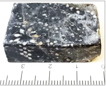
Fig. 2 - Limestone sample with Holosporella magna Bucur & Fucelli n. sp. in transverse and tangential sections, as well as numerous fragments of laterals spread in the rock. Sample FA-81, Ne-vada (U.S.A.).
Discussion
Griphoporella curvata is one of the most
fre-quent species identified in the Tethyan Upper Triassic. Barattolo et al. (1993) studied thoroughly this species, designated a neotype and gave the fol-lowing emended diagnosis (Barattolo et al. 1993, p. 33): “Cylindrical or slightly club-shaped thallus. Primary branches only, arranged in very close, alter-nate whorls. The branches are phloiophorous, with a subterminal narrowing; their transverse section is subcircular. The inclination of the branches is 45°-60° in the proximal portion then it gradually incre-ases outwards up to 70°-80°. The distal portion of the branches form a cortex with polygonal meshes
horizontally compressed. Reproductive organs unknown (not calcified) probably situated in the central stem or in the primary branches (endospore or cladospore). The calcification constitutes a calca-reous skeleton continuous, very thin and enveloping to a various degree different portions of the pri-mary branches. As a consequence, the pores show different morphologies, both in different specimens and in different portions of the same specimen”. The specimens identified in Japan (Pl. 8A-F) as well as rare fragments of thallus from Sonora (Mexico) (Pl. 10A) correspond to this diagnosis. Senowbari-Daryan et al. (2011) described Griphoporella curvata from Upper Triassic Nayband Formation in Iran.
A-F - Griphoporella curvata (Güm-bel). A) longitudinal, slightly oblique section; B) fragment in longitudinal section show-ing the calcareous wall and the distal end of the later-als; C) oblique section; D, E) transverse sections. A, thin section CP-94a; B, D, F, thin section CP-94b; C - thin sec-tion CP-261; E, thin secsec-tion JC-231a; Sambosan, Shikoku Island, Japan.
G - Gyroporella sp., fragment in transverse section. Thin section JC-231a; Sambosan, Japan.
H, I - Clypeina cf. besici (Pantić). Transverse-oblique (H) and transverse (I) sections. Thin section JC-231a; Sambosan, Shikoku Island, Japan. J - Physoporella jomdaensis Flügel &
Mu., longitudinal-oblique section. Thin section JC-231a; Sambosan, Shikoku Island, Japan.
Most of the illustrated specimens show a thin cal-careous envelope covering the external part of the laterals. The laterals have also frequently a vesicu-liform shape (e.g., Fig. 4F, Fig. 5E, H, J, Fig. 6E-G, Fig. 7H, etc. in Senowbari-Daryan et al. 2011). These authors (Senowbari-Daryan et al. 2011) re-cognized two types of Griphoporella: with open, and with closed pores. Senowbari-Daryan & Hamadani (2000) also described specimens with open pores. In discussing the specimens with closed pores, Se-nowbari-Daryan et al. (2011) noted also the presen-ce of closed pores in the type specimen of
Macropo-rella retica (Zanin-Buri, 1965), a species synonymized
by Barattolo et al. (1993) with G. curvata. In fact, the appearance of such closed pores in the type spe-cimen of M. retica represents an effect of sectio-ning, many pores in this specimen being open to the exterior. In our opinion, the specimens illustrated by Senowbari-Daryan et al. (2011) have laterals of vesiculiform shape characteristic for a Gyroporella and should be assigned to this genus (compare also Fig. 3d in Gümbel, 1872 with fig. 4F in Senowbari-Daryan et al. 2011).
Griphoporella minuta Bucur & Peybernes n. sp.
Fig. 3, Pl. 9A-Z
Origin of the name: from the latin minuta, meaning small. Holotype: specimen in oblique section illustrated in Pl. 9D,
thin section CP-259a [MHNG-GEPI-2020-0008].
Paratypes: specimens in Pl. 9A, B, E, F, H, I, R, S, X, thin
section CP-259a [MHNG-GEPI-2020-0008].
Type locality: Sambosan, Shikoku Island (Japan). GPS
co-ordinates: 33° 34.452′N; 133° 42.733′E.
Type level: Upper Triassic.
Material and repository: About 30 specimens identified in
one thin section (CP-259a) stored in the Museum d’Histoire Naturel-le de Genève under the number MHNG-GEPI-2020-0008.
Diagnosis: Thallus cylindrical bearing pholiophorous
pri-mary laterals disposed in whorls with euspondyle arrangement. Large central cavity and very thin calcareous wall. Calcification around the distal part of the primary laterals but probably not reaching their most distal end.
Dimensions (in mm): L (maximum observed) = 3.500 D = 0.360-0.740 (mean = 0.550) d = 0.220-0.470 (0.360) d/D% = 52.38-72.30 (65.10) e - (D-d)/2 = 0.065-0.135 (0.095) p (distal) = 0.050-0.090 (0.069) h = 0.080-0.100 (0.094) w = 11-17 (15) Description
The thallus is cylindrical, but a slightly club-shaped form is not excluded (Pl. 9A, L, Q). The laterals are phloiophorous with a thin proximal part and a larger distal part. Some of them seem to present a constriction in their distal portion just before the final enlargement (Pl. 8H, M, O, arrows) a feature characteristic of the genus Griphoporella Pia, emend Barattolo et al. (1993) which
differen-Fig. 3 - Bioclastic grainstone containing numerous specimens of Griphoporella minuta Bucur & Peybernes n. sp. Sample CP-259a, Sambosan, Shikoku Island (Japan).
tiates this genus from Salpingoporella Pia in Trauth, 1918. The laterals are arranged in close alternating whorls (Pl. 9B, N) (euspondyle arrangement). They probably represent only the distal calcified part of much longer laterals (see Barattolo et al. 1993 p. 40-41, Text figs. 6-7, reconstruction of Griphoporella
curvata). As a consequence of this type of
calcifica-tion the central cavity is very large (more that 65 % of the thallus diameter). The outline of the central cavity is smooth, as emphasized by longitudinal (Pl. 9A), oblique (Pl. 9D, M) and transverse (Pl. 9T, X) sections.
Comparisons: Four valid species are known
from the Triassic (Griphoporella bechstedti, G. curvata,
G.? guembeli and G. kuenzelsanensis) and six from the Jurassic-Cretaceous (G.? aurigerica, G. ellenbergeri, G.
minima, G. perforatissima, G. jurassica and G. cretacea)
(Granier & Deloffre 1993; Granier & Grgasovič 2000; Bucur & Schlagintweit 2009). Griphoporella
minuta n. sp. is morphologically similar to G. curvata
from the Upper Triassic. The consistent difference between the two species is related to the general dimensions. As emphasized by Table 2, the largest specimens of G. minuta have smaller dimensions that the smallest specimens of G. curvata. A compa-rable species regarding the dimensions is
Griphopo-rella minima Nikler & Sokač from the lower part of
the Upper Jurassic of Velebit (Croatia) (Nikler & Sokač 1967). It differs from G. minuta n. sp. by the type of calcification (yellowish cryptocrystalline cal-cite in G. minima) and by the shape and number of laterals. Griphoporella kuenzelsauensis (Flügel &
Hag-dorn, 1993) is another small species having general dimensions close to that of Griphoporella minuta n. sp. The differences consist in the shape of the later-als (cylindrical, slightly enlarged distally, but occa-sionally also narrowed distally in G. kuenzelsauensis; see Fig. 6 in Pl. 2, holotype in Flügel & Hagdorn 1993), as well as in the shape of the thallus (sup-posed moniliform in G. kuenzelsauensis). The shape of the thallus and general dimensions differentiate
G. minuta n. sp. from the other species mentioned
above.
Micropalaeontological assemblage: n. sp.
has been found in a bioclastic grainstone conta-ing microproblematica (Baccanella floriformis), small
foraminifers, ostracodes, sponge fragments, echino-derms and undetermined shell fragments.
Palaeoenvironment: biotic content and
mi-crofacies of associated samples (e.g., CP259B is a sponge-algae boundstone) suggest deposition in a reefal environment.
Genus Physoporella Steinmann, 1903, emend. Grgasović, 1995
Physoporella jomdaensis Flügel & Mu, 1982
Pl. 8J
Discussion
Only one specimen of Physoporella was found in the thin section JC-231a from Shikoku Island (Ja-pan). It shows the characteristic cylindrical thallus with a relatively large central cavity and pyriform laterals with horn-like distal ends. The laterals are arranged in alternating whorls. The Japanese speci-men is a little bit smaller than the type specispeci-mens from Tibet, but it fits the dimensions given by Grgasović (1997) for Physoporella jomdaensis from
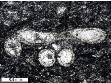
Fig. 4 - Collarecodium? nezpercae Bucur & Rigaud n. sp. showing a very discrete marginal zone probabily corresponging to the cor-tical zone of the alga. Specimens from the Black Marble Quarry (sample MQ-7D), Oregon (U.S.A.)
(Gümbel, 1872) in Barattolo et al. 1993 & Peybernes n. sp L(max.) > 20 3.400 D 0.730-3.120 (1.860) 0.360-0.0.740 (mean 0.550) d 0.420-2.600 (1.370) 0.220-0.470 (0.360) (D-d)/2 0.100-0.360 (0.240) 0.065-0.135 (0.095) p 0.0630-0.110(0.089) 0.050-0.090 (0.069) h 0.075-0.120 (0.091) 0.080-0.100 (0.094) w 16-21 (18.6) 11-17 (15)
Tab. 2 - Comparative dimensions (in mm) of Griphoporella curvata (Gümbel, 1872) and Griphoporella minuta Bucur & Peybernes n. sp.
Croatia (see table 3 for comparisons). As noted by Grgasović (1997, p. 207) the differences in dimen-sions are consistent with the limits of variation known in the other species of the genus Physoporella.
Stratigraphical range: Physoporella jomdaensis
was described by Flügel & Mu (1982) from Carnian limestone of eastern Tibet. The Croatian specimens of Žumberak have a Norian age (Grgasović 1997).
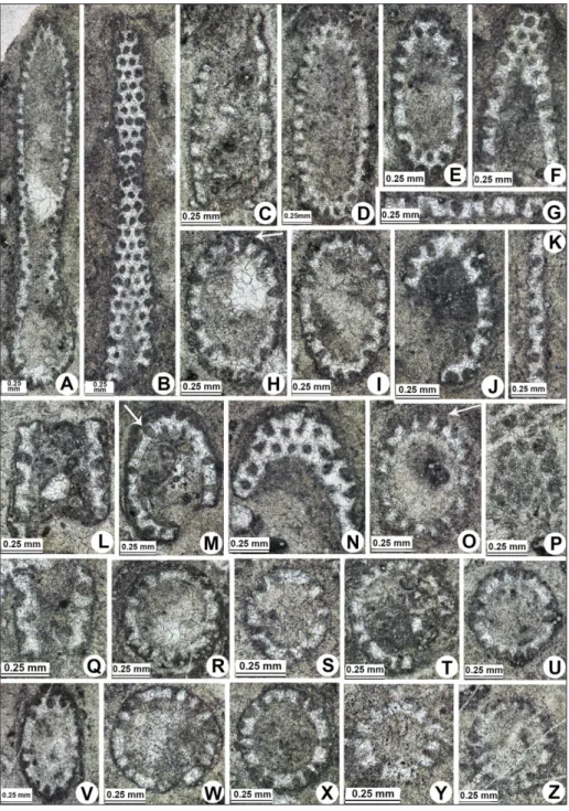
Plate 9
A-Z - Griphoporella minuta Bucur & Peybernes n. sp. A) lon-gitudinal-oblique section; B) longitudinal-tangen-tial section. C, G, K, L, Q) fragments in longitudinal section. D, E, H-J, M-P, V) oblique sections. A-Z, thin section CP-259a; Sambosan, Shikoku Island, Japan. D - holotype; A, B, E, F, H, I, R, S, X - paratypes.
Flügel & Mu 1982 Grgasović 1997 This paper
L (max. observed) 11.7 4.9 6.600 D 1.5-1.75 0.85-1.06 1.100 d 0.8-1.3 0.47-0.68 0.700 d/D % 63-77 49-64 (57) 63.6 p 0.15-0.22 0.11 0.150 h 0.12-0.25 0.23-0.25 0.200 w 15 14 15-16 (estimated)
Tab. 3 - Comparative dimensions (in mm) of Physoporella
jomdaen-sis Flügel & Mu, 1982 from
The limestone from this locality of the Sambosan Accretionary Complex (Shikoku Island, Japan) is broadly dated as Upper Triassic (Carnian-Rhaetian) (Peybernes et al. 2016b).
Genus Salpingoporella Pia in Trauth, 1918 emend. Carras et al., 2006
Salpingoporella sp.
Pl. 10H
Discussion
One specimen of small dimension (D = 0.32 mm; d = 0.21 mm) identified in a sample from
So-nora (Mexico) has phloiophorous laterals arranged in euspondyle whorls, corresponding to characteris-tics of the genus Salpingoporella.
Family Selletonellaceae Korde, 1950, emend Bassoullet et al., 1979
Genus Gyroporella Gümbel, 1882 Gyroporella div. sp.
Pl. 6I-K; Pl. 8G; Pl. 10E-G, I, J
Discussion
Several Gyroporella specimens have been
A - Griphoporella curvata (Gümbel), fragment in oblique section. Thin section EH-306A; So-nora, Mexico.
B-D - Macroporella? sp., longitudi-nal oblique (B) and oblique (C, D) sections. B, C, thin section EH-306A; D, thin section EH-216b; Sonora, Mexico.
E-G, I, J - Gyroporella div. sp., oblique sections. E, thin section EH-212A; F, thin section EH-311A; G, thin section EH-306A; I, thin section 509; J, thin section EH-216b; Sonora and Baja Cali-fornis, Mexico.
H - Salpingoporella sp., oblique sec-tion. Thin section EH-216A; Sonora, Mexico.
identified in samples coming from Far East Russia (Pl. 6I (Gy)-K), Japan (Pl. 8G) and from Mexico (Sonora, Pl. 10E-G, J, and Baja California Sur, Pl. 10I). They probably correspond to 2 or 3 different species. Some specimens (Pl. 6J, K; Pl. 8G; Pl. 10I) have laterals with a short tubular proximal part and a relatively large vesicular distal part, while other specimens (Pl. 10E-G, J) have slender and more nu-merous laterals, with a longer tubular proximal part and a small vesicular distal part. Due to the scarcity of the available material and the diagenetic altera-tion, a specific determination was not possible.
Genus Macroporella Pia, 1912 emend. Bassoullet et al., 1978
Macroporella? sp.
Pl. 10B-D
Discussion
Different sections of a dasycladalean alga found in Sierra del Alamo (Sonora, Mexico) present phoiophorous laterals which apparently are not set in verticils (whorls) but are arranged randomly and very close of each other. This tight arrangement causes a mutual pressure of the laterals at their dis-tal part, and hence the polygonal outline (Pl. 10C, D). The strong recystallisation prevents a good ob-servation of the laterals morphology.
Dimensions (in mm):
D = 0.500-0.740 d = 0.200-0.400 p = 0.030-0.050
A dasycladalean alga with a close morphology was described by Flügel & Mu (1982) as
Salpingo-porella? tibetica. The euspondyle arrangement of the
laterals of this last species is not very convincing, and it could also belong to the genus Macroporella. However, its general dimensions are three times the dimensions of the Mexican species.
Family Polyphysaceae Kützing, 1843
Genus Clypeina Michelin (1845) emend. Bassoullet et al., 1978
Clypeina cf. besici Pantić, 1965 ex Granier & Deloffre, 1994
Pl. 8H, I
Discussion
Two specimens found in the thin section JC-231a from Shikoku Island, Japan belong most prob-ably to Clypeina besici Pantić.
Dimensions (in mm):
D = 0.760-0.850 d = 0.200-0.400 w = 14
Forma-genus Patruliuspora Barattolo, Ionesi & Ţibuleac, 2019
Patruliuspora pacifica Bucur, Del Piero & Peyrotty n. sp.
Pl. 11A-O
Origin of the name: From the occurrence of the alga in
limestone located on both sides (North America and Far East Russia) of the Pacific Ocean.
Holotype: specimen in Pl. 11D, thin section WH-259
[MHNG-GEPI-2020-0024].
Partypes: Pl. 11A-C, E-N, thin sections WH-232b
[MHNG-GEPI-2020-0017], WH-235a [MHNG-GEPI-2020-0021], WH-235b [MHNG-GEPI-2020-0022], WH-259 [MHNG-GEPI-2020-0024], WH-260 [MHNG-GEPI-2020-0025], GP-206C [MHNG-GE-PI-2020-0004], GP-206C-2 [MHNG-GEPI-2020-0005].
Type locality: Lime Peak, Yukon (Canada). Type level: late Norian.
Material and repository: More than 120 specimens
identi-fied in 15 thin sections, stored in the Museum d’Histoire Naturelle de Genève under the numbers MHNG-GEPI-2020-0004, 0005, 0006, 0007, 0016-0026.
Diagnosis: Cyst aggregates (gametophores) of ovoid
sha-pe containing small ovoid to spherical cysts (gametangia), arranged more or less on the margins of the aggregates; consequently, the in-ternal part of the aggregate is generally devoid of cysts.
Description
The gametophores have an ovoid slightly elongated form (Pl. 11A, B, D, G). The gametangia are also ovoid (Pl. 11D, G-J, N), sometimes ovoid-elongated (Pl. 11M), rarely close to a spheroidal shape (Pl. 11E, F). The gametangia are arranged in the marginal zone of the gametophore; conse-quently, the central part of the gametophore is fre-quently devoid of cysts as illustrated by longitudi-nal and oblique (Pl. 11A, G, H) as well as transverse (P. 11I, K, N) sections. The gametangia have a rela-tively thick calcified wall. In some specimens (e.g., Pl. 11E, O) the cyst wall is very thick, representing about 15-20% from cyst diameter, and the calcite has a brown color. In other cases (e.g., the Russian specimens, Pl. 11N) the cyst wall is much thinner.