UNIVERSITY OF SASSARI
PhD School in
Biomolecular and Biotechnological Sciences
Curriculum: Biochemistry and Molecular Biology Director: Prof. Claudia Crosio
“XXVI Ciclo”
Physiopathology of ALS:
from oxidative stress to RNA metabolism.
PhD Student: Sonia Esposito
Supervisor: Prof. Claudia Crosio
Sonia Esposito Physiopathology of ALS: from oxidative stress to RNA metabolism. I PhD Thesis in Biomolecular and Biotechnological Science
University of Sassari
INDEX
CHAPTER 1: INTRODUCTION.
1.1 Amyotrophic Lateral Sclerosis (ALS)……….. 1.2 Aetiology of ALS……….. 1.2.1 ALS environmental factors……… 1.2.2 ALS genetic factors………. 1.3 ALS1/Superoxide Dismutase 1 (SOD1)………. 1.3.1 Structure and functions………. 1.3.2 SOD1 mutations………. 1.4 ALS6/ Fused in sarcoma/Translocated in liposarcoma (FUS/TLS)……….
1.4.1 Structure and functions………. 1.4.2 FUS mutations………. 1.5 ALS10/Transactive response DNA-binding protein-43 (TARDBP-43/TDP43)……….
1.5.1 Structure and functions………. 1.5.2 TDP43 mutations……… 1.6 ALS experimental models………. 1.6.1 Cellular and animal models for SOD1……… 1.6.2 Cellular and animal models for FUS……… 1.6.3 Cellular and animal models for TDP-43……… 1.7 Physiopathological mechanisms of ALS……….. 1.7.1 Oxidative Stress………..
1.7.2 Glutamate Excitotoxicity………
1.7.3 Mitochondrial Dysfunction……….. 1.7.4 Impaired Axonal Transport……….. 1.7.5 Inflammatory cascades and the role of non-neuronal cells………. 1.7.6 Protein aggregation……….. 1.7.7 Misregulation in RNA processing………. 1.7.8 Autophagy dysfunction……….. 1.7.9 Apoptotic pathways……….. 2 3 3 4 6 6 6 9 9 10 11 11 13 14 14 16 18 19 20 21 21 22 23 24 25 26 28
Sonia Esposito Physiopathology of ALS: from oxidative stress to RNA metabolism. II PhD Thesis in Biomolecular and Biotechnological Science
University of Sassari
1.8 ALS therapeutic approaches………
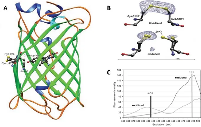
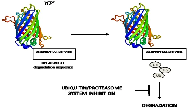
1.9 High−throughput screening (HTS) approach based on multiple fluorescent probes to identify new drugs for treatment of Amyotrophic Lateral Sclerosis………. 1.9.1 Redox sensitive Green Fluorescent Protein (roGFP)……….. 1.9.2 Yellow Fluorescent Proteinu (YFPu)………. 1.9.3 DsRed-LC3-GFP reporter……… 1.9.4 CFP-DEVD-GFP: FRET (Fluorescent Resonance Energy Transfer) reporter………….
CHAPTER 2: AIMS OF THE PROJECT. CHAPTER 3: MATERIALS AND METHODS.
3.1 Materials……….. 3.2 Methods………...
CHAPTER 4: RESULTS.
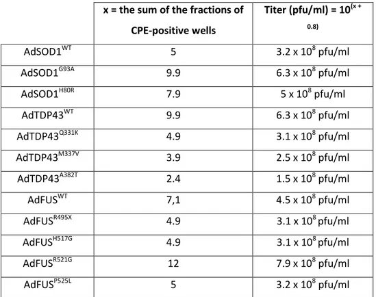
4.1 GFP-based reporters to monitor biochemical alteration in cellular models for ALS: a possible and straightforward read-out for high-throughput screening (HTS) assay………. 4.1.1 Adenoviral delivery of ALS-causative genes………. 4.1.2 Characterization of fALS cellular models……… 4.1.3 Construction and characterization neuronal cell lines stably expressing fluorescent reporter genes to assess in vivo different parameters of cell toxicity induced by ALS−causative genes………. 4.1.3.1 SHSY5Y cells stably expressing redox−sensitive GFP (roGFP) to monitor oxidative stress either in the cytoplasm or in the mitochondria……… 4.1.3.2 Construction and characterization of SHSY5Y cells stably expressing YFPu to monitor alterations in the activity of ubiquitin−proteasome system……… 4.1.3.3 Construction and characterization of SHSY5Y cells stably expressing DsRed-LC3-GFP to monitor autophagy……… 4.1.3.4 Construction and characterization of SHSY5Y cells stably expressing CFP-DEVD-YFP to monitor caspase-3 activation……… 4.1.4 Measure cell toxicity induced by ALS causative genes expression using
fluorescent markers………. 4.2 Study of the pathophysiological mechanisms responsible for cellular toxicity of pathological forms of two RNA-binding protein, FUS and TDP-43, in familial forms of ALS……… 29 30 31 32 33 34 36 39 42 49 50 52 56 56 59 60 61 62 68
Sonia Esposito Physiopathology of ALS: from oxidative stress to RNA metabolism. III PhD Thesis in Biomolecular and Biotechnological Science
University of Sassari
4.2.1 Identification of the RNA target of FUS……….. 4.2.1.1 CLIP (CrossLinking ImmunoPrecipitation) to identify RNA partners of FUS.. 4.2.1.2 R521G-FUS mutant does not mediates alternative splicing of its mRNA
targets………. 4.2.2 Study of the relationship between aggregation, proteins localization,
autophagy and cell death alterations in the presence of pathological forms of TDP43………. 4.2.2.1 TDP43 A382T is delocalized into the cytoplasm………. 4.2.2.2 Mutation A382T induces shuttling nucleus/cytoplasm………. 4.2.2.3 Mutation A382T promotes TDP-43 aggregation……… 4.2.2.4 Overexpression of WT or mutant TDP43 not induces PARP or LC3 cleavage……….
CHAPTER 5: DISCUSSION.
5.1 ALS cellular models for high-throughput screening (HTS) assay………... 5.1.1 Oxidative stress, roGFP and ALS-causative genes……… 5.1.2 Protein aggregation, YFPu and ALS-causative genes……….. 5.2 TDP43 A382T pathological mutation: relationship between aggregation, localization and cell death……….
CHAPTER 6: BIBLIOGRAFY. 68 68 72 73 73 75 75 76 79 79 80 81 83
Sonia Esposito Physiopathology of ALS: from oxidative stress to RNA metabolism. Pag. 1 PhD Thesis in Biomolecular and Biotechnological Science
University of Sassari
CHAPTER 1:
INTRODUCTION.
Sonia Esposito Physiopathology of ALS: from oxidative stress to RNA metabolism. Pag. 2 PhD Thesis in Biomolecular and Biotechnological Science
University of Sassari
1.1 Amyotrophic Lateral Sclerosis (ALS).
Amyotrophic lateral sclerosis (ALS; also known as Lou Gehring’s disease) is the most common adult-onset motor neuron disease, described for the first time by Jean-Martin Charcot in 1874 1. The pathology incidence and prevalence is around 2.5 and 4-6 cases per 100,000 people each year, respectively, with a slightly higher incidence of disease in men. The hallmark of this disease is the selective degeneration of cortical, bulbar and spinal motor neurons, except for those that control the bladder and the eye movement (Fig. 1). This leads to generalised muscle weakness leading to progressive paralysis of voluntary muscles until death caused by respiratory failure. The average age of onset is 50-55 years with a typical survival time of 3-4 years after diagnosis, but this parameter is influenced by age at symptom, clinical presentation, age at respiratory dysfunction and nutrition state of the patient 2,3. ALS may present as a predominantly lower motor neuron (LMN) form designates progressive muscular atrophy (PMA), a predominantly upper lower motor neuron (UMN) form called primary lateral sclerosis (PLS), or more commonly with mixed UMN and LMN deficits 4.
Figure 1: Motor neurons selectively affected in ALS. Degeneration of motor neurons in the motor cortex leads to clinically apparent signs of upper motor neuron abnormalities. Degeneration of motor neurons in the brain stem and spinal cord causes muscle atrophy, weakness and fasciculation 1.
Although the majority of cases are classed as sporadic ALS (sALS), approximately 10% of ALS cases are inherited (familial ALS, fALS), with multiple autosomal dominant and recessive forms. Sporadic and familial ALS share common clinical and neuropathological manifestations, and ALS patients show some degree of heterogeneity as far as symptoms, age of onset and disease duration are concerned, furthermore all genes found mutated in fALS cases have also been found mutated in sALS 5,6.
Sonia Esposito Physiopathology of ALS: from oxidative stress to RNA metabolism. Pag. 3 PhD Thesis in Biomolecular and Biotechnological Science
University of Sassari
The causes of the majority of ALS cases are still obscure. Although numerous pathological mechanisms have been elucidated, ALS remains an invariably fatal disease in the absence of any effective therapy. The heterogeneity of the disease and the failure to develop satisfactory therapeutic protocols reinforce the view that ALS is a multi-factorial and multi-systemic disease. Up to now, Riluzole is the only approved medication as ALS treatment, it has the effect to slow the disease in humans but only slightly, prolonging survival by 3 months and not clearly improving the quality of life of patients 7. Numerous other treatments are currently in different phases of clinical trials and include anti-inflammatories, neurotrophic, anti-oxidants and anti-apoptotic molecules 8.
1.2 Aetiology of ALS.
ALS, like many other neurodegenerative diseases, presents an extremely complex aetiology determined by environmental and genetic factors. In fact, in most cases to disease development contribute both genetic and environmental factors, and only a small percentage of cases can be attributed simply one of the two factors.
The pathogenic mechanisms that underlie ALS remain mostly unclear, even if different alterations of cellular function in ALS motor neurons, including protein misfolding, RNA metabolism dysfunction, mitochondrial defects, oxidative stress, altered axonal transport, excitotoxicity, insufficient growth factor signaling and inflammation have been identified 9,10.Several factors are proposed to instigate these phenomena, including various environmental risk factors (for example, insecticides, pesticides and cigarette smoking)11,12 latent infections by viral and non-viral agents 13, toxins and autoimmune reactions 14.
Lately there has been rising interest the role played by non-neuronal neighbouring cells in the pathogenesis of motor neuron damage. In fact ALS is considered also a glial pathology and disruption of glial cells/motoneurons communication contribute to neurodegeneration and the propagation of motor neuron injury 15.
1.2.1 ALS environmental factors.
Given that sALS represents approximately 90% of all ALS cases, it has been hypothesized that exposure to different environmental factors may interact with individual genetic susceptibility playing a remarkable role in the pathogenesis of the disease.
Firstly it was hypothesized that cigarette smoking and many chemicals contains, could contribute to ALS development through lipid peroxidation via formaldehyde exposure 11,16. Second it appears that life style as well as particular occupation could be associated to ALS for their continued exposure to toxic agents. In particular, consumption of food with high concentrations of the
Sonia Esposito Physiopathology of ALS: from oxidative stress to RNA metabolism. Pag. 4 PhD Thesis in Biomolecular and Biotechnological Science
University of Sassari
neurotoxic aminoacid (β-methyl-amino-L-alanine)17, use of cholesterol-lowering drugs 18, and environmental toxicants such as heavy metals 19, pesticides or herbicides 12,20 may be potential risk factors. Evidences in several case-control studies as well as in experimental studies showed that exposure to these toxicants could induce progressive brain damage by increase of ratio between reactive oxygen species (ROS) and reactive nitrogen species (RNS)21, decrease activation of astrocytes with neuroprotective effects 22 and inhibition of acetylcholinesterase, resulting in excessive stimulation of cholinergic receptors and excitotoxicity 23. Also intensive and strenuous physical exercise of professional and amateur football players (Italian and English soccer players)24,25 as well as military service (Gulf War veterans in the US Army)26 could represent a risk factor for ALS linked to the intermittent occupational hypoxia resulting in the production of ROS27 or to the repeated head injuries 28.
In short, as just described over the years a growing list of potential environmental risk factors for ALS has been proposed, but at present there is no ascertained causal link between any of them and ALS pathogenesis 29.
Recently, it has been evidenced the involvement of epigenetic factors on the progression of many human pathologies, including neurodegenerative diseases such as ALS. The epigenetic modifications could provide a possible link between the environmental risk factors and the gene expression alterations responsible for the ALS onset 30. In fact, it has been proposed that epigenetic silencing of genes important to motor neurons functions could underlie ALS development. In support to this hypothesis, it has been shown that treatment with histone deacetylase enzymes (HDAC) inhibitors significantly prolong survival, improves motor performance and reduce the loss of motor neurons in mouse models of ALS 31,32.
1.2.2 ALS genetic factors.
In the last years, there has been a rapid progress in our knowledge of genetic causes for ALS. Familial ALS can be inherited as an autosomal dominant or autosomal recessive trait. Up to date, more than 20 loci have been identified by genetic analysis, and several have been assigned to specific genes (Table 1).
Sonia Esposito Physiopathology of ALS: from oxidative stress to RNA metabolism. Pag. 5 PhD Thesis in Biomolecular and Biotechnological Science
University of Sassari
Table 1: The genetics of fALS. More than 20-ALS genes have been identified in fALS. These genetic mutations represent different molecular pathways of motor neuron degeneration. Abbreviations: AD: Autosomal dominant; AR: Autosomal recessive; VAPB: Vesicle associated membrane protein associated protein B; VCP: Valosin Containing Protein; SIGMAR1:Sigma Non Opioid Intracellular Receptor; C9ORF72: Chromosome 9 open reading frame 72; DAO:D-Amino Acid Oxidase; FTD: Frontal-temporal dementia; UMN: upper motor neuron; LMN: lower motor neuron 33.
Linkage studies indicated that both types of inheritance are represented by more than one genetic entity. The relationship between the genetic subtypes and the pathological subtypes as well as clinical phenotype has become more and more clear. Although in many cases the function of mutant protein is not yet clearly identified or many different functions have been proposed, the available data shows that fALS proteins are involved in several cellular processes, from RNA processing to antioxidant response, axonal and vesicular transport and angiogenesis 3,33.
Sonia Esposito Physiopathology of ALS: from oxidative stress to RNA metabolism. Pag. 6 PhD Thesis in Biomolecular and Biotechnological Science
University of Sassari
1.3 ALS1/Superoxide Dismutase 1 (SOD1).
1.3.1 Structure and functions.
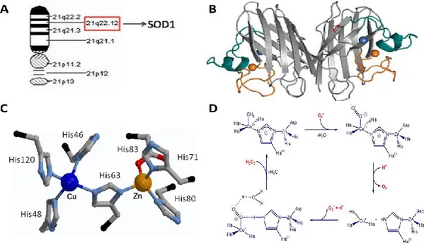
Eukaryotic copper, zinc superoxide dismutase (SOD1) is a 32-kDa homodimeric metalloenzyme, encoded on chromosome 21 (21q22.1) and found predominantly in the cytosol, but also in the mitochondrial intermembrane space, nucleus and peroxisomes. SOD1 is an abundant protein in the CNS, accounting for about 1% of brain protein, but it is also ubiquitously expressed in all other tissues 34,35. Each subunit of SOD1 forms an eight-stranded -barrel and contains an active site that binds a catalytic copper ion (binding residues: His46, His48, His63 and His120), a structural zinc ion (binding residues: His63, His71, His80 and Asp83) and a disulphide bond. Through cyclical reduction and oxidation (dismutation) of copper, SOD1 converts the superoxide anion to dioxygen and hydrogen peroxide preventing the further generation of reactive oxygen species (ROS)36,37,38 (Fig. 2).
Figure 2: Structure and function of SOD1. A) SOD1 gene position on chromosome 21. B) Tridimensional structure of human SOD1. C) Active site of Cu/Zn SOD1. D) Mechanism cyclic oxidation-reduction catalysed by SOD1.
1.3.2 SOD1 mutations.
About 20% of all fALS and 1-4% of sALS cases carry mutations in the locus ALS1 on chromosome 21 that codes for SOD1 (Cu, Zn superoxide dismutase 1, OMIM #105400). After the first report in 1993, more than 150 different mutations have been identified in all five exons of SOD1 predominantly in missense mutations, but also a small percentage of nonsense mutations, insertions and deletions. Most mutations act dominantly except two, N86S and D90A, that are
Sonia Esposito Physiopathology of ALS: from oxidative stress to RNA metabolism. Pag. 7 PhD Thesis in Biomolecular and Biotechnological Science
University of Sassari
linked to recessive inheritance 39,40 although the former mutation was described in rare juvenile onset ALS (Fig.3).
Figure 3: Schematic diagram of human SOD1 primary sequence with exons, introns, metal binding domains (Cu, Zn), intramolecular disulphide bond (SH) and mutations linked to sporadic and familial ALS. Mutations are distributed throughout all exons with high prevalence in exons 4 and 5. No clear structure–function relationship to fALS appears evident. Mutation legend: grey, missense; purple, insertion; red, deletion; blue/green, silent; (D) in frame deletion; X, truncation 41.
Since some mutations concern the metal binding residues at the active site, while others may concern correct folding or stability of the homodimer, the biochemical and biophysical properties of ALS-associated mutant SOD1 proteins are rather heterogeneous 42. In fact, studies on recombinant mutant SOD1 proteins have been proved that ALS-associated SOD1 mutations can be attributed at two mutant classes, the "wild type like" (WTL) SOD1 mutants which retain the ability to bind copper and zinc and exhibit normal specific activity, indicate a native-like structure with only subtle changes to the backbone fold, in contrast the "metal-binding region" (MBR) SOD1 mutants that are deficient in copper and zinc and exhibit severe thermal destabilization and structural disorder of conserved loops near the metal-binding sites 43. G93A and H80R SOD1 mutants belong respectively to the two classes of mutants described above.
There is no clear correlation between enzyme activity, clinical progression and disease phenotype
44
. ALS cases linked to SOD1 mutants show ample variation in the phenotype in the age of onset, severity, rate of disease progression and duration. However, patients carrying the A4V missense mutation (the most common SOD1 mutation in North America) have shorter survival with death occurring in less than one year after the diagnosis and limited upper motor neuron involvement 45, while D90A mutation (the most common SOD1 mutation worldwide and in the sporadic
Sonia Esposito Physiopathology of ALS: from oxidative stress to RNA metabolism. Pag. 8 PhD Thesis in Biomolecular and Biotechnological Science
University of Sassari
population) correlates with non-penetrant or slowly progressive disease. Remarkably, 2–3% of the population, in northern Scandinavia, is heterozygous for the D90A mutation, which is a benign polymorphism 46. However, homozygous individuals for D90A in that region develop slowly progressive motor neuron disease with prominent corticospinal signs and prolonged survival of more than a decade. By contrast, patients who are D90A heterozygotes outside this genetic pool develop classic ALS with survival times of 3–5 years 47.
It is important to note that some SOD1 mutations can remain asymptomatic throughout life, suggesting that not all SOD1 mutations cause ALS onset and some are rather polymorphisms. The clinical phenotype associated with a certain mutation in the SOD1 gene is obviously not only dependent on the mutation itself, but may also be influenced by the genetic background of the patient as well as by environmental factors 48.
Up to date, there is no conclusive explanation about how the SOD1 mutations cause ALS onset. At first, it was hypothesized that mutations would impair the enzymatic activity of the SOD1, thus resulting in increased cellular levels of reactive oxygen species (ROS), oxidative stress and then neuronal death 49. However, results obtained through both in vitro and in vivo experimental models has provided numerous evidences that led to abandon this initial hypothesis in favour of a new hypothesis that result into the acquisition of a novel toxic function to motor neurons (gain-of function hypothesis). In fact, it has been demonstrated that the overexpression of SOD1 WTL-mutants in cell models induces apoptosis in neurons 50 and that SOD1 knockout mice do not develop motor neuron disease 51.
To date, different hypothesis have been made to explain the SOD1 toxicity. The aggregation hypothesis is particularly attractive because protein aggregates are frequently associated with neurodegenerative diseases as ALS 5. Numerous studies revealed that SOD1 mutant is incline to misfolding and to form cytoplasmic aggregates. In turn, these aggregates could lead to cell death by sequestering other cytoplasmic proteins essential for neuronal survival, by clogging the ubiquitin/proteasome system, by chaperones depletion, or by disrupting mitochondria, cytoskeleton and/or axonal transport 52. Another hypothesis was that the misfolding of SOD1 induced by mutations would allow the access of abnormal substrates such as peroxynitrite to the catalytic site leading to the nitration of tyrosine residues 53.
Sonia Esposito Physiopathology of ALS: from oxidative stress to RNA metabolism. Pag. 9 PhD Thesis in Biomolecular and Biotechnological Science
University of Sassari
1.4 ALS6/Fused in sarcoma/Translocated in liposarcoma (FUS/TLS).
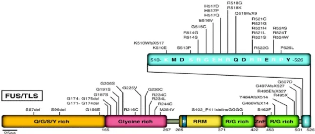
1.4.1 Structure and functions.FUS/TLS (Fused in sarcoma/Translocated in liposarcoma, also called FUS) was originally discovered as a result of characteristic chromosomal translocations in myxoid liposarcoma 54. The gene is located in chromosome 16 (16p11.2) and consists of 15 exons that encode for a 526 amino acid protein, member of the TET family, a family of proteins implicated in various roles of gene expression 55,56. It is characterized by an N-terminal transactivating domain enriched for glutamine, glycine, serine, and tyrosine (QGSY region), a glycine-rich region (GRR) followed by a RNA recognition motif region (RRM) which contains a nuclear export signal (NES) near its 5′ end, a multiple arginine/glycine/glycine (RGG rich) repeats containing a zinc finger (ZnF) domain, and finally a nuclear localization signal (NLS)57,58 (Fig. 4).
Figure 4: Genomic and structural organization of human FUS/TLS gene and protein. FUS/TLS gene is encoded by 15 exons that cover an 11.6 kb region on chromosome 16p11.2. Protein coding exons (filled boxes) and non-coding exons (open boxes) are drawn to scale. SYGQ rich: serine–tyrosine–glycine–glutamine rich domain; G rich: glycine rich domain; RRM: RNA recognition motif; ZnF: cysteine2/cysteine2 zinc finger motif; RGG rich: arginine–glycine–glycine rich domain 59.
FUS is an ubiquitously expressed and highly conserved multifunctional protein, mostly localized into the nucleus and with low levels in the cytoplasm. The precise role of FUS is not fully elucidated but it is now known that it is involved in multiple levels of RNA processing including transcription, splicing, transport and translation 60. FUS acts also as a transcription regulator. It interacts with several gene-specific transcription factors such as NF-B and it is also associated with the general transcriptional machinery and may influence transcription initiation and promoter selection by interacting with RNA polymerase II and the TFIID complex 61,62. Less is known about the role of FUS in splicing regulation. Proteomic analysis identified this protein as part of the spliceosome machinery 63. It has been shown that FUS associates in vitro with large transcription–splicing complexes that bind the 5’ splice sites of pre-mRNA 64 and has been also proposed to directly bind pre-mRNA 3’ splice site 65. FUS seems to play roles in micro-RNA processing. It has been found (by mass spectrometry) associated with Drosha, the nuclear RNase
Sonia Esposito Physiopathology of ALS: from oxidative stress to RNA metabolism. Pag. 10 PhD Thesis in Biomolecular and Biotechnological Science
University of Sassari
III-type protein that mediates the first step in miRNA maturation 66. FUS plays also a cytosolic role in regulation of RNA subcellular localization, translation and decay. In particular, FUS was shown to relocalize to the cytoplasm upon inhibition of RNA polymerase II transcription 67 and increasing evidences suggest that it is integral component of RNA stress granules (SGs), cytoplasmic microscopically visible foci consisting of mRNA and RNP complexes that stall translation under stress conditions 68. Furthermore, several lines of evidence suggest that FUS is required for the maintenance of genomic integrity and may have a role in DNA double-strand break repair. Indeed, FUS promotes the annealing of homologous DNA and the formation of DNA D-loops, an essential step in DNA repair by homologous recombination 69.
1.4.2 FUS mutations.
About 5% of all fALS and 1-2% of sALS cases are due to mutations in the locus ALS6 on chromosome 16 that codes for FUS (Fused in Sarcoma, OMIM #608030). After the first report in 2009 by two independent groups 70,71, a total of 47 mutations have been described in ALS patients, among these the majority are missense mutations, but they also include deletions (only R495X), truncations and in-frame insertions72. Almost all of the pathological mutations show an autosomal dominant inheritance pattern except for one recessive mutation (H517Q) found in a family of Cape Verdean origin 70. Most of FUS ALS-linked mutations are clustered at the C-terminal domain of the protein and in particular in the Nuclear Localization Signal (all 5 the arginines in this region have been identified to be mutated in ALS). The majority of the other ALS associated FUS mutations occur in the GRR (Fig.5). The redistribution of the protein from the nucleus to the cytoplasm is related to the onset and progression of the disease and mutations that affect amino acid residues responsible for the binding to Transportin receptors (thus affecting the nuclear import of the protein) result in a more severe disease progression 73.
The site and age of disease onset are variable within families, and incomplete penetrance has been documented for several FUS/TLS mutations, which may account, at least in part, for the absence of family history in sporadic patients. Interestingly, several patients harbouring the same R521C mutation developed an unusual presentation including an early-onset drop-head syndrome
74,75
Sonia Esposito Physiopathology of ALS: from oxidative stress to RNA metabolism. Pag. 11 PhD Thesis in Biomolecular and Biotechnological Science
University of Sassari
Figure 5: FUS mutations in ALS. Forty-seven mutations have been identified in FUS/TLS in sporadic and familial ALS patients. Most mutations are clustered in the last 17 amino acids and in the glycine-rich region and the putative prion domain comprises amino acids 1–239 76.
This is an atypical phenotype, as only 1% of ALS patients present with severe weakness of neck extensor muscles in the early stage of the disease course 77. Most patients with FUS develop a classical ALS phenotype without cognitive defect. The truncating mutation R495X and the missense mutation K510R, discovered in four German ALS families, were associated with an aggressive disease course and with a mild phenotype with disease duration ranging from 6 to 8 years, respectively 78. Analysis of brain and spinal cords from patients with FUS mutations revealed normal nuclear levels in the nuclei of many neurons and glial cells, but with FUS cytoplasmic aggregations in neurons 71,70. It has not been reported if FUS inclusions are also present in glial cells. Cytoplasmic inclusions of FUS protein are absent in controls, in patients with SOD1 mutation and in sporadic ALS cases that are presumably positive for TDP-43 aggregations. Importantly, TDP43 positive inclusions are absent in FUS mutant patients, implying that neurodegenerative processes driven by FUS mutations are independent of TDP-43 aggregation 71.
1.5 ALS10/Transactive response DNA-binding protein-43 (TARDBP-43/TDP43).
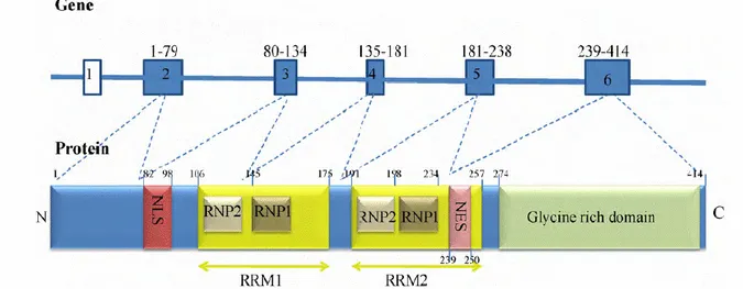
1.5.1 Structure and functions.TAR-DNA binding protein (TARDBP or TDP43) was identified in 1995 as a 43-kDa cellular factor binding TAR regulatory sequence of LTR in HIV-1 virus genome. The gene is located on chromosome 1 (1p36.22) and consists of 6 exons that encode for a 414 amino acid protein, member of the family hnRNPs, RNA binding proteins family 60 containing a nuclear export signal (NES) and a nuclear localization signal (NLS), that enable it to shuttle between the nucleus and the cytoplasm, potentially to transport bound mRNAs 79, two RNA recognition motifs (RRM1 and 2),
Sonia Esposito Physiopathology of ALS: from oxidative stress to RNA metabolism. Pag. 12 PhD Thesis in Biomolecular and Biotechnological Science
University of Sassari
able to interacts with RNA and DNA, and a C-terminal glycine-rich region which is able to mediate protein–protein interactions 80,81 (Fig. 6).
Figure 6: Schematic diagram of the TARDBP gene and TDP-43 protein. Exon 1 of TARDBP is non-coding and exons 2–6 are protein coding. TDP-43 contains five known functional domains: nuclear localisation signal (NLS), nuclear export signal (NES), two RNA recognition motifs (RRM1 and RRM2) and a glycine-rich C-terminal region. Each RRM has two highly conserved regions, ribonucleoprotein 1 (RNP1) and ribonucleoprotein 2 (RNP2) 82.
TDP-43 is predominantly localized into the nucleus with low levels in the cytoplasm. TDP-43 is a multifunctional protein involved in various steps of RNA processing including transcription, pre-mRNA splicing, pre-mRNA transport and translation, although the exact cellular functions remain elusive 82. TDP43 acts as a transcription factor and has been shown to bind to chromosomally integrated TAR DNA to repress transcription of HIV-1 80. It has also been shown to bind to the promoter of the mouse SP-10 gene, which is required for spermatogenesis. The C-terminal domain of TDP43 is involved with exon skipping and splicing inhibitory activity through the interaction with other hnRNP family proteins 60. TDP43 also plays a role in RNA post-transcriptional regulation. TDP43 is recruited to stress granules (mRNA and RNP complexes where protein synthesis is temporarily arrested), indicating that TDP43 may play a protective role against cellular insult 83. TDP43 may play a role in microRNA biogenesis and processing as it has been found to associate with Drosha, the RNase III enzyme responsible for initiating the processing of miRNA 60. Independent studies using knockout mouse models have shown that TDP43 protein is essential for normal prenatal development and viability 84. Knockdown of TDP43 in human cells resulted in cell cycle disruption and increased apoptosis, thereby demonstrating a critical role for TDP43 in cell survival possibly through maintaining genomic stability 85. TDP43 silencing in human embryonic kidney (HEK293) and neuronal (SH-SY5Y) cells has resulted in downregulation of HDAC6, a protein necessary for protein aggregate formation and degradation 64.
Sonia Esposito Physiopathology of ALS: from oxidative stress to RNA metabolism. Pag. 13 PhD Thesis in Biomolecular and Biotechnological Science
University of Sassari
1.5.2 TDP43 mutations.
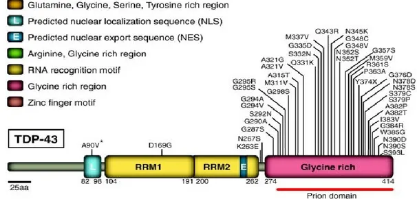
In early 2008, the successful identification of dominant mutations as a primary cause of ALS provided evidence that aberrant TDP43 can directly trigger neurodegeneration. Up to date, more than 40 mutations have been found in the locus ALS10 (OMIM #612069) of ALS patients, accounting for about 5 % fALS cases and 2% of sALS 86. Most of the mutations identified are localized in the glycine-rich region encoded by exon 6 (Fig. 7). All mutations are dominant missense changes with the exception of a truncating mutation at the extreme C-terminal of the protein (Y374X) 87. Several variants lying in the non-coding regions of the TARDBP gene have been identified in patients but further studies are necessary to prove their pathogenic effect 88,89.
Figure 7: TDP43 mutations in ALS. Forty-four mutations have been identified in TDP43 in sporadic and familial ALS patients, with most lying in the C-terminal glycine-rich region. All are missense mutations, except for the truncating mutation TDP43Y374X. Abbreviations: NLS, nuclear localization signal; NES, nuclear export signal 76.
Most patients with TDP43 mutation develop a classical ALS phenotype without cognitive deficit, with some variability within families in the site and age of onset 90,91. The A382T mutation, first identified in two fALS cases from France 92, has the highest prevalence since it was then described in 48 fALS families and 47 sALS. Interestingly, the A382T mutation was identified as a founder mutation in ALS patients from Sardinia, where 40 fALS and 30 sALS cases shared this mutation. The mutational frequency for A382T is about 30% in Sardinia (50%–80% fALS, 9%–23% sALS) 93,94. Geographically, most of the mutations identified were in ALS patients from Europe. However, a considerable number of ALS patients with TDP43 mutations have been identified in patient cohorts from Japan, Australia, North America, and China, suggesting that TDP43 mutations are a global cause of ALS 95.
Postmortem analysis of patients with TDP43 mutations found a pattern similar to the TDP43 pathology described in sporadic ALS and FTLD patients. TDP43 inclusions are not restricted to
Sonia Esposito Physiopathology of ALS: from oxidative stress to RNA metabolism. Pag. 14 PhD Thesis in Biomolecular and Biotechnological Science
University of Sassari
motor neurons but can be widespread in brain in ALS patients with or without dementia 96,97. A very curious, and mechanistically unexplained, aspect of TDP43 pathology is a significant TDP-3 nuclear clearance in a proportion of neurons containing cytoplasmic aggregates, suggesting that pathogenesis may be driven, at least in part, by loss of one or more nuclear TDP43 functions 98,99. Although a plethora of early reports did not establish how prominent this nuclear clearing was and whether it escalates in frequency during disease progression, further efforts, especially the work of Giordana et al. 97, have established that the cytoplasmic redistribution of TDP43 appears to be an early event in ALS. The pre-inclusions and the larger TDP43 inclusions are only partially labelled with anti-ubiquitin antibodies 97, whereas intranuclear and cytoplasmic inclusions are strongly recognized by phosphorylation-specific antibodies to TDP43 100,101. Interestingly, the composition of TDP43 inclusions seems to differ between cortical brain and spinal cord in ALS patients, as inclusions from cortical regions are preferentially labelled by C-terminal antibodies, whereas spinal cord inclusions display equivalent immunoreactivity between N- and C-terminal-specific antibodies
99
. In accordance with this, spinal cord extracts showed an absence or only weak accumulation of 25 kDa CTFs compared with brain extracts 101.
1.6 ALS experimental models.
In order to better understand the pathophysiological processes of the ALS onset, have been created over the years several model systems such as cell lines and transgenic animals, expressing different ALS-causative genes (SOD1, FUS and TDP43) in the wild type form or with the several pathological mutations.
1.6.1 Cellular and animal models for SOD1.
Over time, several cellular models stably or transiently espressing the SOD1 gene linked to ALS have been generated. These have helped to elucidate the molecular cascade of events able to induce cellular toxicity and the cellular mechanisms of disease 35.
The first model of motor neuron degeneration SOD1-related was created inhibiting chronically SOD1 expression through the use of antisense oligodeoxynucleotides or diethyldithiocarbamate in organotypic cultures of spinal cord. This study showed that chronic inhibition of SOD1 was able to induce the apoptotic degeneration of spinal neurons, including motor neurons, and that the degeneration increased when it was also inhibited glutamate transport 102. The same results were obtained in a study conducted on PC12 cells (rat pheochromocytoma cells), where it has been observed apoptotic cell death after downregulation of the SOD1 expression 103. Recently it was shown that the expression of SOD1-G93A, in co-cultures of human glioblastoma and
Sonia Esposito Physiopathology of ALS: from oxidative stress to RNA metabolism. Pag. 15 PhD Thesis in Biomolecular and Biotechnological Science
University of Sassari
neuroblastoma cells, induces caspase-1 activation, release of cytokines and activation of apoptotic pathways, as also confirmed for other pathological mutants related to fALS (G37R, G85R and I113T) 15.
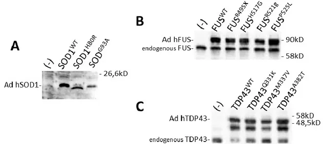
A limit of transient or stable transfection experiments both in human and mouse cells is the low endogenous gene expression level due at the low efficiency of conventional transfection methods. In recent studies it was tried to solve this problem by infecting different cellular models with replication-deficient recombinant adenoviruses, encoding for wild type SOD1 or with pathological mutations, to obtain similar expression levels both exogenous and endogenous SOD1 proteins. In an in vitro cell culture system, it has been revealed that infection of mouse NSC-34 motor neuron-like cells with adenovirus encoding for mutant SOD1-G93A gene increased cellular oxidative stress, mitochondrial dysfunction, cytochrome c release and motor neuron cell death 104.
The animal models if compared to cellular models allow to more closely mimic the human disease and to follow the trend of disease in order to develop effective therapies. Up to date, several transgenic mouse and rat strains were created by the introduction of the sequence coding for human SOD1 under the control of a promoter that enables ubiquitous expression of the transgene. Unlike SOD1 knockouts, transgenic mice and rats with human fALS–SOD1 added to their own enzyme exhibit the ALS-like clinical features: hind limb weakness and muscle tremor followed by more severe manifestations such as progressive motor paralysis, inability to walk, drink and eat, and death within some week. Differences in the age of disease onset among these strains seem to depend primarily on the transgene copy number, mice with high transgene copy numbers are affected by early onset, while mice carrying small transgene copy numbers are affected by late-onset, slowly progressive disease 105,106. Moreover, several biochemical alterations observed in patients are, in most cases, preserved in these models such as the appearance of oxidative-stress markers, alterations of mitochondria in motor neurons and muscle and the activation of phosphorylation cascades 107. Several transgenic lines have been developed, carrying mutations at different positions in SOD1. There are, however, some differences between the various mutant SOD1 mouse strains depending especially on the mutation and copy number of the transgene 37. For example, the progression of the disease is much more rapid in transgenic mice for the G86R mutation of the mouse SOD1 (three days) or for the human G85R mutation (7–14 days) than in mice that are transgenic for human SOD1-G93A (60-110 days) 106.
Transgenic mice carrying 20 copies of SOD1 with the G93A mutation are the most extensively studied. The progression of symptoms in these mice is preceded and paralleled by a sequence of structural and functional alterations of motor neurons. As for other diseases, the great advantage
Sonia Esposito Physiopathology of ALS: from oxidative stress to RNA metabolism. Pag. 16 PhD Thesis in Biomolecular and Biotechnological Science
University of Sassari
of animal models that mimics human pathology is the possibility to follow the stepwise progression of the disease for the design of potential therapeutic strategies. However, the expression levels of mutant SOD1, in heterozygous ALS patients, needed to exert its toxic effects is much lower than for transgenic rodents. In fact, in these models is necessary an increase about 10-30 fold of mutant protein level to induce the pathological phenotype. This is due to differences between mice and man and it should not be overlooked when using mice models 5.
1.6.2 Cellular and animal models for FUS.
To date, the role of FUS in ALS pathogenesis has not been established. It has been suggested that the toxicity of FUS could be the result of a gain-of-function for this protein, instead if mutated is possible a loss of function probably through its sequestration in stress granules. The generation of cellular and animal models of FUS could be provide several key insights into understanding the molecular mechanisms that might contribute to ALS pathogenesis.
Due to ALS-linked mutations, FUS becomes redistributed to the cytoplasm, a consistent observation well documented in human FUS-related ALS patients 70,71. These observations suggest that the cytoplasmic localization of mutant FUS might be a pre-requisite for causing toxicity. In several cell culture experiments have been analyzed the effects on subcellular localization of FUS carrying different C-terminal NLS mutations. Strong cytoplasmic localization was seen in transfected murine N2a cells when a consensus nucleotide of the NLS was mutated (the strongest effect corresponded to human FUS amino acids K510, R522 and P525). Mutations in the NLS that are not part of the consensus sequence gave weaker cytoplasmic localization 108. Studies on stable HEK-293 cell lines that express GFP-tagged FUS constructs, including wild type FUS or mutants, revealed a mutant-specific response to cellular stress that leads to the incorporation of cytoplasmic fALS-linked FUS protein into stress granules. The same results have been confirmed injecting mRNAs encoding GFP-FUS (WT or mutants) in zebrafish eggs at the 1–2 cell stage 109. Several groups have generated a yeast model of FUS-related neurodegeneration by expressing WT and disease associated FUS. Yeast cells expressing the C-terminal fragment with the zinc finger and GRR domain of FUS with ALS-associated mutations causes cytotoxicity under certain conditions 110. Another study has demonstrated that FUS localized to the cytoplasm and nucleus when expressed starting from a weak promoter, suggesting that the FUS expression level is an important determinant of localization and aggregation of FUS in yeast cells. It is possible that the non-canonical NLS of FUS might not be functional in yeast cells or that predominant nuclear localization of FUS requires additional factors such as post translational modifications 110,111.
Sonia Esposito Physiopathology of ALS: from oxidative stress to RNA metabolism. Pag. 17 PhD Thesis in Biomolecular and Biotechnological Science
University of Sassari
Drosophila models of FUS related neurodegeneration were recently generated that recapitulate several features of the human disease 112,113. In one study, ectopic expression of FUS with ALS linked mutations in the fly eyes caused moderate to severe external eye degeneration when compared to ectopic expression of FUS WT. Interestingly, targeted expression of FUS in the fly brain and motor neurons led to the pupal lethality and larval locomotor defects. Conditional expression of mutant FUS in neurons drastically reduced the life span and adult climbing abilities as compared to FUS WT expressing animals 114. Recently, it has been demonstrated that knocking down endogenous FUS in zebrafish leads to defects such as motor neuron degeneration, hyperbranched axons from motor neurons and behavioural phenotypes such as a defect in touch-evoked escape response, suggesting that loss of FUS function can lead to neurodegeneration 115. These observations are further supported by another study where knocking out endogenous cabeza (the fly homologue of human FUS) can cause motoneurons degeneration in Drosophila 113. Interestingly, the motor phenotypes caused by zebrafish FUS knockdown can be rescued by expressing human FUS WT, suggesting that human and zebrafish FUS are functionally related proteins. Furthermore, the expression of the ALS-linked mutation R521H also leads to motor neuron phenotypes, indicating that FUS with disease-causing mutations acquires a toxic gain of function. Two independent groups have generated FUS knock out (KO) mouse models long before the identification of ALS-linked mutations in FUS. The FUS KO mice were smaller in size as compared to non-transgenic animals, they have shown a chromosomal abnormalities indicating that FUS plays an important role in the stability of the genome and a complete male sterility and reduced fertility in females indicating that FUS is required for normal development and fertility. Many of them died within 16 h of birth, suggesting that normal levels of FUS are required for viability of neonatal animals 116,117. Studies on rat models have demonstrated that overexpression of FUS WT was enough to cause neurodegeneration at an advanced age with a moderate but significant loss of neurons in the brain. However, expression of mutant FUS led to severe neurodegeneration at an early age as compared to FUS WT expressing rats 118. Moreover, only rats espressing mutant FUS have shown paralysis and accumulation of ubiquitinated proteins in the cortex and spinal cord. Interestingly, the ubiquitinated inclusions were only present in the FUS expressing cells, but there was no colocalization between FUS and ubiquitinated inclusions. The ubiquitinated inclusions have showed reactivity with a mitochondrial marker protein in this rat models of mutant FUS, suggesting that damaged mitochondria might be ubiquitinated and subsequently degraded 118.
Sonia Esposito Physiopathology of ALS: from oxidative stress to RNA metabolism. Pag. 18 PhD Thesis in Biomolecular and Biotechnological Science
University of Sassari
1.6.3 Cellular and animal models for TDP43.
More and more groups are engaging in research that examines the role of TDP43 in the pathogenesis of ALS. An increasing number of disease cellular or animal models have been set up to provide insights into the molecular mechanism of ALS with TDP43 proteinopathy. Nonetheless, the exact role of TDP43 in the pathogenesis of FLTD/ALS still has not been fully unravelled and probably both a gain of toxic properties and a loss of function via its sequestration in aggregates could be possible.
In several cell culture experiments, the expression of TDP43 proteins carrying mutations that disrupt its NLS led to localization primarily within the cytoplasm 119. A study on rat primary cortical neurons, through an automated microscopy system for long-term visualization, has shown that overexpression of wild-type TDP43 led to increased cytoplasmic localization of TD-43 and cell death independently of mutation, pathogenic TDP43 mutations increased the proportion of cytoplasmic TDP43 120. As reported in different studies the overexpression of WT or mutant hTDP43 in transgenic mice induced motor dysfunction, such as gait disorders. In particular, it has been shown that moderate expression of hTDP43 (about 2.5 fold) can promote cytoplasmic TDP43 expression, TDP43 truncation, TDP43 phosphorylation and TDP43 aggregation, which mimics the TDP43 proteinopathy in CNS of ALS. Besides TDP43 proteinopathy, abnormal mitochondrial aggregation is observed in hTDP-43-overexpressing transgenic mice, indicating the involvement of TDP43 in mitochondrial trafficking 121,122. The overexpression of WT-hTDP43 or mutant hTDP43 in rat models increases cytoplasmic TDP43 expression. However, only rats expressing mutant hTDP43 (M337V-hTDP43) exhibited significant motor dysfunction, which is different from other mouse models. M337V-hTDP43 expression in rat also reproduces the biochemical features of ALS including hyper phosphorylation of TDP43, formation of TDP43 inclusion and expression of truncated TDP43. Most importantly, the ubiquitous expression of M337V-hTDP43 in rat is seen to cause more severe neurodegeneration in the motor system than in the CNS 123. Similar results were also obtained non-human primate models 124 and on non-mammalian transgenic animal models as C. elegans 125, Zebrafish 126 and Drosophila 127.
Fragments (20–25 kDa) containing the carboxy-proximal portion of TDP43 accumulate in detergent (sarkosyl)-insoluble fractions derived from patient CNS tissues. The truncated TDP43 (~25 KDa) has been involved in disease progression by gain of toxic function 98. When expressed in cells, the 25 kDa CTFs recapitulate some of the pathological features such as increased cytoplasmic accumulation, insolubility, hyperphosphorylation, polyubiquitination and cytotoxicity 99,128.
Sonia Esposito Physiopathology of ALS: from oxidative stress to RNA metabolism. Pag. 19 PhD Thesis in Biomolecular and Biotechnological Science
University of Sassari
without cytoplasmic TDP43 inclusions and trigger the processing of endogenous TDP43, which suggest that TDP43 inclusions are independent of cognitive dysfunction 129,130. Caspase-3, which is activated during apoptosis, has been proposed to be the main protease generating the 25 kDa CTF in cells 128,131. The role of TDP43 ubiquitination in disease pathogenesis remains unknown, although it is likely to be a late event, since in patients the pre-inclusions and most TDP43 inclusions are either weakly or not at all ubiquitinated 97,100. Nevertheless, extensive ubiquitination of the pathologic CTFs in cells 132 suggests that cellular degradation machineries such as the ubiquitin-proteasome system (UPS) and/or autophagy 133 may be involved in removing TDP43 aggregates.
To investigate the linkage between the loss-of-function of TDP43 and pathological features ALS, TDP43 knockout models have been created. TDP43 is important for early embryonic development, in fact TDP43 knockout in mice would result in peri-implantation lethality 84. Targeted depletion of TDP43 in motor neurons could trigger mice with age-dependent progressive motor neuron degeneration, muscle atrophy and motor dysfunction, which are reminiscent of human ALS patients. These findings indicated that TDP43 might be important for the long-term maintenance of motor neuron 134135. Therefore, the mouse model with depletion of TDP43 expression in motor neuron not only has a longer lifespan compared to the whole body knockout, but also could be a valuable model for studying the role of TDP43 in motor neurons 136.
Further studies on the molecular mechanism of ALS with TDP43 proteinopathy are needed.
1.7 Physiopathological mechanisms of ALS.
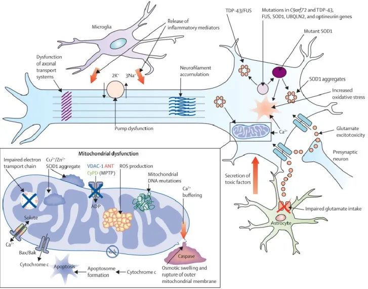
Current hypotheses on biology underlying both sporadic and familiar ALS forms in humans outline a complex model in which non-competing mechanisms including not only gene mutations and environmental factors but also oxidative stress, protein aggregation, excitotoxicity, changes in the ubiquitin-proteasome system and autophagy pathways, defects in RNA metabolism, abnormal axonal transport, as well as neuroinflammatory processes involving astrocytes and microglia, are likely to converge in various unfortunate patterns to mediate selective motor neuron degeneration (Fig. 8).
Sonia Esposito Physiopathology of ALS: from oxidative stress to RNA metabolism. Pag. 20 PhD Thesis in Biomolecular and Biotechnological Science
University of Sassari
Figure 8: Proposed mechanisms underlying neurodegeneration in ALS. Many of these pathways are mechanisms of cell death common to a range of neurological disorders, although in the case of amyotrophic lateral sclerosis (ALS), have been derived from studies undertaken predominantly using the SOD1 mouse model. Pathophysiological mechanisms involving more recent genetic discoveries, particularly the C9orf72 hexanucleotide repeat expansion, have yet to be elucidated. Neurodegeneration in ALS might reflect combinations of glutamate excitoxicity, generation of free radicals, mutant SOD1 enzymes, along with mitochondrial dysfunction and disruption of axonal transport processes through accumulation of neurofilament intracellular aggregates. Mutations in several ALS-related genes are associated with the formation of intracellular aggregates, which appear harmful to neurons. Activation of microglia results in secretion of proinflammatory cytokines resulting in further toxicity. Failure of cytoplasmic mitochondria induces increased susceptibility to glutamate-mediated excitotoxicity, perturbations in motor neuronal energy production, and apoptosis. Mitochondrial dysfunction is associated with increased production of reactive oxygen species (ROS). Cytoplasmic aggregates of SOD1 might directly inhibit conductance of VDAC1, thereby reducing the supply of ADP to mitochondria for ATP generation. Δ ψ =mitochondrial membrane potential 39.
1.7.1 Oxidative Stress.
Oxidative stress arises when the levels of ROS/RNS exceed the quantities required for normal redox signalling. There has been particular interest in the role of oxidative stress in ALS but the exact role of ROS/RNS in disease processes is uncertain 137. ROS are able to cause permanent oxidative damage to several cellular components such as proteins, DNA, lipids, and cell membranes 138. They has been detected in the spinal cord and cerebrospinal fluid (CSF) of sALS
Sonia Esposito Physiopathology of ALS: from oxidative stress to RNA metabolism. Pag. 21 PhD Thesis in Biomolecular and Biotechnological Science
University of Sassari
patients 139. SOD1 and its antioxidant properties have been studied extensively from the perspective of redox regulation in ALS 140. The first hypothesis proposes that mutations in SOD1 alter its ability to act as an antioxidant but further evidences showed that disease onset is not associated with its enzymatic activity 141. However, mutant SOD1 could accept peroxynitrite or hydrogen peroxide as a substrate, and thereby catalyse the nitration of tyrosine residues of SOD1 and hydroxyradicals 53,142, in fact high levels of nitrotyrosine and nitrated proteins have been found in the CSF of both sALS and fALS patients 139.The second hypothesis suggests that mutant SOD1 fails to bind zinc correctly, allowing the rapid reduction of the SOD1-bound copper which catalyses the formation of superoxide anion rather than dismutation 143.
Mutant TDP-43 may be itself an inducer of oxidative stress, as suggested by studies in neuronal cells in vitro in which this protein was shown to down regulate heme oxygenase-1 144 and in yeast, in which TDP-43 expression increased markers of oxidative stress and induced cell death 145.
1.7.2 Glutamate Excitotoxicity.
Another component of neuronal degeneration in many neurodegenerative disorders is excessive glutamate-induced stimulation of postsynaptic glutamate receptors resulting in increased calcium intake and neuronal injury 146. Glutamate uptake from most synapses in the CNS is controlled by excitatory amino acid transporter 2 (EAAC2; also known as SLC1A2 or GLT1) 147. Increased levels of intracellular glutamate and decreased uptake of glutamate from the synapse have been observed in ALS patients 148. Indeed, in most cases of sALS, and in transgenic rodent ALS models, there is a profound reduction in the expression and activity of EAAT2 in the cortex and spinal cord 149.
Excitotoxicity has provided one of the few examples of a mechanistic link between mutant SOD1 mediated the sporadic form of the disease. The presence of mutant SOD1 increases the sensitivity of motor neurons to glutamate toxicity 150, causes alteration in AMPA receptor subunit expression
151
, and causes reduced expression of the major glutamate reuptake transporter EAAT2 107. Whether as a primary or a propagating process, it appears that glutamate toxicity plays a contributory role to the injury of motor neurons in ALS.
1.7.3 Mitochondrial Dysfunction.
Numerous studies have focused on the role of the mitochondrion in the pathogenesis of neurodegenerative diseases like ALS. Indeed, mitochondrial alterations are observed in the spinal cord cells of SALS patients and role of mitochondria has been well studied in relation to ALS pathogenesis 152. It has been demonstrated presence of abnormal and degenerate mitochondria
Sonia Esposito Physiopathology of ALS: from oxidative stress to RNA metabolism. Pag. 22 PhD Thesis in Biomolecular and Biotechnological Science
University of Sassari
either in ALS patients 153 as well as in cellular and animal models of disease 154,155, although how is unclear relationship between non-functioning mitochondria and ALS onset.
In particular, expression of mutant SOD1 in motor neuron cell line (NSC34) results in the development of swollen mitochondria, impaired activity of complexes II and IV of the mitochondrial respiratory chain, impaired cellular bioenergetic status, and alteration in the mitochondrial proteome 156. Similarly, G93A-SOD1 mice show reduced respiratory chain activity and reduced ATP synthesis 157. Furthermore, recent studies have shown that overexpression of TDP-43 causes mitochondrial dysfunction and induces mitophagy in cell culture 158.The presence of ROS and impairment of the mitochondrial respiratory chain have also been observed in TDP-43 models 144.
Significantly, any pathological variations in regulation of the electron transport chain would result in more oxidative stress 159 causing further cellular redox dysregulation, leading to a potential vicious cycle of damage and degeneration.
1.7.4 Impaired Axonal Transport.
Axonal transport is a key mechanism required for cellular viability in neuronal cells. The transport of molecules and organelles is a fundamental cellular process that is particularly important for the development, function and survival of neurons. Several findings indicate that dysfunction of axonal transport might contribute to the demise of motor neurons in ALS 155. Firstly, neurofilaments (NF) accumulation in motor neurons is another histopathological hallmark of ALS
160
and transgenic mice that overexpress NF subunits in motor neurons develop a motor neuron disease with impaired axonal flow, as axonal defects cause delay in transportation of components required for the maintenance of axon 155. Second, both slow and fast anterograde transport are slowed in transgenic G93A-SOD1 and G37R ALS mice prior to disease onset and these deficits are exacerbated. as the disease progresses 161. Tird, retrograde transport is also disrupted in ALS mice
162
. A recent study demonstrated that oxidised wild-type SOD1 immunopurified from sALS patient tissues inhibited kinesin-based axonal transport in a manner similar to mutant SOD1 in fALS providing evidence for common pathogenic mechanisms in both sALS and fALS 163. It has been showed that NF aggregations are associated with SOD1 and nitric oxide synthase activities leading to nitrotyrosine formation on NF 164. Nitrotyrosine can inhibit phosphorylation of NF subunits and may alter axonal transport and initiate motor neuron death 164. Taken together, these findings suggest a relation between redox regulation and axonal transport dysfunctions in ALS.
Sonia Esposito Physiopathology of ALS: from oxidative stress to RNA metabolism. Pag. 23 PhD Thesis in Biomolecular and Biotechnological Science
University of Sassari
1.7.5 Inflammatory cascades and the role of non-neuronal cells.
Recently there has been increasing interest in the possibility that non-neuronal cells, including activated microglia and astrocytes, may contribute to the pathogenesis or propagation of the disease process in ALS. Several studies on mouse models have indicated that expression of mutant SOD1 in neurons alone is insufficient to cause motor neuron degeneration and that participation of non-neuronal cells may be required 15.
Astrocytes have many functions relevant to motor neuron physiology. First, they express the most important glutamate transporter EAAT2/GLT-1, thus contributing to the clearance of this neurotransmitter; deficiency of astroglial EAAT2/GLT-1 causes severe motor neuron loss 165 and alteration of this transporter has been repeatedly invoked as a cause contributing to ALS 9. Second, astrocytes are the major source of both trophic 166 and toxic factors 15 for motor neurons. Several cytokines have been proposed to play a role in ALS as reinforcing signals from glia cells, including interleukin-6 (IL6), tumour necrosis factor α (TNFα) and transforming growth factor β1 (TGFβ1) that were found increased in cerebrospinal fluid, plasma and epidermis from ALS patients, although with sometimes conflicting results 167. In addition, the production of nitric oxide and the activation of cyclooxygenase type 2 (COX2) aggravate the toxic effects of mutant SOD1 in several experimental models for ALS. The production of all those proinflammatory mediators may be secondary to the induction of the transcription factor NF-κB, which is activated in the presence of reactive oxygen species (ROS) and by many other different signalling molecules associated with ALS onset and progression 168. NF-κB activation has been observed in astrocytes from ALS patients and in human cells expressing mutant SOD1 169. NF-κB also regulates the expression of COX2 that may cause an increase in the synthesis of prostaglandins, which trigger astrocytic glutamate release and induce free radical formation, thus contributing to both excitotoxicity and oxidative damage. Indeed, treatment with COX2 inhibitors markedly protects motor neurones and significantly prolongs survival of ALS mice 170. Surprising NF-κB downregulation in astrocytes, via expression of the dominant negative IκB-alpha-AA, fails to influence onset, severity, or progression of disease in a mutant SOD1-based ALS mice model 171.
Recently it has been shown that also microglia play a critical role as resident immunocompetent and phagocytic cells within the CNS. Activation is associated with transformation to phagocytic cells capable of releasing potentially cytotoxic molecules including reactive oxygen species, nitric oxide, proteases, and proinflammatory cytokines such as interleukin-1B, tumour necrosis factor a (TNFα), and interleukin 6 (IL-6) 172. Given this, there is little doubt that activated microglia can inflict significant damage on neurons, but their role is complex and they are capable of stimulating
Sonia Esposito Physiopathology of ALS: from oxidative stress to RNA metabolism. Pag. 24 PhD Thesis in Biomolecular and Biotechnological Science
University of Sassari
neuroprotective as well as neurotoxic effects. Proliferation of activated microglia is a prominent histological feature in the spinal ventral horn both in mutant SOD1 transgenic mice and in human ALS 173. In the mice, microglial activation is present before the onset of significant motor neuron loss or motor weakness. Various inflammatory cytokines or enzymes are upregulated in the spinal cord of ALS patients (IL-6, IL-1β, cyclo-oxygenase 2 (COX2), and prostaglandin E2 (PGE2)) or, in the spinal cord of mutant SOD1 mice (IL-1β, TNFα, COX2, PGE2) 174. Microglia appear to mediate the toxicity to neurons in patients with ALS by releasing factors that enhance glutamate toxicity 175. In a recent study D’Ambrosi and colleagues have investigated about how mutant SOD1 affects P2 receptor-mediated proinflammatory microglial activation, considering that extracellular ATP is one of the most widespread microglia alarm signal endogenous to the CNS, and that ATP signaling evokes many proinflammatory functions of microglia. They observed up-regulation of P2X(4), P2X(7), and P2Y(6) receptors and down-regulation of ATP-hydrolysing activities in mutant SOD1 microglia. This potentiation of the purinergic machinery reflected into enhanced sensitivity mainly to 2'-3'-O-(benzoyl-benzoyl) ATP, a P2X(7) receptor preferential agonist, and translated into deeper morphological changes, enhancement of TNF-α and cyclooxygenase-2 content, and finally into toxic effects exerted on neuronal cell lines by microglia expressing mutant SOD1 176. The purinergic activation of microglia may thus constitute a new route involved in the progression of ALS to be exploited to potentially halt the disease.
1.7.6 Protein aggregation.
Protein aggregates are often found in spinal motor neurons of all types of ALS patients. These inclusions contain many different proteins, some of which may have an intrinsic tendency to aggregate following mutation (such as SOD1, TDP-43 and FUS) while others may be simply entrapped in aggregates 177. Now, there is hardly a general consensus on the assumption that the toxic function gained by SOD1 mutants is related by their tendency to aggregate and to mislocalize, as other ALS mutant proteins. This hypothesis is supported by the presence of intracellular cytoplasmic (but also mitochondrial) inclusions enriched in SOD1 protein in cellular models and in spinal cord motor neurons in animals as well as in motoneurons of ALS patients 177,178
. Over the years, several experimental evidences have been proposed to explain how aggregation of mutant SOD1 could be a major contributor to induce cellular toxicity in ALS patients, from the ability to sequester proteins required for normal motor neuron function 179, to the capacity to reduce proteasome activity needed for normal protein turnover and to inhibit
Sonia Esposito Physiopathology of ALS: from oxidative stress to RNA metabolism. Pag. 25 PhD Thesis in Biomolecular and Biotechnological Science
University of Sassari
specific organelles function (as the mitochondria) by aggregation out or within the same organelles 156,180.
The fact that protein aggregation is a common theme in ALS and that the cellular degeneration in ALS depends upon the sensitiveness of motor neurons to protein aggregation is suggested also by the observation that, likewise to SOD1, also TDP43, FUS aggregate in tissues from ALS patients and models 181. Anatomo-pathological observations of ALS-affected tissues showed that these two proteins abnormally aggregate as cytoplasmic inclusions (positive for ubiquitin, but negative for SOD1) and that ALS-related mutations seem to enhance the rate of aggregation 111,182. Recently, it has been identified that aggregation of FUS and TDP43 relies on prion-like domains and that aggregated FUS or TDP43 can sequester native protein 183.
The formation of such intracellular aggregates may depend on the accumulation of misfolded proteins generated either as a direct consequence of mutation, or as a consequence of oxidative stress. In both instances, impairment of the ubiquitin-proteasome response (UPR) seems to play a major role. Not only ubiquitin-immunoreactive inclusions are the most frequently reported inclusions in all forms of ALS, but aggregates may be also reactive to p62, a protein which has a part in the formation of the sequestosome and in autophagy 184.
1.7.7 Misregulation in RNA processing.
Two of the lately identified genetic factors in ALS, TDP43 and FUS, encode for proteins with an established role in RNA metabolism. Thus, RNA dismetabolism seems to be an important potential physiopathological mechanism in motoneurons degeneration. Since RNA metabolism involves several processes, such as pre-mRNA splicing, mRNA transport, translational regulation, or mRNA decay, the precise RNA pathway that is affected in ALS disease is still unclear, and it is not completely clear whether defects in RNA processing have a causative, direct role in the pathogenesis 3.
Under stress, a cell’s top priority is to conserve energy and divert cellular resources toward survival and eventual recovery. In eukaryotic cells, a powerful way to conserve resources is rapidly assembly of non-translating mRNAs and their associated RNA-binding proteins into aggregate-like structures called RNP granules, including stress granules (SGs) 185. In normal cells, TDP43 and FUS are nuclear proteins that can quickly be shuttled to the cytoplasm upon stress induction where they can rapidly associated with SGs 73,186. Once the instigating stress resolves, SGs dissolve and TDP43 and FUS return to the nucleus. Thus, the nuclear-cytoplasmic shuttling of TDP43 and FUS, as well as their association with cytoplasmic SGs, are physiological and reversible processes.