UNIVERSITÀ DEGLI STUDI DI SASSARI
Biomedical Science department
PhD Course in Life Sciences and Biotechnologies
Eradication of persistent HBV genomes using
SaCas9/3xgRNAs based gene therapy
Thesis coordinator:
Prof. Caterina Serra
Department of Biomedical Science
University of Sassari
Thesis advisor:
Dr. Kamel Khalili
Director of Center for Neurovirology
Lewis Katz School of Medicine, Temple University
Thesis co-advisor: Ph.D student
Prof. Antonina Dolei
Gabriele Ibba
Department of Biomedical Science
University of Sassari
Index
1. Introduction
1.1. Hepadnaviridae
1.2. HBV: structure and characterization
1.3. The HBV genome
1.4. The viral replication cycle
1.5. Immunopathogenesis of chronic Hepatitis B Virus
1.6. Acute and chronic HBV infection diagnosis
1.7. Therapy of chronic HBV infection
1.8. Hepatitis B Virus Vaccines
1.9. Animal models for Hepatitis B research
1.10 CRISPER/CAS9 system
3. Materials and Methods
4. Results
4.1. CRISPR/CAS9 design: in silico definition of the twelve most fitting gRNAs
4.2 Cloning of the gRNA expressing cassettes targeting HBV genome into pX601-SaCas9-AAV vector and verification final pX601-HBV3xgRNAs-shRNA construct.
4.3 Biological validation of pX601-HBV3xgRNAs-shRNA construct on chronically HBV infected cells (HepG2.2.15)
4.4 Off-target genes indel mutation analysis
4.5 Expression of SaCas9/gRNA suppresses HBV infection of human cell line
4.5.1 Analysis of SaCas9/gRNA mediated HBV genome cleavage efficiency.
4.5.2 Quantification of intracellular viral DNA levels.
4.5.3 Quantification of viral RNA expression after CRISPR/Cas9 treatment.
5. Discussion
6. Acknowledgment
7. References
Introduction
1.1. Hepadnaviridae
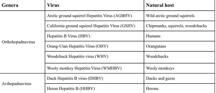
The human Hepatitis B virus (HBV) is a member of the Hepadnaviridae family, genus Orthohepadnavirus; its genome is a single molecule of linear, partially double-stranded DNA. The full-length virus genome is sized 3.2 kb. The Hepadnaviridae family has a recurrent structure and may conduct to persistent virus infections. In Table 1 are reported the members of the Hepadnaviridae family and their natural hosts. (6; 83).
Genera Virus Natural host
Orthohepadnavirus
Arctic ground squirrel Hepatitis Virus (AGBHV) Wild arctic ground squirrels California ground squirrel Hepatitis Virus (GSHV) Chipmunks, squirrels, woodchucks
Hepatitis B Virus (HBV) Humans
Orang-Utan Hepatitis Virus (OHV) Orangutans
Woodchuck Hepatitis virus (WHV) Woodchucks
Wooly monkey Hepatitis Virus (WMHBV) Wooly monkeys
Avihepadnavirus
Duck Hepatitis B virus (DHBV) Ducks and geese
Heron Hepatitis B (HHBV) Herons
1.2. HBV: structure and characterization
The HBV virion also referred as "Dane particle" (20), is the smallest enveloped animal virus, with a diameter of 42 nm (Fig. 1). The envelope is the outer part of the virion, which consists of a phospholipid bilayer and three proteins in a non-glycosylated and glycosylated form: the large (L), the middle (M) and the small (S) proteins. Under the envelope, there is the capsid, composed by the HBV core protein (HBcAg), which contains the viral DNA and the reverse transcriptase (RNA-dependent DNA-polymerase). The core protein is highly conserved between all HBV genotypes; depending on which genotype, it consists of 183 or 185 amino acids (11). The fundamental unit of HBV is made up by an oligomerization between several HBV core protein dimers. In relation to the number of dimers which form the capsid, there are two different types of viral particles: the particle with an icosahedral symmetry T = 3, with a diameter of 30 nm and composed of 90 dimers, and the particle with the icosahedral symmetry T=4, with a diameter of 34 nm and 120 dimers.
Virus particles are present in the peripheral blood of HBV patients up to 10 11/1014particles/ml (79).
Empty spherical or filamentous particles, made of surplus envelope proteins (HBsAg), are present in even higher quantities (Fig. 1) and play a part in the relationship between host and virus.
Figure 1: Schematic representation of different forms of infectious and non-infectious
hepatitis B virus particles. On the top: infectious Dane particle with PreS1, PreS2 and S proteins. The HBV genome is contained in the capsid, composed of the HBc protein. On the bottom: Non-infectious filamentous and spherical particles.
1.3. The HBV genome
The hepatitis B virus genome is a partially double-stranded circular DNA, of approximately 3,200 nucleotides in length. It is one of the animal smallest replication-competent virus genomes. The virus has been classified according to 8 genotypes (from A to J), each with a distinct geographic distribution (52). The viral genome contains a full-length negative-sense strand, that is complementary to the pre-genomic viral RNA, and a shorter positive strand, approximately 2.8 kb long. The viral genome harbors four overlapping open reading frames encoding: S (surface), C (core), P (polymerase), and X genes (Fig. 2). The HBV polymerase (P) protein is covalently joined to the 5` end of the minus strand (27). The P-polymerase and core open reading frames are in the same orientation, but the S translational reading frame overlaps the pol-reading frame. The HBV genome evolves quickly over time, because of the high error rate of the reverse transcriptase. It is estimated a nucleotide substitution rate of 1.4-3.2 × 10⁻⁵/site per year (14).
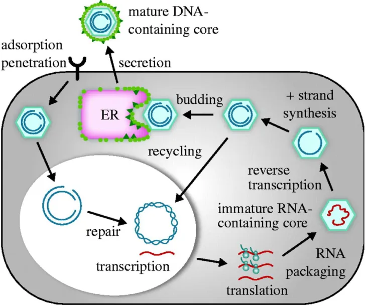
1.4. The viral replication cycle
The viral replication takes place mainly in hepatocytes. Other types of cells have been found to allow HBV replication, even if to lesser extents (70; 72). Viral entry of Hepadnaviridae into hepatocytes depends on the N-terminus of the large (L) surface antigen (30; 49; 96). Recently, hepatitis B virus attachment and entry into hepatocytes has been visualized (64; 82). Simultaneously, another group of researchers identified the sodium taurocholate co-transporting polypeptide (NTCP) as a hepatocyte receptor, which allows the entry of the virus (95). After the viral entry, the nucleocapsid, containing the relaxed circular partially double-stranded DNA (rcDNA), with its covalently linked polymerase, is released into the cytoplasm. The nucleocapsid moves to the cell’s nucleus, along microtubules. Accumulation of the nucleocapsids in proximity to the nuclear membrane facilitates the interactions with adaptor proteins of the nuclear pore complex. After the release of rcDNA into the nucleoplasm, the positive strand is “repaired” by the cellular DNA polymerase kappa (76) and converted into covalently closed circular (ccc) DNA. In this form, the cccDNA may serve as a viral persistence reservoir, and is refractory to current antiviral therapies. This plasmid-like DNA serves as a template for transcription of viral RNAs (9). The transcripts from the cccDNA consist of two species of 3.5 kb long molecules (pre-genomic and pre-core RNAs) and 2.4 kb, 2.1 kb and 0.7 kb long subgenomic RNAs (50). After transportation into the cytoplasm, the viral RNAs are processed by ribosomes into the core protein, the surface S, M and L proteins, and the X-protein. The surface proteins differ in their N-terminal sequences, that are longer in the case of L and M proteins, and have a common S domain. M has an additional preS2 domain, and L has additional preS2 and preS1 domains. These proteins assemble the viral envelope. Further, surface proteins can be secreted as non-infectious filamentous and spherical particles that do not contain a functional nucleocapsid. The pre-genomic (pg) RNA is a template for both core and viral DNA-polymerase. The viral DNA-polymerase binds the pgRNA and induces the
packaging of pgRNA into the core protein, which forms the basis of the capsid, and also plays an active role in binding and packaging of pgRNA (10). After transcription of pgRNA into the minus strand DNA, the capsid interacts with L-proteins and finally acquires the envelopment of the core with the surface proteins.
1.5. Immunopathogenesis of chronic Hepatitis B Virus
The hepatitis B virus is spread by parenteral and mucosal contacts with contaminated blood or other HBV-infected body fluids (i.e. vaginal fluid and semen). The incubation time can vary from about 30 to 180 days. The 65% of infections in young persons and adults are asymptomatic, while in 35% of infected patients, the infection results in liver inflammation. Symptoms of the acute hepatitis are tiredness, dark urine, icterus and an enlarged liver. The symptomatic acute infection continues for two to three weeks; after this period, a convalescence phase occurs. If the expression of HBsAs persists for more than six months, the acute disease will evolve into a chronic infection (5/10% of all infected adults). The risk of contracting a persistent infection is patient age-related. Infection in newborns turns in 90% of cases to a chronic disease (up to 98% of newborns infected peripartum). In young children, chronic manifestation occurs in 50% of infections and 60% of the chronically infected persons live normally, without symptoms, with a chronic persistent hepatitis (CPH). The remaining chronic infections result in chronic active hepatitis (CAH). CAH can spontaneously evolve to CPH (66). Once into the human body, the virus reaches and infects the hepatocytes in the liver. Activation of the innate immune response against HBV is weak. Nevertheless, liver pathology and HBV clearance are mediated mostly by adaptive immune response (14). Before virus replication in hepatocytes, most probably the Kupffer cells recognize HBV patterns and activate the NF-ĸB pathway, which induces the production of proinflammatory cytokines (37). Further, interleukin-6 seems to be responsible for HBV suppression.
In the early steps after hepatocytes infection, HBV activates mitogen-activated protein kinase (MAPK) and c-Jun N-terminal kinase (JNK), which downregulate the expression of hepatocyte nuclear factor 4α (HNF4α) and HNF1α, the key transcription factors regulating HBV gene expression (77). Moreover, the lack of interferon response in the first steps of the infection is responsible for HBV expansion in the liver. It has been demonstrated in transgenic mice that
interferon (IFN)-α and β reduce the expression of HBV genes in the early infection phase (94). As stated previously, disease pathogenesis and viral clearance are mediated mostly by the adaptive immune response. The HBV-specific CD8 T cell response is especially vigorous and multispecific in patients with self-limited infection, but mostly weakly detectable in patients who develop chronic hepatitis B virus infection (14). Nonetheless, a study suggests that induction of an effective CD8 T cell-specific immune response to HBV is dependent on early CD4 T cell priming, which might be controlled by the load of viral inoculum (3). In acute hepatitis infection, HBV is detectable already in the incubation period. Interferons upregulate the expression of major histocompatibility complex (MHC) class I antigens on the cell surface, which allows the elimination of infected cells, while the MHC class II immune response is directed against the capsid proteins (HBcAg and HBeAg). The immune response to HBsAg is lower than that against capsid proteins. Neutralizing antibodies (anti-HBs) block the spread of HBV and infection of hepatocytes. These antibodies to HBsAg protect also from reinfection with the virus (66).
The chronic HBV infection is due mainly to the lack of HBV-specific adaptive T-cell immune response and HBsAg-specific humoral immune response.
1.6. Acute and chronic HBV infection diagnosis
A series of blood tests are available for HBV diagnosis. These tests are based on the evaluation of several virological and serological markers (Table: 2), which are also essential to distinguish between acute and chronic viral infection (51).
Marker
HBV marker for:
HBsAg HBV infection (acute and chronic)
HBeAg High level HBV replication and infectivity (also marker for treatment response)
HBV DNA Level of HBV replication
(also primary marker for treatment response)
Anti-HBc (IgM) Acute HBV infection
(sometimes also in flare of chronic infection)
Anti-HBc (IgG) Recovered chronic HBV infection
Anti-HBs Recovered HBV infection and marker of HBV vaccination (The level reflects vaccine efficacy)
Anti-HBe Low-level HBV replication and infectivity (also marker for treatment response)
Anti-HBc IgG and (and no) Anti-HBs and HBsAg neg
Recovered HBV infection
Anti-HBc (IgG) and HBsAG (6 > Months) Chronic HBV infection
Anti-HBc (IgG and/or anti-HBs and HBV-DNA Latent or occult HBV infection
Table 2: HBV diagnostic markers
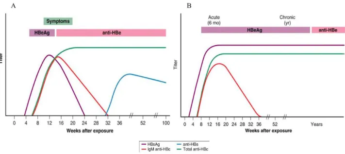
A series of the blood tests are available for the HBV diagnose. These tests are based on the evaluation of several virological and serological markers (Table: 2) which are also essential to distinguish between acute and chronic viral infection(51). The typical pattern of serological markers during the course of acute HBV infection is presented in figure 4A. The first serological marker of acute HBV infection is HBsAg, which is detectable six weeks after infection. From week eight onward, anti-HBc-IgM and HBeAg are slight expressed. IgG antibodies against HBcAg can be first
detected after the peak of viremia, and corresponds to the increase of transaminases. The first indicative parameters of the recovery from infection are the decrease of viral load and, short afterward, the reduction of HBsAg and HBeAg levels in peripheral blood (27).
The chronic HBV infection starts with a serological pattern similar to that of the acute disease (Fig. 4). However, chronically infected subjects do not loose HBV DNA, and rarely HBsAg and HBeAg. If HBsAg is detectable in the host for over six months, it is confirmed a diagnosis of chronic HBV infection. The viral DNA is a marker of active HBV, in both acute and chronic infections. Anti-HBc is an important parameter to define the infection rate of HBV in a community, but it is not used to produce vaccines, since the corresponding antibody is not neutralizing, and is detectable only after infection with HBV.
1.7. Therapy of chronic HBV infection
Chronically HBV-infected patients are at risk of developing liver cirrhosis and hepatocellular carcinoma (HCC), especially if the load of HBV DNA is high. HCC occurs predominantly in populations with a high prevalence of HBV. Currently, therapy is recommended for patients with evidence of ongoing chronic hepatitis B disease (i.e., elevated aminotransferase levels, positive HBV DNA findings, negative or positive HBeAg). Thirty percent of the chronic infections can be treated successfully with this medication. Because of that, therapy with peg-IFNα has become the first-line treatment (84).
Further, treatment with nucleoside analogs (NUCs) has been currently approved. In the past, the most commonly used nucleoside analog was lamivudine (3TC; 2’,3’-Dideoxy-3’- Thiacytidine). Lamivudine is an inhibitor of pyrophosphorolysis activity of the viral reverse transcriptase, and has been developed, and used, for the treatment of HIV infection. In contrast to retroviral infection, the HBV pre-genomic RNA is transcribed into the DNA before the release of the virus particle into the plasma. Since HBV particles half-life is about 1.5 days in the plasma, the decrease of the HBV load can be achieved very fast after medication. However, treatment with NUCs cannot eradicate the virus, due to the presence of viral cccDNA in the nucleus of hepatocytes. Further, only one/fifth of patients lose HBeAg one year after therapy. The same proportion of patients develops resistance to lamivudine, due to mutation in the tyrosine-methionine-aspartate-aspartate (YMDD) locus of the HBV polymerase gene, resulting in a viral rebound (91). The rise of resistance to lamivudine led to the development of new less resistance-susceptible NUCs, like entecavir and tenofovir (Tab. 3). Currently, the first choice therapy drugs approved globally for the HBV treatment are tenofovir diisopropyl, fumarate pegylated interferon, and entecavir. A final therapeutic option for patients with hepatocarcinoma and liver failure is the liver transplantation. Unfortunately, the treatment of HBV-infected patients after transplantation may be difficult, due to the parallel immunosuppressive
treatment drugs and the reinfection of the new organ, mediated by the infectious virus circulating in the blood (86).
Drug
Structure
Lamivudine Cytidine analogue
Adefovir Adenosine analogue
Entecavir Guanosine analogue
Telbivudine Thymidine analogue
Tenofovir Adenosine analogue
1.8. Hepatitis B virus vaccines
In the 1980s, spherical and filamentous HBsAg particles purified from plasma of HBsAg carriers have been used as the first vaccine against HBV (62). Second generation vaccines were based on recombinant, non-glycosylated small surface proteins (S), produced in yeast (63). These vaccines have been proven to be very immunogenic, and are still used in vaccination programs in more than 150 countries, worldwide. However, the seroprotection rate for this kind of vaccines changes in relation to the age at immunization. It was estimated that second generation vaccines do not give the immunity in the 4-8% of subjects treated in the first 30 years of life ;this percentage increases up to 10% in people treated thereafter (63; 2).
A new generation of vaccines was developed, which contain, not only the S protein, but also the middle (M) and the large (L) surface proteins (36). This third generation of vaccines is produced in mammalian Chinese hamster ovary (CHO) cells and can induce an immune response, which occurs earlier and is stronger than that induced by S alone (79). Even in many so-called “non-responders” to S protein, a protective immune response was induced (80).
1.9. Animal models for Hepatitis B research
Chimpanzees are the only permissive animals for hepatitis B virus infection. Conversely, their use for research analysis is rigorously restricted by the high costs and ethical limitations. In general, most of the progress against the hepatitis B virus is based on infection studies with HBV-related animal models. The Woodchuck hepatitis virus (WHV) is member of the same Hepadnaviridae family (55), and induces persistent infections of hepatocytes, viremia, and antigenemia in woodchuchs. Therefore, WHV infection of woodchucks is a relevant model to study pathogenesis and immune reactions caused by HBV in humans (80; 81). Most preclinical testing of anti-HBV treatments is carried out in WHV-infected woodchucks (55).
The genome of WHV is, similar to that of HBV, circular and partially double-stranded, with a length of approximately 3.3 kb. Also the WHV envelope proteins consist of the small, the large, and the middle proteins. The WHV genome is contained in the capsid, constituted by the core protein (WHcAg). Similar to HBV core, the WHV core was shown to induce a strong humoral and cellular immune response. Furthermore, protection against the virus was observed in woodchucks immunized with WHcAg (30). Moreover, viral-like-particles (VLPs) based on the WHcAg were described (4). However, neutralizing antibodies to the PreS1 epitopes of the WHV were not examined, sofar. If a protective effect is proven, this type of chimeric VLPs might be considered as a prototype of a new HBV prophylactic and therapeutic vaccine for the use in humans.
1.10 CRISPER/CAS9 system
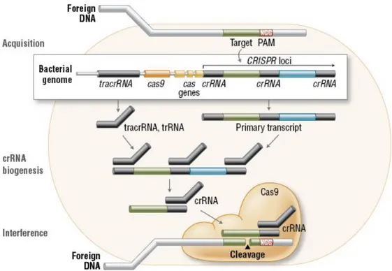
The CRISPR system is the adaptive immune mechanism present in the majority of characterized Archaea. The concept behind the CRISPR system is based on the acquisition of invading bacteriophage DNA and then transcription of it into CRISPR RNAs (crRNAs). The resulting crRNAs will be used as a guide by a nuclease to the cleavage of invading RNA or DNA (25). This CRISPR immune system works in cooperation with many different Cas proteins. CRISPR systems have been divided into two major classes, according to the difference in their elements and mechanisms of action. In the class 1 systems, RNA-guided target cleavage (types I, III, and IV) use a complex of several Cas proteins to degrade foreign nucleic acids. Class 2 systems use a single large protein for the same purpose, called Cas9 (Type II); Cpf1 in type V is needed to mediate the cut of invading nucleic acids. (82). During the first steps, or acquisition stage, the immune response system captures fragments of viral DNA of invading plasmids or phages (Fig. 5). These DNA fragments are called protospacers, and their insertion is catalyzed into the microbe CRISPR locus in the form of spacers between crRNA repeats. In the second step, the system prepares itself for the defense, by expressing the Cas proteins and transcribing the CRISPR array into its long precursor: CRISPR RNA (pre-crRNA); then the pre-crRNA will be cleaved into crRNA by Cas proteins (82).
Figure 5: Incorporating process of foreign genome fragments inside the CRISPR locus.
Credits Biolabs, New England.
The resulting crRNA is a guide that contains a targeting sequence, able to recognize the invading genome. In the third step, Cas proteins, recognizing the appropriate target with the guidance of the crRNA, mediate the cleavage of the foreign genome, thus protecting the host cells from infection. The action of many CRISPR systems depends on the presence of PAM sequence (protospacer-adjacent motif), sequence-specific protospacer-adjacent motif, adjacent to the crRNA target site in the invading genome (83). The lack of the PAMs in the host genome at the CRISPR locus allows type I and type II CRISPR systems to discriminate between self and not-self sequences (87). The editing of genomic information at the DNA level requires a molecular machine, composed of two main parts: a domain to bind the DNA and mediates the sequence-specific DNA recognition
engineered to mark precise genomic sites for gene editing purposes (88).
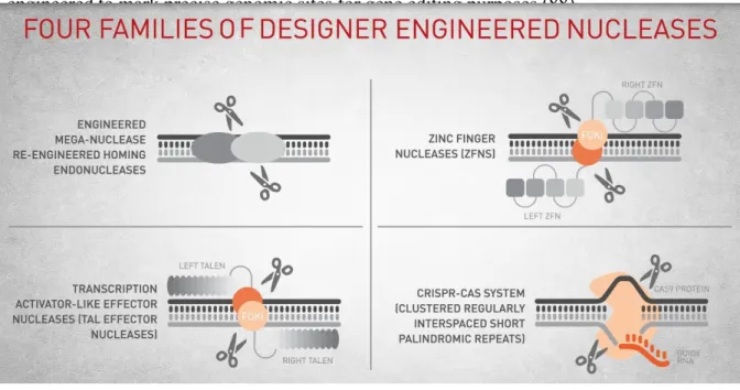
Figure 6: Four families of designer nucleases
Meganucleases are a group of nucleases, which recognize long nucleotide sequences and induce a double strand break at their target site. The long recognition sequence allows Meganucleases to edit DNA in a more accurate way, since the target sequence may occur just once in the invading genome. Engineered Meganucleases can be developed to specifically alter different sequences through usually labor-intensive procedures, as protein engineering, structure-based design, and molecular evolution. (33). Zinc-finger nucleases (ZFNs) and the transcription activator-like effector nucleases (TALENs) are other examples of programmable genome editing machines (9). In contrast to previously cited genome-editing machines, Cas9 is an RNA-guided nuclease, whose target specificity derives from the interaction between Cas9 and PAM and the pairing between guide RNAs and the related target DNA site (42). Thus, Cas9 can be programmed, by changing its guide RNA sequence, to target almost everywhere in the host genome, in a simple and relatively cheap way, making it an ideal platform for high-throughput applications. With the aim to make the genome editing easier, in the last decade, the CRISPR/CAS9 bacterial immune system was engineered in a simple RNA-guided platform, which was used for this work.
2. Aim of the thesis
HBV infection remains an important global health problem, concerning 248 million people worldwide (72). Despite an effective vaccine, more than 780,000 people die yearly because of HBV-related secondary diseases, primarily cirrhosis, and hepatocellular carcinoma. Antiviral therapies, such as nucleoside analogs (NA) and interferon-α (IFNα), are effective but rarely can eliminate established, persistent HBV infections (13). IFNα may help degrade nuclear viral DNA, but the effect is limited, and less than 10% of patients show a sustained virological response, measured as loss of hepatitis B surface antigen (HBsAg) (17). Nucleoside analogs are effective inhibitors of the HBV reverse transcriptase and can prevent the release of infectious virus from HBV-infected cells. However, cessation of treatment often results in viral relapse, since NAs alone have little or no effect on the elimination of the replicative template of HBV, i.e. the covalently closed circular DNA (cccDNA) (56). Consequently, to remove completely HBV infection and induce a complete remission, there is an imperative need for a different approach criterion, to repress HBV replication and eliminate cccDNA, the latent viral reservoir. The aim of the present thesis is to verify whether Type II bacterial clustered regularly interspaced palindromic repeat (CRISPR)-associated (Cas) 9-based genome editing technologies may provide a potential solution to this issue.
3. Materials and Methods
Cloning of CRISPR/SaCas9 Constructs.
To create all in SaCas9/gRNA/shRNA construct targeting HBV genome we used existing pX601-AAV-CMV::NLS-SaCas9-NLS-3xHA-bGHpA;U6::Bsa1-sgRNA plasmid (Addgene #61591) consisting of adapted to use in mammalian cells Staphylococcus aureus derived SaCas9/gRNA system. Protospacer regions corresponding to selected target sites were ordered as a pair of 5’-G(N19)-3’ complementary oligonucleotides containing Bsa1 overhangs at their respective 5’ ends (Table 4). After annealing and phosphorylation using T4 polynucleotide kinase (NEB) double stranded protospacers were ligated into Bsa1 digested, dephosphorylated with Calf Intestine Phosphatase (CIP, NEB) pX601 backbone plasmid. Bacterial clones were screened for the presence of gRNA protospacer inserts by PCRs using top, forward gRNA oligos in combination with reverse primer from scaffold gRNA segment of U6-gRNA cassette (Table 4). Successful clones were further verified by sequencing using the same reverse primer. To create HBV3xgRNA construct motif 2 and 3 gRNA expressing cassettes were PCR amplified from their respective pX601 plasmids using primers containing Xba1 (in forward) and Spe1 (in reverse) restriction sites and ligated into Xba1 digested pX601-HBVmotif1 plasmid in two cycles of Xba1 restriction digestion/ligation. In the final step, to add to our construct HBV X shRNA expressing cassette, we used Xba1/Spe1 extended oligos containing minimal 24bp U6 promoter allowing direct cloning of annealed double-stranded hairpin coding sequence into Xba1 digested pX601-HBV3xgRNAs
Purpose
Name
Sequence
gRNAs prorospacers gRNA HBV m1 f CAAGAATCCTCACAATAC gRNA HBV m1 r GTATTGTGAGGATTCTTG gRNA HBV m2 f GGACGTCCTTTGTTTACG gRNA HBV m2 r CGTAAACAAAGGACGTCC gRNA HBV m3 f GTCCTTTGTTTACGTCCCGTCGGCG gRNA HBV m3 r CGCCGACGGGACGTAAACAAAGGAC Cleavage Detection HBV 68-89 cut f TCCAGTTCAGGAGCAGTAAACC HBV 1476-96 cut r AGAAGGGGACGAGAGAGTCTC qPCR analysis HBV X1805 f TCACCAGCACCATGCAAC HBV X1896 r AAGCCACCCAAGGCACAG HBV pol 2270 f GAGTGTGGATTCGCACTCC HBV pol 2392 r GAGGCGAGGGAGTTCTTCT qPCR references Hs b-globin f CCCTTGGACCCAGAGGTTCT Hs b-globin r CGAGCACTTTCTTGCCATGA h/m b-actin f CTACAATGAGCTGCGTGTGGC h/m b-actin r CAGGTCCAGACGCAGGATGGC gRNA verificationgRNA scaffold r CTCGCCAACAAGTTGACGAGATAA
pX6O1 U6 XbaI f CTATCTAGAGAGAGGGCCTATTTCCCATG
FM1chr20f AAGTGGGGCATTGGTGACAT
FM1chr20r TAAGGACAGTGGGCTCTGGA
FM1chr13F AGCCTGCTCAGAACATGGTTA
FM1chr13R TTGCACACCAGTAGAACATCAGA
FM2chr14R GCCTAGGGTAGACCTGGAAGA FM2chr19F GAAGTGGGTGGGAATGAGCG FM2chr19R TAATTGGCCAAGGCTGAGAGA FM3chr6F CTCGAGCTCGCCGAAGATGT FM3chr6R CTGCCCGCCTGCAAACTTC FM3chr12F TTGGCTTCGGCTTTATGGCT FM3chr12R GATGCAGCCGCACAAAGAAC
Table 4: List of the primers used in the study.
Cell Culture.
HepG2.2.15 and TC120 cell line cells were cultured in Dulbecco’s Modified Eagle’s Medium (DMEM) (Life Technologies, NY) supplemented with 10% fetal bovine serum (FBS), 2 mM glutamine and 400 μg/ml of Gentamycin (Life Technologies, NY). To promote cell attachment all culture dishes and plates were precoated with poly-D-lysine prior plating cells. For puromycin selection, cells were incubated in growth medium containing 3ug/ml of puromycin (Sigma-Aldrich). The medium was changed every day for one week to achieve maximum selection strength.
Antibodies.
To detect NLS-SaCas9-NLS-3xHA HA-tag antibody was used (1:1000, Abcam) for Western blot loading control anti-tubulin clone B512 from (1:5000, Sigma Aldrich ).
Transfection.
Cells were plated in 6 well plates at density 150000 cells per well. Next day cells were transfected using Lipofectamine 2000 reagent (Invitrogen) according to manufacturer protocol. Briefly, 7.5ul Lipofectamine 2000 was resuspended in 100ul of Opti-MEM medium (Gibco) and incubated for 5 minutes. Meantime, plasmid DNA mixtures were prepared: 2ug of control empty pX601 or
pX601-HBV3xgRNAs/shRNA together with 0.5ug of pKLV-U6gRNA(BbsI)-PGKpuro2ABFP (Addgene #50946, to provide puromycin resistance for selection and BFP for transfection efficiency control) plasmids were added to 100ul of Opti-MEM medium mixed and then combined with 100ul Lipofectamine 2000/Opti-MEM and incubated for 15 minutes at room temperature (DNA:Lipofectamine ratio: 1:2.5). Next to DNA/Lipofectamine complexes (200ul) were vortexed and added dropwise into 800ul Opti-MEM per well in culture plates. After 4 hours incubation, 1ml/well of growth medium was added and left overnight. Next day medium was replaced with fresh growth medium and cells were incubated for another 48h before harvesting.
Viral DNA extraction and analysis.
Cell pellets were collected and DNA was extracted using NucleoSpin kit (Macherey-Nagel) according to the manufacturer’s protocol, and the final product was eluted in 60 μl of water. For standard PCRs, 250ng of genomic DNA was used. Reaction mixtures were prepared using Fail Safe Kit enzyme mix, PCR buffer J (Epicenter) and primers designed to amplify the targeted region of HBV genotype D (see Table 4 ). Quantification of HBV intracellular DNA was performed with 50ng of genomic DNA per well using SybrGreen real time PCR (Roche) with primer sets specific to pol and X viral genes and human beta-globin as a reference (Table 4).
Analysis of RNA
Total RNA was extracted from cell pellets using RNAesy kit (Qiagen) according manufacturer protocol. Next 2.5ug of RNA was used for reverse transcription reactions using M-MLV reverse transcription (Invitrogen) and different reverse primers depending on the purpose of experiment. For detection/verification of gRNAs expression in transfected cells, gRNA scaffold reverse primer was used (Table 4) followed by standard PCR using top gRNA specific oligo as a forward primer
polymerase and reference human beta-actin (Table 4) were used in SybrGreen real time PCR reactions (Roche).
Quantification of virus level in cell culture supernatants
SybrGreen real time PCR was used to quantify viral DNA levels in supernatants of infected cells. Culture medium was collected and spun down for 10 minutes at 3000 RPM to remove floating cells and cell debris. Next supernatants were incubated for 5 minutes at 95oC to denature/destroy infective viral particles. Standard curve was prepared using serial dilutions of PCR amplified fragment of HBV genome spanning core and X genes (primers Table 4). qPCR reactions were performed using 5ul of deactivated, ten times diluted in water supernatants and HBV X specific primers.
CRISPR/Cas9 design and validation. Using the CRISPR online design tool available on (http://www. benchling.com), 12 single guide RNAs (sgRNAs) were generated, targeting the HBV genome (Fig. 1). Target sequences were chosen in order to maximize conservation across viral genotypes, and minimize homology to the human genome. Based on these criteria, only guides targeting pol, pres1 gene and derivates, and X ORFs were designed.
Off-target analysis. To verify specificity of our SaCas9/gRNAs we performed PCR/sequencing analysis of the top predicted off target regions in human genome (Table 4). Sets of primers were designed to amplify these regions followed by subcloning into pCR2.2 TA vector (Invitrogen) and Sanger sequencing.
4. Results
4.1. CRISPR/Cas9 design: in silico definition of the twelve most fitting gRNAs
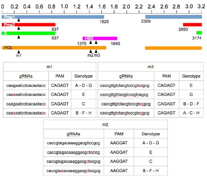
For the eradication of the HBV virus we initially selected set of 12 candidate gRNAs, targeting the most representative Hepatitis B virus genes. To design these gRNAs we used the CRISPR designer
Figure 7 : Sequence and location in the HBV genome of the 12 candidate gRNAs designed by
position.
tool from Benchling web site. The HBV genotype A genome was used as an input sequence and screened for the presence of 20 nucleotide protospacer regions followed by NNGRRT protospacer adjacent motifs (PAMs) which are specifically recognized by SaCas9 endonuclease. The twelve gRNAs showed (Fig. 7) are the gRNAs with the highest “on target” “off target“ score. Finally, three gRNAs were chosen based on the most conserved region among ten reported HBV genotypes in NCBI. All three gRNAs are targeting viral polymerase gene (P). Additionally, because of overlap of reading frames, the m1 gRNA targets also the surface protein gene (S), while the m2 and m3 gRNAs target the viral trans-activator protein gene (X). In order to block viral expression in treated cells, to improve gene editing efficiency of SaCas9/gRNAs complexes, a shRNA expressing cassette against X mRNA was added (Fig. 8). All the gRNAs and the shRNA were cloned into a single pX601 vector. The pX601 plasmid is an AAV delivery vector, containing a 1 kb shorter orthologue of canonical Streptococcus pyogenes Cas9 (SpCas9), derived from Staphylococcus aureus (SaCas9). Shorter SaCas9 gene allows the combining of up to four different gRNA cassettes in a single “all in” vector, without exceeding the restrictive cargo size of AAV, which is around 4.5 kb.
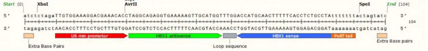
Figure 8 : Sequence of short hairpin RNA against Hepatitis B transactivator X. The shRNA
4.2 Cloning of the gRNA expressing cassettes targeting HBV genome into pX601-SaCas9-AAV vector and verification final pX601-HBV3xgRNAs-shRNA construct.
After the bioinformatics analysis, pairs of sense and antisense oligonucleotides, matching selected target protospacer regions and containing Bsa1 overhangs on 5’ ends, were ordered, annealed and cloned into a Bsa1 restriction site, located between U6 promoter and scaffold crRNA sequence in gRNA expressing cassette of pX601 plasmid. To create a multiplex “three in one” gRNAs construct, every single U6-gRNAs cassette was PCR amplified using primers with Xba1/Spe1 extensions at their respective 5’ ends. Next the amplicons were cloned, by restriction digestion followed by ligation into pX601-HBVmotif1plasmid XbaI restriction site. The same process was used to add the shRNA-expressing cassette. The final construct is shown in Fig.9.
Figure 9: Map of pX601-HBV3xgRNAs-shRNA construct targeting the Hepatitis B Virus
genome. gRNA protospacer regions in red, shRNA for HBX in green, NLS-SaCas9-NLS-3xHA in brown-orange.
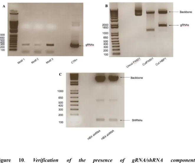
The presence of all three gRNA and single shRNA expressing cassettes in the final construct was verified in set of gRNA specific PCRs and by restriction digestions as shown in the Fig 10.
Figure 10. Verification of the presence of gRNA/shRNA components in
in standard PCRs using U6 promoter forward and reverse primers specific to each of cloned gRNAs (A). Additionally restriction digestion was performed using Sac1/ Spe1 restriction enzymes to confirm existence of gRNAmotif2/motif3/shRNA insert upstream of SaCas9 gene (B). Finally HBX shRNA presence was verified by Xba1/Spe1 restriction digestion ©. In the next step we checked if the final construct was able to express all the components of SaCas9/gRNA gene editing platform. The pX601-HBV3xgRNA-shRNA construct was transfected into TC620 cells and 48h later total RNA and proteins were extracted. gRNAs expression was verified in reverse transcription followed by PCR using forward primers specific to each gRNA and scaffold RNA reverse primer (Fig. 11 A). To detect NLS-SaCas9-NLS-3xHA protein expression Western blot analysis was performed using HA-tag antibody (Fig. 11 B).
Figure 11: Confirmation of the correct SaCas9/gRNAs expression from
scaffold as a reverse primers. NLS-SaCas9-NLS-3xHA protein expression was verified in Western blot using HA-tag antibody (B).
4.3 Biological validation of pX601-HBV3xgRNAs-shRNA construct on chronically HBV infected cells (HepG2.2.15)
To test the ability of our construct to induce site specific cleavage and excision of HBV genome we used the chronically HBV-infected HepG2.2.15 cell line. 70% confluent cell cultures were transfected with pX601-HBV3xgRNAs-shRNA plasmid, as reported in Materials and Methods. Two days after transfection cells were harvested and genomic DNA was prepared. Next the targeted region of the virus was PCR amplified and resolved by agarose gel electrophoresis. As shown in Fig. 12 we were able to detect two distinct HBV specific PCR products: 1454 bp and 355 bp long. Longer, 1454bp band corresponds to unmodified full length (in case of control untreated cells) and single cut/end-joined region of HBV genome (in case of SaCas9/gRNAs treated cells). Shorter, 355bp band represents double cutted/end-joined truncated form of viral sequence and is present exclusively in SaCas9/gRNA treated cells.
Figure 12: Detection of the SaCas9/gRNAs induced excision of the HBV genome. The cleavage
region was PCR amplified using two primers: forward, annealing 144 nucleotides upstream of the motif 1 and reverse, 191 downstream of motif 2 target site. Amplification using these primers yielded two products: full length 1454 bp long, representing the uncut/singly cut and end-joined HBV genomes and short 355 bp one corresponding to double cleaved/end joined viral sequences.
The truncated double cleaved/end-joined band was purified from the gel, cloned and sent for Sanger sequencing. The obtained sequences were aligned using Clustal-Omega software using Hepatitis B genotype D sequence as a reference (Fig. 13). All clones showed perfect CRISPR/Cas9 mediated signature-cleavage three nucleotides from PAM at target sites for motifs 1 and 2. At the target motif 3 no any cleavage was detected since this gRNA was designed to targets exclusively HBV genotype A and in present in HepG2.2.15 HBV genotype D there are 5 mismatches at this target sites
providing additional prove of SaCas9/gRNA specificity (Fig. 13).
Figure 13: Verification of SaCas9/gRNA mediated excision of HBV sequences. The targeted
region of HBV genome was PCR amplified and resolved in agarose gel. Truncated PCR products representing double cleaved/end-joined viral sequences (345bp band) were purified, subcloned in TA vector and sequenced. Representative three truncated sequences are shown in relation to full length intact viral sequence as a reference. PCR primers are shown in green, target sequences in red followed my PAMs in yellow. The canonical, 3 nucleotides from PAM sequences, SaCas9/gRNAs
mediated cleavage sites were detected with deletion of 1216 bp long viral DNA fragment between target sites motif 1 and 2. There was no cut at target site motif 3 since HBV genotype D present in HepG2.2.15 cells carries 5 mismatches in this region.
4.4 Off-target analysis
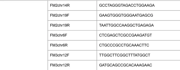
To verify specificity of our excision strategy in targeting the viral genome, we performed analysis of the predicted/possible off targets sites in the human genome. The closest to target sequences hits had at least 3 mismatches (Table 4) making cleavage at these sites highly improbable and un-efficient.
Table 4: List of top predicted off-target sites for HBV specific gRNAs in human genome.
With the Primer-Blast tool from the NCBI website, we designed primer pairs for PCR amplification of every genomic region with an off-target score even or above 0.5. After purification and subcloning into TA vector, amplified predicted off-target regions were sent for Sanger sequencing. No indel mutations were detected in the selected off-target genes.
4.5 Expression of Cas9/gRNA suppresses Viral replication cycle
analysed in standard PCRs for detection of targeted region of HBV genome (A for 3 and B for 7 days timepoint). To allow semi-quantification of excision efficiency, PCRs for human beta-actin were performed as a reference genomic DNA loading control for 7 days timepoint (B). The intensities of PCR bands from agarose gels were analysed using ImageJ software (C) and plotted after normalizing to beta-actin levels (D).
To verify the real effectiveness of our construct in blocking viral replication, we set another experiment. HepG2.2.15 cells were transfected with pX601-HBV3xgRNAs-shRNA or control, empty pX601 plasmids. Additionally we added to transfection mixtures pKLV-puro-BFP-empty vector in ratio 4:1 (2ug pX601: 0.5 ug pKLV) to permit monitoring of transfection efficiency by BFP fluorescence microscopy and to allow puromycin selection of transfected cells (since pX601 AAV vector does not contain any fluorescent label or selection marker). Half of transfected cells were left untreated and were harvested after 3 days. Rest of the cells were selected for one week under rigorous puromycin regiment (3ug/ml, medium changed every day) in order to remove untransfected cells and promote stable expression of SaCas9/gRNA in transfected cells. For both populations, we checked the viral integrity and expression at DNA, mRNA and viral release level. First, genomic DNA was used in standard PCRs with primers specific to targeted region of HBV as was done previously. Again we were able to detect two distinct HBV specific amplification products full length 1454bp and truncated 355bp (Fig 15 A/B). We noticed significant reduction of full length band intensity in treated cells which is a direct result of SaCas9/gRNA mediated cleavage and degradation of episomal HBV genomes. Additionally, as was shown before, in the line corresponding to treated cells we could detect characteristic truncated 355bp long band representing double cutted and end-joined viral genome. The ImageJ analysis of band intensities for day 7 timepoint indicated drastic, 50% drop in the level of the full-length HBV DNA in the cells treated with pX601-HBV3xgRNA-shRNA construct (Fig. 15C, D).
4.5.2 Quantification of intracellular viral DNA levels.
Figure 16: Quantification of intracellular HBV DNA levels in treated cells. Genomic DNA from transfected HepG2.2.15 cells was subjected to SybrGreen real time PCR reactions using primer sets specific to HBV pol and as a reference human beta-globin genes.
To quantify HBV DNA affected by cleavage, we performed a qPCR assay on genomic DNA extracted from HepG2.2.15 treated cells. Using primers specific to HBV pol and reference human beta-globin genes we were able to detect significant, close to 30% drop in intracellular viral DNA levels at seven days post-transfection time point (Fig 16). The levels of viral DNA at day 3 were
for this time point. It is important to note that the primers used in qPCR can not discriminate between episomal and integrated HBV DNA and they anneal outside of the targeted region of viral genome.
4.5.3 Quantification of viral RNA expression after CRISPR/Cas9 treatment
Figure 17: Quantification of intracellular viral RNA levels. Total RNA was extracted from cells
transfected with empty pX601 (SaCas9, no gRNA) and pX601-HBV3xgRNAs-shRNA (SaCas9 and gRNAs) at three days post-transfection and after one-week selection with puromycin. After reverse transcription using oligo-dT primers, SybrGreen real time PCRs were performed on diluted cDNA samples using primer sets specific to HBV pol and human beta-actin as a reference.
in the decrease of viral RNA levels. To quantify viral RNA levels in treated cells total RNA was extracted and subjected to reverse transcription reaction followed by SybrGreen real time PCR assay using primers specific to HBV pol and human beta-actin as a reference. As shown in figure 17 we observed progressive, time dependent reduction of intracellular HBV RNA levels in treated cells. At 3 days after transfection the decrease reached 30% and at 7 days the levels went down to 50% of control, SaCas9/gRNA untreated control.
4.5.4 Checking Hepatitis B virus release from treated cells.
Final step in viral replication cycle is release of progeny viral particles from infected cells. Viral pregenomic DNA is packaged into viral capsids by interactions with viral core proteins then enveloped and released from infected cells. We used SybrGreen real time PCR to measure the levels of viral DNA in supernatants from treated cells which should correspond with viral particles release. As shown in Figure 18 we observed drastic, more than 95%, depletion of viral DNA levels in the supernatants from treated cells at 3 days post-transfection. At day 7 time point viral DNA levels in supernatants were generally very low and only minimal decrease of was observed in treated versus untreated cells.
using HBV X gene specific primers and standard prepared from serial dilutions of PCR amplification product corresponding to X gene of HBV.
5. Discussion
The Hepatitis B virus is still a significant threat for 240 million of people in the world. In this work of thesis, we are describing a novel, CRISPR/SaCas9-based gene therapy, directed against the persistent HBV DNA genome conserved among all ten HBV genotypes spanning five of the total six viral genes: PreS1, PreS2, S, transactivator X and polymerase. Successful SaCas9/gRNAs-mediated cleavage at these target sites will have different consequences depending on the timing of the cleavage reactions, cellular DNA repair mechanisms and the form of viral genome. Cleavage of episomal cccDNA ordinary will lead to its linearization and degradation by cellular exo- and endonucleases. Less frequent end-joining repair and re-circularization will result in InDel mutations at the cut sites, in case of single cuts, or excisions/deletions of longer fragments, in case of two or more simultaneous cuts, both resulting in defective viral genomes. In case of much less frequent integrated form of HBV genome, the SaCas9/gRNAs-mediated cleavage will result exclusively in end-joining, InDels and deletions at cut sites. Since PCR primers do not distinguish between episomal cccDNA and integrated HBV genome forms, the products of PCR amplification shown in figures 12 and 15 represent a mixture of both forms. The full length 1454bp top band consists mostly of episomal cccDNA, since it is the predominant form of viral genome present in the infected cells. As mentioned above, the Cas9/gRNAs activity will cause fragmentation/linearization and subsequent degradation of cccDNA, which can be observed as a decrease (up to 50% in case of 7 days time point) in the intensity of this band, in the sample of treated cells (Fig. 15 A and B lane 2, top bands). On the other hand, the cleavage of the integrated viral genome is promptly repaired by the cellular double strand break repair pathways, mostly by
corresponding to integrated HBV genome will contain InDel mutations at repaired cut sites, which will disrupt or completely block viral gene expression. In case of successful simultaneous cleavage at two sites, the DNA fragment located between them gets edited out leaving truncated defective viral genome, detected as a shorter 354bp PCR product in SaCas9/gRNAs treated cells (see Fig 12 and 15, line 2). All mentioned above consequences of SaCas9/gRNAs mediated targeting and cleavage of viral genomes in infected cells will ultimately culminate in suppression of viral expression. Degradation of viral genomes will result in drop in viral RNA and proteins levels. Additionally expression from mutated/truncated sequences will lead to defective viral mRNAs and proteins as a result of premature transcription terminations and shifted open reading frames. As anticipated we observed significant decrease in viral RNA expression levels in SaCas9/gRNAs treated cells as shown in Fig. 17 which mirrors detected depletion of viral DNA. The decrease was greater in cells selected for one week with puromycin what can be explained by longer period of SaCas9/gRNA expression in the treated cells and death of untransfected (=untreated) cells. The last stage of viral replication cycle is release of the progeny viral particles from infected cells. Here again, consistently with diminished intracellular viral DNA and RNA levels we detected repression of viral release as measured by qPCR specific to viral DNA in supernatants from gene therapy treated cells. Surprisingly viral DNA level in supernatants of puromycin selected cells was very low in both control (SaCas9 only) and treated cells (SaCas9/gRNAs). Puromycin is aminonucleoside that inhibits translation by disrupting peptide transfer on ribosomes. Inhibitory effect on HBV virion release was not reported before and warrants further studies. Overall our data provide for the first time prove of successful targeting and cleavage of HBV genome by shorter Staphylococcus aureus derived Cas9/gRNA gene editing platform. Recently other groups reported successful using of canonical SpCas9/gRNA gene editing techniques to target HBV genome (Ramanan et al., 2015). Our approach combining triple gRNAs and shorter SaCas9 in single AAV delivery vector provides more robust and applicable system to use in clinical settings. To provide suitable for in vivo
delivery system we prepared our SaCas9/gRNA construct using as a backbone plasmid AAV delivery vector pX601. Adeno-associated virus (AAV) vectors are the most commonly used delivery vehicles in vivo, because of their low immunogenic potential, reduced oncogenic risk from host-genome integration, broad-range of serotype specificity, low toxicity and sustained gene expression. The AAV-mediated gene delivery system is already used, for treating human diseases caused by a gene loss or mutation, including cancer, heart failure, neurodegenerative diseases, cystic fibrosis, Canavan's disease, arthritis and muscular dystrophy, among others (41, 65, 67). By creating and validating in chronic hepatitis cell culture model our pX601-HBV3xgRNAs/shRNA construct we successfully completed first, in vitro phase of the project. In the next, pre-clinical stage we are planning to confirm anti-HBV activity of our unique SaCas9/triple gRNA design in in vivo settings, using immunodeficient mice model and AAV-mediated delivery. If successful our approach should provide base for developing gene therapy for treatment chronic HBV infection.
6. Acknowledgments
I would like to use this opportunity to express my special gratitude, appreciation, and thanks to all the people that directly made possible this work.
Firstly, I would like to thanks my coordinator Professor Caterina Serra and my co-advisor, professor Antonina Dolei, of the Virology Laboratory at the Department of Biomedical Science (University of Sassari). They supported me during these past three years, encouraged my research and enabled me to grow as a scientist.
My sincere gratitude goes to Dr. Kamel Khalili, who provided me the chance to join his team as a research scholar at Center for Neurovirology (Lewis Katz School of Medicine) at Temple University, Philadelphia. The outstanding team, the laboratories and the challenges I had the opportunity to face, made priceless the period spent at the Center for Neurovirology, professionally and personally speaking. A special thanks go to my tutor, Dr. Hassen Wollebo, who introduced me to the field of CRISPR/Cas9 system, and for his endless patience, and to Dr. Rafal Kaminski, as an experienced scientist and as a friend.
I have to thank all the wonderful people I knew in this "journey," starting from the ones I met in Sassari: Elena, Claudia, and Maurizio and the team I joined in America, especially Anna, Martina, Tijana, Pietro, and Workinae.
Finally, I assert my sincere gratitude to my parents, to my sisters and of course to my girlfriend, that supported me through my life. Their infinite assistance and patience made all this possible.
Università degli Studi di Sassari
Corso di Dottorato di ricerca in
Scienze della vita e biotecnologie
La presente tesi è stata prodotta durante la frequenza del Corso di Dottorato di ricerca in Scienze della vita e biotecnologie dell’Università degli Studi di Sassari, A.A. 2013/2014 - XXIX ciclo, con il sostegno di una borsa di studio finanziata con le risorse dell’INPS – Gestione Ex INPDAP nell’ambito delle Iniziative Accademiche Homo Sapiens Sapiens.
This thesis has been produced during the frequency of the Ph.D. Programme in Life Sciences and Biotechnology of the University of Sassari, A.A. 2013/2014 - XXIX cycle, with the support of a scholarship funded by the INPS resources - Gestione EX INPDAP as part of Academic Initiatives
7. REFERENCES
1. Aa, Evelyn Van Der, Sonja I. Buschow, Paula J. Biesta, Harry L. A. Janssen, and Andrea M. Woltman. "The Effect of Chronic Hepatitis B Virus Infection on BDCA3 Dendritic Cell Frequency and Function." Plos One 11.8 (2016). Print.
2. André, F.e. "Overview of a 5-year Clinical Experience with a Yeast-derived Hepatitis B Vaccine." Vaccine 8 (1990). Print.
3. Asabe, S., S. F. Wieland, P. K. Chattopadhyay, M. Roederer, R. E. Engle, R. H. Purcell, and F. V. Chisari. "The Size of the Viral Inoculum Contributes to the Outcome of Hepatitis B Virus Infection." Journal of Virology 83.19 (2009): 9652-662. Print.
4. Billaud, J.-N., D. Peterson, M. Barr, A. Chen, M. Sallberg, F. Garduno, P. Goldstein, W. Mcdowell, J. Hughes, J. Jones, and D. Milich. "Combinatorial Approach to Hepadnavirus-Like Particle Vaccine Design." Journal of Virology 79.21 (2005): 13656-3666. Print.
5. Biolabs, New England. "CRISPR/Cas9 and Targeted Genome Editing: A New Era in Molecular Biology." CRISPR/Cas9 and Targeted Genome Editing: A New Era in Molecular Biology | NEB . Web. 26 Mar. 2017.
6. Blumberg, B. "Australia Antigen and the Biology of Hepatitis B." Science 197.4298 (1977): 17-25. Print.
7. Blumberg, Baruch S. "A Serum Antigen (Australia Antigen) in Down's Syndrome, Leukemia, and Hepatitis." Annals of Internal Medicine 66.5 (1967): 924. Print.
8. Boch, J., H. Scholze, S. Schornack, A. Landgraf, S. Hahn, S. Kay, T. Lahaye, A. Nickstadt, and U. Bonas. "Breaking the Code of DNA Binding Specificity of TAL-Type III Effectors." Science 326.5959 (2009): 1509-512. Print.
9. Bock, Claus-Thomas, Peter Schranz, Claus Hobe Schröder, and Hanswalter Zentgraf. "Hepatitis B Virus Genome Is Organized into Nucleosomes in the Nucleus of the Infected Cell." Virus Genes 8.2 (1994): 215-29. Print.
10. Bock, C.thomas, Susanne Schwinn, Stephen Locarnini, Janet Fyfe, Michael P. Manns, Christian Trautwein, and Hanswalter Zentgraf. "Structural Organization of the Hepatitis B Virus Minichromosome." Journal of Molecular Biology 307.1 (2001): 183-96. Print.
11. Chain, Benjamin M., and Richard Myers. BMC Microbiology 5.1 (2005): 33. Print.
12. Chang, Shau-Feng, Hans Jürgen Netter, Michael Bruns, Ralf Schneider, Kai Frölich, and Hans Will. "A New Avian Hepadnavirus Infecting Snow Geese (Anser Caerulescens) Produces a Significant Fraction of Virions Containing Single-Stranded DNA." Virology 262.1 (1999): 39-54. Print.
13. Chen, Jieliang, and Zhenghong Yuan. "Interplay between Hepatitis B Virus and the Innate Immune Responses: Implications for New Therapeutic Strategies." Virologica Sinica 29.1 (2014): 17-24. Print. 14. Chisari, F.v., M. Isogawa, and S.f. Wieland. "Pathogenesis of Hepatitis B Virus Infection." Pathologie
Biologie 58.4 (2010): 258-66. Print.
15. Cossart, Y. "Virus-Like Particles In Serum Of Patients With Australia-Antigen-Associated Hepatitis." The Lancet 295.7651 (1970): 848. Print.
16. Crowther, R.a. "The Leeuwenhoek Lecture 2006. Microscopy Goes Cold: Frozen Viruses Reveal Their Structural Secrets." Philosophical Transactions of the Royal Society B: Biological Sciences 363.1502 (2008): 2441-451. Print.
17. Dahmen, U., J. Li, O. Dirsch, Y. L. Gu, S. Polywka, L. Doebel, K. Shen, and C. E. Broelsch. "Adoptive Transfer of Donor-derived Immunity by Liver Transplantation: A Potential Avenue to Prevent Hepatitis B Virus Reinfection." Journal of Viral Hepatitis 10.1 (2003): 31-36. Print.
18. Dahmen, Uta, Olaf Dirsch, Jun Li, Melanie Fiedle, Mengj Lu, Kai Rispeter, Martha Picucci, Christoph E. Broelsch, and Michael Roggendorf. "Adoptive Transfer Of Immunity: A New Strategy To Interfere With Severe Hepatitis Virus Reinfection After Woodchuck Liver Transplantation." Transplantation 77.7 (2004): 965-72. Print.
19. Dahmen, Uta, Jun Li, Yi Gu, Lothar Doebel, Li Ming Fan, Susanne Polywka, Olaf Dirsch, and Christoph E. Broelsch. "The Efficiency of Humoral Immune Transfer Depends on Both the Graft and the Immunosuppressive Treatment." Transplant International 16.3 (2003): 161-67. Print.
20. Dane, D.s., C.h. Cameron, and Moya Briggs. "Virus-Like Particles In Serum Of Patients With Australia-Antigen-Associated Hepatitis." The Lancet 295.7649 (1970): 695-98. Print.
21. Das, Abhishek, and Mala K. Maini. "Innate and Adaptive Immune Responses in Hepatitis B Virus Infection." Digestive Diseases 28.1 (2010): 126-32. Print.
Immunology 179.1 (1990). Print.
23. "Experiments on Transfer of Cutaneous Sensitivity to Simple Compounds." Journal of Allergy 13.5 (1942): 530-31. Print.
24. Ferrari, C., A. Penna, A. Degliantoni, and F. Fiaccadori. "Cellular Immune Response to Hepatitis B Virus Antigens." Journal of Hepatology 7.1 (1988): 21-33. Print.
25. Garneau, Josiane E., Marie-Ève Dupuis, Manuela Villion, Dennis A. Romero, Rodolphe Barrangou, Patrick Boyaval, Christophe Fremaux, Philippe Horvath, Alfonso H. Magadán, and Sylvain Moineau. "The CRISPR/Cas Bacterial Immune System Cleaves Bacteriophage and Plasmid DNA." Nature 468.7320 (2010): 67-71. Print.
26. Gasiunas, G., R. Barrangou, P. Horvath, and V. Siksnys. "Cas9-crRNA Ribonucleoprotein Complex Mediates Specific DNA Cleavage for Adaptive Immunity in Bacteria." Proceedings of the National Academy of Sciences 109.39 (2012). Print.
27. Gerlich, Wolfram H. Hepatitis B Virus: Selected Topics ; 15 Tables . Basel: Karger, 1995. Print. 28. Ghadirian, E., and E. Meerovitch. "Passive Transfer of Immunity against Hepatic Amoebiasis in the
Hamster by Cells." Parasite Immunology 5.4 (1983): 369-76. Print.
29. Gilbert, Luke A., Matthew H. Larson, Leonardo Morsut, Zairan Liu, Gloria A. Brar, Sandra E. Torres, Noam Stern-Ginossar, Onn Brandman, Evan H. Whitehead, Jennifer A. Doudna, Wendell A. Lim, Jonathan S. Weissman, and Lei S. Qi. "CRISPR-Mediated Modular RNA-Guided Regulation of Transcription in Eukaryotes." Cell 154.2 (2013): 442-51. Print.
30. Glebe, D., H. Lorenz, W. H. Gerlich, S. D. Butler, I. A. Tochkov, B. C. Tennant, P. Cote, and S. Menne. "Correlation of Virus and Host Response Markers with Circulating Immune Complexes during Acute and Chronic Woodchuck Hepatitis Virus Infection." Journal of Virology 83.4 (2008): 1579-591. Print.
31. Glebe, D., M. Aliakbari, P. Krass, E. V. Knoop, K. P. Valerius, and W. H. Gerlich. "Pre-S1 Antigen-Dependent Infection of Tupaia Hepatocyte Cultures with Human Hepatitis B Virus." Journal of Virology 77.17 (2003): 9511-521. Print.
32. Glebe, Dieter, Stephan Urban, Eva V. Knoop, Nilgün Ça ǧ, Peter Krass, Stefanie Grün, Aiste Bulavaite, Kestutis Sasnauskas, and Wolfram H. Gerlich. "Mapping of the Hepatitis B Virus Attachment Site by Use of Infection-Inhibiting PreS1 Lipopeptides and Tupaia Hepatocytes."
Gastroenterology 129.1 (2005): 234-45. Print.
33. Grizot, S., J. C. Epinat, S. Thomas, A. Duclert, S. Rolland, F. Paques, and P. Duchateau. "Generation of Redesigned Homing Endonucleases Comprising DNA-binding Domains Derived from Two Different Scaffolds." Nucleic Acids Research 38.6 (2009): 2006-018. Print.
34. Hong, Hyo Jeong, Chun Jeih Ryu, Hyangsuk Hur, Seho Kim, Han Kyu Oh, Mee Sook Oh, and Song 35. Yong Park. "In Vivo Neutralization of Hepatitis B Virus Infection by an Anti-preS1 Humanized
Antibody in Chimpanzees." Virology 318.1 (2004): 134-41. Print.
36. Hourvitz, A., R. Mosseri, A. Solomon, Y. Yehezkelli, J. Atsmon, Y. L. Danon, R. Koren, and D. Shouval. "Reactogenicity and Immunogenicity of a New Recombinant Hepatitis B Vaccine Containing Pre S Antigens: A Preliminary Report." Journal of Viral Hepatitis 3.1 (1996): 37-42. Print. 37. Hösel, Marianna, Maria Quasdorff, Katja Wiegmann, Dennis Webb, Uta Zedler, Mathias
Broxtermann, Raindy Tedjokusumo, Knud Esser, Silke Arzberger, Carsten J. Kirschning, Anja Langenkamp, Christine Falk, Hildegard Büning, Stefan Rose-John, and Ulrike Protzer. "Not Interferon, but Interleukin-6 Controls Early Gene Expression in Hepatitis B Virus Infection." Hepatology 50.6 (2009): 1773-782. Print.
38. Ilan, Y. "Adoptive Transfer of Immunity to Hepatitis B Virus after T Cell-depleted Allogeneic Bone Marrow Transplantation." Hepatology 18.2 (1993): 246-52. Print.
39. Ishino, Y., H. Shinagawa, K. Makino, M. Amemura, and A. Nakata. "Nucleotide Sequence of the Iap Gene, Responsible for Alkaline Phosphatase Isozyme Conversion in Escherichia Coli, and Identification of the Gene Product." Journal of Bacteriology 169.12 (1987): 5429-433. Print.
40. Iverson, M. "Role of Contrasuppression in the Adoptive Transfer of Immunity." Journal of Experimental Medicine 158.3 (1983): 982-87. Print.
41. Janson, Christopher, Scott Mcphee, Larissa Bilaniuk, John Haselgrove, Mark Testaiuti, Andrew Freese, Dah-Jyuu Wang, David Shera, Peter Hurh, Joan Rupin, Elizabeth Saslow, Olga Goldfarb, Michael Goldberg, Ghassem Larijani, William Sharrar, Larisa Liouterman, Angelique Camp, Edwin Kolodny, Jude Samulski, and Paola Leone. "Gene Therapy of Canavan Disease: AAV-2 Vector for Neurosurgical Delivery of Aspartoacylase Gene ( ASPA ) to the Human Brain." Human Gene Therapy 13.11 (2002): 1391-412. Print.
Dual-RNA-Guided DNA Endonuclease in Adaptive Bacterial Immunity." Science 337.6096 (2012): 816-21. Print.
43. Joung, J. Keith, and Jeffry D. Sander. "TALENs: A Widely Applicable Technology for Targeted Genome Editing." Nature Reviews Molecular Cell Biology 14.1 (2012): 49-55. Print.
44. Kianianmomeni, Arash. "Genome Editing Using Engineered Zinc Finger Nucleases." Clinical Biochemistry 44.13 (2011). Print.
45. Klug, A., J. Miller, and A. D. Mclachlan. "Repetitive Zn 2 -binding Domains in the Protein Transcription Factor IIIA from Xenopus Oocytes." Biochemical Society Transactions 14.2 (1986). Print.
46. Küpeli, Serhan, Hasan Özen, Duygu Uçkan, Mualla Çetin, Murat Tuncer, Ebru Aypar, and Ilhan Tezcan. "Changes in Hepatitis B Virus Serology in Bone Marrow Transplanted Children." Pediatric Transplantation 6.5 (2002): 406-10. Print.
47. Lachmann, S., H. Meisel, C. Muselmann, D. Koletzki, H.r. Gelderblom, G. Borisova, D.h. Krüger, P. Pumpens, and R. Ulrich. "Characterization of Potential Insertion Sites in the Core Antigen of Hepatitis B Virus by the Use of a Short-Sized Model Epitope1." Intervirology 42.1 (1999): 51-56. Print.
48. Lai, Ching-Lung, Rong-Nan Chien, Nancy W.y. Leung, Ting-Tsung Chang, Richard Guan, Dar-In Tai, Keng-Yeen Ng, Pui-Chee Wu, Julie C. Dent, Judy Barber, Sally L. Stephenson, and D. Fraser Gray. "A One-Year Trial of Lamivudine for Chronic Hepatitis B." New England Journal of Medicine 339.2 (1998): 61-68. Print.
49. Leistner, Corinna M., Stefanie Gruen-Bernhard, and Dieter Glebe. "Role of Glycosaminoglycans for Binding and Infection of Hepatitis B Virus." Cellular Microbiology 0.0 (2007). Print.
50. Liang, T. Jake. "Hepatitis B: The Virus and Disease." Hepatology 49.S5 (2009). Print. 51. Liang, T. Jake. "Hepatitis B: The Virus and Disease." Hepatology 49.S5 (2009). Print.
52. Lin, Tzu-Chieh, Nikroo Hashemi, Seoyoung C. Kim, Yea-Huei Kao Yang, Kazuki Yoshida, Sara Tedeschi, Rishi Desai, and Daniel H. Solomon. "The Practice Pattern of Hepatitis B Testing in Rheumatoid Arthritis Patients: A Cross-national Comparison between US and Taiwan." Arthritis Care & Research (2017). Print.
of Nickel Sensitization." Clinical Experimental Allergy 33.7 (2003): 992-98. Print.
54. Lindhout, B. I., P. Fransz, F. Tessadori, T. Meckel, P. J.j. Hooykaas, and B. J. Van Der Zaal. "Live Cell Imaging of Repetitive DNA Sequences via GFP-tagged Polydactyl Zinc Finger Proteins." Nucleic Acids Research 35.16 (2007). Print.
55. Lu, M., X. Yao, Y. Xu, H. Lorenz, U. Dahmen, H. Chi, O. Dirsch, T. Kemper, L. He, D. Glebe, W. H. Gerlich, Y. Wen, and M. Roggendorf. "Combination of an Antiviral Drug and Immunomodulation against Hepadnaviral Infection in the Woodchuck Model." Journal of Virology 82.5 (2007): 2598-603. Print.
56. Lucifora, J., Y. Xia, F. Reisinger, K. Zhang, D. Stadler, X. Cheng, M. F. Sprinzl, H. Koppensteiner, Z. Makowska, T. Volz, C. Remouchamps, W.-M. Chou, W. E. Thasler, N. Huser, D. Durantel, T. J. Liang, C. Munk, M. H. Heim, J. L. Browning, E. Dejardin, M. Dandri, M. Schindler, M. Heikenwalder, and U. Protzer. "Specific and Nonhepatotoxic Degradation of Nuclear Hepatitis B Virus CccDNA." Science 343.6176 (2014): 1221-228. Print.
57. Luo, Ying, Chung Mau Lo, Cindy K. Cheung, George K. Lau, Sheung Tat Fan, and John Wong. "Identification of Hepatitis B Virus–specific Lymphocytes in Human Liver Grafts from HBV-immune Donors." Liver Transplantation 13.1 (2006): 71-79. Print.
58. Maeng, Cheol-Young, Chun Jeih Ryu, Philippe Gripon, Christiane Guguen-Guillouzo, and Hyo Jeong Hong. "Fine Mapping of Virus-Neutralizing Epitopes on Hepatitis B Virus PreS1." Virology 270.1 (2000): 9-16. Print.
59. Maeng, Cheol-Young, Chun Jeih Ryu, Philippe Gripon, Christiane Guguen-Guillouzo, and Hyo Jeong Hong. "Fine Mapping of Virus-Neutralizing Epitopes on Hepatitis B Virus PreS1." Virology 270.1 (2000): 9-16. Print.
60. Marion, P. L., L. S. Oshiro, D. C. Regnery, G. H. Scullard, and W. S. Robinson. "A Virus in Beechey Ground Squirrels That Is Related to Hepatitis B Virus of Humans." Proceedings of the National Academy of Sciences 77.5 (1980): 2941-945. Print.
61. Marraffini, L. A., and E. J. Sontheimer. "CRISPR Interference Limits Horizontal Gene Transfer in Staphylococci by Targeting DNA." Science 322.5909 (2008): 1843-845. Print.