Advances in Neurobiology 14
Hardy J. Rideout Editor
Leucine-Rich Repeat
Kinase 2
(LRRK2)
Volume 14
Series Editor
Arne Schousboe
Editor
Leucine-Rich Repeat
Kinase 2 (LRRK2)
ISSN 2190-5215 ISSN 2190-5223 (electronic) Advances in Neurobiology
ISBN 978-3-319-49967-3 ISBN 978-3-319-49969-7 (eBook) DOI 10.1007/978-3-319-49969-7
Library of Congress Control Number: 2017932445 © Springer International Publishing AG 2017
This work is subject to copyright. All rights are reserved by the Publisher, whether the whole or part of the material is concerned, specifically the rights of translation, reprinting, reuse of illustrations, recitation, broadcasting, reproduction on microfilms or in any other physical way, and transmission or information storage and retrieval, electronic adaptation, computer software, or by similar or dissimilar methodology now known or hereafter developed.
The use of general descriptive names, registered names, trademarks, service marks, etc. in this publication does not imply, even in the absence of a specific statement, that such names are exempt from the relevant protective laws and regulations and therefore free for general use.
The publisher, the authors and the editors are safe to assume that the advice and information in this book are believed to be true and accurate at the date of publication. Neither the publisher nor the authors or the editors give a warranty, express or implied, with respect to the material contained herein or for any errors or omissions that may have been made. The publisher re-mains neutral with regard to jurisdictional claims in published maps and institutional affiliations.
Printed on acid-free paper
This Springer imprint is published by Springer Nature The registered company is Springer International Publishing AG
The registered company address is: Gewerbestrasse 11, 6330 Cham, Switzerland
Hardy J. Rideout
Biomedical Research Foundation of the Academy of Athens (BRFAA) Athens, Greece
This volume represents a comprehensive compilation of up-to-date knowledge on LRRK2 biology and its link to Parkinson’s disease (PD), written by authorities in the field.
The connection between LRRK2 and PD, of course, starts from genetics. Monfrini and di Fonzo go into exhaustive detail in systematically analyzing the multitude of published studies in various ethnic backgrounds. They categorize the nucleotide changes into definitely pathogenic, possibly pathogenic, and risk factor variants. They make the important points of incomplete penetrance, the link to spo-radic disease, and the variable neuropathological picture, in which the only unifying feature is the nigral neurodegeneration. They conclude with recommendations for genetic testing that have to be highly individualized and adjusted to the specific ethnic background.
Kestenbaum and Alcalay pick up the torch and delve more deeply into the clini-cal aspects of LRRK2-associated PD. There are inconsistencies in the published literature, but LRRK2-associated PD is largely indistinguishable clinically from the idiopathic form; some subtle differences may exist, however, for example, with less olfactory dysfunction and possibly cognitive deficits reported in LRRK2 carriers. It is interesting that there is substantial variability in the clinical phenotype, even among carriers of the same mutation. The important point is made that such vari-ability may depend in part on the variable involvement of extranigral regions, which, in turn, may relate to the neuropathological correlate of synucleinopathy. In any case, the fact that clinically LRRK2-associated PD is so similar to the sporadic condition underscores its relevance to the study of PD at large.
Several key functions of LRRK2 are regulated by its phosphorylation status, including its dimerization, interactions with other proteins such as 14-3-3, and its turnover. Nichols introduces a comprehensive review of this process, focusing in particular on the potential enzymes that regulate this phosphorylation and the inter-play between phosphorylation and ubiquitination; this interinter-play provides a potential link between phosphorylation and dephosphorylation of LRRK2 to its degradation. The complicated relationship of particular PD-associated mutants with kinase
activity and phosphorylation at specific sites is analyzed in detail, providing links to the disease which may form the basis of targeted therapies.
Nguyen and Moore provide a comprehensive review of the importance of LRRK2 GTPase activity. They review the evidence that this activity, mediated by the corre-sponding Roc-COR tandem domain, is closely linked to both physiologic and path-ological effects of LRRK2, in part through modulation of LRRK2 kinase activity. Given the accumulating evidence that the relevant domain is important for mutant LRRK2 pathogenic effects, there is an opportunity of targeting GTPase activity as a therapeutic strategy in PD.
Manzoni and Lewis discuss the evidence that LRRK2 modulates autophagy pathways. Indeed, multiple studies have shown an association of LRRK2 overex-pression or downregulation with an alteration in macroautophagy or chaperone- mediated autophagy indices. However, the direction of the effect is quite variable, depending on the study, and the molecular pathway involved has not been identified. Consequently, a unifying picture regarding the reciprocal relationship of LRRK2 and autophagy has not yet emerged. Identifying this link and its mechanistic under-pinnings may be very important in tying LRRK2 with other PD-associated proteins, which may also relate to autophagy.
In addition to the regulation of LRRK2 dimerization by its phosphorylation, the GTPase activity of LRRK2 is also closely linked to this process, which is the subject of the chapter by Civiero, Russo, Bubacco, and Greggio. The ROC domain of LRRK2, as in other Roco proteins, is essential for dimerization, and dimerization influences GTPase activity. Dimerization appears to occur in a cellular context in conjunction with altered localization close to membranes and is associated with enhanced LRRK2 activity. Such data suggest the possibility that inhibiting LRRK2 dimerization may have therapeutic potential.
The physiologic and pathological relationship of LRRK2 to the immune system is reviewed by Dzamko. Cells of the adaptive and innate immune system, in particu-lar under proinflammatory conditions, express LRRK2, which appears to have a physiologic role in inflammatory responses. This may be quite relevant not only to Parkinson’s disease but also to other disorders of the immune system. Indeed, genetic evidence exists linking LLRK2 polymorphisms to inflammatory bowel dis-ease and to susceptibility to leprosy. The mechanisms that may underlie such links are succinctly reviewed, and aspects of immune responses and LRRK2 intracellular effects are touched upon, which may be context-dependent. This aspect of LRRK2 biology may be very important not only as a therapeutic target but also when con-sidering LRRK2-targeted therapies, as one has to bear in mind potential untoward side effects in the immune system.
Taymans reviews the complicated issue of the multiple phosphorylation sites present on LRRK2 and their regulation by phosphatases, highlighting the role of at least one, PPA1, in heterologous phosphorylation in the ANK-LRR interdomain region. Other phosphatases are likely to regulate other heterologous but also auto-phosphorylation events, and this deserves further study. Such knowledge is essen-tial, as PD-associated LRRK2 mutants display in general altered phosphorylation status at the various sites compared to the WT protein, while the employed kinase
inhibitors lead to general hypophosporylation of LRRK2. In this context, LRRK2 phosphatases may be important as therapeutic targets and biomarker indices.
In the final section of this volume, the chapters focus on neurotoxic mechanisms of mutant LRRK2, modeling this as well as basic LRRK2 functions in different in vivo settings, and potential therapeutic strategies to reverse LRRK2-mediated neurodegeneration. First, Xiong, Dawson, and Dawson go on to review the available LRRK2 animal models. They describe in great detail the models developed in vari-ous organisms, mainly Drosophila, C. elegans, mice, and rats. The overarching con-clusion is that LRRK2 knockout leads to no discernible effects on the dopaminergic system, although it may have some impact on nonneuronal tissues. In contrast, over-expression of mutant forms of LRRK2 in some models leads to dopaminergic neu-rodegeneration, supporting a gain of function toxic effect that may depend on both cell-autonomous and non-cell-autonomous mechanisms.
Rideout and Re go on to dissect the relationship between mutant LRRK2 and cell death pathways. Such a relationship may underlie the neurotoxic effects seen upon mutant LRRK2 overexpression in various cell culture systems. Rideout and Re review studies that have linked LRRK2 to both the extrinsic and, more recently, the intrinsic apoptotic cell death pathways and suggest a link to necroptosis. This latter link derives from the potential functional homology of LRRK2 to RIP kinase pro-teins and, in particular RIP1 and RIP3, components of the necroptosis pathway. Although the idea that mutant LRRK2 may trigger cell death through necroptosis is just a hypothesis, it is an intriguing one. Non-cell-autonomous processes mediating various forms of neuronal death through microglial cells may also come into play and complicate the picture even further. Assuming direct links between pathological LRRK2 and components of the cell death machinery, such links may be therapeuti-cally tractable.
The aforementioned link of LRRK2 to autophagy may play a particular role in its relationship with α-synuclein, the other major protein involved in autosomal dominant PD and in Lewy body formation. Daher goes over this and other possible mechanisms through which this interrelationship may occur, placing major empha-sis on inflammation and vesicular trafficking, as well as on the particular brain region studied, in order to disentangle the discrepancies that exist in the literature. In part through Daher’s own work in the lab of Andy West, evidence suggests that LRRK2 proinflammatory effects mediate dopaminergic neurotoxicity induced by α-synuclein. Deciphering the pathway that connects these two molecules will be a very important goal for future studies.
In his chapter discussing the toxic effects of LRRK2, Cookson argues that the unifying feature that ties all alterations in LRRK2 to PD, be they point mutations, risk factors, or GWAS hits, is the fact that they lead to increased signaling of the molecule and, therefore, to a toxic gain of activity of the WT protein. Some linger-ing inconsistencies in this all-encompasslinger-ing view are, however, pointed out, sug-gesting that excessively low LRRK2 levels may also be detrimental. Cookson suggests that the underlying effects are mediated in both neuronal and nonneuronal, and in particular microglial, cells, likely consisting of modulation of vesicular
dynamics or the cytoskeleton. This conceptual framework ties together neatly vari-ous aspects of LRRK2 pathobiology.
The many issues that have hampered the development of inhibitors of LLRK2 kinase activity, despite intense ongoing activity, are reviewed by Hatcher, Choi, Alessi, and Gray from a chemical perspective. Chemical structures and molecular models of the compounds are presented. Selectivity of the developed compounds in relation to other kinases; stoichiometry; ability to target LRRK2 mutants; on-target undesired effects, mainly in the lung; and brain penetration form only a part of the considerations that have to be addressed in each case. Furthermore, there has been a difficulty in assessing biological effects, due to the, up till recently, paucity of identification of biologically relevant LRRK2 substrates. Despite this, selective compounds with prospects of clinical development are beginning to emerge.
Overall, the chapters in this volume provide a rich source of information on most current aspects of LRRK2 biology, presented in a critical fashion by leaders in the field. Every chapter ends with future perspectives, such that the reader will get a sense not only of current knowledge but also of the major questions that lie ahead and will occupy the field for years to come. It is hoped that such insights will soon enable LRRK2-based therapeutics to be applied to the clinical setting.
Leonidas Stefanis Professor of Neurology and Neurobiology
University of Athens Medical School Director, 2nd Department of Neurology Hospital Attikon, Athens, Greece
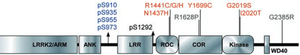
The PARK8 locus on chromosome 12, identified from linkage analyses of a large Japanese family with individuals from multiple generations diagnosed with the pro-gressive neurodegenerative disorder Parkinson’s disease (PD), was first reported in 2002 by Funayama and colleagues. It was not until two years later that independent groups led by A. Singleton and T. Gasser cloned the responsible gene within this locus known as leucine-rich repeat kinase 2, or LRRK2. Typically, PD develops sporadically with the greatest risk factor being age. However, for approximately 10% of cases, a classical Mendelian inheritance with both autosomal dominant and recessive transmissions is present. The six clearly pathogenic single amino acid substitutions that are causative for PD collectively comprise the most frequent genetic causes of PD, with the most common mutation being Gly2019Ser.
The protein encoded by the LRRK2 gene is a large 2527 amino acid multi- domain protein comprised of several well-defined protein interaction domains as well as a Ser/Thr kinase domain and a small GTPase-like domain (ROC; Ras of complex proteins). The kinase and GTPase domains are interrupted by a COR (C-terminal of ROC) domain characteristic of the ROCO protein family. Likewise, the domain structure and kinase domain in particular of LRRK2, and the related LRRK1, bear significant similarity to the receptor-interacting protein kinase (RIPK) family. Clearly, its complex domain structure and widespread expression, including, in addition to the brain, high levels in the kidney and lung as well as circulating immune cells, predicts a wide range of cellular functions and activities. In the short period of time since its identification, our understanding of LRRK2 biology, as well as our tools to investigate its function, is rapidly evolving. Historically, only the G2019S mutant form of LRRK2, when overexpressed in cell lines, would exhibit a significant alteration in kinase activity, displaying approximately two- to fivefold increases in autophosphorylation or, later on, phosphorylation of model peptide substrates. Multiple studies of the remaining pathogenic LRRK2 mutations reported mixed effects on kinase function. Recently, however, multiple members of the large Rab GTPase family of proteins were identified as true physiological phospho- substrates of LRRK2; and strikingly, virtually all of the pathogenic LRRK2 mutants, as well as several risk factor variants, show elevated phosphorylation of these
substrates. While this represents an important step in our understanding of LRRK2 function, what is still lacking is a better understanding of how mutations in LRRK2 alter its function in such a way, in what cell types, and in response to what external stimuli that results in the progressive loss of dopamine neurons of the substantia nigra pars compacta—the neurodegenerative hallmark underlying the motor symp-toms of PD. With a prominent focus on its role in PD, but always in the larger con-text of a broader range of activities of LRRK2, this book was envisioned to provide a window into the current state of understanding of this complex protein by some of the leaders in the field of LRRK2.
In Part I, we begin with discussions of the genetic and clinical considerations of LRRK2-associated PD, with contributions by Di Fonzo and colleagues and Alcalay and colleagues, respectively. In a gene of this size, there are, expectedly, dozens of sequence variants that show varying degrees of association with developing PD. However, relatively few such mutations, located primarily within the central ROC-COR-kinase signaling core of the protein, demonstrate clear pathogenicity. Even among carriers of those specific mutations, there is considerable variability in the penetrance across different ethnic backgrounds. Despite this, the clinical fea-tures of LRRK2-associated PD are remarkably similar to the much more common idiopathic manifestation of the disease; however, some important clinical and path-ological differences have been reported among the distinct mutations, including altered progression of motor symptoms as well as the absence of classical Lewy body-like inclusions in some cases.
LRRK2 possesses an extraordinarily broad range of cellular functions, dictated not only by its expression in specific cell types but also by a complicated and coor-dinated regulation of its activity. This aspect of LRRK2 biology is covered in detail in Part II of the book. One regulatory mechanism occurs via the phosphorylation of LRRK2, by itself and other kinases, at multiple domains throughout the protein (discussed in the chapter by Nichols). This is in turn kept in check via the action of specific cellular phosphatases (Taymans). While the kinase activity of LRRK2 has received considerable attention in terms of substrate profiling, its requirement for neurodegeneration, and the obvious opportunity for targeted therapeutic strategies, our understanding of the GTPase function of LRRK2, in terms of its reciprocal regulation of kinase activity as well as its activity underlying the pathological effects of mutant LRRK2, is rapidly increasing. This is discussed in detail in the chapter by Moore and colleagues. One of the earliest systems shown to be affected by LRRK2 function, or dysfunction, is autophagic/lysosomal protein degradation. Interestingly, as is the case for many proteins, including another dominantly inherited gene linked to PD, α-synuclein, in addition to being degraded in part through by the autophagic machinery, LRRK2 (particularly mutant forms of the protein) can also modulate the activity of multiple forms of autophagy (Lewis and colleagues).
An important regulatory protein interacting with LRRK2 is 14-3-3. It binds to a cluster of phosphorylated residues located in the N-terminal region of the protein. While the phosphorylation of these residues is dependent upon LRRK2 kinase activity, pharmacological inhibition leads to dephosphorylation at these sites; they are not true autophosphorylation sites within LRRK2. Multiple other kinases,
downstream of LRRK2 activity, have been identified that can phosphorylate these residues. The phospho-dependent binding of 14-3-3 at these sites appears to play a critical role in the subcellular localization of LRRK2. Upon dissociation, LRRK2 redistributes into discrete filamentous structures of unknown function, similar to those seen upon overexpression of certain mutant forms of LRRK2. Although it remains unclear what the composition of these structures is, there is strong evidence supporting the existence of LRRK2 in a dimeric state. Further, it is believed that, at least in terms of kinase activation, the LRRK2 dimer is the active conformation. A comprehensive review of the evidence, and implications, of LRRK2 dimer forma-tion is provided in the chapter by Greggio and colleagues. Beyond its role in neuro-degeneration, LRRK2 plays an important role in many other pathways as well as a result of its prominent activity in the immune system (Dzamko).
The final section of the book focuses on modeling LRRK2 neurodegeneration, the potential links to other PD-related proteins, the mechanisms of neurotoxicity and cellular implications of LRRK2 dysregulation, and efforts to develop therapeu-tically viable inhibitors of LRRK2 activity. Despite the generation of many in vivo models of mutant LRRK2 overexpression, including traditional transgenic models, BAC transgenic lines, and knockin lines expressing mutant LRRK2, as well as viral models, the plurality of in vivo models fails to show evidence of a progressive loss of dopaminergic neurons (Dawson and colleagues). That is not to say that gains have not been made from these efforts; far from it. Using specific promoters, or viral vector approaches, the progressive degeneration of dopaminergic neurons can be elicited by overexpression of mutant forms of LRRK2; and other neuronal patholo-gies have been reported in lines even in the absence of neuronal loss. Conversely, clues to the function of LRRK2 have been discerned from knockout models in non-neuronal tissues; and its important interaction in the neurodegenerative phenotype triggered by α-synuclein overexpression or inflammatory insults (Daher) has been discovered in LRRK2-deficient rats. This is highlighted by the critical discussion of non-cell-autonomous effects of LRRK2, as well as its role in vesicular trafficking (Cookson). While the nature of cell death observed in isolated neuronal cell models of LRRK2 neurodegeneration appears to be apoptotic, whether the same is true at the systems level remains to be seen. The similarity of LRRK2 to the RIP kinase protein family and its interaction with, and activation of, extrinsic death pathway components raise the possibility that other modes of cell death may contribute to the loss of neurons in PD (Rideout). Finally, the book closes with a discussion of the efforts to develop small molecule inhibitors of LRRK2 kinase activity that could potentially be utilized in the clinic (Gray and colleagues). Multiple cellular and in vivo models indicate that mutant LRRK2-induced neuronal death is dependent upon its kinase activity, although the substrates of this activity linked to cell death remain unknown. These efforts, both academic- and industry-wide, have resulted in the discovery of ever more potent and selective inhibitors of LRRK2 kinase activity that are currently being evaluated in safety studies.
This book would not be possible without the time and effort of the contributing authors. I am personally grateful for their generosity, support, and enthusiasm for this project. I would also like to acknowledge the publisher Springer-Nature and
especially would like to thank Simina Calin and Jeffery Taub for their support and guidance. It is my hope and belief that this book will serve as an in-depth introduc-tion and snapshot of the current state of the art in LRRK2 biology and, as high-lighted in each of chapters’ discussion of future directions, will stand as a foundation for the next steps taken in this exciting field.
Part I Clinical and Genetic Considerations of LRRK2 Associated Parkinson’s Disease
1 Leucine-Rich Repeat Kinase (LRRK2) Genetics
and Parkinson’s Disease ... 3 Edoardo Monfrini and Alessio Di Fonzo
2 Clinical Features of LRRK2 Carriers with Parkinson’s Disease ... 31
Meir Kestenbaum and Roy N. Alcalay
Part II Fundamentals of LRRK2 Biology
3 LRRK2 Phosphorylation ... 51
R. Jeremy Nichols
4 Understanding the GTPase Activity of LRRK2:
Regulation, Function, and Neurotoxicity ... 71
An Phu Tran Nguyen and Darren J. Moore
5 LRRK2 and Autophagy ... 89
Claudia Manzoni and Patrick A. Lewis
6 Molecular Insights and Functional Implication
of LRRK2 Dimerization ... 107
Laura Civiero, Isabella Russo, Luigi Bubacco, and Elisa Greggio
7 LRRK2 and the Immune System ... 123
Nicolas L. Dzamko
8 Regulation of LRRK2 by Phosphatases ... 145
Part III LRRK2 Neurodegeneration, Modeling, and Therapeutic Options
9 Models of LRRK2-Associated Parkinson’s Disease ... 163
Yulan Xiong, Ted M. Dawson, and Valina L. Dawson
10 LRRK2 and the “LRRKtosome” at the Crossroads
of Programmed Cell Death: Clues from RIP Kinase Relatives ... 193
Hardy J. Rideout and Diane B. Re
11 Interaction of LRRK2 and α-Synuclein in Parkinson’s Disease ... 209
João Paulo Lima Daher
12 Mechanisms of Mutant LRRK2 Neurodegeneration ... 227
Mark R. Cookson
13 Small-Molecule Inhibitors of LRRK2 ... 241
John M. Hatcher, Hwan Geun Choi, Dario R. Alessi, and Nathanael S. Gray
Roy N. Alcalay Department of Neurology, Columbia University Medical Center, New York, NY, USA
Dario R. Alessi MRC Protein Phosphorylation and Ubiquitylation Unit, University of Dundee, Sir James Black Centre, Dundee, DD1 5EH, UK
Luigi Bubacco Department of Biology, University of Padova, Padova, Italy
Hwan Geun Choi New Drug Development Center, Daegu Gyeongbuk Medical Innovation Foundation, Daegu, South Korea
Laura Civiero Department of Biology, University of Padova, Padova, Italy
Mark R. Cookson Laboratory of Neurogenetics, National Institute on Aging, National Institutes of Health, Bethesda, MD, USA
João Paulo Lima Daher Faculty of Medicine, School of Medical Sciences, University of New South Wales, Sydney, NSW, Australia
Neuroscience Research Australia, Randwick, NSW, Australia
Ted M. Dawson Department of Pharmacology and Molecular Sciences, Johns Hopkins University School of Medicine, Baltimore, MD, USA
Neuroregeneration and Stem Cell Programs, Institute for Cell Engineering, Johns Hopkins University School of Medicine, Baltimore, MD, USA
Department of Neurology, Johns Hopkins University School of Medicine, Baltimore, MD, USA
Solomon H. Snyder Department of Neuroscience, Johns Hopkins University School of Medicine, Baltimore, MD, USA
Valina L. Dawson Solomon H. Snyder Department of Neuroscience, Johns Hopkins University School of Medicine, Baltimore, MD, USA
Adrienne Helis Malvin Medical Research Foundation, New Orleans, LA, USA Department of Physiology, Johns Hopkins University School of Medicine, Baltimore, MD, USA
Neuroregeneration and Stem Cell Programs, Institute for Cell Engineering, Johns Hopkins University School of Medicine, Baltimore, MD, USA
Department of Neurology, Johns Hopkins University School of Medicine, Baltimore, MD, USA
Nicolas L. Dzamko School of Medical Sciences, University of NSW, Kensington, NSW, Australia
Neuroscience Research Australia, Randwick, NSW, Australia
Alessio Di Fonzo IRCCS Foundation Ca’ Granda Ospedale Maggiore Policlinico, Dino Ferrari Center, Neuroscience Section, Department of Pathophysiology and Transplantation, University of Milan, Milan, Italy
Nathanael S. Gray Department of Cancer Biology, Dana Farber Cancer Institute, Boston, MA, USA
Elisa Greggio Department of Biology, University of Padova, Padova, Italy
John M. Hatcher Department of Cancer Biology, Dana Farber Cancer Institute, Boston, MA, USA
Meir Kestenbaum Department of Neurology, Columbia University Medical Center, New York, NY, USA
Patrick A. Lewis School of Pharmacy, University of Reading, Reading, UK Department of Molecular Neuroscience, UCL Institute of Neurology, London, UK
Claudia Manzoni School of Pharmacy, University of Reading, Reading, UK Department of Molecular Neuroscience, UCL Institute of Neurology, London, UK
Edoardo Monfrini IRCCS Foundation Ca’ Granda Ospedale Maggiore Policlinico, Dino Ferrari Center, Neuroscience Section, Department of Pathophysiology and Transplantation, University of Milan, Milan, Italy
Darren J. Moore Center for Neurodegenerative Science, Van Andel Research Institute, Grand Rapids, MI, USA
An Phu Tran Nguyen Center for Neurodegenerative Science, Van Andel Research Institute, Grand Rapids, MI, USA
Diane B. Re EHS Department and Motor Neuron Center, Columbia University, New York, NY, USA
Hardy J. Rideout Division of Basic Neurosciences, Biomedical Research Foundation of the Academy of Athens, Athens, Greece
Isabella Russo Department of Biology, University of Padova, Padova, Italy
Jean-Marc Taymans Université de Lille, Inserm, CHU Lille, UMR-S 1172— JPARC Jean-Pierre Aubert Research Center, Neurosciences and Cancer, Lille, France
Yulan Xiong Neuroregeneration and Stem Cell Programs, Institute for Cell Engineering, Johns Hopkins University School of Medicine, Baltimore, MD, USA Department of Neurology, Johns Hopkins University School of Medicine, Baltimore, MD, USA
Department of Anatomy and Physiology, Kansas State University, College of Veterinary Medicine, Manhattan, KS, USA
Clinical and Genetic Considerations
of LRRK2 Associated Parkinson’s Disease
3 © Springer International Publishing AG 2017
Leucine-Rich Repeat Kinase (LRRK2)
Genetics and Parkinson’s Disease
Edoardo Monfrini and Alessio Di Fonzo
Abstract The discovery of LRRK2 mutations as a cause of Parkinson’s disease
(PD), including the sporadic late-onset form, established the decisive role of genet-ics in the field of PD research. Among LRRK2 mutations, the G2019S, mostly lying in a haplotype originating from a common Middle Eastern ancestor, has been identi-fied in different populations worldwide. The G2385R and R1628P variants repre-sent validated risk factors for PD in Asian populations. Here, we describe in detail the origin, the present worldwide epidemiology, and the penetrance of LRRK2 mutations. Furthermore, this chapter aims to characterize other definitely/probably pathogenic mutations and risk variants of LRRK2. Finally, we provide some general guidelines for a LRRK2 genetic testing and counseling. In summary, LRRK2 discov-ery revolutionized the understanding of PD etiology and laid the foundation for a promising future of genetics in PD research.
Keywords Leucine-rich repeat kinase 2 • LRRK2 • Dardarin • Parkinson’s disease
• PARK8 • Parkinson’s disease genetics • Familial Parkinson’s disease • LRRK2 mutations
Until the discovery of leucine-rich repeat kinase 2 (LRRK2) mutations as a genetic cause of Parkinson’s disease (PD), the hereditary influences on PD were limited to observation of rare autosomal dominant familial cases harboring highly penetrant SNCA (alpha-synuclein) mutations and juvenile or young onset autosomal recessive forms carrying PRKN, PINK1, and DJ-1 mutations. This scenario was more sugges-tive of a minor role played by genetic factors in PD, especially considering the com-mon sporadic late-onset form. The innovative finding of LRRK2 low penetrant mutations in common forms of PD revolutionized this outdated view.
E. Monfrini • A. Di Fonzo (*)
IRCCS Foundation Ca’ Granda Ospedale Maggiore Policlinico, Dino Ferrari Center, Neuroscience Section, Department of Pathophysiology and Transplantation, University of Milan, Milan, Italy
Genetic Contribution in Etiology of PD
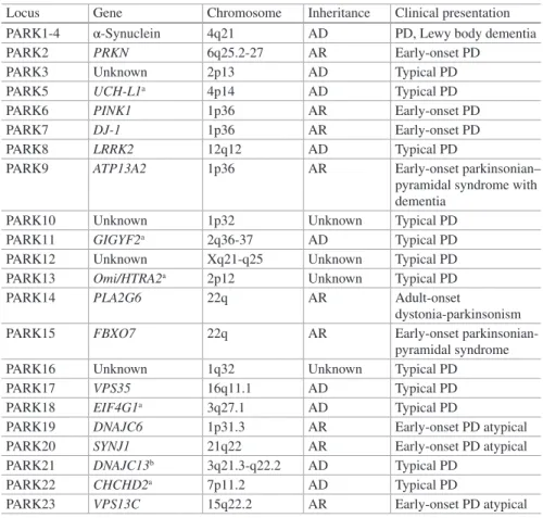
Epidemiological studies reveal that 10–15% of PD have a positive familial history for the disease, while the majority of cases are sporadic. Through linkage analysis and positional cloning approaches, five genes have been definitely implicated in the etiology of PD. Mutations in the SNCA [1, 2], LRRK2 [3, 4], and VPS35 [5, 6] genes cause autosomal dominant forms, whereas mutations in the PRKN [7], DJ-1 [8], and PINK1 [9] genes cause autosomal recessive forms of PD. Furthermore, mutations in the ATP13A2 [10], PLA2G6 [11], FBXO7 [12, 13], DNAJC6 [14], and SYNJ1 [15] have been reported as rare causes of early-onset parkinsonism with atypical clinical features which might be mechanistically distinct from classical PD. Finally, muta-tions in UCH-L1 [16], Omi/HtrA2 [17], GIGYF2 [18], EIF4G1 [19], and DNAJC13 [20] genes have also been described in PD cases, but their role in the disease remains uncertain. Another three PD loci have also been mapped (PARK3, PARK10, PARK12, PARK16) [21–23], but the defective genes remain unknown (Table 1.1).
Table 1.1 List of loci, genes, patterns of inheritance, and clinical presentations of genetic forms
of Parkinson’s disease
Locus Gene Chromosome Inheritance Clinical presentation PARK1-4 α-Synuclein 4q21 AD PD, Lewy body dementia PARK2 PRKN 6q25.2-27 AR Early-onset PD
PARK3 Unknown 2p13 AD Typical PD PARK5 UCH-L1a 4p14 AD Typical PD
PARK6 PINK1 1p36 AR Early-onset PD PARK7 DJ-1 1p36 AR Early-onset PD PARK8 LRRK2 12q12 AD Typical PD
PARK9 ATP13A2 1p36 AR Early-onset parkinsonian– pyramidal syndrome with dementia
PARK10 Unknown 1p32 Unknown Typical PD PARK11 GIGYF2a 2q36-37 AD Typical PD
PARK12 Unknown Xq21-q25 Unknown Typical PD PARK13 Omi/HTRA2a 2p12 Unknown Typical PD
PARK14 PLA2G6 22q AR Adult-onset
dystonia-parkinsonism PARK15 FBXO7 22q AR Early-onset
parkinsonian-pyramidal syndrome PARK16 Unknown 1q32 Unknown Typical PD PARK17 VPS35 16q11.1 AD Typical PD PARK18 EIF4G1a 3q27.1 AD Typical PD
PARK19 DNAJC6 1p31.3 AR Early-onset PD atypical PARK20 SYNJ1 21q22 AR Early-onset PD atypical PARK21 DNAJC13b 3q21.3-q22.2 AD Typical PD
PARK22 CHCHD2a 7p11.2 AD Typical PD
PARK23 VPS13C 15q22.2 AR Early-onset PD atypical
aNot confirmed by other studies
In addition to the Mendelian forms of PD, genetic risk factors for the disease have been investigated in several candidate genes and, more recently, in genome- wide association studies [24]. With the exceptions of SNCA, microtubule-associated protein tau (MAPT), and HLA region [25–32], none of the loci reported have so far been convincingly replicated in independent studies.
Another exception is represented by the glucocerebrosidase gene (GBA) involved in a recessive neurometabolic disease (Gaucher’s disease). Screening of PD patients for GBA mutations showed a higher number of heterozygous mutations carriers as compared to healthy controls. Mutations have been found in about 2–4% of Caucasian PD patients and less than 1% of controls [33].
The LRRK2 Gene: Mapping and Cloning
Although the discovery of mutations in the SNCA, PRKN, PINK1, and DJ-1 genes clearly contributed to our understanding of the pathogenesis of PD, they were iden-tified in a limited number of PD cases, often with early-onset or pathologically atypical features.
A new locus for PD, termed PARK8, was identified in a large family with auto-somal dominant PD, known as the “Sagamihara family” from the region in Japan where the family originated from [34]. The clinical features in affected individuals of the kindred were reported to resemble very closely classical PD, with an average of symptoms onset at 51 ± 6 years. A pattern of “pure nigral degeneration” without Lewy bodies (LB) was found at autopsy in six PD patients examined, another carrier of the disease haplotype developed multiple system atrophy type P-like pathology, and one showed classical LB pathology [35]. In this family, a genome-wide linkage scan yielded significant evidence for linkage of PD to the centromeric region of chromosome 12 (12p11.2-q13.1). The haplotype analysis suggested an incomplete penetrance of the mutation [34, 35]. In 2004 the linkage to PARK8 was confirmed in two Caucasian families, “family A” (a German–Canadian kindred) and “family D” (from Western Nebraska) with dominantly inherited neurodegeneration [36], and thereafter in several Basque PD [37] families suggesting PARK8 to be a rela-tively common locus and refining the critical region. A wide clinical–pathological spectrum was shown in these families, including typical PD but also dementia and amyotrophy, diffuse LB and tau pathology, nigral degeneration without inclusions, and atypical, ubiquitin-positive inclusions [38].
In 2004 two independent groups, by positional cloning, identified mutations in a gene at that time annotated as DKFZp434H2111, which cosegregated with PD in several PARK8-linked pedigrees [3, 4]. The gene was renamed LRRK2 (leucine-rich repeat kinase 2) and the encoded protein LRRK2 or dardarin (from the Basque term dardara, meaning tremor, since resting tremor was a consistent clinical feature of the Basque patients who carried LRRK2 mutations).
Subsequently, early in 2005, several groups identified a single LRRK2 mutation (c.G6055A) leading to a G2019S substitution in the encoded protein, which was
present in familial and sporadic PD with unprecedented high frequency [39–42]. The following years have seen an explosion of research into the LRRK2 gene in PD and related disorders. The I2020T mutation was detected as the cause of disease in the original “Sagamihara family” [43].
The G2019S Mutation
Prevalence of G2019S Across Populations
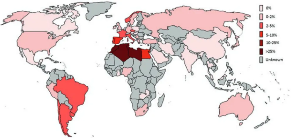
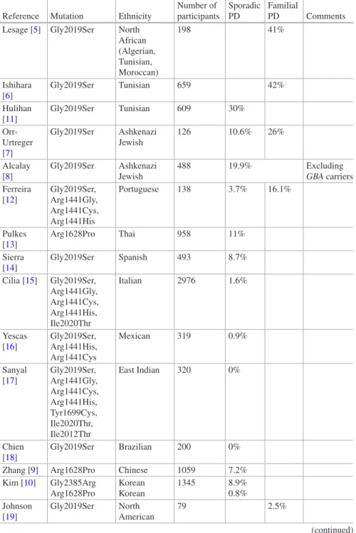
G2019S is particularly important among the PD-causing mutations in LRRK2. This mutation was identified by several groups as a common cause of the disease, being detected initially in ~5–6% of large cohorts of familial PD in Europe and the USA [39, 41] and in ~1–2% of sporadic PD from the UK [40]. Due to the unprecedented high frequency in familial and late-onset classical parkinsonism, which in the past would have been identified as “idiopathic PD,” this specific mutation has been extensively studied worldwide (Fig. 1.1).
So far, large screenings revealed that the frequency of G2019S is population specific. The G2019S mutation has been reported at the highest frequency (up to 37%) among familial PD cases of North African descent and in familial Ashkenazi Jewish patients (23%) [44, 45]. Similar frequencies were replicated in independent studies on PD cases from Tunisia [46–48] and in Ashkenazi Jews [48–51]. Remarkably, the frequency of this mutation was considerably high among sporadic cases (41% North Africans and 13% Ashkenazi Jews) and rarely identified in healthy controls too (3% North Africans and 1.3% Ashkenazi Jews). Other studies reported the presence of the G2019S among 1–2% of healthy North Africans, Ashkenazi, and
Fig. 1.1 Rough estimates of worldwide G2019S prevalence in PD patients (familial and
Sephardic Jewish subjects [49, 50, 52, 53]. So far the G2019S mutation has not been found in sub-Saharan Africans, with exceptions of South Africans where the mutation was present in subjects with European and Jewish ancestry only [54–57]. Little is known about the prevalence in Middle Eastern populations. G2019S is rare in Turkey [58] and has not been identified so far in Yemenite Jews [51] and in Iran [59].
In Western Europe there is a south–north gradient of frequency. The G2019S is found in 9–16% of familial and 3–4% of sporadic PD patients in Portugal [60, 61]; it accounts for 6–16% of familial and 2–6% of sporadic PD in different regions of Spain: Catalonia [62], Cantabria [63, 64], Asturias [65], Galicia [63], and Basque regions (patients without Basque ancestry) [66], while it is less common in patients of Basque origin (1–2%) [66].
In Italy, the G2019S mutation has been reported up to 6–7% of familial and ~1–2% of sporadic cases [39, 67–71]. Similarly, in France the mutation accounts for ~3.5% of familial and ~1.9% of sporadic cases [44, 72–75]. Two independent screening in Sardinians, an isolated population, reported a frequency of ~1.5% in both familial and sporadic cases [76, 77]. Interestingly, the mutation that appeared to be common in the western Mediterranean basin is instead very rare in Greece and Crete [78–81].
A slightly lower frequency was reported in UK screenings of PD patients of Caucasian ethnicity (2.5% familial and 0.3–1.6% sporadic) [40, 82, 83] and also in populations of Celtic and Baltic origin (Ireland 1.1% of familial PD [42, 84], Norway ~1.5% of familial PD [85], and Sweden 1.4% of sporadic cases [86]). Mutation analyses in more than 300 familial and 1200 sporadic PD in Germany sug-gested a very low frequency of this mutation (0.8% of familial cases [87, 88] 0.2– 0.9% of sporadic) [87–89], as well as in Belgium [90], the Netherlands [91], Denmark [92], and Austria [93].
In Poland, Serbia, Hungary, Czech Republic, and Slovakia, the G2019S appeared to be rare (or found in a single subject) [87, 94–98]. On the contrary, four studies have been performed in Russia, where the mutation accounts for 4–7% of familial and 1% of sporadic cases [99–102]. However, subjects were included from a mixed ethnic background, since at least two PD families and one sporadic case reported their ethnic origin as Ashkenazi Jews [101].
An analogous observation can be done when analyzing patients from the USA, where the frequency of the G2019S in Caucasian PD reaches 2–3.5% in familial and 0.5–1.6% in sporadic cases [46, 49, 103–109]; it seems to be rare among American Indians and Afro-Americans (but the sample size for these two ethnic groups is still insufficient to make firm conclusions) [108, 109], whereas it was reported to be higher when patients of Ashkenazi Jewish ancestry were included [49]. In Canadian PD patients, the G2019S is rare/absent [110, 111].
Four different populations of South America, where the Spanish, Portuguese, and Italian ethnic backgrounds are strong, have been studied for this mutation. In Uruguay [112], Chile [113], and Argentina [114], G2019S accounts for 3.5–5.5% of familial and 2.9–4.2% of sporadic cases. The 5.45% of a PD cohort from Argentina was found to carry the mutation, all of them being of Jewish ancestry. While in Peru, the G2019S appears to be rare [112]. Controversial results came from large screening
in Brazil (from 3 to 6.8% in familial and 0 to 1.7% in sporadic cases), probably due to the high degree of ethnic heterogeneity within the study cohorts [115–117].
G2019S is rare/absent among Chinese patients with familial and sporadic PD [118–121], as well as in Korea [122, 123] and in India [121, 124]. So far, only three patients have been reported with this mutation in Japan (0.7% of sporadic cases) [48, 125].
Finally, the mutation is present in Australia, among PD patients with European ancestry (2–6% of familial, 0.4% of sporadic PD) [126, 127], while it has not been identified in Australian Aboriginal.
Taken together, these data show that a single LRRK2 mutation represents the most frequent known genetic determinant of PD. The frequency of the G2019S mutation varies widely across populations, indicating that ethnicity is an important factor. For some populations, independent studies on the prevalence of the mutation are already available, and often, the reported results are consistent. These observa-tions imply that most neurologists who treat patients with movement disorders will see patients with LRRK2-related PD that may be addressed to genetic testing. This estimation could be even higher if we include the other LRRK2 definitely patho-genic mutations.
Origins of the G2019S Mutation
So far three different haplotypes have been identified in patients carrying the G2019S mutation.
Haplotype 1
The first studies on unrelated carriers of the G2019S of European or Middle Eastern– North African origin revealed that all shared the same haplotype, consistent with a common founder [42, 44, 45, 70, 128, 129].
Subsequently, the same haplotype has been identified among subjects carrying the G2019S mutation from Italy (independent subset) [71], France [47], Germany [87], Russia [99] Sardinia [76, 77], Spain [65], Portugal [61, 70], Brazil [70], Chile [113], Uruguay and Peru [112], and Australia [126].
According to a general rule in population genetics, the geographic center of the origin of a mutation corresponds to the area where that mutation is most frequent [130]. The highest prevalence of the G2019S mutation has been reported in Berbers [52], followed by North African Arabs, Ashkenazi, and Sephardic Jews. The fre-quency data combined with the identification of a common haplotype among these populations support the hypothesis that the mutation of haplotype I originated in North Africa or in the Middle East and then spread to other countries following the patterns of migration.
Further studies provided important insights on the estimated age of the common founder for the haplotype 1 carriers. Analyzing the haplotypes of European and Ashkenazi Jews [129] and Tunisian G2019S carriers [131], the age estimated of the common ancestor (using the 30-year intergeneration interval) was 2250 (95% CI 1650–3120) and 3120 (95% CI 2340–4620) years ago, respectively. A third study, on Ashkenazi Jews only, estimated a more recent founder approximately 1525– 1830 years ago (150–450 A.D.) [132]. This estimation would fit with the absence of the G2019S in Yemenite Jews [51]. The Yemenite Jews evolved completely separate from all of the other Jewish populations. Most of them arrived in Yemen in the early second century A.D. (~160 A.D.). Finally, a multicentric study proposed a consen-sus of haplotype 1 origin, estimating the founding mutational event in Ashkenazi Jews ancestors in a period ranging from 4500 to 9100 years. In this scenario, being the Ashkenazi Jews history more recent (at most 2000 years old), it is possible that the G2019S have arisen at least 4000 years ago in the Near East and then Ashkenazi ancestors may have kept the mutation through the different diasporas. Thereafter, the mutation may have been reintroduced by gene flow from Ashkenazi Jews to other European and North African populations [133].
Haplotype 2
A different G2019S haplotype was identified in three families from Western Europe, which appeared to share a more recent founder than haplotype 1. The geographic origin of this haplotype is less certain [129].
Haplotype 3
The third haplotype has been found in Japanese patients carrying the G2019S mutation [125]. This haplotype differs across the markers closest to the mutation, which would suggest an independent origin of the mutation in Japanese and European populations rather than a single ancient founder. Interestingly, the haplo-type 3 has also been observed in a single sporadic Turkish patient [134]. This may be the result of a common ancestry (plausibly explained by the large centuries-long migration of the Turkic people across Central Asia) or coincidental presence of Japanese ancestors.
Incomplete Penetrance of G2019S
Incomplete penetrance was already suspected for the mutations underlying the PARK8 locus at the time of the linkage studies. Most of the penetrance analyses have been performed on the frequent G2019S mutation.
Analyses performed on Ashkenazi Jews from the USA revealed a lifetime pene-trance of 31.8% [45]. A slighter lower penetrance (24–26% at 80 years) was esti-mated in independent groups of US Ashkenazi Jews [49, 135].
The International LRRK2 Consortium performed a penetrance study on the larg-est dataset of G2019S carriers. By analyzing a large sample of PD patients, they calculated a 28% risk of PD at 59 years, 51% at 69 years, and 74% at 79 years for LRRK2 G2019S carriers without differences in penetrance by sex or ethnic group [136]. Interestingly a penetrance study in Tunisian G2019S PD cases, after stratify-ing by homozygous (n = 23) and heterozygous carriers, reported a penetrance con-sistently higher in homozygotes in each age group. Considering possible biases in estimating penetrance only from families, this finding, if true, would indicate a gene dosage effect, although the age of onset was not dissimilar between the two groups [46]. However, subsequent studies collecting clinical data of homozygous carriers showed no phenotype differences between heterozygous and homozygous carriers ruling out a gene dosage effect [44, 137, 138].
The reduced penetrance of this frequent mutation is in keeping with the LRRK2 G2019S being the most important genetic determinant, known so far, of sporadic PD. Penetrance can also be expressed in terms of risk (calculated as odd ratio) to develop the disease. For an Ashkenazi Jew who carries the G2019S, the risk of developing PD increases ~18-fold [45]. By analyzing the G2019S in North Africans, a lifetime odds ratio for developing PD of 48.6 (CI 11.2–211.0) [44] has been calculated.
Nevertheless, additional studies in different populations are warranted before G2019S genetic counseling can be implemented, since the precise estimation of the penetrance in some countries is still controversial.
Dissimilar results across the abovementioned Ashkenazi Jews from the USA and other G2019S carriers might be influenced by different methodological approaches (e.g. including only patients with both parents genotyped, excluding patients with GBA mutations, etc.) or by additional genetic or nongenetic factors that can act as modifiers.
The analysis of candidate genes involved in neurodegeneration as potential genetic modifiers of LRRK2 has been reported. The first to be explored was PRKN, since patients who simultaneously harbored PRKN mutations and LRRK2 G2019S have been mentioned in several studies [61, 73, 99, 139–141]. However, the clinical and cosegregation analysis of patients carrying heterozygous PRKN mutations and the G2019S revealed that the combination of the two does not influence the symp-toms or the age at disease onset [142].
Polymorphic variations in the microtubule-associated protein tau (MAPT) have been proposed to be significantly associated with age of disease onset in individuals with LRRK2 mutations [143]. Moreover, SNCA variants have been found as deter-minant of age of onset in G2019S carriers [144]. It is a common observation among neurologists of the different penetrance of LRRK2 mutations in affected families, implying the great importance of genetic modifiers. Further analyses, especially on large samples and families carrying the G2019S, are warranted to identify genetic factors that can act as modifiers of LRRK2 mutations.
The R1441 Mutational Hot Spot
The LRRK2 R1441 residue is the second most common spot of pathogenic LRRK2 mutations, after G2019S. Three non-synonymous substitutions (R1441C, R1441G, and R1441H) and the synonymous R1441R have been reported in several patients.
R1441C: The Second Most Frequent Pathogenic LRRK2 Mutation
This mutation (c. 4321C > T) represents the second known most common mutation of the LRRK2 gene. The R1441C was identified as causative mutation of the PARK8-linked “family D” (Western Nebraska) [4]. Cosegregation was reported also in smaller PD families from Germany [4, 87], Italy [69], Belgium [90], the USA [42, 145], and Iran [59]. The mutation has also been reported in a few other families, but additional affected relatives were not available for cosegregation anal-ysis [62, 69, 90, 146]. The R1441C is also found among sporadic cases and has been reported in patients from Italy [70], Sardinia [77], Russia (Slavic origin) [100], China [147], and Belgium [90]. The variant was absent in large cohorts of ethnically matched controls (>1000 German, 530 Italian, 208 Sardinian, 400 Chinese, 178 Belgian, and 300 American). Interestingly, the R1441C has been found to be more common than G2019S in southern Italy [148].
Haplotype analysis of LRRK2 R1441C carriers from 20 families of different geo-graphical areas revealed in total four classes of haplotypes. Only for the two major haplotypes, the phase could be established [149]. A first haplotype was identified in the Italian carriers, as well as in German, Spanish, and American patients. A second haplotype was present in the American family D (Western Nebraska) and in Belgian R1441C families. A German and an Irish patient shared a third haplotype for which phase could not be unambiguously determined. Finally, a Chinese proband carried alleles that could not be assigned to any of three previous haplotype classes.
The phenotype associated with this mutation is similar to that of classic PD [149]. The mutation exhibits incomplete penetrance, which could explain its pres-ence in sporadic cases, but calculations performed so far must be interpreted with caution as only a small number of R1441C mutation carriers have been identified until now.
R1441G: A Founder Pathogenic Mutation in the Basques
The LRRK2 R1441G (c. 4321C > G) was initially described in patients with autoso-mal dominant late-onset PD in PARK8-linked families of Basque ethnicity [3]. The Basques are a homogeneous ethnic group who historically were isolated by linguis-tic and geographical barriers. The first report on the frequency of this mutation in Basque PD (~8% of familial cases) [3] and the absence in other large populations screened (except for a US patient reported to be of Hispanic descent [50]) suggested that this variant was population specific. Further studies investigated the prevalence
of this mutation in Basque. One group detected the R1441G in 16.4% and 4.0% of familial and sporadic Basque PD, respectively [150], while a more recent study reported a prevalence of 46% in familial Basque patients and 2.5% of sporadic cases [66]. It has also been identified at lower frequencies in patients from nearby prov-inces in Spain who did not report Basque ancestry (6% of non-Basques living in the Basque countries [66], 2.7% in Asturias [151], 0.7% in Catalonia [62], two families from the neighboring region of Navarre, and one from La Rioja [63]), while it is rare in Cantabria [64]. Haplotype analysis on R1441G carriers from Basque and neigh-borhood regions [63, 150, 152] indicates that this mutation occurred in a single com-mon ancestor, which in one study was estimated to have lived 1350 (95% CI, 1020–1740) years ago [152]. Since the Basque population has a history of emigra-tion to Europe and North, Central, and South Americas, it would not be surprising to find isolated cases in those countries. However, a single case from Uruguay and a family from Japan carrying the R1441G have been reported with a different hap-lotype than the Basque, suggesting in these cases independent mutational events [112, 153].
R1441H and R1441R: Uncommon but also Likely Pathogenic
This variant, c.G4322A on LRRK2 cDNA, occurs immediately adjacent to the two pre-viously reported pathogenic mutations, c.C4321T (R1441C) and c.C4321G (R1441G), resulting in a different substitution of the same amino acid residue (R1441H).
R1441H has been described in a US PD family, but only the proband and an unaffected sibling were available for testing [146]. It was also reported in PD fami-lies from Crete [81], Portugal [61], and Taiwan [84], all not large enough to demon-strate definitive cosegregation with the disease.
Haplotype analysis of the abovementioned R1441H carriers showed diversity suggesting a number of independent founders [154]. Subsequently, the R1441H mutation has been identified in two cases from Australia, both of British origin and with a possible common haplotype, although in these cases the phase was not assessed [126]. A further proof in favor of a pathogenic role of this variant came from the identification of R1441H in two slightly larger French families [72].
R1441H was not found in 281 Americans, 300 Cretans, 200 Portugueses, 174 Europeans, and a set of 1000 control samples (600 North Americans, 200 Taiwaneses, 200 Norwegians, 200 Irish, and 200 Spanish). Moreover several studies screened by sequence the LRRK2 exon 31 in a large sample of healthy controls (>3000 Caucasian [3, 4, 69, 90]) in order to check for the R1441C and R1441G, and none reported mutation in the adjacent nucleotide.
The clinical presentation of affected R1441H carriers appears to be similar to typical Parkinson’s disease with an age at onset range of 32–66 years. All display levodopa-responsive parkinsonism; however, the disease in one of the siblings from the Greek R1441H family appeared to transition into a progressive supranuclear palsy-like disorder [81].
To further highlight the nature of codon 1441 as a mutational hot spot, two groups reported a R1441R (c.C4323T) in a sporadic PD patient [101] from Russia
and a PD patient with ascertained LB pathology who additionally developed demen-tia and dysautonomia (PDD) [155]. As for the R1441H, we can indirectly assume that the variant is rare in the Caucasian population, since sequencing controls for the other mutations at the same codon did not reveal any R1441R carriers. This variant is predicted to lead to a synonymous substitution, which would suggest a nonpatho-genic role. Moreover, being the nucleotide change close to the splice site, cDNA analysis from the brain of the PDD patient was performed and did not reveal any aberrations on the LRRK2 transcript [155]. Taken together these results suggest that R1441R is likely to represent a rare but nonpathogenic polymorphism.
Mutations in LRRK2 are associated with pleomorphic pathology, although the Lewy bodies (LB)-positive pathology is the most common pattern, particularly for the G2019S mutation [38, 40, 82, 156, 157].
In a large screen of 405 LB-positive brains, eight (~2%) have been found to be carriers of the G2019S mutation, including four with brainstem type, three with transitional type, and one with diffuse LB pathology. In two G2019S-positive brains, Alzheimer-type pathology was also present, and it was of enough severity to make a concomitant pathological diagnosis of Alzheimer’s disease [157].
A further study on 80 brains with PD or LB dementia screened for the G2019S mutation, and three were found to be carriers. Typical brainstem-type LB-positive pathology was found in one, while the Lewy body variant of Alzheimer’s disease was diagnosed in the second. The third brain showed only cell loss in the substantia nigra and locus coeruleus, but no α-synuclein inclusions were detected. There were only rare tau-positive tangles and occasional plaques. No other ubiquitin-positive inclusions were present either [156].
In family D (Western Nebraska), all R1441C carriers examined showed substan-tia nigra neuronal loss. Two cases had LB pathology, one brainstem type, and the other one diffuse type. The third case had “nonspecific” substantia nigra degenera-tion with ubiquitin-positive neuronal inclusions. The final case had PSP-like changes with tau-immunoreactive neuronal and glial lesions [4].
The neuropathological examination of R1441G Basque carriers displayed “non-specific” nigral degeneration in the substantia nigra without α-synuclein, tau, or ubiquitin inclusions [158].
Japanese cases with the I2020T mutation were found to display hyperphosphory-lated tau aggregates [159]. Therefore, despite LBs represent the predominant fea-ture in neuropathological studies, the overall LRRK2-associated pathology has revealed great variability, probably recapitulating the heterogeneity of PD itself, which can be a more complex disease than what we thought until now.
The Other LRRK2 Variants: Which Are Pathogenic?
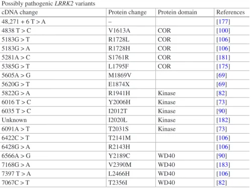
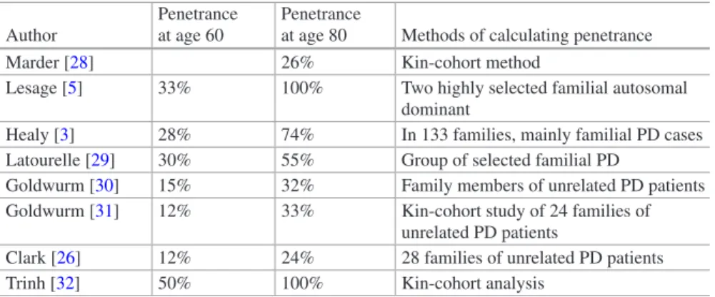
Besides the most recurrent G2019S and R1441C/R1441G/R1441H, more than 50 different LRRK2 sequence variants have been reported in familial and sporadic PD cases so far; moreover, few novel LRRK2 substitutions have been found in healthy control subjects only (Fig. 1.2).
Fig . 1.2 Schematic representation of the LRRK2 gene, the dardarin protein, and its kno wn functional domains. LRRK2 83 coding variants and three putati ve splice v
ariants are grouped according to e
vidence of pathogenicity (see main te
The Y1699C and I2020T mutations are considered as definitely pathogenic. The Y1669C was identified in two independent large families, the Lincolnshire kindred [3, 82] (family PL) of European ancestry, and “family A” (German–Canadian) [4,
38]. The I2020T was identified in “family 32” [4] and “T10738” [89], both of German ancestry. Additionally the same mutation was identified segregating in the large PARK8-linked Sagamihara kindred [43] and in two smaller Japanese families coming from the neighborhood of the Sagamihara region [48].
The role of several other variants remains unclear, since often no family mem-bers were available to assess cosegregation and a limited number of ethnically matched controls were screened. Overall, the criteria that may be applied to con-sider the pathogenicity of the LRRK2 variants should concon-sider several standpoints: frequency in healthy subjects, cosegregation in families, confirmation in indepen-dent studies, and pathogenic consequences on cellular and animal models.
Association Studies on LRRK2
In the past few years, many groups put special effort in search of common risk fac-tors for complex diseases. Among these, PD and other neurodegenerative disorders have been extensively studied. However, even using high-throughput techniques allowing to genotype hundreds of thousands of SNPs and covering the whole genome in cases and controls (genome-wide association studies, GWA), no repro-ducible risk loci have been reported so far.
One caveat is that the GWA approach can be problematic because the massive number of statistical tests performed presents an unprecedented potential for false- positive results.
After the discovering of mutations in the LRRK2 gene, several studies aimed to explore whether common variant of this gene could represent a risk factor for PD.
Two association studies on LRRK2 have been performed in Caucasians. The first enrolled 340 PD patients and 608 controls from Germany. 121 SNPs (81 tagging SNPs) were genotyped attempting to represent the complete DNA variation of the LRRK2 gene [160]. The second study analyzed four common coding SNPs (L953L, R1398H, G1624G, and T2397M) in 250 controls and 121 unrelated PD, mostly with early-onset and positive family history [141]. Neither of these studies revealed any evidences of association between PD and the LRRK2 SNPs at both allelic and genotypic levels.
In 2005, one study performed in Singapore yielded a significant association. A set of 21 tagging SNPs covering the LRRK2 gene were genotyped in 466 sporadic PD and 374 control individuals all of Chinese ancestry. The authors identified a common haplotype that was highly overrepresented within cases (p = 0.005) and, when present in two copies, significantly increased the risk of PD (OR = 5.5, 95% C.I. = 2.1–14.0, P = 0.0001) [161]. However, no LRRK2 variants within the risk haplotype were reported as the biologically relevant factors.
The G2385R Variant
The LRRK2 G2385R represents the first common genetic risk factor for PD in the Asian population. This variant was first reported in a small PD family from Taiwan [84]. Evidence for cosegregation with PD in that family was limited due to the small pedigree size; however, the mutation was reported to be absent in 200 ethnically matched controls and, therefore, interpreted as putatively pathogenic. At that time very limited data were available on the nature and frequency of LRRK2 mutations and on the polymorphism content of the gene in patients from Asia.
Several groups conducted a mutational screening of three known PD-causing mutations (I2012T, G2019S, and I2020T) which appeared to be very rare or absent in Asian PD patients [43, 118, 120, 121]. A sequence of the whole LRRK2 in Chinese Han patients revealed four coding variants (A419V, P755L, M1869V which were novel substitutions, and the G2385R) that were tested for association with PD in 608 Chinese Han cases and 373 ethnically matched controls.
The heterozygosity for the G2385R variant was significantly higher among PD cases than controls (10% vs 4% p 0012). This suggested that the G2385R variant, or another variant in linkage disequilibrium, is associated with PD in the Taiwanese population.
Since then, several association studies on Asian populations from Taiwan, Singapore, Mainland China, Korea, and Malaysia replicated this finding with a sim-ilar size effect. Interestingly the association was also reported in Japanese PD patients and controls, giving a risk of developing PD increased of ~twofold [125,
166] (Table 1.2).
Two groups performed a haplotype analysis of G2385R carriers in a cohort of Chinese Han from Taiwan [165, 168]. A single common haplotype shared by carri-ers has been identified, likely originated from a single ancestor who lived approxi-mately 4800 years ago. Also all Japanese G2385R carriers shared the same haplotype, with a set of markers (D12S2516, D12S2519, and D12S2521) which overlapped with the Chinese haplotype. This might suggest that the G2385R of Chinese Han and Japanese ancestry has arisen from a common ancestor [125].
The R1628P Variant
The LRRK2 R1628P has been identified in a multicentric study which combined 1986 Chinese individuals from three independent centers in Taiwan and Singapore and so far represents the second most frequent genetic risk factor for PD in Asia [184]. This variant was approximately twice as frequent in affected individuals as control subjects (~6% of PD and ~3.5% of controls, odds ratio 1.84, 95% C.I.: 1.20–2.83, nominal p value = 0.006) [184].
Table 1.2 Association studies on Asian populations (from Taiwan, Singapore, China, Korea,
Japan, and Malaysia) showing the G2385R variant as significantly associated with Parkinson’s disease
Geographical
location Ethnicity PD Controls OR (95% CI) References Taiwan Chinese Han 61/608 (10%) 18/373
(4.8%) 2.20 (1.28–3.78) [162] Singapore Chinese 37/495 (7.5%) 18/494 (3.6%) 2.14 (1.20–3.81) [163] Taiwan Chinese Han 27/305 (9%) 1/176 (0.5%) 17.00
(2.29– 126.20) [164] Taiwan Chinese 34/410 (9.3%) 13/335 (3.9%) 2.24 (1.16–4.32) [165] Japan Japanese 52/448 (11.6%) 22/457 (4.8%) 2.60 (1.55–4.35) [166] Singapore Malay 2/98 (2%) 2/173 (1.2%) 1.75 (0.25–12.85) [167] Indian 0/66 (0%) 0/133 (0%)
Shanghai Chinese Han 14/235 (6%) 0/214 (0%) 28.08 (1.66– 473.72) [168] Mainland of China Chinese Han 71/600 (11.8%) 11/334 (3.3%) 3.94 (2.06–7.55) [169] Japan and USA Japanese 69/601
(11.5%) 101/1628 (6.2%) 1.96 (1.42–2.70) [170] Japan Japanese 30/229 (13.1%) 23/358 (6.4%) 2.06 [171] Korea Koreans 82/923 (8.9%) 21/422 (5%) 1.83 [172] Asia Taiwanese 369 (NA) 300 (NA) 1.62 [173]
Korean 844 (NA) 587 (NA) 1.87 Japanese 173 (NA) 95 (NA) 1.44
Malaysia Malaysian 695 (NA) 507 (NA) 2.22 [174] Total 479/5018 (9.6%) 230/5097 (4.5%) 2.23 (1.89–2.62) p value < 0.0001
This finding was replicated in two independent Chinese Han cohorts from Singapore [185] and Taiwan [186]. On the contrary, the R1628P is rare in Japan and in non-Chinese Asians [170, 184, 187].
Haplotype analysis strongly indicates that carriers of the R1628P variant share an extended haplotype, indicative of a founder effect [184]. The mutation has been estimated to arise ~2500 years ago and, in contrast to the older G2385R, has remained confined to subjects of Chinese Han ethnicity.
Like for the G2385R, the clinical phenotype of the affected R1628P carriers is that of typical late-onset L-dopa-responsive PD [184, 186, 187].
Taken together, these studies indicate for the first time that common population specific genetic risk factors for PD exist. The association of both LRRK2 variants with PD in Asia has been extensively confirmed in independent dataset of patients. These findings open several opportunities of studies for researchers and clinicians. Discovering how those variants can increase the risk of death of dopaminergic neu-rons might provide important insight into the pathogenesis of the disease. Other interesting prospects can be provided in clinical practice, for example, studying the effect of neuroprotective drugs in large cohorts of asymptomatic carriers of these two LRRK2 variants, in order to explore whether the risk of developing PD would decrease in the treated subjects.
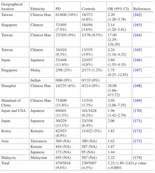
In conclusion, the LRRK2 gene displays a high polymorphic content in terms of single nucleotide substitutions. No deletions or duplications have been identified until now. Variants identified in patients are located in almost all exons. However, most of them still lack a definite proof of pathogenicity (Tables 1.3). This has direct
Table 1.3 LRRK2 genetic variants associated with Parkinson’s disease that are possibly
pathogenic, but need more evidence to be definitely associated with the disease Possibly pathogenic LRRK2 variants
cDNA change Protein change Protein domain References 28G > A E10K LRRK2 repeats [175] 155C > T S52F LRRK2 repeats [72] 632C > T A211V LRRK2 repeats [176] 1000G > A E334K LRRK2 repeats [175] 1088A > G N363S LRRK2 repeats [72] 1630A > G K544E LRRK2 repeats [176] 2134A > G M712V LRRK2 repeats [106] 2242119_2242122delGTAA – LRRK2 repeats [104] 2769G > C Q923R LRRK2 repeats [115] 2789A > G Q930R LRRK2 repeats [89] 2918G > A S973N LRRK2 repeats [93] 3200G > A R1067Q LRRK2 repeats [177] 3287C > G S1096C LRRK2 repeats [89] 3333G > T Q1111H LRRK2 repeats [175] 3364A > G I1122V LRRK2 repeats [4] 3574A > G I1192V LRRK2 repeats [175] 3451G > A A1191T LRRK2 repeats [178] 3494 T > C L1165P LRRK2 repeats [179] 3287G > C S1228T LRRK2 repeats [89] 3974G > A R1325Q [90] 4111A > G I1371V [69] 4309A > C N1437H Roc domain [180] 4324G > C A1442P Roc domain [126] 4402A > G K1468E Roc domain [90] 4448G > A R1483Q Roc domain [90] 45,361 + 3A > G – [146]
![Table 3.1Phosphosites of LRRK2 LRRK2 P-sitePeptide sequenceGloeckner et al. [52]Nichols et al](https://thumb-eu.123doks.com/thumbv2/123dokorg/5514927.64031/71.659.99.593.88.822/table-phosphosites-lrrk-lrrk-p-sitepeptide-sequencegloeckner-nichols.webp)
