ORIGINAL ARTICLE
Imaging of Bronchial Pathology in Antibody Deficiency:
Data from the European Chest CT Group
Katharina Schütz1&Diana Alecsandru2,3&Bodo Grimbacher3,4&Jamanda Haddock5&Annemarie Bruining6& Gertjan Driessen7,8&Esther de Vries9,10&Peter M. van Hagen11&Ieneke Hartmann12&Francesco Fraioli13,14& Cinzia Milito15&Milica Mitrevski15&Isabella Quinti15&Goffredo Serra13&Peter Kelleher16&Michael Loebinger17& Jiri Litzman18&Vera Postranecka19&Vojtech Thon18,20&Judith Babar21&Alison M. Condliffe21&Andrew Exley22& Dinakantha Kumararatne23&Nick Screaton24&Alison Jones25&Maria P. Bondioni26&Vassilios Lougaris27& Alessandro Plebani27&Annarosa Soresina28&Cesare Sirignano29&Giuseppe Spadaro30&Nermeen Galal31& Luis I. Gonzalez-Granado2&Sabine Dettmer32&Robert Stirling33&Helen Chapel34&Mary Lucas34&Smita Patel34& Claire-Michele Farber35&Isabelle Meyts36&Arpan K. Banerjee37&Scott Hackett38&John R. Hurst39&Klaus Warnatz4& Benjamin Gathmann40&Ulrich Baumann1&for the Chest CT in Antibody Deficiency Group
Received: 16 July 2017 / Accepted: 26 November 2018
# Springer Science+Business Media, LLC, part of Springer Nature 2018
Abstract
Studies of chest computed tomography (CT) in patients with primary antibody deficiency syndromes (ADS) suggest a broad range of bronchial pathology. However, there are as yet no multicentre studies to assess the variety of bronchial pathology in this patient group. One of the underlying reasons is the lack of a consensus methodology, a prerequisite to jointly document chest CT findings. We aimed to establish an international platform for the evaluation of bronchial pathology as assessed by chest CT and to describe the range of bronchial pathologies in patients with antibody deficiency. Ffteen immunodeficiency centres from 9 countries evaluated chest CT scans of patients with ADS using a predefined list of potential findings including an extent score for bronchiectasis. Data of 282 patients with ADS were collected. Patients with common variable immunodeficiency disorders (CVID) comprised the largest subgroup (232 patients, 82.3%). Eighty percent of CVID patients had radiological evidence of bronchial pathology including bronchiectasis in 61%, bronchial wall thickening in 44% and mucus plugging in 29%. Bronchiectasis was detected in 44% of CVID patients aged less than 20 years. Cough was a better predictor for bronchiectasis than spirometry values. Delay of diagnosis as well as duration of disease correlated positively with presence of bronchiectasis. The use of consensus diagnostic criteria and a pre-defined list of bronchial pathologies allows for comparison of chest CT data in multicentre studies. Our data suggest a high prevalence of bronchial pathology in CVID due to late diagnosis or duration of disease. Keywords Chest CT . CVID . primary antibody deficiency . bronchiectasis . bronchial pathology
Introduction
Primary antibody deficiency syndromes (ADS) are a hetero-geneous group of immune disorders characterised by
subnormal immunoglobulin levels and/or the inability to mount specific antibody responses [1]. Common variable im-munodeficiency disorders (CVID) are the most frequent hu-moral immunodeficiency requiring immunoglobulin replace-ment therapy with an incidence of approximately 1:25,000– 1:50,000 live births [2]. Agammaglobulinaemia, x-linked agammaglobulinaemia (XLA) together with other variants, forms the second largest group affecting three to six per mil-lion live births [3]. Recurrent bacterial infections of the respi-ratory tract are a major part of morbidity in both conditions, although the frequency and severity are reduced by immuno-globulin replacement therapy [2,4–10]. Airway infections are predominantly caused by encapsulated bacteria and can lead
Claire-Michele Farber is deceased.
Electronic supplementary material The online version of this article (https://doi.org/10.1007/s10875-018-0577-9) contains supplementary material, which is available to authorized users.
* Ulrich Baumann
Extended author information available on the last page of the article https://doi.org/10.1007/s10875-018-0577-9
to persistent structural lung disease such as bronchiectasis [11, 12]. Presence of bronchiectasis is strongly associated with mortality in CVID and XLA [13,14].
Early detection of the presence or progression of structural lung disease is essential to develop preventive or therapeutic strategies in this setting. Imaging techniques, in particular computed tomography (CT), are considered the gold standard for diagnosing structural lung disease [15,16].
Chest X-ray (CXR) and pulmonary function tests (PFT) including spirometry, gas transfer and body plethysmography are readily available and can be repeated frequently, due to lower or absent dose of ionising radiation compared to se-quential CT imaging.
However, both methods lack sensitivity to detect structural lung disease in patients with antibody deficiency [11,17] or other conditions, such as cystic fibrosis (CF) [18].
A sizeable body of literature reports bronchial morbidity in patients with antibody deficiency based on chest CT findings [17,19]. However, these studies were almost exclusively single-centre studies and are not easily comparable, since they used differing reporting systems and nomenclatures [11,20, 21]. The performance of multicentre studies therefore de-mands consensus data definition, reporting and scanning methodology to afford internal validity.
The difficulties of providing a standardised evaluation of chest CT scans in antibody deficiencies may have contributed to the apparent paucity of clinical studies describing preven-tive or therapeutic interventions in this patient group [22].
The present study is the result of an international multicentre and multidisciplinary collaboration aiming to cre-ate a common platform of chest CT findings from patients with primary antibody deficiencies. Based on a large number of chest CT studies, the distribution, variety and extent of pulmonary pathology was assessed. Herein, we report the findings on bronchial pathology in conjunction with clinical data and pulmonary function testing. Based on these findings, we propose a method to document bronchial pathologies for multicentre use such as patient registries.
Methods
Development of a Consensus List of CT Findings
In the Chest CT in Antibody Deficiency Group, immunolo-gists, radiologists and pulmonologists agreed a list of chest CT findings with the aim of collecting pathologies of potential relevance for patients with PAD. The list was based upon clinical experience and review of the literature. In order to use radiological terminology that is both widely accepted and defined by an image repository, the syllabus of the Fleischner Society [15] was used. A short form of definitions
and exemplary images was provided to the participating cen-tres (http://www.chest-ct-group.eu/imagerepository).
The Chest CT Group criteria scored 16 items (Table 1, online repository), including 4 on bronchial pathologies. Bronchial wall thickening was defined as a diameter of a bronchial wall being larger than one-third of the outer diame-ter of the accompanying bronchial ardiame-tery, of more than a fifth of the outer diameter of the bronchus. Bronchiectasis was defined as an airway lumen larger in diameter than the outer diameter of the accompanying bronchial artery. Two items were scored only as present or absent (bronchial wall thicken-ing, atelectasis). Mucus plugging was additionally scored for the distribution pattern (central or peripheral). Bronchiectasis was scored for distribution and extent, the latter comprising a simplified bronchiectasis score (0, 1, 2 or 3,≥ 4 lobes affected or cystic changes in≥ 2 lobes).
Chest CT Scans and Data Collection
Chest CT scans were performed according to local guidelines of the participating centres and evaluated locally. All chest CTs were acquired at full inspiratory capacity by using a thin slice protocol of acquisition.
The diagnosis of the immunodeficiency was based on the diagnostic criteria of the European Society for Immunodeficiencies (ESID) [23]. Only CT scans that were performed in patients with a stable clinical condition were included. The radiologists were requested to employ chest CT scoring system provided by the Chest CT in Antibody Deficiency Group in addition to their local practice.
CT findings along with clinical data were documented with the ESID online registry [24]. Some centres preferred to send the data with an anonymised chest CT documentation sheet (TableI, online repository) to the study centre. All data were stored in a database in an anonymised fashion.
Clinical Data Collection
Similarly, clinical data were collected with a second documen-tation sheet with items identical to the ESID registry (TableII, Clinical Data Sheet in the online repository). The clinical data sheet comprised data on lung function (spirometry, body plethysmography and carbon monoxide diffusion capacity), pattern of cough and use of antibiotics.
Since lung function data were only available as “percent-age of predicted for height and weigth” in several centres, data were collected accordingly (not as absolute measurements). Cough lasting longer than 8 weeks was defined as chronic cough [25]. Duration of disease was calculated as time interval from date of onset of disease-specific symptoms to date of CT scan. The interval from date of diagnosis to CT scan was considered“duration of therapy” based on the assumption that
a diagnosis of CVID or XLA is generally followed by a rapid initiation of immunoglobulin replacement therapy.“Delay of diagnosis” was the time from onset of symptoms to diagnosis.
Quality Assurance, Data Processing and Statistical
Analysis
Written informed consent was obtained for documentation within the ESID Registry [2]. Data were transferred into a relational database (Microsoft Access V2010 Microsoft, Redmont, WA (USA)) and evaluated anonymously. The inter-rater reliability of the CT findings as documented in the chest CT pathology form was assessed by calculation of the intra-class correlation coefficient (ICC) between independent radiologists. Descriptive data were calculated as mean and standard deviation, or, if appropriate, as median and interquar-tile range. Categorical data were reported as frequencies and percentages. Groups were compared using thet test unless the data were not normally distributed. In this case, the following nonparametric methods were used. Categorical variables were analysed with Chi-square tests. Correlations were calculated as Pearson’s coefficient and with linear regression analysis. Explanation of variance was calculated using linear regression analysis. Dependence of variables on parametric data was assessed by logistic regression analysis. The influence of sev-eral variables was assessed by conditional forward and back-ward logistic and linear regression analysis. Ap value < 0.05 was considered statistically significant. Differences in preva-lence of parameters between sexes were calculated by Mann-WhitneyU Test. All statistical analyses were performed with the Statistical Software Package for the Social Sciences (SPSS; V 24, IBM, Armonk, New York (USA)).
Results
Study Population
Fifteen centres in 9 countries contributed chest CT findings of 282 patients (TableIIIin online repository). Clinical data were available in 192 patients. Diagnoses were CVID (232 patients (82%)), XLA (28 patients (10%)) and other PAD (22 patients (8%)). Data of the latter group are not included in this manu-script due to their lack of homogeneity.
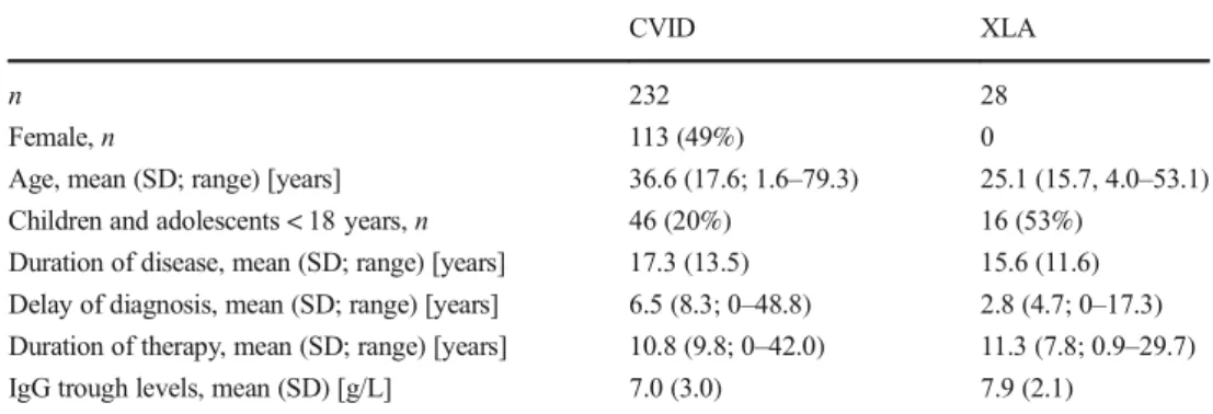
Baseline characteristics of the CVID and XLA patient co-horts and data on clinical history are given in Table1. For age distribution, see Fig.Iin the online repository.
Quality Assurance and Data Quality
The CT pathology documentation form was assessed for inter-rater reliability based on assessment by 4 radiologists of 21 randomly chosen CT studies. Rating was in good or very good
agreement, as shown by intra-class correlation coefficients (ICC) (Calder et al. Paediatric Radiology 2014 44:1496– 1506), ICC was 0.79 (p < 0.001) for atelectasis, 0.957 (p < 0.001) for mucus plugging and 0.917 (p < 0.001) for bronchiectasis severity. Rating of bronchial wall thickening, however, was not reliable (ICC 0.332,p = n.s.).
In the main study, rating on the four items on bronchial pathologies was given in 94.9% (SD 2.1%) of the CT scans. The highest rate of missing data was present in the item termed“mucus plugging”, with missing data in 7.8% of the cases. Anthropometric data were available from 64%, spirom-etry from 62%, cough frequency from 64%, antibiotic treat-ment regimen from 72% and body plethysmography from 28% of the patients.
Bronchial Pathology in CVID
Eighty percent of the CVID patients had radiological evidence of some form of bronchial pathology. Bronchiectasis had the highest prevalence of all bronchial pathologies and was re-ported in 61% of the CVID cohort, followed by bronchial wall thickening (44%), atelectasis (32%) and mucus plugging (29%). Mucus plugging was more frequent in the periphery (20%) than in a distributed pattern (central and peripheral, 9%, Fig.1). The prevalence of bronchial pathologies did not differ between sexes. Bronchiectasis was not associated with other bronchial pathology. Of the patients with bronchiectasis, 64% had no evidence of mucus plugging, 60% had no atelectasis and 43% had no evidence of bronchial wall thickening.
Impact of Age and Duration of Disease on Bronchial
Pathology in CVID
The prevalence of bronchiectasis was lowest in the patient group undergoing CT at < 20 years at 44% and increased steadily with age to 79% in the age group ≥ 60 years (Fig.2a). Extent of bronchiectasis showed an age-related in-crease (R2
= 0.029;F = 6.6; p = 0.01, Fig.2b–d). Patients ≥ 60 years had the highest proportion of extensive disease (three or more lobes affected and/or cystic changes) with 36% of this age group affected (Fig.2d).
In contrast to bronchiectasis, prevalence of bronchial wall thickening, atelectasis and mucus plugging did not rise with age nor with duration of disease or of therapy. Bronchiectasis was associated with bronchial wall thickening, atelectasis and mucus plugging only in younger age groups (Table IVin online repository).
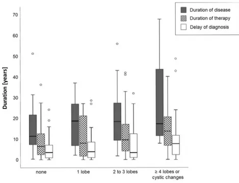
In multiple regression analysis, duration of the disease was a predictor for the presence and extent of bronchiectasis, but age, sex and duration of therapy were not. Each year of disease was associated with an additional risk of bronchiectasis by 4.4% (p = 0.07) and an increase of the severity score by 0.025 (p < 0.001) (Fig.IV, online repository).
Patients with a longer delay of diagnosis had a higher ex-tent of bronchiectasis, although this association was compar-atively weak (F = 6.14, p = 0.015 in analysis of variance, Fig.5). Patients with advanced bronchiectasis tended to have higher trough IgG levels (rpearson= 0.19,p = 0.048).
Bronchial Pathology in XLA
The prevalences of bronchiectasis, atelectasis and mucus plugging, but not of bronchial wall thickening, were higher in the XLA cohort as compared to the CVID cohort (Fig.1a, b). The extent of bronchiectasis was also more strongly related to age (rPearson= 0.6, p < 0.001, Fig.IIin online repository)
and to duration of disease (rPearson= 0.7,p < 0.001) than in
CVID patients. Again, duration of therapy correlated less strongly than age or duration of disease with the extent of bronchiectasis in XLA patients, but more so than in CVID patients (rPearson= 0.55, p = 0.017). Bronchiectasis was also
associated with bronchial wall thickening (rPearson= 0.44,
p = 0.018), but not to mucus plugging or atelectasis in this cohort.
Lung Function
CVID patients showed mild obstructive lung disease in the older age groups without restriction, FEV1was 87.8 (19.6)
% predicted in patients < 20 years and FEV1(FEV1,Fig.3a)
72.9 (26.3) % predicted in patients≥ 60 years. There was a similar age-dependent decline in maximal expiratory flow at 25% of vital capacity (MEF25), and total lung capacity (TLC),
but not of vital capacity (VC). However, explanation of vari-ance of all parameters by age was weak (FEV1:R2= 0.041,
p = 0.016, F = 5.994; MEF25:R 2
= 0.053,p = 0.01, F = 6.882; TLC:R2= 0.072,p = 0.028, F = 5.061). In XLA patients, ad-vance of lung disease with age was more obvious. FEV1
de-clined from 95.7 (8.7) % predicted in patients aged less than 20 years to 44.0 (23.2) %predicted in patients ≥ 40 years. Accordingly, linear regression analysis showed a stronger re-lation between age and decline in lung function parameters in XLA patients (vital capacity (VC):R2= 0.351,p = 0.005, F = 10.285; FEV1: R2= 0.529,p < 0.001, F = 22.439; MEF25:
R2
= 0.637, p = 0.002, F = 17.511; TLC: R2= 0.072, p = 0.028,F = 5.061).
CT Findings and Lung Function
Presence of bronchial wall thickening, bronchiectasis or mucus plugging was associated with a lower FEV1in CVID patients
(TableVonline repository, Fig.3b). The combination of bron-chiectasis and bronchial wall thickening showed a further de-cline (n = 54, FEV1of 69.3 (23.3) % % predicted). Patients
with a severe lung disease as indicated by spirometry (FEV1
< 40% predicted) had a high prevalence of bronchiectasis (89%). However, normal FEV1(> 80% predicted) did not Table 1 Characteristics of the
study population, sorted by the two main diagnosis groups
CVID XLA
n 232 28
Female,n 113 (49%) 0
Age, mean (SD; range) [years] 36.6 (17.6; 1.6–79.3) 25.1 (15.7, 4.0–53.1) Children and adolescents < 18 years,n 46 (20%) 16 (53%)
Duration of disease, mean (SD; range) [years] 17.3 (13.5) 15.6 (11.6) Delay of diagnosis, mean (SD; range) [years] 6.5 (8.3; 0–48.8) 2.8 (4.7; 0–17.3) Duration of therapy, mean (SD; range) [years] 10.8 (9.8; 0–42.0) 11.3 (7.8; 0.9–29.7)
IgG trough levels, mean (SD) [g/L] 7.0 (3.0) 7.9 (2.1)
CVID common variable immunodeficiency disorders, XLA X-linked agammaglobulinaemia, SD standard deviation
0 20 40 60 80 100 Mucus plugging
Atelectasis Bronchial wall thickening Bronchiectasis %
a
0 20 40 60 80 100 pathological normalb
%Fig. 1 Prevalence of bronchial pathology in patients with CVID (a) and XLA (b)
preclude the presence of bronchiectasis. Further, 59% of the patients with a normal lung function had bronchiectasis (Fig. 3). Thus, spirometry was a poor predictor for presence (sensi-tivity 48.9%) or absence of bronchiectasis (specificity 68.8%, TableIVonline repository). Findings of bodyplethysmography and carbon monoxide diffusion capacity were not better asso-ciated with structural bronchial pathology.
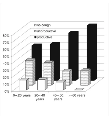
Cough
The majority of CVID patients for whom clinical data were available (n = 147) had occasional (53.1%) or recurrent or chronic cough (34.7%). Prevalence of chronic cough
increased with age and rose from 18% in the age group < 20 years to 38.5% in the age group ≥ 60 years (R2= 0.054, F = 8.315, p = 0.005). Quality of cough also changed with age; a higher proportion of patients had productive cough and frequency of cough with increasing age (79% in age group≥ 60 years). In this age group, all patient suffered from cough (Fig.4). Of the patients who coughed chronically, 75% had evidence of bronchiectasis. Nevertheless, 60% of the sub-jects with occasional cough also had bronchiectasis. Almost all patients with radiological evidence of bronchiectasis had some sort of cough (92.7%). These patients were twice as much likely to have productive rather than unproductive cough (66.7 vs. 33.3%). Patients with bronchiectasis and
global prevalence <20 y 20-4 0 y 40-6 0 y >60 y 0 20 40 60 80 100
a
% 1 lobe <20 y 20-40 y 40-60 y >60 y 0 20 40 60b
% 2 or 3 lobes <20 y 20-40 y 40-6 0 y >60 y 0 20 40 60c
%4 lobes or more or cystic changes
<20 y 20-40 y 40-60 y >60 y 0 20 40 60
d
%Fig. 2 Prevalence, extent and age distribution of bronchiectasis in 232 CVID patients. a Global prevalence (any bronchiectasis), b 1 lobe affected, c 2 or 3 lobes affected and d 4 or more lobes affected or cystic changes. Lingula counted as a separate lobe. The extent score correlated significantly with age (rPearson= 0.171,p = 0.01) < 20 y 20-40 y 40-60 y > 60 y 50 60 70 80 90 100 110 120 FE V1 ]. d e r p[ % Age < 40 40- <60 60- <80 80- <100 >100 0 20 40 60 80 100 FEV1% [pred.] B r o n chi ec ta si s (% )
a
b
Fig. 3 a Mean (SD) forced expiratory volume in 1 s as percentage of predicted value (FEV1% [pred.]) in 232 CVID patients stratified in age groups. FEV1% predicted declined significantly with age (rPearson= −0.203, p = 0.016). b Prevalence of bronchiectasis stratified by age
groups. Prevalence and extent of bronchiectasis increased with deteriorating FEV1(rPearson=−.22, p = 0.009 and rPearson=−.322, p < 0.001) for prevalence and extent of bronchiectasis with FEV1% predicted
productive cough had a more compromised lung function test (mean FEV1= 71.9 (26.1) % predicted) than those with
bron-chiectasis and unproductive cough (mean FEV1= 86.2 (25.0)
% [pred.],p = 0.033).
Use of Antibiotics
Use of antibiotics varied considerably. Intermittent antibiotic therapy was more frequent (47.7%) than maintenance (26.8%) therapy inn = 163 CVID patients, for whom data were avail-able. Usage varied also for patients with chronic cough (n = 51): intermittent 51.8% and maintenance 40.8% therapy and for patients with bronchiectasis (n = 95): intermittent 44.2%, and maintenance therapy 33.7%. Courses of antibiotic therapy were used more commonly as the proportion of patients with bronchiectasis rose: 51.2% of the patients who received no antibiotic therapy (n = 42) had bronchiectasis, 59.2% of the patients with intermittent therapy (n = 71) had bronchiectasis, and 72.7% of the patients with mainentance antibiotic therapy (n = 44) had bronchiectasis. Our data did not discriminate be-tween prophylactic and therapeutic use of antibiotics (Fig.5).
Discussion
The purpose of this study was to identify the range and extent of bronchial pathology as detected by chest CT in antibody deficient patients. A multicentre approach was used, as anti-body deficiencies are a relatively rare condition. CT findings
provide primarily qualitative data, which makes multicentre studies difficult to accomplish in the absence of pre-agreed criteria. With the Chest CT in Antibody Deficiency Group, we set up a catalogue of pathologies that were reported in the literature or seen in our own patients. In order to compile data that is comparable, we agreed upon common radiological terminology, set up an image repository and agreed upon com-mon definitions.
We found a high overall prevalence of bronchial pathology, with bronchial wall thickening and bronchiectasis present in 52 and 61% of the CVID patients, respectively. The present study is the first multicentre study to also assess extent of bronchiectasis in children and adults with antibody deficiency. While the prevalence of bronchiectasis increased with age, it was already present in our youngest age group (< 20 years) at 43%. Duration of disease, however, was the best predictor for presence and extent of bronchiectasis, with age and sex having no additional impact. Importantly, also delay of diagnosis cor-related significantly with the extent of bronchiectasis. Atelectasis and mucus plugging were reported less frequently, but also at sizeable proportions of the patients.
As expected, the prevalence of bronchiectasis increased with age which did not apply to the bronchial wall thickening, atelectasis and mucus plugging. While bronchiectasis corre-lated to the other pathologies at younger age groups (Table4, online repository), in the older age groups bronchiectasis ap-peared to be more the accumulation of damage acquired in past and present inflammatory processes. Age as well as du-ration of disease accounted for more of the variation in bron-chiectasis and lung function in XLA than in CVID. This may reflect the earlier and more homogeneous onset of immuno-deficiency in XLA [8] compared with the predominantly adult onset of CVID [11], although differences in the pathogenesis of lung disease cannot be excluded.
Similar to prevalence and extent of bronchiectasis, spirom-etry values tended to be more pathological at higher age groups. Along with bronchiectasis, prevalence of chronic and productive cough increased with age, reaching 100%, in the oldest age group (> 60 years). Cough frequency correlated better to bronchiectasis than spirometry. XLA patients had more advanced bronchial disease in the older age groups when compared to the CVID cohort. This is consistent with data from an Italian cohort [8] who had a cumulative risk of devel-oping structural lung disease of 92% by the age of 25 years, which was higher than in the Italian CVID cohort that had a prevalence of bronchiectasis of 54% at an average age of 41 years [26].
Our data show a rate of bronchiectasis in CVID patients (61%) in the same range as reported with the as yet largest CVID cohort of Italian patients (54%) [26]. In smaller chest CT studies, bronchiectasis rates varied between 29 and 78% (summarised in [17]). A meta-analysis of other studies summarised data from 587 CVID patients published in 26
Fig. 4 Prevalence of productive and unproductive cough of 120 CVID patients stratified in age groups. Productive cough was more frequent with age (rPearson= 0.222,p = 0.012)
studies. The authors reported an overall prevalence of 73% of pulmonary pathologies, mainly bronchiectasis and bronchial wall thickening.
The present study has several limitations: First, it was not designed as a cross-sectional cohort study to assess the prev-alence of particular pathologies. The participating centres var-ied in their policies to perform chest CT between clinical grounds and routine use. Since some centres performed chest CTs only on clinical grounds, the study is likely to overesti-mate prevalence and extent. The relatively high prevalence of bronchiectasis in children and adolescents (43% in the age group < 20 years) may be partly explained by the fact that the majority of the CT studies in this age group were per-formed in a centre that perper-formed CT on clinical grounds.
Second, we employed no training or quality control mea-sures for our raters. Although we used an internationally ac-cepted vocabulary [15], published an image repository on our website, we cannot be sure that all raters shared similar levels of expertise. Appreciating this, we designed the list of CT findings of this study to be as simple as possible, indicating merely the presence or absence for most pathologies. A study on inter-rater reliability with our list of CT findings showed very high rates of inter-rater agreement for all findings, in particular for the bronchiectasis score. Rating of bronchial wall thickening, however, was unreliable which is well recognised in the literature [27].
Despite these limitations, our data may nevertheless be meaningful. Our CVID study cohort has an age and sex dis-tribution that is close to the disdis-tribution of the ESID registry. Also, the size of the compiled cohort is larger than previous reports in the literature.
Bronchiectasis is the finding most frequently reported in previous studies. Our data on the prevalence of bronchiectasis (61%) are in the same range compared to the as yet largest CVID cohort study (54%) [26]. The latter study is likely to give the most accurate estimate on prevalence of bronchiecta-sis for it was based on regular chest CT scans. Other studies based on smaller cohorts reported bronchiectasis rates be-tween 29 and 78% (summarised in [17]).
Chest CT scans identified a high proportion of respiratory pathology which did not appear to be identified by symptoms or lung function. This applied in particular to patients with low-grade bronchiectasis in which spirometry tended to be normal (Fig. III in online repository). Also, the decline in FEV1with age was relatively small in our cohort, compared
to other conditions with chronic lung disease, such as primary ciliary dyskinesia (PCD) [28,29]. However, spirometry ap-peared to better discriminate between prevalence and absence of bronchiectasis in our patient group than reported in PCD or CF [30,31]. While a sensitivity of 49% and a specificity of 68.8% for detection and exclusion of bronchiectasis are far from satisfactory, the use of spirometry in routine manage-ment in patients with ADS may be at least as adviseable as in PCD or CF. Although spirometry may not detect mild bron-chiectasis, it is likely to be a meaningful parameter for ad-vanced stage of bronchial disease. In addition, any decline in spirometry in a given patient, even within the normal range, may indicate progressing lung damage and hence should prompt further evaluation. The higher susceptibility to irradi-ation damage in some subgroups of CVID also supports the notion to regularly monitor pulmonary disease without use of ionising radiation. Particular attention should be paid to
Fig. 5 Severity of bronchiectasis in relation to duration of disease, duration of medical attention following diagnosis of CVID (“duration of therapy” and “delay of diagnosis”). Boxplots indicate median and interquartiles, whiskers range. Open dots show outliers. Delay of diagnosis correlates significantly with severity of bronchiectasis (p = 0.03)
children and adolescents. Although bronchiectasis may be overestimated in this age group, the true prevalence of bron-chiectasis is likely to be high enough to warrant high priority for prevention of development of structural lung disease. Spirometry needs to be complemented by more sensitive func-tional tests. The multiple breath washout technique may be particularly promising for detecting bronchiectasis, as shown in CF and other conditions [30].
One important finding of this study is the observation that patients with a delay of diagnosis correlated with advanced formation of bronchiectasis in CVID. This finding argues that awareness of primary immunodeficiencies and early diagnosis may be particularly beneficial.
Prevalence and extent of bronchiectasis also increased with the years of disease, suggesting a repeated or continuous bur-den of bronchial inflammation throughout the course of disease.
Cough turned out to be more closely related to bronchial disease than parameters of spirometry. Again, patient selection may have biased this surprisingly high proportion of patients with clinical evidence of lung disease. However, cough and other clinical parameters, e.g. sputum volume, colour, fre-quency of chest exacerbations or frefre-quency of antibiotic ther-apy, may be valuable tools in future interventional trials. Our findings also argue that we do need better monitoring strate-gies for development of pulmonary patholostrate-gies before chronic or productive cough develops.
Therapeutic regimens for antibiotic treatment of CVID pa-tients with bronchiectasis, pathologic spirometry or produc-tive cough differ substantially in the present study as in others [9,11]. Chapel et al. found no clear evidence that bronchiec-tasis can be prevented by prophylactic maintenance antibiotic therapy. Bondioni et al. recommended early detection of pul-monary changes to adjust antibiotic therapy [21,32,33].
Evidence for the benefit of antibiotic therapy or other in-terventions to prevent or ameliorate progress of bronchiectatic lung disease in other conditions is conflicting. Among the reasons for the lack of efficacy trials in immunodeficiency is the difficult choice of outcome parameters. FEV1and other
lung function parameters show relatively little changes over time rendering them less sensitive than desirable. Magnetic resonance imaging (MRI) has made substantial progress in the detection of pulmonary pathology but is less widely avail-able [34]. Chest CT is still considered the gold standard for detection of structural bronchial pathology [17] and sensitive for changes over time [21].
Our bronchiectasis score was designed for use in routine care without prior training of the raters. This score is clearly too crude to specifically assess progress of bronchiectasis. More detailed extent scores for bronchiectasis in CVID were applied in two single-centre studies [20,21,35], demonstrat-ing that progress of lung disease is detectable by chest CT at intervals as short as 5 years.
Given the size and the relevance of pulmonary morbidity in primary antibody deficiencies, the present study argues to op-timise the use of chest CT. First, there is a need for a detailed score on bronchial and other pulmonary pathologies for inter-ventional trials [36]. Second, chest CT scans which are per-formed as part of routine care in primary antibody deficiencies should be documented in a uniform manner in a patient reg-istry along other clinical and immunological data. Documentation should provide more quantitative data than the one used in the present study, but still be compatible with routine care. The Chest CT Group has elaborated a proposal for severity graded documentation of bronchial pathology (TableVI, online repository). Since this will be more prone to variation between different raters, we plan to implement quality control measures that include rating of test images.
In summary, chest CT is a highly sensitive method for as-sessment of structural abnormalities of the bronchial airways. If it is complemented by lung function and clinical parameters, it can provide essential information on the progress and nature of lung disease in patients with antibody deficiencies. However, rating of CT findings for cohorts requires a consensus as to how the findings are documented. The present study shows how a multidisciplinary and multicentre approach can come into op-eration and affords a rationale how to shape future steps towards a better management of lung disease in antibody deficiency.
Acknowledgments The research leading to these results has received funding from the European Community’s Seventh Framework Programme (FP7/2007-2011) under grant agreement no201549 (EURO-PADnet). Studies contributed by Voijtech Thon were supported by CETOCOEN PLUS (no. CZ.02.1.01/0.0/0.0/15_003/0000469 by MEYS).
Compliance with Ethical Standards
Conflict of Interest The authors declare that they have no conflict of interest.
Publisher’s Note Springer Nature remains neutral with regard to juris-dictional claims in published maps and institutional affiliations.
References
1. Chapel H. Classification of primary immunodeficiency diseases by the International Union of Immunological Societies (IUIS) expert committee on primary immunodeficiency 2011. Clin Exp Immunol. 2012;168(1):58–9.
2. Gathmann B, Mahlaoui N, CEREDIH, Gerard L, Oksenhendler E, Warnatz K, et al. Clinical picture and treatment of 2212 patients with common variable immunodeficiency. J Allergy Clin Immunol 2014;134(1):116–126.
3. Conley ME, Howard VC. X-Linked Agammaglobulinemia. In: Pagon RA, Adam MP, Bird TD, Dolan CR, Fong CT, Smith RJH, et al., editors. GeneReviews(R). Seattle: University of Washington, Seattle; 1993.
4. Orange JS, Belohradsky BH, Berger M, Borte M, Hagan J, Jolles S, et al. Evaluation of correlation between dose and clinical outcomes in subcutaneous immunoglobulin replacement therapy. Clin Exp Immunol. 2012;169(2):172–81.
5. Orange JS, Grossman WJ, Navickis RJ, Wilkes MM. Impact of trough IgG on pneumonia incidence in primary immunodeficiency: a meta-analysis of clinical studies. Clin Immunol. 2010;137(1):21–30. 6. Baumann U, Miescher S, Vonarburg C. Immunoglobulin replace-ment therapy in antibody deficiency syndromes: are we really doing enough? Clin Exp Immunol. 2014;178(Suppl 1):83–5.
7. Quinti I, Soresina A, Spadaro G, Martino S, Donnanno S, Agostini C, et al. Long-term follow-up and outcome of a large cohort of patients with common variable immunodeficiency. J Clin Immunol. 2007;27(3):308–16.
8. Plebani A, Soresina A, Rondelli R, Amato GM, Azzari C, Cardinale F, et al. Clinical, immunological, and molecular analysis in a large cohort of patients with X-linked agammaglobulinemia: an Italian multicenter study. Clin Immunol. 2002;104(3):221–30. 9. Lucas M, Lee M, Lortan J, Lopez-Granados E, Misbah S, Chapel
H. Infection outcomes in patients with common variable immuno-deficiency disorders: relationship to immunoglobulin therapy over 22 years. J Allergy Clin Immunol. 2010;125(6):1354–1360.e4. 10. Aghamohammadi A, Allahverdi A, Abolhassani H, Moazzami K,
Alizadeh H, Gharagozlou M, et al. Comparison of pulmonary dis-eases in common variable immunodeficiency and X-linked agammaglobulinaemia. Respirology. 2010;15(2):289–95. 11. Thickett KM, Kumararatne DS, Banerjee AK, Dudley R,
Stableforth DE. Common variable immune deficiency: respiratory manifestations, pulmonary function and high-resolution CT scan findings. QJM. 2002;95(10):655–62.
12. Kainulainen L, Nikoskelainen J, Vuorinen T, Tevola K, Liippo K, Ruuskanen O. Viruses and bacteria in bronchial samples from pa-tients with primary hypogammaglobulinemia. Am J Respir Crit Care Med. 1999;159(4 Pt 1):1199–204.
13. Chapel H, Lucas M, Lee M, Bjorkander J, Webster D, Grimbacher B, et al. Common variable immunodeficiency disorders: division into distinct clinical phenotypes. Blood. 2008;112(2):277–86. 14. Winkelstein JA, Marino MC, Lederman HM, Jones SM, Sullivan
K, Burks AW, et al. X-linked agammaglobulinemia: report on a United States registry of 201 patients. Medicine (Baltimore). 2006;85(4):193–202.
15. Hansell DM, Bankier AA, MacMahon H, McLoud TC, Muller NL, Remy J. Fleischner society: glossary of terms for thoracic imaging. Radiology. 2008;246(3):697–722.
16. Hampson FA, Chandra A, Screaton NJ, Condliffe A, Kumararatne DS, Exley AR, et al. Respiratory disease in common variable im-munodeficiency and other primary imim-munodeficiency disorders. Clin Radiol. 2012;67(6):587–95.
17. Touw CM, van de Ven AA, de Jong PA, Terheggen-Lagro S, Beek E, Sanders EA, et al. Detection of pulmonary complications in common variable immunodeficiency. Pediatr Allergy Immunol. 2010;21(5):793–805.
18. Loeve M, Krestin GP, Rosenfeld M, de Bruijne M, Stick SM, Tiddens HA. Chest computed tomography: a validated surrogate endpoint of cystic fibrosis lung disease? Eur Respir J. 2013;42(3): 844–857.
19. Maglione PJ, Overbey JR, Radigan L, Bagiella E, Cunningham-Rundles C. Pulmonary radiologic findings in common variable im-munodeficiency: clinical and immunological correlations. Ann Allergy Asthma Immunol. 2014;113(4):452–9.
20. Gregersen S, Aalokken TM, Mynarek G, Kongerud J, Aukrust P, Froland SS, et al. High resolution computed tomography and
pulmonary function in common variable immunodeficiency. Respir Med. 2009;103(6):873–80.
21. Bondioni MP, Soresina A, Lougaris V, Gatta D, Plebani A, Maroldi R. Common variable immunodeficiency: computed tomography evaluation of bronchopulmonary changes including nodular lesions in 40 patients. Correlation with clinical and immunological data. J Comput Assist Tomogr. 2010;34(3):395–401.
22. Verma N, Thaventhiran A, Gathmann B, ESID Registry Working Party, Thaventhiran J, Grimbacher B. Therapeutic management of primary immunodeficiency in older patients. Drugs Aging. 2013;30(7):503–12.
23. http://esid.org/Working-Parties/Clinical/Resources/Diagnostic-criteria-for-PID2, latest access 22.8.2018.
24. http://esid.org/Working-Parties/Registry-Working-Party, latest access 22.8.2018.
25. Morice AH, Fontana GA, Sovijarvi AR, Pistolesi M, Chung KF, Widdicombe J, et al. The diagnosis and management of chronic cough. Eur Respir J. 2004;24(3):481–92.
26. Quinti I, Soresina A, Guerra A, Rondelli R, Spadaro G, Agostini C, et al. Effectiveness of immunoglobulin replacement therapy on clin-ical outcome in patients with primary antibody deficiencies: results from a multicenter prospective cohort study. J Clin Immunol. 2011;31(3):315–22.
27. Calder AD, Bush A, Brody AS, Owens CM. Scoring of chest CT in children with cystic fibrosis: state of the art. Pediatr Radiol. 2014;44(12):1496–506.
28. Maglione M, Bush A, Nielsen KG, Hogg C, Montella S, Marthin JK, et al. Multicenter analysis of body mass index, lung function, and sputum microbiology in primary ciliary dyskinesia. Pediatr Pulmonol. 2014;49(12):1243–50.
29. Harun SN, Wainwright C, Klein K, Hennig S. A systematic review of studies examining the rate of lung function decline in patients with cystic fibrosis. Paediatr Respir Rev. 2016;20:55–66. 30. Gustafsson PM, De Jong PA, Tiddens HA, Lindblad A.
Multiple-breath inert gas washout and spirometry versus structural lung dis-ease in cystic fibrosis. Thorax. 2008;63(2):129–34.
31. Boon M, Vermeulen FL, Gysemans W, Proesmans M, Jorissen M, De Boeck K. Lung structure-function correlation in patients with primary ciliary dyskinesia. Thorax. 2015;70(4):339–45.
32. Altenburg J, de Graaff CS, Stienstra Y, Sloos JH, van Haren EH, Koppers RJ, et al. Effect of azithromycin maintenance treatment on infectious exacerbations among patients with non-cystic fibrosis bronchiectasis: the BAT randomized controlled trial. JAMA. 2013;309(12):1251–9.
33. Rogers GB, Bruce KD, Martin ML, Burr LD, Serisier DJ. The effect of long-term macrolide treatment on respiratory microbiota composition in non-cystic fibrosis bronchiectasis: an analysis from the randomised, double-blind, placebo-controlled BLESS trial. Lancet Respir Med. 2014;2(12):988–96.
34. Milito C, Pulvirenti F, Serra G, Valente M, Pesce AM, Granata G, et al. Lung magnetic resonance imaging with diffusion weighted imaging provides regional structural as well as functional informa-tion without radiainforma-tion exposure in primary antibody deficiencies. J Clin Immunol. 2015;35(5):491–500.
35. Serra G, Milito C, Mitrevski M, Granata G, Martini H, Pesce AM, et al. Lung MRI as a possible alternative to CT scan for patients with primary immune deficiencies and increased radiosensitivity. Chest. 2011;140(6):1581–9.
36. de Jong PA, Tiddens HA. Cystic fibrosis specific computed tomog-raphy scoring. Proc Am Thorac Soc. 2007;4(4):338–42.
Affiliations
Katharina Schütz1&Diana Alecsandru2,3&Bodo Grimbacher3,4&Jamanda Haddock5&Annemarie Bruining6& Gertjan Driessen7,8&Esther de Vries9,10&Peter M. van Hagen11&Ieneke Hartmann12&Francesco Fraioli13,14& Cinzia Milito15&Milica Mitrevski15&Isabella Quinti15&Goffredo Serra13&Peter Kelleher16&Michael Loebinger17& Jiri Litzman18&Vera Postranecka19&Vojtech Thon18,20&Judith Babar21&Alison M. Condliffe21&Andrew Exley22& Dinakantha Kumararatne23&Nick Screaton24&Alison Jones25&Maria P. Bondioni26&Vassilios Lougaris27& Alessandro Plebani27&Annarosa Soresina28&Cesare Sirignano29&Giuseppe Spadaro30&Nermeen Galal31& Luis I. Gonzalez-Granado2&Sabine Dettmer32&Robert Stirling33&Helen Chapel34&Mary Lucas34&Smita Patel34& Claire-Michele Farber35&Isabelle Meyts36&Arpan K. Banerjee37&Scott Hackett38&John R. Hurst39&Klaus Warnatz4& Benjamin Gathmann40&Ulrich Baumann1
1
Paediatric Immunology Unit, Department of Paediatric Pulmonology, Allergology and Neonatology, Hanover Medical School, Carl-Neuberg Str. 1, 30625 Hannover, Germany 2
Primary Immunodeficiencies Unit, Pediatrics, Hospital 12 Octubre, Madrid, Spain
3
Clinical Immunology, Royal Free Hospital, London, UK 4
Centre for Chronic Immunodeficiency, University Medical Center of Freiburg, Freiburg, Germany
5
Radiology, Royal Free Hospital, London, UK
6 Dutch Cancer Institute, Antoni van Leeuwenhoek Hospital, The Hague, The Netherlands
7 Paediatric Immunology, Erasmus MC Sophia Children’s Hospital, Rotterdam, The Netherlands
8
Paediatrics, Juliana Children’s Hospital/Haga Teaching Hospital, The Hague, The Netherlands
9 Jeroen Bosch Academy, Jeroen Bosch Hospital, ‘s-Hertogenbosch, The Netherlands
10
Tranzo, Tilburg University, Tilburg, The Netherlands 11
Immunology and Internal Medicine, Erasmus MC, Rotterdam, The Netherlands
12 Department of Radiology, Erasmus MC Sophia Children’s Hospital, Rotterdam, The Netherlands
13
Radiology, Università degli Studi di Roma La Sapienza, Rome, Italy
14
Institute of Nuclear Medicine, University College London, London, UK
15 Department of Molecular Medicine, Sapienza University of Rome, Rome, Italy
16
Immunology Section Department of Medicine, Imperial College London, London, UK
17
Department of Respiratory Medicine, Royal Brompton Hospital, London, UK
18 Department of Clinical Immunology and Allergy, Faculty of Medicine, Masaryk University, St Anne’s University Hospital, Brno, Czech Republic
19
Department of Radiology, Faculty of Medicine, Masaryk University, St Anne’s University Hospital, Brno, Czech Republic 20
RECETOX, Faculty of Science, Masaryk University, Brno, Czech Republic
21 Radiology, Addenbrooke’s Hospital, Cambridge, UK 22
Immunology, Papworth Hospital, Cambridge, UK 23
Immunology, Addenbrooke’s Hospital, Cambridge, UK 24 Radiology, Papworth Hospital, Cambridge, UK 25
Paediatric Immunology, Great Ormond Street Hospital, London, UK 26 Department of Radiology, University of Brescia, Brescia, Italy 27
Pediatrics Clinic and Institute for Molecular Medicine A. Nocivelli, Department of Clinical and Experimental Sciences, University of Brescia and ASST-Spedali Civili of Brescia, Brescia, Italy 28
Pediatrics Clinic, ASST-Spedali Civili, Brescia, Italy
29 Radiology, IBB-CNR University of Naples Federico II, Naples, Italy 30
Immunology, University of Naples Federico II, Naples, Italy 31
Paediatric University Hospital, Cairo, Egypt 32
Diagnostic Radiology, Hanover Medical School, Hanover, Germany
33
Allergy, Immunology and Respiratory Medicine, The Alfred Hospital, Melbourne, Australia
34 Primary Immunodeficiency Unit, Nuffield Department of Medicine, University of Oxford, Oxford, UK
35
Immunology, Cliniques Universitaires de Bruxelles Hôpital Erasme, Brussels, Belgium
36
Paediatric Immunology and Pulmonology, University Hospitals, Leuven, Belgium
37 Radiology, Heartlands Hospital, Birmingham, UK 38
Paediatric Immunology Department, Heartlands Hospital Birmingham, Birmingham, UK
39
UCL Respiratory Medicine, University College London, London, UK
40 ESID Registry Working Party, University Hospital Freiburg, Freiburg, Germany