UNIVERSITÀ DEGLI STUDI DI CAMERINO
School of Advanced Studies
Doctoral Course in
Chemical and Pharmaceutical Sciences and Biotechnology
– Pharmaceutical Sciences Cycle XXXISearching for effective natural products against
Human African Trypanosomiasis (HAT)
with special reference to African natural resources
PhD Candidate
Supervisors
Stephane Landry Ngahang Kamte
Prof. Riccardo Petrelli
Prof.ssa Loredana Cappellacci
Coordinator
Table of contents
LIST OF PUBLICATIONS ... 3 ABSTRACT ... 5 CHAPTER 1. - Searching for effective natural products against Human African Trypanosomiasis
(HAT) with special reference to african natural resources. ... 7 CHAPTER 2. - Phytochemical analysis and bioactive constituents of the Mexican sunflower
Tithonia Diversifolia (Asteraceae) ... 75
CHAPTER 3. - Identification of Skinonin derivatives from Onosma Visianii roots extract as potential antitrypanosomal drugs or botanical insecticides ... 137 CHAPTER 4. - Searching for highly effective trypanocidal compounds in the essential oils from the Apiaceae family... 172 CHAPTER 5. - A neglected vegetable, Smyrnium Olusatrum, as potential weapon against a Neglected Tropical Disease, African Trypanosomiasis ... 198 CHAPTER 6. - Trypanosoma Brucei inhibition by essential oils from medicinal and aromatic plants traditionally used in Cameroon ... 221
LIST OF PUBLICATIONS
This thesis is based on the following publications, which are appropriately adapted in every Chapter:
1. Orsomando, G., Agostinelli, S., Bramucci, M., Cappellacci, L., Damiano, S., Lupidi, G., Maggi, F., Ngahang Kamte, S.L., Biapa Nya, P.C., Papa, F., Petrelli, D., Quassinti, L., Sorci, L., Vitali, L.A., Petrelli, R. (2016). Mexican sunflower (Tithonia diversifolia, Asteraceae) volatile oil as a selective inhibitor of Staphylococcus aureus nicotinate mononucleotide adenylyltransferase (NadD) Ind.Crop.Prod. 85, 181-189.
2. Petrelli, R., Ranjbarian, F., Dall'Acqua, S., Papa, F. Iannarelli, R., Ngahang Kamte, S.L., Vittori, S., Benelli, G., Maggi, F., Hofer, A., Cappellacci, L. (2017). An overlooked orticultural crop, Smyrnium olusatrum, as a potential source of compounds effective against African trypanosomiasis. Parasitol. Int. 66, 146-151.
3. Ngahang Kamte, S.L., Ranjbarian, F., Campagnaro, G.D., Biapa Nya, P.C. Mbuntcha, H., Woguem,V., Womeni, H.M., Azefack Tapondjou, L., Giordani, C., Barboni, L., Benelli, G., Cappellacci, L., Hofer, A., Petrelli, R., Maggi, F. (2017). Trypanosoma brucei inhibition by essential oils from medicinal and aromatic plants traditionally used in Cameroon (Azadirachta indica, Aframomum melegueta, Aframomum daniellii, Clausena anisata, Dichrostachys cinerea and Echinops giganteus). Int. J. Environ. Res. Public Health 74, 1-16.
4. Sut, S., Dall'Acqua, S., Baldan, V., Ngahang Kamte, S.L., Ranjbarian, F., Biapa Nya, P.C., Vittori, S., Benelli, G., Maggi, F., Cappellacci, L., Hofer, A., Petrelli, R. (2018). Identification of tagitinin C from Tithonia diversifolia as antitrypanosomal compound using bioactive-guided fractionation. Fitoterapia 124, 145-151.
5. Pavela, R., Dall'Acqua, S., Sut, S., Baldan, V. Ngahang Kamte, S.L., Biapa Nya, P.C., Cappellacci, L., Petrelli, R., Nicoletti, M., Canale, A., Maggi, F., Benelli, G. (2018). Oviposition inhibitory activity of the Mexican sunflower Tithonia diversifolia (Asteraceae) polar extracts against the two-spotted spider mite Tetranychus urticae (Tetranychidae). Physiol. Mol. Plant
Pathol. 101, 85-92.
6. Ngahang Kamte, S.L., Ranjbarianb, F., Cianfaglione, K., Sut, S., Bruno, M., Afshar, F.H., Iannarelli, R., Benelli, G., Cappellacci, L., Hofer, A., Maggi, F., Petrelli, R. (2018). Identification of highly effective antitrypanosomal compounds in essential oils from the Apiaceae family.
Comments on contribution
1. Collected the plant material and performed the hydrodistillation. Conducted test for antitrypanosomal activity.
2. Collected the plant material, performed the hydrodistillation and the chemical and gas-chromatography analysis.Conducted test for antitrypanosomal activity.
3. Collected the plant material and performed the hydrodistillation. Took part in all data analysis and was one of the main contributors to writing the manuscript.
4. Collected the plant material and performed the fractionation of the extracts. Performed and analysed all of the IC50 determinations on T. brucei and mammalian cells.
5. Collected the plant material and performed the preparation of the crude extracts and the isolation of sesquiterpene lactones.Contributed to writing the manuscript.
6. Collected part of the plant material and performed the hydrodistillation and chemical analysis of essential oils. Performed and analysed all of the IC50 determinations on T. brucei and mammalian
cells. Contributed to writing the manuscript.
Publications not included in the thesis
1. Pavela, R., Maggi, F., Ngahang Kamte, S.L., Rakotosaona, R., Rasoanaivo P., Nicoletti, M., Canale, A., Benelli, G. (2017). Chemical composition of Cinnamosma madagascariensis (Cannelaceae) essential oil and its larvicidal potential against the filariasis vector Culex quinquefasciatus Say. S. Afr. J. Bot. 108, 359-363.
2. Benelli, G., Pavela, R., Lupidi, G., Nabissi, M. Petrelli, R., Ngahang Kamte, S.L., Cappellacci, L., Fiorini, D., Sut, S., Dall'Acqua, S., Maggi, F. (2018). The crop-residue of fiber hemp cv Futura 75: from a waste product to a source of botanical insecticides against Culex quinquefasciatus, Spodoptera littoralis and Musca domestica. Environ. Sci. Poll. Res. 25, 1-11.
ABSTRACT
For centuries, African natives have been facing various infectious tropical illnesses, among which African trypanosomiases are some of the most frequent relevant parasitic diseases. African trypanosomiases, commonly called sleeping sickness in humans (HAT; Human African Trypanosomiasis) and Nagana in domestic livestock, affect a huge number of people living in poverty in 36 sub-Saharan countries, resulting in a key socioeconomic impact. After a century of outbreaks, due to political instability and lack of funding, around 70 million people and 50 million cattle are still at risk of exposure in Africa. Trypanosomiasis is transmitted by the bite of insects from the Glossina spp (Glossinidae) and is fatal in humans, if untreated. While taking a blood meal, infected Glossina flies can spread extracellular protozoans from the species Trypanosoma brucei. There are three morphologically indistinguishable subspecies of T. brucei. The subspecies T. b.
gambiense is responsible for a chronic form of the human disease, while T. b. rhodesiense causes an
acute form, which more rapidly leads to death. Both subspecies are infective to humans, whereas T.
b. brucei is only infective to animals. During the early stage of the disease or hemolymphatic phase,
the parasite is restricted to the blood and lymph and after months or years it invades the central nervous system resulting in various neurological symptoms including sleeping disturbance. As for other neglected tropical diseases, the chemotherapeutical arsenal against HAT is based on limited, expensive and often toxic medicines that are administered parentally in a context of poverty and lack of qualified personnell in healthcare centers. The few drugs that are available are pentamidine and suramin for the early stage disease and eflornithine (also in combination with nifurtimox) and melarsoprol for the late stage when the parasite infects the brain. Overall, the situation described above highlights the critical nature of this phenomena and the urgent need to explore new sources of potentially effective and safe compounds for therapy. In this scenario the naturally-occurring products may play a crucial role as source of bioactive drug candidates.
With this vision in mind, in Chapter 2 I performed a complete phytochemical analysis on both polar and volatile compounds of T. diversifolia collected from a geographically isolated population living in Dschang, Cameroon and I assessed their biological activities (antitrypanosomal and amtimicrobial activities). The main secondary metabolites occurring in the T. diversifolia methanolic extract were isolated by column chromatography and structurally elucidated by MS and NMR techniques. Tagitinins C emerged as the most active compound against T. brucei (TC221) with an IC50 value of 0.0042 μg/mL. This activity was 4.5 times better than that of the reference
the NAD biosynthetic enzyme NadD from S. aureus (IC50 of ∼60 g/mL). Besides its extensive
utilizations in the traditional medicine, the plant is believed to have a great potential in agriculture. For this reason, I decided to evaluate the T. diversifolia polar extracts against the two-spotted spider mite Tetranychus urticae (Tetranychidae), which is one of the most economically important arthropod pests worldwide. The ethyl acetate extract resulted as the most active oviposition inhibitor, with an ED50 value of 44.3 µg.cm-3 and an ED90 of 121.5 µg.cm-3.
In Chapter 3, I investigated a lipophilic extract of Onosma visianii roots containing 12% of shikonin derivatives. The phytochemical investigation of the lipophilic extract resulted in the isolation of 12 naphtoquinone derivatives which were evaluated against Trypanosoma brucei. Isobutylshikonin and isovalerylshikonin emerged as the most active naphtoquinone derivatives, showing an IC50 of 3.3 and 2.7 g/mL, respectively. Furthermore, isovalerylshikonin provided an
inhibition of Glossina palpalis acetylcholinesterase (gpAChE) (IC50 = 7.1 μg/mL), stronger than
isobutyrylshikonin (IC50 = 91.3 μg/mL), with a significant tse-tse fly versus human selectivity (SI =
7.2).
In Chapter 4, I oriented my attention to the Apiaceae family, which is a class of aromatic plants rich of EOs. Four out of nine Apiaceae EOs resulted active against T. brucei showing an IC50
in the range 2.7-10.7 g/mL. Terpinolene, one the major isolated component of these oils, was particularly active with an IC50 value of 0.035 g/mL (0.26 µM) and a selectivity index (SI) of 180.
As part of the extended family of naturally-occurring products, sesquiterpenes hold promising inhibitory effects against the bloodstream forms of T. brucei. For this reason, in Chapter
5, I decided to explore the potential of Smyrnium olusatrum EOs obtained and its main oxygenated
sesquiterpenes, namely germacrone, isofuranodiene, and β-acetoxyfuranoeudesm-4(15)-ene, as potential inhibitors of T. brucei. The EOs obtained efficiently inhibited the growth of parasite with IC50 ranging from 1.9 to 4.0 g/mL. Among the isolated main EOs components, isofuranodiene
exhibited a significant and selective inhibitory activity against T. brucei (IC50 = 0.6 g/mL, SI =
30).
In Chapter 6, I finally selected six medicinal and aromatic plants traditionally used in Cameroon to treat several disorders, including infections and parasitic diseases. Then I evaluated the activity of their EOs against T. brucei TC221 and their selectivity against Balb/3T3 cells, used as counter-screen for cytotoxicity. The most relevant outcomes showed that the EOs from A. indica,
A. daniellii and E. giganteus were the most active ones, with IC50 values of 15.21, 7.65 and 10.50
g/mL, respectively.
Overall, the results of my PhD thesis provided new insights into the potential of naturally-occurring compounds as valuable sources for the development of innovative trypanocidal drugs or botanical insecticides.
Searching for effective natural products against
Human African Trypanosomiasis (HAT)
with special reference to African natural resources
“The forest not only hides man’s enemies
but it is full of man’s medicine, healing power and food.”
African proverb
Chapter 1 Outline
Abbreviation... 9
1.1 Epidemiology of Human African Trypanosomiasis (HAT) ... 10
1.1.1 Economic burden and public health impact in Africa ... 16
1.2 Historical perspective on African Trypanosomiasis: the journey so far ... 17
1.2.1 Prehistory... 17 1.2.2 Antiquity ... 18 1.2.3 Middle Ages ... 18 1.2.4 Modern Times ... 19 1.3 Biology ... 22 1.3.1 The Vector ... 22
1.3.2 The parasite life cycle... 24
1.3.3 The parasite architecture (morphology) ... 26
1.3.4 The Variant Surface Glycoproteins (VSG) ... 27
1.4 Mode of trasmission ... 28
1.5 Clinical features of HAT disease ... 30
1.6 Diagnosis ... 32
1.7 Chemotherapy for HAT ... 35
1.7.1 Chemotherapy for first or early stage of HAT ... 36
1.7.1.1 Pentamidine ... 36
1.7.1.2 Suramin ... 37
1.7.2 Chemotherapy for second or late stage of HAT ... 38
1.7.2.1 Melarsoprol ... 38
1.7.2.2 Eflornithine ... 40
1.7.2.3 Nifurtimox-Eflornithine combination therapy (NECT) ... 41
1.8. Drug candidates in clinical trials for HAT ... 43
1.8.1 Fexinidazole ... 43
1.8.2 Oxaborole (SCYX-7158) ... 45
1.9 Natural products as a source of trypanocidal compounds... 46
1.9.1 Antiparasitic drugs from natural origin ... 47
1.9.2 Screening of natural product for antitrypanosomal activity ... 48
Abbreviation
BBB = Blood brain barrier
CATT = Card agglutination trypanosomiasis test CNS = Central nervous system
CPDD = Consortium for parasitic drug developement CSF = Cerebrospinal fluid
CTC = Capillary tube centrifugation DFMO = DL-α-difluoromethylornithine
DMC = Dichloretamethane
DNDi = Drug for neglected disease initiative ES = Ectoperitrophic space
GAPDH = Glyceraldeyde-3-phosphate dehydrogenase gHAT = Gambiense human african trypanosomiasis HAT = Human african trypanosomiasis
L6 cells = Rat skeletal myoblast cells
MAECT = Mini anion exchange centrifugation MIC = Minimum inhibitory concentration MRC-S =
NECT = Nifurtimox eflornithine combination therapy ODC = Ornithine decarboxylase
OAU = Organisation of African Unity
PATTEC = Pan African tsetse and trypanosomiasis eradication campaign PF = Procyclic form
PM = Peritropic matrix
QBC = Quantitative buffy coat RDT = Rapid diagnostic tests
rHAT = Rhodiesiense human african trypanosomiasis TPP = Target product profile
1.1 Epidemiology of Human African Trypanosomiasis (HAT)
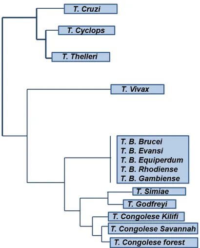
The protozoal parasite trypanosomes are grouped in the order “kinetoplastida” because of the presence of a kinetoplast. The genus Trypanosoma is divided in two main groups based on the mode of transmission by their insect vectors: Stercoraria and Salivaria (Figure 1). The infection to the vertebrate of Stercoraria parasites is via feces. The causative agent of Chagas’ disease,
Trypanosoma cruzi, is a representative example of this group. On the other hand, in the Salivarian
parasitesthe infectionof the vertebrate occurs via saliva when the vector bites the host’s skin.
Fig. 1. Classification of trypanosomes.
Human African Trypanosomiasis (HAT) also known as sleeping sickness is a vector borne neglected tropical disease caused by two parasites from the genus Trypanosoma and the specie T.
brucei namely Trypanosoma brucei gambiense (T.b. gambiense) and Trypanosoma brucei rhodesiense (T.b. rhodesiense). T.b. gambiense is considered the slow-progressing form and is
endemic in western and central Africa, whereas T.b. rhodesiense is the faster progressing form found in eastern and southern Africa (Simarro et al., 2010). Both forms are classically transmitted by the bite of blood-sucking vector tsetse flies (Diptera, genus Glossina). T.b. gambiense can also be transmitted by other routes which are poorly documented, one of them is congenitally (Lestrade-Carluer De Kyvon, 2016).
T.b. gambiense is also called Gambian HAT (Figure 2) and it is considered a chronic illness
that can take years or decades before fatality (Checchi et al., 2008). It is an anthroponotic disease with a minor incidence for animals (livestock and wildlife) and it is responsible for 98% of HAT cases reported in the last few decade (Franco et al., 2014).
T.b. rhodesiense, also called Rhodesian HAT (Figure 2), is an acute zoonotic disease,
causing death within weeks or months upon infection, mainly affecting livestock and game animals (Odiit et al., 1997). Humans represent the main reservoir host for T.b. gambiense, whereas domestic cattle and wild animals are considered the main animal reservoir of T.b. rhodesiense.
Animal African Trypanosomiasis (AAT, also called Nagana, from the Zulu word ‘N’gana’ which means ‘powerless/useless’), is caused by trypanosome species T. vivax, T. congolense, and T.
brucei brucei. Nagana is widespread in sub-Saharan Africa and it is cyclically transmitted by the
same vector responsible of human-infective trypanosomes, the tsetse fly. Trypanosomes can be also transmitted mechanically when the flies begin a blood meal on an infected host and they end it on another one. The time between the two meals is short enough to ensure the survival of parasites in the insect mouth. This distinctive feature (mechanical transmission) has allowed T. vivax to be
spread outside the limits of the “African tsetse fly belt” and the parasite is now present in 13 South American countries where it is considered an emerging disease (especially on cattle farming). Unlike other trypanosomes, T. vivax completes its short life cycle in the insect proboscis and this is the reason why can also be transmitted mechanically (Moloo et al. 2000). Furthermore, among African trypanosomes, T. vivax is the most phylogenetically distinct species as shown in Figure 3.
Historically, the impact of AAT has been so deep, that it has influenced the migration routes of cattle-owning tribes that avoided the “tsetse fly belt” (Figure 4), as well as the movements of early European and Arab colonizers who depended on horses and oxen in Africa.
Fig. 2. Distribution of the main foci of the two forms of HAT in sub-Saharan Africa with incidences and risk
for travelers. The yellow line divides the areas in which T.b. gambiense prevails and those in which T.b. rhodesiense predominates.
Fig. 3. Phylogenetic tree based on SSU rRNA sequences from trypanosome species. Adapted from (Cortez et
Fig. 4. The “flies belt” : distribution of tsetse and cattle raising area in Africa.
According to WHO reports (WHO 2013), if we take into the accounts the Disability Adjusted Life Year (DALY) guidance (i.e., loss of healthy life, premature mortality and disability) HAT can be considered the third most important contributor to the global burden of the parasitic diseases after schistosomiasis and malaria. The distribution of the disease overlaps with the presence of the vector. It occurs in 36 sub-Saharan countries (where is endemic) on an area of 1,55 million Km2 between 14°N and 20°S latitude (Simarro et al., 2012) (Figure 5). The disease is mostly present in rural area with suitable environment for tsetse flies development. It is estimated that 69 million persons are at risk of HAT (of which 57 million are exposed to T.b. gambiense ) as well as
cases were caused by T.b. rhodesiense mostly in eastern and southern Africa with Malawi and Uganda hosting more than 80% of the cases (Büscher et al., 2017). Thanks to the public private partnership established in 2000, the number of cases was reduced by 73% in little more than a decade reversing the epidemiological trend (WHO 2013). The prevalence of the disease is higher in the adult population and there is a relationship with the type of activity. For example fishermen which are in close contact with the vector appeared to be more prone to infection (Büscher et al., 2017). Cases are detected either by a passive case detection in which the patients report themselves to the healthcare services or by active screening carried by health professionals in remote areas with mobile teams.
Fig. 5. Geographic distribution of HAT cases reported in the period 2010–2014 (Büscher et al., 2017).
According to WHO reports, the number of HAT cases reported globally decreased from 37,991 in 1998 to 7216 cases in 2012 (five folds). Encouraged by this important decrease linked to the intensified control efforts, HAT has been included in the WHO NTD roadmap, a program targeted for elimination of HAT as a public health problem by the year 2020 and zero transmission by the year 2030 (WHO 2013) (Figure 7). The successes of this ambitious approach will rely on commitment of both international players and the national control programs of endemic countries. However, active case detection through mass community screening and related trends should be
interpreted carefully, as the number of cases are strictly related to the intensity and the quality of screening efforts.
1.1.1 Economic burden and public health impact in Africa
According to the Global Burden of Disease the estimates of “years lived with disability” (YLDs) for HAT, range from 2,000 to 25,000 per year (Sutherland et al., 2015). However, the decreasing trend in HAT case numbers is reflected in the recent Global Burden of Disease estimates. In 2010 the HAT-related burden was estimated to cause a loss of 560,000 DALYs per year, with a 72.5 % reduction from the 1990 DALY estimate (Murray 2012). Although the global HAT elimination appears to be on track, sleeping sickness remains a major public health problem in rural poor communities. For example, in the Democratic Republic of Congo (DRC), one of the most affected country, the number of years of life lost (YLL) per death caused by HAT was estimated at 27 years (Lutumba et al. 2007 ).
A recent study showed that HAT resulted in exorbitant indirect health care costs (ranging from US$ 60–170) for people already living on less than US$1 per day (Bukachi et al., 2017). Morbidity from HAT temporarily removes adults from their regular employment, causing shifting of their household roles. This phenomena negatively impact on children which are forced to be absent from school to provide household work. In addition, the socio-economic effects of HAT are exacerbated by coping strategies with negative consequences on people. For this reason, continuous sensitization about HAT risks at all the community levels is critical in making progress towards the goal of HAT elimination by 2020.
1.2 Historical perspective on African Trypanosomiasis: the journey so far
The economic and cultural development of sub-Saharan African has been severely repressed by African trypanosomiasis. Different parameters including climate, environmental and socio-economic changes have influenced the spread of the disease through history. History has also demonstrated that African trypanosomiasis prevented the introduction of stock farming in endemic areas and the problem is still present today. A consequence of this is that much of sub-Saharan Africa has not been converted yet into grassland for cattle breeding, since the disease can be controlled but can’t be completely eradicated. The disease affecting cattle, Nagana, has been recognized since antiquity and interestingly, humans are resistant to these species of trypanosomes such as T. congolense, T.vivax, and T. brucei brucei. The trypanosome lytic factors circulating in human blood, which brings attention to the long evolution of humans in the presence of these parasites in Africa, are responsible of this resistance. On the other hand, HAT is a relatively recent event in human evolution and the infectivity of T. brucei rhodesiense to humans is due to a serum-resistance associated (SRA) gene. After this event, the mutate gene has been spread through sub-Saharian Africa by genetic exchange (Gibson, 2005).
This chapter examines the contribution of these parameters to our understanding of the epidemiology and history of human sleeping sickness in sub-Saharan Africa.
1.2.1 Prehistory
Phylogenetic reconstruction research suggests that Salivarian trypanosomes emerged 300 million years ago and then probably became a gut parasite of some early insects (Haag et al., 1998). The bloodsucking insect tsetse fly emerged 35 million years ago and transmitted the trypanosome to mammals, which evolved 180 million years ago.
The long coexistence of both tsetse flies and wild animals may explain why most African wildlife species are tolerant of trypanosomiasis. In contrast, domestic animals have yet been incapable to develop tolerance or resistance to trypanosome infections.
Around 1.8. million years ago, concomitantly with serious climate changes, the hominids moved from the rainforest to the savannah (open plains of East Africa) and came into close contact
1.2.2 Antiquity
As shown in Figure 8, a four thousand years old veterinary papyrus describes a disease similar to nagana (animal trypanosomiasis) infecting cattle in ancient Egypt (Steverding et al., 2008). It is also reported that an ointment made from the fat of particular birds was used as treatment against the bite of flies. This suggests the presence of sleeping sickness in that region meanwhile in the sub-Saharan region, the spread of trypanosomiasis was unintentionally slowed as the border of the villages were cleared from vegetation as a protection from foreign invasion and as adjustment of the stream course of the Nile River. This gradual eradication of the tsetse fly allowed the ancient Egyptians to raise pure breeds of zebu cattle. However, the failure in horse breeding, introduced in Egypt after the 16th century BC, may be also due to trypanosome infections.
Fig. 8. A section of a damaged veterinary papyrus about a cattle disease from the 2nd millennium BC. The papyrus explains the different symptoms of the animal trypanosomiasis called ushau.
1.2.3 Middle Ages
There are only a few reports on the occurrence of trypanosomiasis in Africa during the Middle Ages and most of them are from the Arabs. For trade purposes, the Arabs made relations with West African kingdoms like Benin, Mali, Ghana and Songhai.
The first case of sleeping sickness report comes from the Arabian geographer Abu Abdallah Yakut, who describes a devastating outbreak in Sudan (at the turn of the 12th century). Then, the famous Arabian writer Ibn Khaldun (around 1373) describes the death of the emperor of Mali, King
Mari Jata, who died of an illness which corresponds to the description of human trypanosomiasis. Both are considered as the first written reports on sleeping sickness (Steverding et al., 2008).
1.2.4 Modern Times
In early Modern Times, the history of HAT is closely linked to the slave trade and the first accounts of sleeping sickness came from ship doctors and slave-trade companies. Since HAT caused increasing losses in slaves, slave-traders pushed their ship doctor to investigate this disease. In 1734, John Aktins published the first accurate medical report on HAT, describing only the neurological symptoms of the late stage of the disease (Cox, 2004). Although in the 19th century HAT became a well-accepted disease, no one had any clue about the nature of the illness (Bruce, 1895). It was the Scottish explorer David Livingston, in 1852, who first suggested that “Nagana” is caused by the bite of tsetse flies. However, it took another 50 years until trypanosomes were identified as the causative agents of Nagana and sleeping sickness, and in 1895 the microbiologist David Bruce discovered T. brucei as the factor for cattle trypanosomiasis (cattle Nagana).
Colonial authorities played an important role to give scientific evidence on the existence of the disease. Physicians were sent in the endemic areas to study the cause of swollen lymph nodes on the neck of some autochthones and the occurrence of neurological symptoms. These studies allowed making a relationship between the distribution of the tsetse fly and the cases of sleeping sickness. It is in 1901 that the British surgeon Robert Forde observed trypanosomes in human blood (Forde, 1902). The colony where it was observed (the Gambia) inspired the name of the species
Trypanosoma gambiense (Dutton, 1902). In the meantime, the two other animal pathogenic
trypanosome species T. congolense and T. vivax were discovered in 1904 and 1905 by Alphonse Broden (Broden, 1904) and Hans Ziemann (Ziemann, 1905), respectively. Subsequently, the second human pathogenic trypanosome species, T. rhodesiense (now T.b. rhodesiense), was uncovered in 1910 by the two parasitologists John William Watson Stephens and Harold Benjamin Fantham (Stephens et al., 1910).
These discoveries opened the way for a better understanding of the pathology, the life cycle of the parasite and for the exploration of possible chemotherapy. In fact, after the huge wave of sleeping sickness epidemics that killed around half of million people in central and West Africa at
In the 20th century, Africa have been plagued by three major sleeping sickness. The first epidemic at the turn of the 20th century, killed about 300 000−500 000 people in the Congo basin, Uganda, and Kenya. In 1916 a small team of chemists in collaboration with the pharmaceutical company Bayer developed the first effective drug (Bayern 205, later named suramin) for the treatment of sleeping sickness. Suramin is still in use in therapy for the treatment of the first phase of T.b. rhodesiense infections, alone or in combination with other drugs (Steverding et al., 2008).
A year earlier, the organo-arsenical tryparsamide was discovered and it was considered the first drug for the treatment of late-stage of HAT. Later on, a third drug, named pentamidine, was introduced into the market by the English chemist Ewins for the treatment of late-stage of HAT. Pentamidine is still regarded as the only effective drug for late stage T.b. rhodesiense sleeping sickness. The second major epidemic occurred between about 1920 and 1940, and as a consequences to these epidemics, severe control measures were introduced (e.g. fly traps, brush clearing, and game destruction).
Since the 1950s, several drugs have become available for chemotherapy of animal trypanosomiasis and these included: the phenanthridine derivatives homidium bromide (Ethidium®, Novidium®) and isometamidium chloride (Samorin®, Trypamidium®), the aminoquinaldine derivative quinapyramine (Anthrycid®) and the aromatic diamidine diminazene aceturate (Berenil®). Vector control and employment of chemotherapy, led to a drastic reduction in the incidence of sleeping sickness at the beginning of the 1960s.
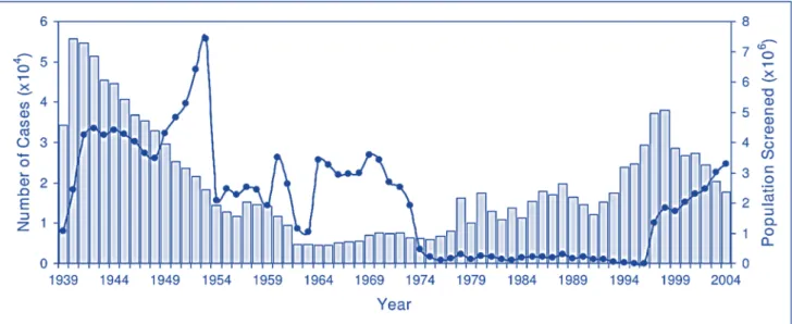
The third major HAT epidemic occurred following the departure of colonial powers (exacerbated by the banning of DDT in the 1970s). By the mid of the 1960s, most of the trypanosomiasis-endemic countries became independent (decolonized) and experienced economic crisis and political instability with negative consequences on their health systems. After that, the control of trypanosomiasis was no longer a priority and specific screening programs were stopped. As result, by the mid of 1970s, there has been a substantial increase in the number of reported cases of sleeping sickness (Figure 9). This was the beginning of the third and most recent sleeping sickness epidemic in the 20th century, mainly affecting Congo, Angola, Sudan, and the West Nile district of Uganda. The scenario remained unchanged until eflornithine (DL-α-difluoromethylornithine, DFMO), a drug initially developed for the treatment of cancer by the Merrell Research Institute in Strasbourg (Meyeskens et al., 1999), was introduced into the clinic for the treatment of late stage T.b. gambiense HAT.
In 2001, a new initiative has been launched by the Organisation of African Unity (OAU) a new initiative, named Pan African Tsetse and Trypanosomiasis Eradication Campaign (PATTEC),
aimed to eliminate the tsetse fly from Africa. It has been proposed to use odour-baited traps, insecticide-treated targets and aerial spraying insecticides in order to eradicate the tsetse fly population. However, in contrast to the Zanzibar project (an island infested with only one tsetse fly species), the PATTEC campaign dealt with a vast expanse of untamed territory (sub-Saharan Africa is ~10 million km2) populated by 7 different Glossina species accepted as vectors for transmission of sleeping sickness.
Since in the past a similar eradication campaign already failed, the scientific community was considerable skeptical about its feasibility, because the tsetse fly infested areas could not be isolated. Furthermore, the huge costs associated with the eradication projects were also matter of concern.
Fig. 9. Number of reported cases of sleeping sickness and population screened, 1939–2004. Light blue
columns, number of reported cases; blue circles, population screened. Figure derived from (Steverding et al., 2008).
1.3 Biology
The Vector and Parasite Life Cycle
1.3.1 The Vector
The commonly called Tsetse fly belongs to the order of Diptera and based on its reproduction through adenotropic viviparity, tsetse fly is categorized as member of the superfamily
Hippoboscidea, the family of Glossinidae and genus of Glossina. Tsetse fly includes 31 species
classified in three species groups (or subgenera): palpalis group (Nemorhina), morsitans group (Glossina) and fusca group (Austenina) (Franco et al., 2014) (Figure 10). The 31 tsetse flies species can be also classified as forest, riverine, or savannah, based on their morphological differences and habitat preference (Cecchi et al., 2008).
Overall, the species of the fusca and the palpalis groups are mainly implicated in the transmission of T.b. gambiense, while species of the morsitans group are involved in the transmission of T.b. rhodesiense (Geiger et al., 2004). The most important species involved are G.
palpalis gambiensis, G. palpalis palpalis, G. fuscipes quanzensis, G. tachinoides, G. fuscipes fuscipes and G. fuscipes martini. G. fuscipes fuscipes is considered the major vector of both forms
of HAT in Uganda.
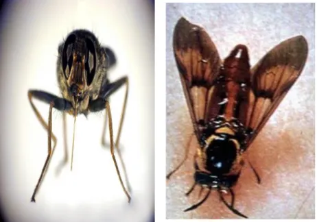
Fig. 10. The figure showed the principal vectors of HAT: Glossina morsitans on the left and Glossina
The insect is characterized by a recognizable proboscis, antenna with branched arista hairs and wings that fold and have a characteristic “hatchet” cell (Figure 11).
Fig. 11. Anatomy of the tsetse fly.
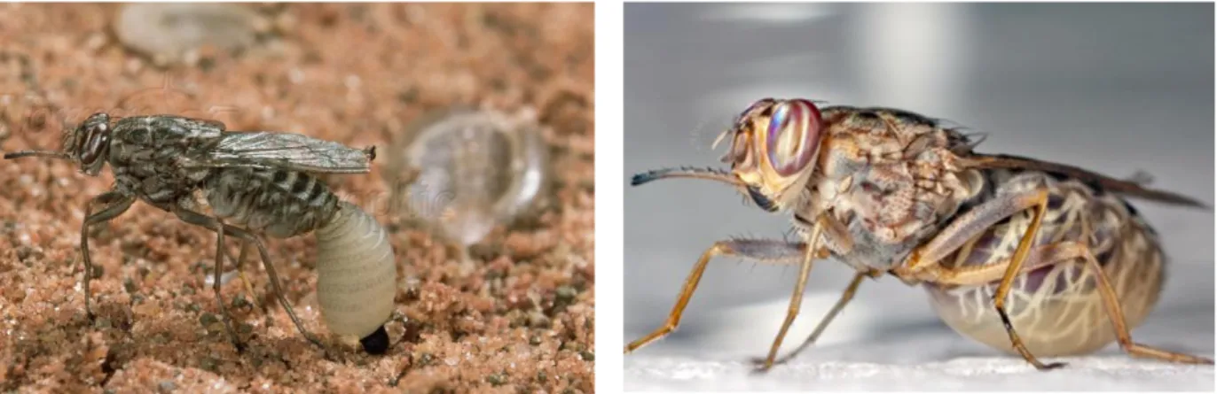
Tsetse flies of both sexes are hematophagous (blood-feeding) and can transmit trypanosomes, which relay on the insect for their cyclical transmission. The insects are viviparous and have a particular reproductive cycle. The female deposits a fully developed larva in humid soil instead of lays eggs (Figure 12). After 20-80 days the larva turns up as an adult. A female fly will only produce three to five such larvae during her 2-3 months lifespan (2 months for males) (Figure 13). Consequently, the growth rate of tsetse populations is fairly low.
Fig. 13. On the left: Tsetse fly female laying larva. On the right: Adult tsetse fly.
Tsetse flies are infected with T. brucei when they ingest trypanosomes in the blood or in the skin of mammals. Once ingested, the “short stumpy” trypanosomes reach the salivary glands of the insects and develop into the human-infective metacyclic forms.
In a normal tsetse flies population, only a small proportion (about 0.01%) carries the metacyclic trypanosomes in their salivary glands, as detected by parasitological methods (dissection and microscopy). The classical dissection/microscopy technique is the only tool available to determine infection rates in the field (Abdi et al., 2017; Wamwiri et al., 2016). However, a tsetse fly feeds every 3 days and can infect several people during its lifetime. Based on this, eliminating the flies (by aerial spraying and installation of impregnated traps) or reducing the contact between them and humans can be considered as effective approaches to block the transmission of T. brucei.
1.3.2 The parasite life cycle
The trypanosomes represent the excellent examples of organisms that display an extreme adaptation to their environment, mostly because they must avoid the immune response of the host. The parasite’s life cycle involves tsetse flies and mammalians and the trypanosomes undergo many morphological changes (Figure 14). Mammalians get infected after the bite of infected blood-sucker insects from the Glossina genus. During a blood meal on the mammalian host, an infected tsetse fly releases in the intradermal tissue the metacyclic trypanosomes. The parasites enter the lymphatic system and pass into the bloodstream (phase 1). Inside the host, they transform into bloodstream trypomastigotes (phase 2), which are carried to other sites throughout the body, and reach other blood fluids (e.g., lymph, spinal fluid). The bloodstream trypanosomes then replicate by binary fission (phases 3 and 4) and get into the insect midgut after another blood meal (phase 5). In the fly’s midgut, the trypanosomes transform in procyclic trypomastigotes, multiply by binary fission
(phase 6) and reach the salivary glands as epimastigotes (phase 7). The epimastigotes will also divide in the salivary glands and transform into metacyclic trypomastigotes responsible for new infection (phase 8).
Fig. 14. Life cycle of Trypanosoma spp. (CDC, 2015).
Injected into human skin, the trypanosome parasites will first proliferate at the site of the tsetse fly bite and divide by binary fission in the interstitial spaces. The local inflammation reaction generated by the buildup of metabolic wastes and cell debris also leads to the formation of a trypanosomal “chancre” (called ‘trypanome’) and some local lymphadenopathy.
Another important factor to taking into the consideration is that in many laboratory experiments used as infection models, is extremely evident that there is a fine tuning between the ability of the tsetse to eliminate the infection and the capacity of the parasite to evade the hostile environment of the fly. That means that tsetse infection by trypanosomes is likely to be a strongly contested process. In the Figure 15 are represented the major bottlenecks affecting the transmission of T. brucei through the tsetse fly midgut. T. brucei long and proliferative slender (LS) and short
(ES). Both survival and establishment of a parasite infection in the midgut are major bottlenecks as highlighted by the arrow at the bottom of the Figure 15. Even in successful midgut colonizations, parasite survival ranges from 1% to 0.013–0.027% by 3 dpi.
Fig. 15. Bottlenecks affecting the transmission of T. brucei through the tsetse fly. The parasite nuclei and
kinetoplasts are represented in red and black.
1.3.3 The parasite architecture (morphology)
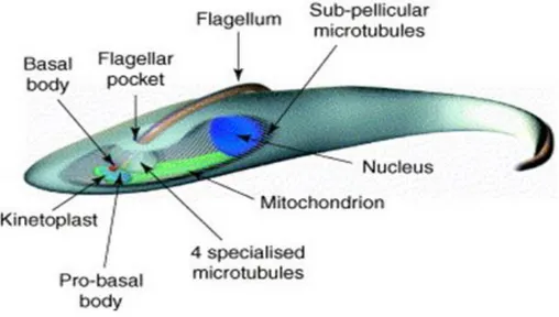
Trypanosomes are among the most ancient eukaryotes. The cell is elongated and shaped by the presence of a microtubules network following the longitudinal axis of the parasite (Mc Kean, 2003). The major organelles are present, such as mitochondria, the nucleus, the golgi apparatus and endoplasmic reticulum. The nucleus is located in the central region of the cell and contains 11 diploid pairs of megabase chromosomes, and approximately100 minichromosomes (Akiyoshi et al., 2013). The kinetoplast (mitochondrial genome) is located at the posterior end of the cell and is linked to the flagellum basal body. The flagellum rises up from the cell and is attached to the cell body along most of its length.
As part of kinetoplastids, trypanosomes possess a mitochondrial DNA which is organized in a large structure also known as kinetoplast. The presence of the flagellum is essential for the parasite motility and is attached to the trypanosome body(Figure 16).
Fig. 16. Schematic representation of procyclic T. brucei on the basis of electron microscopy studies (Mc
Kean, 2003)
1.3.4 The Variant Surface Glycoproteins (VSG)
Some trypanosome species are not infective to human and are infective only to animals. This is due to the presence in the human serum of the trypanolytic factors (TNF1 and TNF2) that provide innate protection. The species responsible for HAT, T.b. gambiense and T.b. rhodesiense, are not sensitive to these factors (Molina-Portela et al., 2008). Another very important feature of the parasite is the presence of the Variant Surface Glycoproteins that start to be expressed on the metacyclic form. These molecules are densely packed glycoproteins from 55 to 65 KDa (Dubois et
al., 2005). The invasion of the host by trypanosomes triggers a humoral immune response. The
VSG is the only antigen that can be targeted by the human’s immune system and only one species is present on the trypanosome surface coat. The parasite escapes the immune response by a continuous change of the VSG coat through an antigenic variation. This phenomenon of antigenic variation renders the development of an effective vaccine improbable (Magez et al., 2010).
1.4 Mode of trasmission
T.b. rhodesiense HAT is a zoonosis and its transmission is predominantly maintained in an
animal reservoir (cattle and wildlife). In countries where the reservoir is mainly wildlife, sporadic transmission can happen to tourists visiting National Parks. Some countries like Malawi, Tanzania, and Zambia have National Park areas within HAT endemic regions. Such “promiscuous” environment makes impossible the eradication of T.b. rhodesiense HAT. On the other hand, in Uganda, all recognised HAT endemic regions are outside National Parks areas, which makes effective control more feasible.
Due to the exponential growth of the African population, most wild animals living outside “Wildlife preserve” have been decimated by hunting and domestic animals represent nowadays the most important reservoirs of the disease. A recent study run in Busoga has shown that T.b.
rhodesiense HAT can be transmitted five/six times more likely through a cattle-fly-human cycle
than by a human-fly-human cycle (Hide et al., 2007) (Figure 17).
Fig. 17. Cattle chronically infected with T.brucei exhibit the same symptoms to those of late stage HAT.
On the contrary, T.b. gambiense HAT is sustained by the human-tsetse fly-human cycle, whereas the role of an animal reservoir is debated. Infrequently, the parasite is transmitted across the placenta or by blood transfusion.
Recently studies using modern molecular tools have confirmed the presence of T.b.
gambiense in pigs in Cameroon and Ivory Coast (Jamonneau et al., 2004; Nkinin et al., 2002 ), and
the infection was also detected in several primates.
Eight out of 24 different wild animal species (e.g. monkeys, ungulates and carnivores) were infected with T.b. gambiense in an HAT focus in Cameroon. Even though T.b. gambiense is commonly maintained by the human reservoir, this does not exclude that in some foci of Gambian HAT both reservoirs may coexist (WHO, 2013).
1.5 Clinical features of HAT disease
Many factors as the parasite subspecies, the host immune response and the stage of the disease can influence the clinical features of HAT (Büscher et al., 2017). Gambiense HAT (gHAT) leads to a chronic infection while rhodesiense HAT (rHAT) is acute. However, both are fatal if untreated or inadequately treated. The fast progression of rHAT to death is complete from few weeks to 6 months; on the other hand, it will take at least 3 years for gHAT to be fatal (Büscher et
al., 2017).
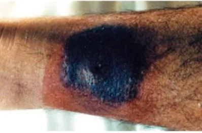
Five-fifthteen days after the tsetse bite, a 3-4 cm local skin reaction called trypanosomal chancre, (Figure 18) coupled to the presence of enlarged posterior cervical lymphadenopathy, indicates the beginning of the disease (Sitch et al., 2002; Büscher et al., 2017). The dermal reaction occurs mostly in rHAT cases and is rare on patients with T.b. gambiense disease. Microscopic examination of the aspirate or palpation of enlarged neck glands in positive cases is generally used for HAT diagnosis (Lutumba et al., 2007).
Fig. 18. Trypanosomal chancre on the dorsal side of the right ankle of a patient with gambiense HAT
(Malvy D. et al., 2011).
The disease affects mainly the lymphoid system, heart, lungs, and brain and evolves in two stages. The first stage or early stage (haemolymphatic stage) is haemato-lymphatic and the parasite is present in the blood and in the lymphatic system. The parasitaemia is generally low (less than 100 parasites per ml of blood) and fluctuating. In the second or late stage (meningo-encephalic stage) parasites will penetrate the blood-brain barrier (BBB). The parasites invade perivascular areas with subsequent infiltration in the white and grey matter of the brain.
The most common symptom in the hemolymphatic phase is fever lasting one day to several weeks. Its irregular or intermittent pattern can be explained by the continuous waves of parasites in the blood (Sitch et al.,2002; Büscher et al., 2017). Other symptoms include headache, pruritus, hepatosplenomegaly and posterior cervical lymphadenopathy. Some endocrine dysfunctions like impotence, infertility and amenorrhea are also noticed (Figure 19).
The second phase occurs within weeks in the rHAT and months in the gambiense form following the invasion of the central nervous system (CNS) by the parasites. Some studies suggest that a high concentration of the parasites in the blood allows the cross of the BBB in an immune mediated way (Mogk et al., 2014). A chronic encephalopathy and sleep-pattern disturbances, with dysregulation of the circadian rhythm, represent the principal manifestations of the second stage and lend HAT its common name “sleeping sickness”. Fever becomes less present and patients can experience mental changes and psychiatric disorders, painful peripheral sensory disturbances, tone and mobility disorders, and difficulties to concentrate. Cardiac disorders are also noticed with electrocardiogram abnormalities like perimyocarditis (Büscher et al., 2017).
1.6 Diagnosis
Symptoms for HAT are nonspecific and can easily be confused with other diseases, for this reason HAT diagnosis is based on laboratory tests. Early diagnosis is very important to increase the perspective of a cure. In fact, targeting the parasite before it get into the central nervous system necessitates a less complex therapy with relatively safer drugs. However, exhaustive screenings require major investment in hospital specialized personnel and material resources. In poor countries like Africa such resources are often limited, especially in rural and remote areas where the disease is most common. As a result, many infected people may die before diagnosis or treatment.
The differentiation between the two stages is done through examination of the cerebrospinal fluid (CSF). Staging is an important factor for HAT case management since drugs like Melarsoprol used for the second stage can cause reactive encephalopathy on patients (Checkley et al., 2007).The detection of the trypanosome parasites and the number of white blood cell (WBC) per µL are the criteria that allow to discriminate stage I and stage II. WHO recommends to consider the presence of trypanosome in the CSF or a count of more than 5 WBC/µL or both as a diagnostic criteria for second stage (Bonnet et al., 2015). The sensitivity and specificity of the criteria are debated due to the possibility of a low number of parasites present in the CSF making them undetectable. In addition, the presence of WBC in the CSF is not specific to HAT, but it is common in other diseases like meningitis. The most widely used screening test in Gambiense HAT is Card-Agglutination trypanosomiasis test (CATT), which was developed almost 40 years ago and still played an important role on the fight against the disease (Büscher et al., 2017) (Figures 20 and 21). The test is based on antigen type LiTat 1.3 (Bonnet et al., 2015) and its sensitivity and specificity vary between 68.8–100 and 83.5–99.3. Although its cost is affordable, limitation factors are represented by: cold chain storage, the 50-dose format that leads to loss of non-used doses and the availability of power sources. The test is carried on blood collected by finger prick and also on plasma or serum. Despite the high sensitivity, some false-positive results are noticed, especially for patients infected with malaria and filariasis. During the last 5 years, rapid diagnostic tests (RDT) were developed based on the antigens LiTat 1.3 and 1.5 and Second- generation RDTs based on recombinant antigens are now in clinical development (Sternberg et al., 2014 ).
The HAT Sero-K-Set and the SD bioline 1.0 were recently developed. These are rapid test made for gambiense HAT diagnostic. They are better than CATT and more suitable for population screening.
Fig. 20. Decision tree of HAT stage diagnosis (Bonnet et al., 2015). CTC (Capillary Tube Centrifugation);
All HAT confirmatory tests are based on microscopic examination for the visualisation of parasites. However, there is no single confirmatory test that has a satisfactory sensitivity, and in practice combinations of several are used. These include: lymphnode aspirates in suspects with cervical adenopathy, capillary tube centrifugation (CTC), quantitative buffy coat (QBC) or the mini anion exchange centrifugation technique (mAECT). The most sensitive test is mAECT that isolates the parasites from venous blood using anion exchange mini columns by anion exchange and concentrate them. Patient blood cells are negatively charged, while trypanosomes remain neutral, so that they can be separated by anion-exchange chromatography at pH 8.
In confirmed HAT patients, an examination of CST by a lumbar puncture, helps to establish the stage of the disease and subsequently to determine the most appropriate treatment. When parasites are visualized in the cerebrospinal fluid, the patient is considered to be in the late stage of the disease. The WHO diagnostic criteria, which are the most widely used guidelines for diagnosing late stages of the disease, required the presence of trypanosomes in the CSF of more than 5 cells per 𝜇L (WHO, 2013). However, the detection of trypanosomes in CSF by microscopy alone has limited sensitivity and may generate false negative results.
Another potential parameter which may assist with late stage diagnosis is the measurement of CSF IgM concentrations, which are increased early when there is CNS involvement.
1.7 Chemotherapy for HAT
Since the development of a preventive vaccine would be an ideal approach to this devastating disease but, unfortunately, trypanosomes are able to evade the host immune system, chemotherapy remains the only available treatment option for fighting the infection.
Current treatments for HAT depend on the stage of the disease but also on the different parasite subspecies. Nowadays, there are four drugs and one drug combination available: pentamidine, suramin, melarsoprol, eflornithine and the nifurtimox-eflornithine combination therapy (NECT) (Figure 22 and Table 1). Three out of the four drugs available (1-3) were developed over 60 years ago and exhibit severe drawbacks.
.
Table 1. Drugs for treating HAT
Disease Stage Drug Year of
Introduction
Route of
administration Shortcomings
Gambiense HAT Early Pentamidine 1941 IM or IV No oral formulation
Eflornithine 1981 IV Expensive
Melarsoprol 1949 IV Arsenical (toxic
encephalopathy)
Late NECT 2009 IV + PO Expensive
Rhodesiense HAT Early Suramin 1922 IV No oral formulation
Late Melarsoprol 1949 IV Arsenical (toxic
encephalopathy)
1.7.1 Chemotherapy for first or early stage of HAT 1.7.1.1 Pentamidine
Pentamidine isethionate (2, Figure 22) is an aromatic diamine drug discovered in 1941. It is recommended by WHO as the first-line treatment for first stage gambiense infection and has been used as such for seven decades. It is also used against antimony-resistant leishmaniasis and against
Pneumocystis jiroveci in AIDS patients (Soeiro et al., 2005).
Pentamidine efficacy against T.b. gambiense is very high, around 90-95%, while it is very limited for rhodesiense infection treatment (Büscher et al., 2017). Although the mechanism of action is unclear, the diamine structure is thought to be responsible for the antiparasitic activity trough parasite’s DNA binding (Wilson et al., 2005). A recent study suggests that pentamidine uptake is by endocytosis with the specific aquaglyceroporin TbAQP2 acting as a high affinity receptor (Song et al., 2016).
It is administered once daily for 7 days intramuscularly or as an intravenous infusion in saline over 2h (Malvy et al., 2011). To avoid the hypoglycemia provoked by this drug, the administration is preceded by the ingestion of sugar, and supine position must be maintained for 1-2 h to avoid hypotension. The intramuscular administration is less suitable for developing countries with lack of healthcare facilities. To make the administration more convenient for patients, a new strategy based on three injections is on the way and comparative clinical trials have been launched.
Pentamidine is generally well tolerated but some adverse effects can be noticed and include pain at the site of injection, hypoglycemia, hypotension, abdominal pain and leucopenia. Resistance to pentamidine has been generated in laboratory strains but is not reported to be a widespread problem in the field (Barrett et al., 2011). The limitation of pentamidine use is the parenteral
administration because of the non-oral bioavailability due to the positively charged amidine group. For decades, researcher have focus their attention on the discovery of new diamines with a better efficacy, oral bioavailability and blood brain barrier penetration. Furamidine (Figure 23) showed a poor oral bioavailability despite good in vitro and in vivo activities on Trypanosoma spp. (Midgley
et al., 2007; Wenzler et al., 2009). The selective and rapid uptake of furamidine allows an increase
in the concentration in several pathogens including Plasmodium falciparum, Leishmania spp.,
Gardia intestinalis and P. jiroveci.
To increase the oral availability, furamidine prodrugs were synthesized. In 2000, the methamidoxime prodrug, parafuramidine (Figure 23), was selected by the consortium for parasitic drug development (CPDD) for clinical development. The first phase of clinical trials, conducted in 2000 on healthy patients for 21 days, was successful. The second lasted 6 years and was conducted on patients at the first stage of the disease. The third phase conducted in collaboration with the Food and drug administration (FDA) aimed to assess the efficacy and safety of parafuramide compared to pentamidine maleate for treatment of first stage sleeping sickness. Unfortunately, there were no significant difference in term of efficacy. Parafuramidine had a better safety profile but some renal post treatment toxicities have led to widespread concerns (Pohlig et al., 2015). For this reason the development was halted in 2008.
Fig. 23. Chemical structures of Furamide and Parafuramide.
1.7.1.2 Suramin
Suramin (1, Figure 22) was first used in 1922 for HAT treatment but developed few years earlier by Bayer chemists from azo dyes, trypan red and trypan blue. It is a polysulphonated naphtylamine-based compound used for the first stage of T. b. rhodesiense. It is also active on T.b.
gambiense form of the disease but not used in therapy because of the high risk of co-infection with
Intravenous administration is the best way of administration, because suramin is poorly absorbed orally and causes serious irritation after intramuscular injections (Babokhov et al., 2013). Due to its high affinity with serum proteins (99.7%) it has a long half-life of around 44-54 days. Low-density lipoprotein (LDL) are uptaken through endocytosis as a sterols source for bloodstream forms and suramin can then enter T.b. rhodesiense (Babokhov et al., 2013).
Suramin targets include specific inhibition of T. brucei glycolytic enzymes. Thanks to its negative charge, it can specifically target the trypanosomal enzyme which has a higher isoelectric points than its mammalian counterpart (Bacchi et al., 2009). Other targets include dihydrofolate reductase, thymidine kinase and pentose phosphate pathway enzymes. The multiple targets aspect of suramin’s mechanism of action may help to explain the low development of resistance. However, a study published in 2018 has revealed the involvement of VSG in the development of resistance to suramin (Wiedemar et al., 2018).
Suramin cannot cross the blood brain barrier probably due to its large size, the presence of tights junctions near the endothelial membrane and the absence of transport vesicles. All these factors contribute to limit the concentration of suramin in the CSF (around 1%) compared to the serum. Suramin is then not effective for second stage disease but can be used in synergy with second stage HAT drugs because of its ability to inhibit the P-glycoprotein leading to the limitation of second stage drug expulsion from CNS (Sanderson et al., 2007). The side effects due to suramin administration include fatigue, neuropathy, renal problems, anaemia and nausea.
1.7.2 Chemotherapy for second or late stage of HAT
1.7.2.1 Melarsoprol
Melarsoprol (3, Figure 22) is an arsenical compound introduced in 1949 and produced by Sanofi-Aventis. Melarsoprol has been also donated, free of charge, to WHO. It is derived from the toxic melamine arsenical called melarsen and was synthesized by the Swiss chemist Friedheim. To increase the antitrypanosomal activity of the molecule, some modifications from melarsen were performed and in 1939 an analogue of melarsen named melarsen oxide was synthesized (Figure 24).
Fig. 24. Chemical structure of Melarsen oxide.
Unfortunately, the increase of activity also led to an increase in toxicity and melarsen oxide could not be used for therapy. The adding of dimercaprol (British Anti-Lewisite), a disulfide chelating agent, significantly reduced the toxicity and the resulting compound was named melarsoprol (Steverding et al., 2008). This latter has been for several decades the only option for second stage HAT treatment for both East and West African forms. After the introduction of eflornithine, melarsoprol is nowadays only recommended for treatment of late stage East African trypanosomiasis caused by T. b. rhodesiense thanks to its ability to cross the blood brain barrier.
The mechanism of action remains unclear but some studies suggest an inhibition of the trypanosomal enzymes pyruvate kinase, phosphofructokinase, fructose-2,6-bisphosphatase and trypanothione reductase (Denise et al., 1999). Arsenical interaction with glycerol-3-phosphate dehydrogenase has been studied and it has been shown that the enzyme binds specifically Cymelarsan, a melarsoprol analogue, used for animal trypanosomiasis (Denise et al., 1999). In addition, the bloodstream forms of trypanosomes are highly dependent on glycolytic and redox processes and any dysregulation of these factors may lead to cell lysis due to lack of ATP. Other studies report that melarsoprol uptake is done through the purine nucleotide transporter P2 and arsenical-resistant trypanosomes lack those transporters (Carter et al., 1993).
Melarsoprol is administered intravenously after dissolution in propylene glycol because it is not soluble in water. Different variants are considered for melarsoprol administration, but the best regiment consists of 2.2 mg/kg per day over 10 days with a cure rate of 93.9% at the beginning and 86.2% after two years of follow-up (Babokhov et al., 2013).
Melarsoprol has a high number of side effects and the most important is the arsenical-induced reactive encephalopathy syndrome (fever, convulsion, neurological disorders, and coma)
considered as an alarm and the treatment should be stopped. The intravenous administration is painful due to the presence of propylene glycol that can destroy the veins.
1.7.2.2 Eflornithine
Eflornithine (α-difluoromethylornithine or DMFO, 4, Figure 22) is the latest antitrypanosomal monotherapy introduced in 1990. It was first developed in the 1970s as an anticancer and ten years later, the scientist Cyrus Bacchi decided to assess its antitrypanosomal activity. He demonstrated that eflornithine was able to cure T.b. brucei infected mice with no signs of side effects. DFMO was then subjected to extensive human clinical trials in Africa.
For a typical-sized individual, the demanding regime is nearly a half-kilogram of drug administered while the patient is hospitalised. Through support from WHO, eflornithine kits for two full treatments weigh 40 kg and cost US$ 1420, and have been made available for distribution in disease endemic countries.
Between 2001 and 2002, 1055 patients (adults and children) diagnosed with second stage HAT received eflornithine for 14 days at different dosages, 400 mg/Kg/day and 600 mg/Kg/day for adults and children, respectively. The study concluded that eflornithine is effective. and relative safety when used as first line treatment for HAT (Priotto et al., 2008). No relapse was observed more than 12 months after the treatment and higher doses were well tolerated by children. The drug is an inhibitor of the enzyme ornithine decarboxylase (ODC) which is responsible for the synthesis of polyamines to facilitate cell division and proliferation. It binds irreversibly to ODC catalytic site. A study assessing the in vivo activity of Eflornithine on metabolism and morphology of T.b. brucei reveals that treated parasites have a short and broader form with multiple kinetoplasts (Bacchi et al., 2009). In addition, trypanosomal ODC activity is reduced by 99% in 12 hours and polyamine levels are significantly reduced. It is only effective on T.b. brucei and T.b. gambiense but not on the T.b.
rhodesiense strains. The selectivity is due to the slow ODC turnover rate of T. brucei gambiense (19
hours) compared to the T.b. rhodesiense and mammalian counterparts with respectively 4.3 hours and 10-30 minutes (Babokhov et al., 2013).
Eflornithine is not a trypanocidal but a trypanostatic molecule which blocks the division of bloodstream trypanosomes and requires a functional immune system to get rid of the parasite. The co-infection with HIV-AIDS or other immuno-suppressant conditions can be a serious problem.
The standard administration of DMFO involves 56 intravenous infusions at 100 mg/kg for adults and 150 mg/kg for children every 6 hours for 7 days. An alternative regiment involving a reduced number of intravenous injections at 100 mg/kg every 6 hours over 7 days was studied in clinical trials as well as the oral administration of Eflornithine. Both studies showed reduced efficacy (Babokhov et al., 2013).
Eflornithine is as an alternative after melarsoprol relapse for gambiense infection treatment. The drug is in fact more effective against T.b. gambiense compared to melarsoprol but less fatal. The fatality rate for eflornithine is estimated to be around 1.4% versus 5% for melarsoprol and the cure rate is 94% for eflornithine and 84% for melarsoprol (Priotto et al., 2008). The side effects for eflornithine include diarrhea, headache, seizures, anemia, and leucopenia, typical of anticancer drugs. Resistance, due to mutations in a putative amino acid transporter, has been shown in vitro.
1.7.2.3 Nifurtimox-Eflornithine combination therapy (NECT)
An important recent advancement in HAT chemotherapy was the introduction of nifurtimox-eflornithine combination therapy (NECT), which is currently the first line of treatment for HAT. The treatment is strongly recommended by WHO and has been included to the WHO Essential Medicine List in 2009. Kits for four full treatment courses weigh 36 kg and cost US$ 1440, and are being widely adopted in disease endemic countries.
Nifurtimox (5, Figure 22), also known as Lampit, is a nitrofuran derivative trypanocide. It was developed by Bayer in 1960 and used as treatment of Chagas disease (South American trypanosomiasis) caused by Trypanosoma cruzi since 1967. The company provides free of charge tablets to WHO and financial assistance to sustain endemic countries like Honduras and El Salvador. The mechanism of action of nifurtimox is unclear, but some studies report a metabolic conversion after enzymatic reduction of the nitro group leading to the production of free radicals. Nitroreductases (NTR) and trypanothione reductases are the enzymes involved in the free radical generation which interfere with parasite membrane, DNA and proteins. The impairment of type I nitroreductase in T. cruzi conferred a resistance to nifurtimox (Wilkinson et al., 2008). Its efficacy against T. brucei infections have been assessed and showed a limited activity against stage II HAT. The low concentration of nifurtimox in the CNS compared to the plasma and the point mutation of
nifurtimox and eflornithine + melarsoprol combination were assessed. The Nifurtimox + eflornithine combination therapy consisting of nifurtimox 15 mg/Kg/day over 10 days orally and eflornithine 400 mg/Kg/day over 7 days IV was shown to be the most potent with 94% cure rate and low fatality rate (Priotto et al., 2008). From 56 intravenous administration of eflornithine monotherapy, the combination allowed a reduction to 14, which makes it easier to administer and less expensive. In rural areas characterized by poverty and a lack of healthcare personnel, it is a huge advancement since it requires less hospitalization and allows a better compliance for patients who can take nifurtimox at home. The side effects for NECT replicates those of eflornithine and nifurtimox but it is generally well tolerated. This includes abdominal pain, vomiting and headache.
Despite this positive advancement with the HAT therapy, the high costs of distribution coupled with intravenous route of administration, makes the therapy way far from ideal chemotherapy. For this reason, DNDi has proposed a target product profile (TPP) for getting a better drug for HAT. The ideal drug would be orally administered, effective against both early- and late-stage disease, safe for children and pregnant women, without drawbacks and inexpensive (less than 30 euros per course). Due to the large distance between the ideal HAT drug and the state-of-art of currently used HAT drugs, there is still much work to be done as drug discovery. Recent advancements and new discoveries that will be discussed in the next chapters fill us with optimism that these goals are achievablein the foreseeable future.
1.8. Drug candidates in clinical trials for HAT
1.8.1 Fexinidazole
Fexinidazole (Figure 25) is a 2-substituted 5-nitroimidazole that was identified as a promising drug candidate for HAT after a screening of around 700 nitroheterocyclic compounds
from diverse sources conducted by the DNDi. Fexinidazole
(1-methyl-2-((p-methylthio)phenoxy)methyl)-5-nitroimidazole was first developed by Hoechst AG (now Sanofi-Aventis) as a broad spectrum antimicrobial agent in the 1970’s.
Fig. 25. Chemical structure of Fexinidazole.
In 2010, Torreele et al. conducted a study to assess the in vitro and in vivo antitrypanosomal activities of fexinidazole and its metabolites fexinidazole sulphoxide and fexinidazole sulphone (Torreele et al., 2010) (Figure 26).