https://doi.org/10.1177/1756284819867839 https://doi.org/10.1177/1756284819867839 Ther Adv Gastroenterol
2019, Vol. 12: 1–14 DOI: 10.1177/ 1756284819867839 © The Author(s), 2019. Article reuse guidelines: sagepub.com/journals-permissions
Therapeutic Advances in Gastroenterology
journals.sagepub.com/home/tag 1
Introduction
Colorectal cancer (CRC) is an important health issue, since one million new cases are diagnosed worldwide each year, with half a million related deaths.1,2 In most cases it is sporadic in nature,
with age being the only major risk factor reported.3 Metabolic syndrome (MetS), that is,
the combi nation of cardiovascular risk factors, such as obesity, hypertension, diabetes and dyslipidemia,4–6 has been identified as a potential
risk factor for cancer,7 including sporadic
CRC.8–10 The prevalence of MetS ranges between
34.8% and 41.9% in the US and 18% and 46% in Europe, depending on age, geographic location
Metabolic syndrome is a risk factor for
colorectal adenoma and cancer: a study in a
White population using the harmonized criteria
Angelo Milano, Maria Antonia Bianco, Luigi Buri, Livio Cipolletta, Enzo Grossi, Gianluca RotondanoΩ, Francesco Tessari, Konstantinos Efthymakis
and Matteo Neri on behalf of NEOCOSM (NEOplasia del COlon e Sindrome Metabolica) Study Group
Abstract
Background: Metabolic syndrome (MetS) has been associated with colorectal adenomas
and cancer. However, MetS definitions have changed over time, leading to a heterogeneity of patients included in previous studies and a substantial inextensibility of observations across time or eastern and western populations. Our aim was to evaluate the association of ‘harmonized’ criteria-defined MetS and its individual components with colorectal neoplasia and cancer in a western population.
Methods: In this multicenter, cross-sectional study, we prospectively evaluated consecutive
outpatients who underwent open-access colonoscopy over a 3-month period. MetS was diagnosed according to the 2009 ‘harmonized’ criteria.
Results: Out of 5707 patients enrolled, we found 213 cancers (3.7%), 1614 polyps (28.3%),
240 nonpolypoid lesions (4.2%), 95 laterally spreading tumors (1.6%). Polyps presented histological low-grade dysplasia in 72.9% of samples, while in 9.8%, high-grade dysplasia or in situ carcinoma was present; dysplasia rates for nonpolypoid lesions were 66.2% (low-grade) and 2.9% (high-grade/in situ carcinoma), while for laterally spreading tumors, 29.6% and 37%, respectively. Overall, MetS prevalence was 41.6%. MetS correlated with both adenomas [odds ratio (OR): 1.76, 95% confidence interval (CI) 1.54–2.00] and cancer (OR: 1.92, 95% CI 1.42– 2.58). MetS was the only risk factor for such colonic lesions in subjects younger than 50 years. For all colonic neoplasia, we found MetS and not its individual components to be significantly associated.
Conclusions: MetS is risk factor for cancer and adenoma in Whites, especially when younger
than 50 years. MetS patients might be considered as a high-risk population also in colorectal cancer screening programs.
Keywords: colon cancer, colorectal adenoma, CRC, metabolic syndrome, MetS Received: 5 February 2019; revised manuscript accepted: 12 July 2019.
Correspondence to:
Matteo Neri Department of Medicine and Aging Sciences and Center of Aging Sciences and Translational Medicine (CeSI-MeT), ‘G.D.’ Annunzio University and Foundation, Chieti, Italy Digestive Endoscopy and Gastroenterology Unit, ‘SS Annunziata’ University Hospital, Chieti, Italy
Angelo Milano Konstantinos Efthymakis
Department of Medicine and Aging Sciences and Center of Aging Sciences and Translational Medicine (CeSI-MeT), ‘G.D.’ Annunzio University and Foundation, Chieti, Italy Digestive Endoscopy and Gastroenterology Unit, ‘SS Annunziata’ University Hospital, Chieti, Italy
Maria Antonia Bianco Livio Cipolletta Gianluca Rotondano
Division of Gastroenterology and Digestive Endoscopy Unit, Hospital ‘A Maresca’, Torre del Greco, Italy
Luigi Buri
Gastroenterology and Digestive Endoscopy Unit, Cattinara Hospital, Trieste, Italy
Enzo Grossi
Medical Department Bracco SpA, Milan, Italy
Francesco Tessari Electronic Data Processing, Idea 99 Srl, Padova, Italy ΩDeceased. Original Research
or demographic features of the population stud-ied,11,12 thus raising relevant public health
impli-cations. In fact, although screening programs based on age and fecal occult blood have proven effective in reducing CRC mortality,13
identifi-cation of other potential risk factors, such as MetS, may further improve cost effectiveness of these screening strategies by favoring a more detailed risk-based stratification of the target populations. Although the concept of metabolic syndrome has been widely accepted for a long time,14,15 there was no largely recognized
inter-national definition until 1998 and onward,16–19
up to the ‘harmonized’ classification in 200920
that deeply revised the concept of MetS. This has led to the identification of heterogeneous target populations based on the adopted defini-tion. Two recent meta-analyses, although show-ing an association between MetS and colon cancer, have calculated I2 values indicative of a
significant inhomogeneity among studies,21,22
thus weakening their conclusions.
The primary objective of this study, therefore, was to prospectively evaluate the association between MetS, defined according to the 2009 cri-teria, and colonic neoplastic lesions in consecu-tive patients undergoing colonoscopy; secondary objectives were to evaluate the role of individual components of MetS in indicating a specific risk for neoplastic disease and, finally, if MetS is a risk factor independent of age.
Methods
Study participants and design
This study has been conducted in 50 open-access endoscopy units, evenly distributed throughout Italy. Every unit prospectively enrolled consecutive outpatients undergoing colonoscopy in a 3-month period.
The indications for performing colonoscopy were abdominal symptoms (impaired bowel habitus, pain, hematochezia, anemia, weight loss, abdomi-nal mass on physical examination), follow up [previous polypectomy, previous colonic surgery, inflammatory bowel disease (IBD)], colon cancer screening [with or without previous positive fecal occult blood (FOBT), voluntary colonoscopy if over 50 years of age, positive family history for CRC]. Patients were excluded in case of failure of cecum intubation, poor bowel cleaning, defined
as persistence of solid or semisolid debris that could not be effectively cleaned,23 age below
30 years, impossibility to obtain anthropometric measurements, polyposis syndrome, advanced neoplastic disease, impossibility to obtain written informed consent. Before implementation, this study was approved by the ethical committee of each participating center and informed consent was obtained from all participants.
For each subject, blood pressure, height, weight, and waist circumference were measured. Interviewers completed a medication history for each participant, including use of insulin, oral hypoglycemic agents, antihypertensive drugs, and hypolipidemic agents. Medical history assessment included also occupation, use of low-dose acetyl-salicylic acid, and hormone replacement therapy in women. Self-reported lack of physical activity was also recorded.24
Laboratory results for fasting plasma glucose, high-density lipoprotein cholesterol (HDL-C), total cholesterol and triglyceride levels were obtained. Since biochemistry data could not be centralized, blood tests had to be performed in sites complying with the updated European guide-lines on accreditation of medical laboratories.
Quality control
Eligible endoscopy units were required to have performed at least 1000 colonoscopies in the pre-vious year and to have already participated in at least one previous multicenter study.25 This
ensured that all endoscopists adhered to previ-ously validated quality criteria regarding colono-scopic examination and identification of superficial lesions, in order to avoid heterogene-ous diagnoses and noncomparable results. In fact, all endoscopists had already been trained to rec-ognize and classify colonic superficial lesions according to the Paris classification.26 A 1-day
start-up preparatory meeting of at least one inves-tigator from each participating unit was held 3 months prior to the initiation of the study, in order to discuss the protocol and instruct all par-ticipants to achieve homogeneous blood pressure and anthropometric measurements.
Blood pressure was obtained sphygmomanometri-cally, according to standard protocols and tech-niques.27 Height was measured without shoes to the
shoes or heavy outer clothing. Circumferences were measured to the nearest centimeter using a stretch-resistant tape that provides constant tension over light clothing. Waist circumference was measured halfway between the lower ribs and the iliac crest at the end of a normal expiration, when the lungs are at their functional residual capacity, while hip cir-cumference was measured at the largest circumfer-ence around the buttocks with the tape parallel to the floor, as per World Health Organization recommendations.28
Outpatients were consecutively enrolled; endo-scopists were blinded to the metabolic status of the examinees.
Definitions
Metabolic syndrome. According to the ‘harmo-nized’ criteria set forth by a joint scientific multi-societal committee in 2009,20 MetS is defined as
the coexistence of three or more of the following risk factors: waist circumference equal or greater than 102 cm in men and equal or greater than 88 cm in women, serum triglyceride concentration of 150 mg/dl or greater; HDL-C concentration of less than 40 mg/dl for men or less than 50 mg/dl for women; blood pressure of 130/85 mmHg or greater; fasting plasma glucose concentration of 100 mg/dl or greater. Individuals who were using antidiabetics, antihypertensive or hypolipidemic drugs were treated as meeting the criteria for the affected variable.
Colonic lesions and histology. Masses or severely excavated and ulcerated lesions were classified as advanced neoplasia/cancer. Colonic superficial neoplastic lesions (SNLs) were classified accord-ing to the Paris classification26 and subgrouped as
polypoid (pedunculated or sessile, 0-Ip and 0-Is, respectively) and nonpolypoid lesions (NPLs) if measuring less than 2.5 mm in height. LSTs (lat-erally spreading tumors) were defined as lesions measuring less than 2.5 mm in height and more than 10mm in width, as in previous studies.25,29,30
For each type of lesion, size and number were recorded; in case of multiple lesions, measure-ments were performed on the largest. Location of colorectal neoplasia was defined according to anatomic site. Cecum, ascending colon, hepatic flexure, transverse colon, and splenic flexure were defined as proximal colon; descending and sig-moid colon were defined as distal colon, whereas the rectum was considered separately. Subjects
who had lesions in both the proximal and the dis-tal colon were defined as having lesions on both sides. A positive colonoscopy was defined as pres-ence of one or more of the aforementioned lesion types.
All detected lesions, where possible, were resected endoscopically. Biopsies were obtained only in case of suspected advanced cancers or nonresect-able lesions that, when confirmed as such, were referred for surgery or palliation. Therefore, his-tological analysis of superficial lesions was based on resected lesions; in case of multiple resections, the most advanced histological pattern was con-sidered for analysis. All specimens were reviewed by an experienced and dedicated pathologist at each center, who was blinded to the patients’ metabolic status.
Histological findings were classified as hyperplas-tic lesions, low-grade intraepithelial neoplasia (LGIN: serrated adenoma, tubular adenoma, tubulovillous adenoma, villous adenoma), high-grade intraepithelial neoplasia (HGIN: SNLs revealing high-grade dysplasia or intraepithelial neoplasia or carcinoma in situ31), while lesions
with invasive features were defined as submucosal carcinoma, as per current literature.25 Lesions
presenting either LGIN or HGIN were subse-quently grouped together under the term ade-noma, for statistical analysis.
Statistical analysis
Descriptive analysis included calculations of rates and proportions for categorical data, as well as means and standard deviations (SDs) for continu-ous data. Differences between means of continucontinu-ous variables were analyzed using the Student’s t test, while the χ2 test was used for categorical variables.
All p values were two tailed, and p values below 0.05 were considered statistically significant. A standard logistic regression model was used to estimate the association between positive colo-noscopy, CRC, SNLs, MetS and demographic, anthropometric, lifestyle and biochemical data by univariate analysis. Significantly associated varia-bles were considered for multivariable analysis, with the additional inclusion of age, sex, absence of physical activity and body mass index (BMI) > 30, unless otherwise specified in indi-vidual tables or figures. Results are presented as odds ratio (OR) with a 95% confidence interval
(95% CI). Analyses were performed using the SPSS version 17.0 statistical package (SPSS, Inc., Chicago, IL).
Results
In the present study, 5707 subjects out of 5824 have been enrolled; 117 were excluded mainly due to missing data. Demographic, anthropomet-ric and laboratory data are shown in Table 1. Indications for colonoscopy were presence of symptoms in 48%, follow up 38%, colon cancer screening 17%. No previous colonoscopy was reported in 62% of cases.
Colonoscopy was positive for SNL/CRC in 35.2% (2010/5707) of the procedures, for a total of 2162 lesions observed. Of these, 90.1% were SNL and 9.8% were cancers. Four cases of familiar adeno-matous polyposis were diagnosed at this stage and therefore excluded. The majority of neoplasia were sessile polyps (62%); 20.8% were peduncu-lated, 15.4% were flat elevated and 1.7% were flat depressed lesions. Detailed histological grading of endoscopic findings is shown in Table 2.
MetS was found in 41.6% of subjects enrolled; it was significantly more prevalent among females (females 43.4% versus males 40.1%; p < 0.05). In both sexes, MetS positively correlated with older age (mean age of MetS versus non-MetS, males 65.3 ± 10.5 versus 58 ± 13.7 years; p < 0.001, females 65.8 ± 10.7 versus 56.1 ± 13.6 years; p < 0.001). Regarding geographic distribution, MetS was found more frequently in individuals from southern Italy (south versus central or north-ern Italy: OR 1.48, 95% CI: 1.11–2.19; p < 0.001). Northern and central populations were similarly affected.
Male sex (OR 1.48, 95% CI: 1.33–1.66), age > 50 years (OR 3.57, 95% CI: 3.01–4.24), absence of self-reported regular physical exercise (OR 1.29, 95% CI: 1.12–1.48), obesity (OR 1.38, 95% CI: 1.20–1.58) and MetS (OR 2.17, 95% CI: 1.94–2.43) were significantly associated to a positive colonoscopy at univariate analysis. At multivariable analysis, MetS (OR 1.84, 95% CI: 1.63–2.08; p < 0.001), age > 50 years (OR 3.00, 95% CI: 2.51–3.59; p < 0.001) and male sex (OR 1.50, 95% CI: 1.34–1.69; p < 0.001) remained statistically significant when corrected for age, sex, absence of physical exercise and obesity. Notably, obesity did not show any
association with colonoscopy outcomes. Overall, MetS significantly associated by multivariable analysis with both adenoma and cancer (respec-tively, OR 1.76, 95% CI: 1.54–2.00 and OR 1.92, 95% CI: 1.42–2.58; p < 0.001), as did age > 50 years (respectively, OR 3.16, 95% CI: 2.56–3.90 and OR 2.83, 95% CI: 1.59–5.04; p < 0.001); this remained true when colonic and rectal lesions were considered separately. Male sex significantly associated with colorectal ade-noma (OR 1.47, 95% CI: 1.30–1.67; p < 0.001) but not cancer, while the opposite was true for subjects reporting no regular physical exercise (OR 1.98, 95% CI: 1.25–3.14; p < 0.01; see Table 3). No differences in terms of anatomical distribution of lesions were observed between sexes; we observed a tendency for a more proxi-mal localization in MetS subjects, although this did not achieve statistical significance. MetS was significantly associated to the presence of both polypoid and NPLs (p < 0.05), but not to their size and number; moreover, no association was found with LSTs.
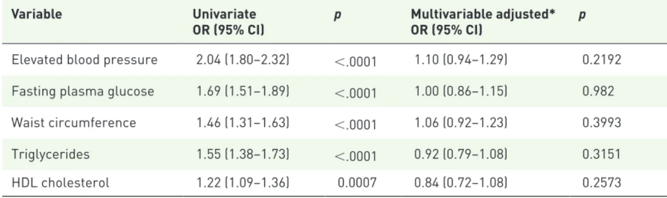
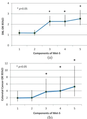
In order to take into account the possibility that colonoscopic neoplasias were more prevalent in symptomatic patients, we performed a multivari-able analysis correcting for all indications for colonoscopy (detailed in the Materials and Methods section), as well as sex, age, absence of physical activity and obesity; MetS was again confirmed as independently associated with ade-nomas (OR 1.78, 95% CI: 1.58–2.00, p < 0.05) and CRC (OR 1.86; 95% CI: 1.40–2.49, p < 0.05). In a subgroup analysis on patients undergoing screening colonoscopies (n = 967), we found 464 lesions (367 adenomas and 24 can-cers). In this subgroup, at multivariable analysis, MetS was again associated to adenomas (OR 2.23, 95% CI: 1.70–2.92, p < 0.05). Association with cancer was not statistically significant (OR 1.19, 95% CI: 0.53–2.70, p > 0.05), possibly due to the low number of occurrences (n = 28). None of the components of MetS was individu-ally associated with a positive colonoscopy in multivariable analysis (Table 4). However, a sta-tistically significant association was observed when at least three components were simultane-ously present, as per MetS definition, in both adenomas and CRC (p < 0.05; Figure 1). MetS as a risk factor for neoplastic lesions was inde-pendent of BMI; its role was maintained in lean (BMI < 25, OR 2.27, 95% CI: 1.84–2.79),
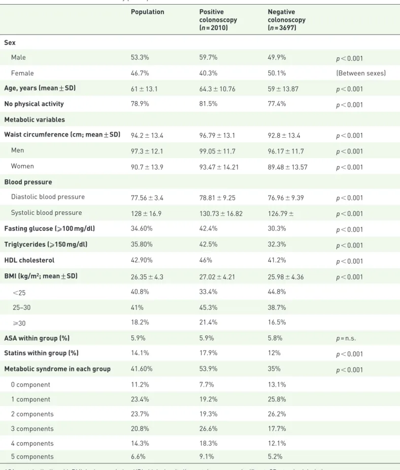
Table 1. Baseline characteristics of study participants. Population Positive colonoscopy (n = 2010) Negative colonoscopy (n = 3697) Sex Male 53.3% 59.7% 49.9% p < 0.001
Female 46.7% 40.3% 50.1% (Between sexes)
Age, years (mean ± SD) 61 ± 13.1 64.3 ± 10.76 59 ± 13.87 p < 0.001
No physical activity 78.9% 81.5% 77.4% p < 0.001
Metabolic variables
Waist circumference (cm; mean ± SD) 94.2 ± 13.4 96.79 ± 13.1 92.8 ± 13.4 p < 0.001
Men 97.3 ± 12.1 99.05 ± 11.7 96.17 ± 11.7 p < 0.001
Women 90.7 ± 13.9 93.47 ± 14.21 89.48 ± 13.57 p < 0.001
Blood pressure
Diastolic blood pressure 77.56 ± 3.4 78.81 ± 9.25 76.96 ± 9.39 p < 0.001 Systolic blood pressure 128 ± 16.9 130.73 ± 16.82 126.79 ± p < 0.001
Fasting glucose (⩾100 mg/dl) 34.60% 42.4% 30.3% p < 0.001 Triglycerides (⩾150 mg/dl) 35.80% 42.5% 32.3% p < 0.001 HDL cholesterol 42.90% 46% 41.2% p < 0.001 BMI (kg/m2; mean ± SD) 26.35 ± 4.3 27.02 ± 4.21 25.98 ± 4.36 p < 0.001 <25 40.8% 33.4% 44.8% 25–30 41% 45.3% 38.7% ⩾30 18.2% 21.4% 16.5%
ASA within group (%) 5.9% 5.9% 5.8% p = n.s.
Statins within group (%) 14.1% 17.9% 12% p < 0.001
Metabolic syndrome in each group 41.60% 53.9% 35% p < 0.001
0 component 11.2% 7.7% 13.1% 1 component 23.4% 19.2% 25.8% 2 components 23.7% 19.3% 26.2% 3 components 20.8% 26.6% 17.7% 4 components 14.3% 18.3% 12.1% 5 components 6.6% 9.1% 5.2%
overweight (BMI ⩾ 25 but <30, OR 1.74, 95% CI: 1.47–2.05), and obese subjects (BMI ⩾ 30, OR 2.55, 95% CI: 1.90–3.41, p < 0.05).
Finally, MetS was an important predictor of a positive outcome at colonoscopy in subjects younger than 50 years, which represented 19.9%
Table 2. Histological patterns of superficial neoplastic lesions (n = 1949).
Polyp NPL LST
n % (within
category) n % (within category) n % (within category)
Hyperplastic 255 15.7% 70 29.1% 5 3.7%
LGIN 1177 72.9% 159 66.2% 45 29.6%
HGIN/Cis 159 9.8% 7 2.9% 36 37.0%
Submucosal cancer 23 1.4% 4 1.6% 9 29.6%
Total 1614 (82.8%) 240 (12.3%) 95 (4.8%)
Cis, Carcinoma in situ; HGIN, high-grade intraepithelial neoplasia; LGIN, low-grade intraepithelial neoplasia; LST, laterally spreading tumor; NPL, nonpolypoid lesion; Submucosal cancer, neoplasia extending beyond the lamina propria.
Table 3. Multivariable-adjusted estimates of odds ratios for superficial neoplastic lesions and colorectal cancer.
Variables Adenoma
OR (95% CI) p CancerOR (95% CI) p
Age > 50 years 3.16 (2.56–3.90) <0.001 2.83 (1.59–5.04) <0.001 Male sex 1.47 (1.30–1.67) <0.001 1.23 (0.93–1.63) n.s. No physical activity 0.99 (0.84–1.16) n.s. 1.98 (1.25–3.14) <0.01 Metabolic syndrome 1.76 (1.54–2.00) <0.001 1.92 (1.42–2.58) <0.001
Obesity 1.05 (0.90–1.23) n.s. 0.92 (0.65–1.3) n.s.
CI, confidence interval; n.s., nonsignificant; OR, odds ratio.
Table 4. Univariate and multivariable analysis of metabolic factors associated with neoplastic findings at colonoscopy.
Variable Univariate
OR (95% CI) p Multivariable adjusted*OR (95% CI) p Elevated blood pressure 2.04 (1.80–2.32) <.0001 1.10 (0.94–1.29) 0.2192 Fasting plasma glucose 1.69 (1.51–1.89) <.0001 1.00 (0.86–1.15) 0.982 Waist circumference 1.46 (1.31–1.63) <.0001 1.06 (0.92–1.23) 0.3993 Triglycerides 1.55 (1.38–1.73) <.0001 0.92 (0.79–1.08) 0.3151 HDL cholesterol 1.22 (1.09–1.36) 0.0007 0.84 (0.72–1.08) 0.2573
*Adjustment was made for each component of MetS, as well as age, sex and absence of physical activity. CI, confidence interval; HDL, high-density lipoprotein; MetS, metabolic syndrome; OR, odds ratio.
of the studied population (555 male, 581 female, mean age 41.6 ± 6.38 years). Although the preva-lence of colonic lesions within this age group was expectedly lower than in the elderly (15.8% versus 40.1%, respectively), in subjects with MetS, it was almost three times higher (2.7% versus 0.8%; p < 0.05). In addition, when analyzed in a multi-variable model corrected for IBD, family history, sex, physical activity, and obesity, MetS was the only factor associated with the presence of colonic lesions (OR 2.59, CI 95%: 1.78–3.79, p < 0.05). Discussion
Previous studies from Asian populations have shown that MetS is a risk factor for colonic ade-nomas.32–34 In one study35 conducted on a Korean
population, MetS was associated to adenomas in the rectosigmoid colon; however, in this study, rectosigmoidoscopy only was performed, and
modified Adult Treatment Panel criteria (using BMI instead of waist circumference) were used to diagnose MetS. Morita and colleagues33
con-firmed the association of MetS with adenomas in the proximal colon for lesions greater than 5 mm. However, this study was restricted to male patients and a positive correlation was found only when the lowest cut-off for waist circumference was applied, thereby increasing the sensitivity of MetS diagnostic criteria. The importance of MetS was also highlighted in a recent 5-year fol-low-up study by Chiu and colleagues, showing that MetS is a significant risk factor for advanced adenoma in negative baseline colonoscopy and low-risk patients.36
Although the association between MetS and colorectal cancer has been previously investi-gated,37–39 available studies are widely
heteroge-neous in terms of the criteria used to diagnose the syndrome; in fact, some studies included fea-tures not previously adopted by any internation-ally formalized definition.37,40–43 Some authors
have used BMI, an indicator of obesity rather than of a multifactorial metabolic disturbance, as a discriminatory component for MetS.39,43–45
Studies were also different in terms of design (cohort or association) and endpoints (mortality or incidence);45–47 variable adjustments for
con-founding factors and ethnicity may also account for disparity. Moreover, although it is known that colon and rectal cancer are different in terms of prevalence, natural history and prognosis, dis-tinctions between them,38,40 or between male and
female sex38,41 have variably been described,
thus, making the results difficult to pool and interpret. Finally, evolving definitions of MetS have previously required mandatory criteria such as insulin resistance16 and waist circumference;18
as such, those individual criteria were, by nature, associated with endoscopic neoplasia because of selection bias.
For these reasons, in this cross-sectional, multi-center, observational study, we have applied the latest ‘harmonized’ criteria for the diagnosis of MetS20 in a well-defined population of 5707
White patients undergoing colonoscopy. This includes also patients with metabolic disturbances preceding prediabetes or diabetes, hypertension or obesity, thus, strengthening the role of MetS as a cluster of factors and not a mere association of unrelated phenotypes. Our data demonstrate that patients affected by MetS have a significantly
Figure 1. Occurrence of superficial neoplastic lesions or colorectal cancer according to the number of components of MetS present.
Occurrence of superficial neoplastic lesions (a) or colorectal cancer (b) according to the number of components of MetS present. For both SNL and CRC, statistical significance was achieved when at least three components were simultaneously present.
CI, confidence interval; CRC, colorectal cancer; MetS, metabolic syndrome; OR, odds ratio; SNL, superficial neoplastic lesions.
increased risk of colonic and rectal adenomas and cancer at endoscopy.
To our knowledge, no consistent data from west-ern countries on the relationship between MetS and colonic lesions are available. Large previous studies on western populations using older or not universally accepted definitions of MetS, many retrospective in nature, have variably reported a positive association between MetS and colonic adenoma or cancer,39,41,43,44,46,48,49 noting
impor-tant differences between sexes. This is the first large prospective cross-sectional study that estab-lishes an association between MetS, as defined by the current ‘harmonized’ criteria, and adenoma/ CRC in a western population. In contrast to pre-vious studies, we have shown that MetS is an independent significant risk factor for neoplastic lesions in the colon and rectum, both in men and women, in a large White population.
Our data demonstrate that patients affected by MetS have a significantly increased risk of colonic and rectal adenomas and cancer at endoscopy. None of the components of MetS when consid-ered individually or in pairs, were associated with an increased risk of neoplasia. On the contrary, simultaneous presence of at least three out of five criteria abruptly increased this risk in a statisti-cally significant fashion, regardless of age. This observation further strengthens the notion that MetS is more than the sum of its parts, potentially indicating an independent underlying pathophys-iological drive for epithelial transformation at any age. A previous large retrospective case-control European study,45 utilizing multiple MetS
defini-tions, including the ‘harmonized’ criteria, found a significant relative risk for colon cancer in affected patients irrespective of the definition used. However, the authors found that this risk was adequately accounted for by the presence of an abnormal glucose metabolism and abdominal obesity, rather than the syndrome itself. Furthermore, cases included only final CRC diagnoses, but not adenomatous lesions or spe-cific endoscopic outcomes.
The potential implication of these findings is that a set of particular dysmetabolic alterations giving rise to MetS might be an important factor that modulates the transition from a hyperproliferative to an adenomatous epithelium.50 Environmental
risk factors and dysmetabolic conditions (obesity, increased visceral adipose tissue, diabetes or
insulin resistance, dyslipidemia) have been largely investigated in relation to CRC.8,51,52 Some have
reported positive associations between CRC and obesity with an incremental risk of 1.24 for men and 1.09 for women per 5 kg/m2 increase.51
Others have shown that fat tissue distribution (waist circumference, hip circumference, waist-to-hip ratio), more than BMI, correlates with the risk of colorectal cancer or adenoma,32,53 and that
a higher visceral adipose tissue volume is associ-ated to an increased risk for adenoma at baseline and follow-up colonoscopy.54,55 Both adult-onset
diabetes mellitus and glucose intolerance have been associated with an increased risk of colon cancer56,57 or adenoma.58,59 Elevated arterial
blood pressure,60 hypertriglyceridemia and
hyper-cholesterolemia61,62 have also been associated
with CRC. The mechanisms underlying these associations are not completely understood, yet insulin resistance has been proposed as a key mechanism in promoting cancer through adi-pokine imbalance, reactive oxygen species and inflammatory cytokine release, such as tumor necrosis factor-alpha and interleukin-6, that act as ancillary pathways in this context.8,50,63–66
In our study, the association between MetS and colonic neoplasia was independent from obesity, as defined by BMI class. In fact, the correlation between this complex metabolic disturbance and colorectal adenoma or cancer was similar in either lean, overweight, or obese patients. Moreover, after accounting for MetS status, BMI-defined obesity showed no residual risk for colonic neoplasia, prob-ably because MetS is a much more specific state of metabolic imbalance, transcending simple over-weight/obesity. It has been noted that while BMI is generally an accurate index of obesity in the general population, it is less well correlated to the meta-bolic disturbances implied by such a status. In fact, it is precisely this drawback of BMI that led to the adoption of waist circumference as a marker of metabolic disturbance and a relevant component of MetS. It has been hypothesized that the partial lack of correlation between these anthropometric indi-ces is due to the fact that waist circumference, a more specific marker of abdominal obesity, is a bet-ter predictor of the metabolically active visceral adi-pose tissue, as opadi-posed to the less active peripheral fat.19,50,54 Previously, colon cancer risk has been
reported as more strongly associated with abdomi-nal obesity, persisting even after adjusting for BMI, while the relation between BMI and colon cancer was notably attenuated after adjustment for waist
circumference.67 Thus, MetS should be considered
more as including a subset of obese individuals, in whom body mass alone may not be a major factor associated with an increased cancer risk. This is of particular relevance to colonic neoplasia in which the role of other risk factors beyond age and genet-ics remains to be elucidated.
One additional interesting observation of our study is that patients younger than 50 years were also at risk for neoplasia at colonoscopy, if affected by MetS. This is in agreement with a Korean study on a prescreening population, showing that colorectal neoplasias were significantly more frequent in sub-jects 45–49 years old, if affected by MetS.68 These
data might suggest that MetS, as defined by the ‘harmonized’ criteria, could be a useful addition to the current screening protocols, in addition to FOBT positivity and age beyond 50 years.
The main limitation of this study is the inclu-sion of consecutive subjects at colonoscopy, with a significant proportion of symptomatic patients. This design was deemed necessary due to local differences in colorectal cancer screen-ing protocols across Italy, which are imple-mented on a regional basis. Yet, the per-protocol and post-hoc analyses confirm the role of MetS as a potential risk factor for colorectal neoplasia independently of symptoms.
In conclusion, MetS is associated with an increased risk for colorectal adenoma and carcinoma at
colonoscopy, seemingly owing to its underlying dysmetabolic status, regardless of sex and age. This could be particularly useful when targeting younger populations for screening. To this day, aside from age and FOBT positivity, no other dependable clinical marker is used for risk stratifi-cation and prevention. Further studies are needed to establish the potential prospective value of MetS as a marker of increased risk for colonic neoplasia, as well as its real-world impact on CRC screening programs and prevention.
Acknowledgments
AM and MN: study concept and design; acquisition of data; analysis and interpretation of data; drafting of the manuscript; final approval of the article. MAB, LB, LC, EG, and GR: study concept and design; acquisition of data; critical revision of the manuscript for important intellectual content; final approval of the article.
FT: study concept and design; statistical analysis; final approval of the article.
KE: acquisition of data; analysis and interpreta-tion of data; drafting of the manuscript; final approval of the article.
NEOCOSM study group participants: study con-cept and design; acquisition of data. Please note that all participants in the NEOCOSM study should have the same credit for co-authorship for bibliometric and indexing purposes.
City Chief of unit Coworkers
Arezzo SBRAMA Fabio AGNOLUCCI Angiolo, AGLIETTI Alessandro Aviano CANNIZZARO Renato CANNIZZARO Renato, MAIERO Stefania Bassano del Grappa MASTROPAOLO Gaetano MASTROPAOLO Gaetano, MONICA Fabio Belluno GERMANA’ Bastianello GERMANA’ Bastianello, LECIS Pier Enrico Brescia CESTARI Renzo CESTARI Renzo, LANCINI Gian Paolo Campobasso INGROSSO Marcello INGROSSO Marcello, MARANGI Stefania
Campobasso CARRATO Alfredo CARRATO Alfredo
Caserta FORTE Giovanni Battista GRIMALDI Enzo, ASTRETTO Silvia Castellana Grotte DI MATTEO Giovanni CUPPONE Renato, BURATTINI Osvaldo
NEOCOSM study group participants.
Catania VIRGILIO Clara VIRGILIO Clara, MIRAGLIA Stefania Catanzaro SACCA’ Natale SACCA’ Natale, RODINO’ Stefano
Chieti NERI Matteo MILANO Angelo, SERIO Mariaelena,
EFTHYMAKIS Konstantinos
Civitanova Marche MOBILI Massimo BALATSINOU Chrysanthi, MANCINI Stefano Como SPINZI Giancarlo MANDELLI Giovanna, TOLDI Anna
Cosenza LEO Pietro LEO Pietro, LEDONNE Ester
Cremona BUFFOLI Federico BUFFOLI Federico, IIRITANO Elena
Cuneo MANCA Aldo ASNAGHI Giuliano
Firenze ANNESE Vito ANNESE Vito, BONANOMI Andrea Giovanni
Forlì RICCI Enrico RICCI Enrico, DE PADOVA Angelo
Genova SAVARINO Vincenzo GEMIGNANI Lorenzo, GIAMBRUNO Elisa Genova COCCIA Gianni COCCIA Gianni, ALLEGRETTI Annaglays
Macerata TOMBESI Giorgio FELICIANGELI Giuseppe
Matera DE MAIO Giovanni DE MAIO Giovanni, CORAZZA Luciano
Merano PIERAMICO Oreste PIERAMICO Oreste
Messina FAMILIARI Luigi FAMILIARI Luigi, PALLIO Socrate
Milano MUTIGNANI Massimiliano MUTIGNANI Massimiliano, Salerno Raffaele
Napoli DI GIORGIO Pietro DI GIORGIO Pietro
Napoli NARDONE Gerardo ROCCO Alba, NARDONE Gerardo
Napoli TEMPESTA Alfonso MARONE Pietro, D’ANGELO Valentina
Napoli ROMANO Marco ROMANO Marco, GRAVINA Antonietta Gerarda Novara DEL PIANO Mario DEL PIANO Mario, TARI Roberto
Osimo TOMARELLI Luigi Maria SILVESTRELLI Marco, BOUSERHAL Toufic Polistena CARDONE Francesco
Carmelo CARDONE Francesco Carmelo
Rionero in Vulture CIUFFI Mario TREMOLATERRA Fabrizio, CIUFFI Mario Roma DI GIULIO Emilio DI GIULIO Emilio, CORLETO Vito
Roma CICALA Michele DI MATTEO Francesco Maria
Roma COSTAMAGNA Guido PETRUZZIELLO Licio, CESARO Paola, COSTAMAGNA Guido
Roma PALLONE Francesco DEL VECCHIO BLANCO Giovanna, COPPOLA Manuela
Roma STROPPA Italo STROPPA Italo
San Giovanni Rotondo ANDRIULLI Angelo ANDRIULLI Angelo, BISCAGLIA Giuseppe
Salerno CORDUA Anna Maria ROMANO Maurizio, BORGHERESI Patrizia San Cataldo SCARPULLA Giuseppe SCARPULLA Giuseppe, CAMILLERI Salvatore San Donato Milanese VECCHI Maurizio VECCHI Maurizio, POLIANI Luca
Siena FROSINI Giorgio FROSINI Giorgio, RENTINI Silvia
Torre del Greco CIPOLLETTA Livio BIANCO Maria Antonia, ROTONDANO Gianluca
Trani GUGLIELMI Francesco
William GUGLIELMI Francesco William, REGANO Nunzia
Trieste BURI Luigi BURI Luigi, SIMETH Catrin
Udine ZILLI Maurizio ZILLI Maurizio, CHECCHIN Davide Vasto SPADACCINI Antonio SPADACCINI Antonio, SILLA Michele Verona GABBRIELLI Armando GABBRIELLI Armando, BERNARDONI Laura
Funding
The author(s) disclosed receipt of the following financial support for the research, authorship, and/or publication of this article: An unrestricted grant for logistical support was provided by Bracco SpA, Italy.
Conflict of interest statement
The authors declare that there is no conflict of interest.
ORCID iD
Konstantinos Efthymakis https://orcid.org/ 0000-0002-9537-6507
References
1. Boyle P and Leon ME. Epidemiology of colorectal cancer. Br Med Bull 2002; 64: 1–25. 2. Curado MP, Edwards B, Shin HR, et al. Cancer
incidence in five continents, Vol. IX. IARC
Scientific Publications No. 160, Lyon: IARC, 2007.
3. Weitz J, Koch M, Debus J, et al. Colorectal cancer. Lancet 2005; 365: 153–165. 4. Grundy SM. Hypertriglyceridemia, insulin
resistance, and the metabolic syndrome. Am J
Cardiol 1999; 83: 25–29.
5. Alexander CM, Landsman PB, Teutsch SM,
et al. NCEP-defined metabolic syndrome,
diabetes, and prevalence of coronary heart disease among NHANES III participants age 50 years and older. Diabetes 2003; 52: 1210–1214.
6. Moller DE and Kaufman KD. Metabolic syndrome: a clinical and molecular perspective.
Ann Rev Med 2005; 56: 45–62.
7. Braun S, Bitton-Worms K and LeRoith D. The link between the metabolic syndrome and cancer.
Int J Biol Sci 2011; 7: 1003–1015.
8. Giovannucci E. Metabolic syndrome,
hyperinsulinemia, and colon cancer: a review. Am
J Clin Nutr 2007; 86: s836–s842.
9. Renehan AG, Tyson M, Egger M, et al. Body-mass index and incidence of cancer: a systematic review and meta-analysis of prospective
observational studies. Lancet 2008; 371: 569–578. 10. Pais R, Silaghi H, Silaghi AC, et al. Metabolic
syndrome and risk of subsequent colorectal cancer. World J Gastroenterol 2009; 15: 5141– 5148.
11. Grundy SM. Metabolic syndrome pandemic.
Arterioscler Thromb Vasc Biol 2008; 28: 629–636.
12. Martínez MA, Puig JG, Mora M, et al. Metabolic syndrome: prevalence, associated factors, and C-reactive protein: the MADRIC (MADrid RIesgo Cardiovascular) study. Metabolism 2008; 57: 1232–1240.
13. Shaukat A, Mongin SJ, Geisser MS, et al. Long-term mortality after screening for colorectal cancer. N Engl J Med 2013; 369: 1106–1114. 14. Kylin E. Studien ueber das
hypertonie-hyperglykemie-hyperurikämiesyndrome. Zentralbl
Inn Med 1923; 44: 105–127.
15. Vague J. La differenciation sexuelle, facteur determinant des formes de l’obesite. Presse Med 1947; 55: 339.
16. Alberti KG and Zimmet PZ. Definition, diagnosis and classification of diabetes mellitus and its complications, part 1: diagnosis and classification of diabetes mellitus provisional report of a WHO consultation. Diabet Med 1998; 15: 539–553. 17. National Cholesterol Education Program
(NCEP) expert panel on detection, evaluation, and treatment of high blood cholesterol in adults (Adult Treatment Panel III). Third report of the National Cholesterol Education Program (NCEP) expert panel on detection, evaluation, and treatment of high blood cholesterol in adults (Adult Treatment Panel III) final report.
Circulation 2002; 106: 3143–3421.
18. Alberti KG, Zimmet P, Shaw J, et al. The metabolic syndrome: a new worldwide definition.
Lancet 2005; 366: 1059–1062.
19. Grundy SM, Cleeman JI, Daniels SR, et al. Diagnosis and management of the metabolic syndrome: an American Heart Association/
National Heart, Lung, and Blood Institute scientific statement [published corrections appear in
Circulation 2005; 112: e297 and Circulation 2005;
112: e298]. Circulation 2005; 112: 2735–2752. 20. Alberti KG, Eckel RH, Grundy SM, et al.
Harmonizing the metabolic syndrome: a joint interim statement of the international diabetes federation task force on epidemiology and prevention; national heart, lung, and blood institute; American Heart Association; World Heart Federation; International Atherosclerosis Society; and International Association for the Study of Obesity. Circulation 2009; 120: 1640–1645. 21. Esposito K, Chiodini P, Capuano A, et al.
Colorectal cancer association with metabolic syndrome and its components: a systematic review with meta-analysis. Endocrine 2013; 44: 634–647. 22. Jinjuvadia R, Lohia P, Jinjuvadia C, et al. The
association between metabolic syndrome and colorectal neoplasm: systemic review and meta-analysis. J Clin Gastroenterol 2013; 47: 33–44. 23. Rex DK, Petrini JL, Baron TH, et al. Quality indicators for colonoscopy. Am J Gastroenterol 2006; 101: 873–885.
24. Furberg AS and Thune J. Metabolic abnormalities (hypertension, hyperglycemia and overweight), lifestyle (high energy intake and physical inactivity) and endometrial cancer risk in a Norwegian cohort. Int J Cancer 2003; 104: 669–676.
25. Bianco MA, Cipolletta L, Rotondano G, et al. On behalf of the flat lesions Italian network (FLIN). Prevalence of non polypoid colorectal neoplasia: an Italian multicenter observational study.
Endoscopy 2010; 42: 279–285.
26. The Paris endoscopic classification of superficial neoplastic lesions: esophagus, stomach, and colon. Gastrointest Endosc 2003; 58(Suppl. 6): S3–S43.
27. Pickering TG, Hall JE, Appel LJ, et al. Recommendations for blood pressure measurement in humans and experimental animals: part 1: blood pressure measurement in humans: a statement for professionals from the subcommittee of professional and public education of the American Heart Association Council on high blood pressure research.
Hypertension 2005; 45: 142–161.
28. World Health Organization. Waist circumference and
waist-hip ratio report of a WHO expert consultation.
Geneva: World Health Organization, 2011. 29. Tamura S, Nakajo K, Yokoyama Y, et al.
Evaluation of endoscopic mucosal resection for laterally spreading rectal tumors. Endoscopy 2004; 36: 306–312.
30. Uraoka T, Saito Y, Matsuda T, et al. Endoscopic indications for endoscopic mucosal resection of laterally spreading tumours in the colorectum.
Gut 2006; 55: 1592–1597.
31. Dixon MF. Gastrointestinal epithelial neoplasia: Vienna revisited. Gut 2002; 51: 130–131. 32. Kim JH, Lim YJ, Kim YH, et al. Is metabolic
syndrome a risk factor for colorectal adenoma?
Cancer Epidemiol Biomarkers Prev 2007; 16:
1543–1546.
33. Morita T, Tabata S, Mineshita M, et al. The metabolic syndrome is associated with increased risk of colorectal adenoma development: the self-defense forces health study. Asian Pac J Cancer
Prev 2005; 6: 485–489.
34. Chiu HM, Lin JT, Shun CT, et al. Association of metabolic syndrome with proximal and synchronous colorectal neoplasm. Clin
Gastroenterol Hepatol 2007; 5: 221–229.
35. Kim MC, Kim CS, Chung TH, et al. Metabolic syndrome, lifestyle risk factors, and distal colon adenoma: a retrospective cohort study. World J
Gastroenterol 2011; 17: 4031–4037.
36. Chiu HM, Lee YC, Tu CH, et al. Effects of metabolic syndrome and findings from baseline colonoscopies on occurrence of colorectal neoplasms. Clin Gastroenterol Hepatol 2015; 13: 1134–1142.e8.
37. Colangelo LA, Gapstur SM, Gann PH, et al. Colorectal cancer mortality and factors related to the insulin resistance syndrome. Cancer Epidemiol
38. Stûrmer T, Buring JE, Lee IM, et al. Metabolic abnormalities and risk for colorectal cancer in the physicians’ health study. Cancer Epidemiol
Biomarkers Prev 2006; 15: 2391–2397.
39. Stocks T, Lukanova A, Johansson M, et al. Components of the metabolic syndrome and colorectal cancer risk: a prospective study. Int J
Obes 2008; 32: 304–314.
40. Trevisan M, Liu J, Muti P, et al. Markers of insulin resistance and colorectal cancer mortality.
Cancer Epidemiol Biomarkers Prev 2001; 10:
937–941.
41. Bowers K, Albanes D, Limburg P, et al. A prospective study of anthropometric and clinical measurements associated with insulin resistance syndrome and colorectal cancer in male smokers.
Am J Epidemiol 2006; 164: 652–664.
42. Russo A, Autelitano M and Bisanti L. Metabolic syndrome and cancer risk. Eur J Cancer 2008; 44: 293–297.
43. Stocks T, Lukanova A, Bjørge T, et al. Metabolic factors and the risk of colorectal cancer in 580,000 men and women in the metabolic syndrome and cancer project (Me-Can). Cancer 2011; 117: 2398–2407.
44. Pelucchi C, Negri E, Talamini R, et al. Metabolic syndrome is associated with colorectal cancer in men. Eur J Cancer 2010; 46: 1866–1872. 45. Aleksandrova K, Boeing H, Jenab M, et al.
Metabolic syndrome and risks of colon and rectal cancer: the European prospective investigation into cancer and nutrition study. Cancer Prev Res 2011; 4: 1873–1883.
46. Ahmed RL, Schmitz KH, Anderson KE, et al. The metabolic syndrome and risk of incident colorectal cancer. Cancer 2006; 107: 28–36. 47. Shen Z, Wang S, Ye Y, et al. Clinical study on
the correlation between metabolic syndrome and colorectal carcinoma. ANZ J Surg 2010; 80: 331–336.
48. Stürmer T, Buring JE, Lee IM, et al. Metabolic abnormalities and risk for colorectal cancer in the physicians’ health study. Cancer Epidemiol
Biomarkers Prev 2006; 15: 2391–2397.
49. Ashbeck EL, Jacobs ET, Martínez ME, et al. Components of metabolic syndrome and metachronous colorectal neoplasia. Cancer
Epidemiol Biomarkers Prev 2009; 18: 1134–1143.
50. Bardou M, Barkun AN and Martel M. Obesity and colorectal cancer. Gut 2013; 62: 933–947.
51. Renehan AG, Tyson M, Egger M, et al. Body-mass index and incidence of cancer: a systematic review and meta-analysis of prospective
observational studies. Lancet 2008; 371: 569–578. 52. Pais R, Silaghi H, Silaghi AC, et al. Metabolic
syndrome and risk of subsequent colorectal cancer.
World J Gastroenterol 2009; 15: 5141–5148.
53. Moore LL, Bradlee ML, Singer MR, et al. BMI and waist circumference as predictors of lifetime colon cancer risk in Framingham study adults. Int
J Obes Relat Metab Disord 2004; 28: 559–567.
54. Nam SY, Kim BC, Han KS, et al. Abdominal visceral adipose tissue predicts risk of colorectal adenoma in both sexes. Clin Gastroenterol Hepatol 2010; 8: 443–450.
55. Kim B, Kim BC, Nam SY, et al. Visceral adipose tissue volume and the occurrence of colorectal adenoma in follow-up colonoscopy for screening and surveillance. Nutr Cancer 2017; 69: 739–745. 56. Le Marchand L, Wilkens LR, Kolonel LN,
et al. Associations of sedentary lifestyle, obesity,
smoking, alcohol use, and diabetes with the risk of colorectal cancer. Cancer Res 1997; 57: 4787–4794.
57. Deng L, Gui Z, Zhao L, et al. Diabetes mellitus and the incidence of colorectal cancer: an updated systematic review and meta-analysis. Dig
Dis Sci 2012; 57: 1576–1585.
58. Kono S, Honjo S, Todoroki I, et al. Glucose intolerance and adenomas of the sigmoid colon in Japanese men (Japan). Cancer Causes Control 1998; 9: 441–446.
59. Elwing JE, Gao F, Davidson NO, et al. Type 2 diabetes mellitus: the impact on colorectal adenoma risk in women. Am J Gastroenterol 2006; 101: 1866–1867.
60. Stocks T, Van Hemelrijck M, Manjer J, et al. Blood pressure and risk of cancer incidence and mortality in the metabolic syndrome and cancer project. Hypertension 2012; 59: 802–810. 61. Liao KF, Lai HC, Lai SW, et al. Association
between rectosigmoid adenomas and cardiovascular risk factors: a hospital-based, cross-sectional study. Ann Acad Med Singapore 2009; 38: 630–636.
62. Tabuchi M, Kitayama J and Nagawa H.
Hypertriglyceridemia is positively correlated with the development of colorectal tubular adenoma in Japanese men. World J Gastroenterol 2006; 12: 1261–1264.
63. Eckel RH, Grundy SM and Zimmet PZ. The metabolic syndrome. Lancet 2005; 365: 1415– 1428.
64. Cowey S and Hardy RW. The metabolic syndrome a high-risk state for cancer? Am J
Pathol 2006; 169: 1505–1522.
65. Hodge DR, Hurt EM and Farrar WL. The role of IL-6 and STAT3 in inflammation and cancer.
Eur J Cancer 2005; 41: 2502–2512.
66. Balkwill F. Tumour necrosis factor and cancer.
Nat Rev Cancer 2009; 9: 361–371.
67. Larsson SC and Wolk A. Obesity and colon and rectal cancer risk: a meta-analysis of prospective studies. Am J Clin Nutr 2007; 86: 556–565. 68. Hong SN, Kim JH, Choe WH, et al. Prevalence
and risk of colorectal neoplasms in asymptomatic, average-risk screenees 40 to 49 years of age.
Gastrointest Endosc 2010; 72: 480–489.
Visit SAGE journals online journals.sagepub.com/ home/tag