R E S E A R C H A R T I C L E
Open Access
Comparison of the effects of hyaluronidase and
hyaluronic acid on probiotics growth
Alessandro Di Cerbo
1*, Maria Aponte
2, Rita Esposito
1, Moreno Bondi
3and Beniamino Palmieri
1Abstract
Background: Hyaluronic acid has several clinical applications. Recent evidences suggested antimicrobial properties against several pathogens. The aim of the present survey was to evaluate the effect of hyaluronic acid, alone or in combination with hyaluronidase, on protechnological or probiotic strains.
Results: The role of hyaluronic acid and hyaluronidase on in vitro growth rate of different lactic acid bacteria was investigated. Standard methods revealed that low concentrations of hyaluronic acid (0.5-0.125 mg ml-1), and hyaluronidase at fixed concentration (1.6 mg ml-1), resulted in an increased bacterial strains growth up to 72 hours whereas higher concentrations of the acid (2 and 1 mg ml-1), and hyaluronidase at the same fixed concentration, reduced the bacterial growth.
Conclusions: Observations might suggest a possible protective role of both hyaluronidase and low doses of hyaluronic acid towards some strains, supporting their in vivo proliferation and engraftment after oral administration. Hyaluronidase introduction into growth medium greatly enhanced the bacterial growth up to 72 hours.
Keywords: Hyaluronic acid, Hyaluronidase, Lactic acid bacteria Background
Hyaluronic acid (HA), a large linear glycosaminoglycan which is mostly present within extracellular matrix and whose molecular weight ranges from 8 × 10⁵(LMWHA) to 2 × 10⁶(HMWHA) Da [1], is a chain of repeating disacchar-ide units of D-glucuronic acid and N-acetyl-D-glucosamine [2]. HA is involved in biological and pathological processes such as cell adhesion, migration, proliferation, differentiation [3], vascular diseases and lymphocyte trafficking [4,5].
HA Anti-inflammatory action [6,7], bacteriostatic effect [8] and antioxidant properties [9] have been recently highlighted with a wide range of potential therapeutic per-spectives such as oral, pneumological, dermatological and urological areas [10]. Healing properties of degradation products of HA achieved by N-acetylglucosaminic bonds breakdown, catalysed by the hyaluronidases, have been also well described in the literature [11]. Hyaluronidase (Hy), “hydrolases” with a molecular weight of approximately 60000 Da, has been widely used in medicine due to its abil-ity to reduce extravasation injuries [12], to temporarily
liquefy hyaluronic acid increasing the permeability of vessel membranes [13] and, as recently observed in Watanabe heritable hyperlipidaemic rabbits, to cause a partial disrup-tion of the atherosclerotic plaque surfaces [14]. The hyal-uronidases can be subdivided into three types [15]: 1) hyaluronate-4-glycanohydrolases (EC 3.2.1.35), that are present in mammalian spermatozoa, lysosomes and the venoms of various insects and snakes; 2) hyaluronate-3-glycanohydrolases (EC 3.2.1.36), that are produced by leeches and some hookworms and 3) bacterial hyaluroni-dases or hyaluronate lyases (EC 4.2.2.1 or EC 4.2.99.1).
Commonly used hyaluronidases are the partially purified bovine and ovine testicular ones. In spite of such a wide employment of both HA and Hy, only a few studies have been conducted to assess their possible combined effects, if any, on protechnological or probiotic bacteria. Based on the survey of Ardizzoni et al. (2011) [8], focused on the in-hibitory effect of HA on a group of pathogenic bacteria and fungal strains, the aim of the present study was to evaluate the effects of HA on potential probiotic Lactic Acid Bacteria (LAB).
* Correspondence:[email protected]
1
Dipartimento di Chirurgia Generale e Specialità Chirurgiche, Università degli Studi di Modena e Reggio Emilia, via del pozzo 71 41124, Modena, Italy Full list of author information is available at the end of the article
© 2013 Di Cerbo et al.; licensee BioMed Central Ltd. This is an open access article distributed under the terms of the Creative Commons Attribution License (http://creativecommons.org/licenses/by/2.0), which permits unrestricted use, distribution, and reproduction in any medium, provided the original work is properly cited.
Results and discussion
LAB engraftment within human gut has been the main challenge of last decade. However, well standardized procedures to achieve a long lasting engraftment still lack. This study, has been focused upon HA- Hy - LAB interaction to promote bacterial engraftment and feeding in order to enhance and prolong their beneficial effects. Firstly, the antimicrobial effect of HA was evaluated by MIC test in MRS agar. Among strains listed in Table 1, no one proved to be inhibited by HA even at a concen-tration of 4 mg ml-1. pH values of HA dilutions ranged from 6.5 to 7.6, corresponding to an HA concentration of 4 and 0.0625 mg ml-1, respectively. Moreover, when Lactobacillus (Lb.) rhamnosus LbGG cells were exposed, for 30 min, to different levels of HA (4–0.0625 mg ml-1
) a slight increase (about 0.5 log CFU ml-1) in microbial counts was recorded (data not shown). In other words, high molecular weight HA did not exert any antimicro-bial activity when tested on several LAB strains, but, on contrary, it seemed to enhance the bacterial viability.
To better understand the - strains viability improve-ment, the ability to ferment HA and its precursor was evaluated by assessment of pH lowering according to a conventional procedure. All tested strains, namely three urease positive streptococci [19] and LbGG, proved to be able to utilize N-acetyl-D glucosamine, but not D-glucuronic acid as well as HA.
LbGG is a probiotic strain able to survive to 30 min of exposure to simulated gastric juice but not to 90 min [20]. Strain’s survival, evaluated in presence of increasing concentration of HA (0.0125-1.6 mg ml-1) to simulated gastric juice for 90 min, highlighted a weak positive gastro-protective effect that appeared directly correlated to HA concentration: 1) At 1.6 and 0.8 mg ml-1 HA a five Log of reduction (from 7 to 2 CFU ml-1) was re-corded; 2) At 0.4 and 0.2 mg ml-1HA a 5.5 Log reduc-tion (from 7 to 1.5 CFU ml-1) was recorded; 3) At HA concentration lower than 0.1 mg ml-1no strain survival
was detected. At the used concentrations, HA is not able to protect the probiotic strain Lb. rhamnosus GG during a 90 minutes long exposition to simulated gastric juice, but further studies would be useful to understand if re-sults may be improved by considering higher concentra-tion of HA.
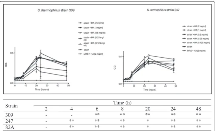
A widely accepted in vitro system, which allows simul-taneous evaluation of several HA doses, was compared with an innovative method based on the old concept of dynamic light scattering. By these two approaches com-parable kinetic curves were obtained. Firstly, tests were performed on three selected urease positive strains be-longing to Streptococcus (St.) thermophilus species in presence of growing concentrations of HA, until 48 h. As shown in Figure 1, each strain displayed a recurrent trend in the O.D. kinetics. In detail, curve profiles dropped after 24 h in all cases, showing a higher marked decrease when HA concentration was higher. When lower concentrations of HA were used, O.D. decrease was limited. Strain 82A behaved as 247 and therefore was not shown.
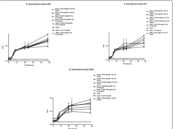
Streptococci were even employed for the same set of trials previously described, but in presence of both HA and Hy. According to obtained data (Figure 2), strains displayed after 24 h a completely different behavior: strains 309 and 247 exhibited an O.D. increase, above all in presence of higher concentrations of HA, indicating a bacterial growth enhancement.
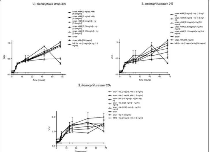
Monitoring strains’ activity up to 72 hours revealed a slight increase of the slope, except in presence of the highest HA concentration. As shown in Figure 3, each strain displayed the same trend at the highest HA con-centration. The curve profile of each strain at 2 mg mL-1 of HA showed a slight decrease after 24 h as for higher HA concentration. At lower HA concentrations both a little O.D. increase for 82A strain and a slight O.D. in-crease for 309 and 247 strains were observed.
These preliminary experiments, demonstrated that bacterial growth may be influenced by HA concentra-tion, by Hy concentration and by both of them.
Standard method indicated that a bacterial growth in-hibition was observable when HA, along with Hy, was used at concentrations ranging from 2 to 1 mg ml-1. When considering higher HA concentrations (ranging from 0.5 to 0.125 mg ml-1), along with Hy, a growth stimulation up to 72 hours was observed. These results provide interesting insights about LAB growth kinetics, and highlight a possible synergistic role of the two chal-lenged molecules that is likely to be related to the ability of LAB strains to use the N-acetyl-D glucosamine monomer as carbon source.
Although speculative, a possible combined role of HA and hyaluronidase on the bacterial growth was already hy-pothesized by Starr et al. (2006) [21]. Hy-Streptococcus (St.)
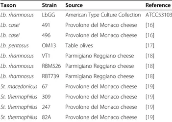
Table 1 Strains used in this study and source of isolation
Taxon Strain Source Reference
Lb. rhamnosus LbGG American Type Culture Collection ATCC53103 Lb. casei 491 Provolone del Monaco cheese [16] Lb. casei 496 Provolone del Monaco cheese [16]
Lb. pentosus OM13 Table olives [17]
Lb. rhamnosus VT1 Parmigiano Reggiano cheese [18] Lb. rhamnosus RBM526 Parmigiano Reggiano cheese [18] Lb. rhamnosus RBT739 Parmigiano Reggiano cheese [18] St. macedonicus 67 Provolone del Monaco cheese [19] St. thermophilus 309 Provolone del Monaco cheese [19] St. thermophilus 247 Provolone del Monaco cheese [19] St. thermophilus 82A Provolone del Monaco cheese [19]
Di Cerbo et al. BMC Microbiology 2013, 13:243 Page 2 of 6
pyogenes was shown to grow with N-acetylglucosamine but not with D-glucuronic acid as a sole carbon source. The same metabolic behavior was recorded in protechnological and probiotic LAB during this study. Only Hy+ strains could grow utilizing HA, as a sole carbon source, suggest-ing that Hy could permit the strain to utilize host HA as an energy source.
In conclusion, especially high HA concentrations seem to inhibit bacterial growth, however when low HA con-centrations are combined with Hy the bacterial growth seems to be enhanced even beyond 72 hours.
Further studies, in order to understand if the effects of HA and Hy are strain specific as they seems to be, are urgently required; specifically, a wider screening of dif-ferent LAB with interesting features, such as urease positive and/or hyaluronidase activity, might help to out-line a new probiotic oral formula with enhanced pre-biotic gut adherence properties and more effective therapeutic effect.
Conclusions
The effect of hyaluronic acid on protechnological or probiotic bacteria has never been evaluated before. In this study, the effect of hyaluronic acid, alone or in
combination with hyaluronidase, on three streptococci and one probiotic Lactobacillus strain was assessed. By obtained evidences, a synergistic role of the two mole-cules was described: when low hyaluronic acid concen-trations are combined with hyaluronidase, the bacterial growth appeared greatly enhanced even beyond 72 hours. This phenomenon could be related to ability of tested strains to metabolize N-acetyl-D glucosamine, one of the precursor of hyaluronic acid.
Methods
Media and reagents
MRS (Oxoid LTD, Basingstoke, Hampshire, U.K.) was employed for bacterial strains growth, strain mainten-ance and viable count assessment. Sterile saline solutions of High Molecular Weight HA (1837 kDa, 8 mg ml-1) where kindly provided by IBSA (Institute Biochemique SA, Lugano, CH). Hyaluronidase solution (Jaluronidasi 100 I.U., 3.2 mg ml-1) was purchased from Farmacia Testi snc, Milan, Italy.
Evaluation of minimal inhibitory concentration for HA
Dilutions for HA MIC determination were performed in sterile deionized water with concentrations ranging from
Strain
Time (h)
2
4
6
8
20
24
48
309
-
-
**
**
**
**
**
247
-
**
**
**
*
**
**
82A
-
**
**
**
*
**
**
Figure 1 Effects of HA onSt. thermophilus strains 309 and 247 until 48 h. Bacteria were employed at a starting concentration of 1 × 10⁶CFU mL-1. Lower panel: statistical significance between HA-treated and untreated strains. **Highly significant (P < 0.01); *significant
0.0625 up to 4 mg ml-1for a total of 7 levels of exposure. 50 μl of each dilution were loaded into wells in MRS agar plates seeded with tested strains. pH values of HA so-lutions were evaluated by means of pH-meter (Beckman PHI43). LAB tested are reported in Table 1.
Tolerance to HA of strain Lb. rhamnosus LbGG (ATCC) was also evaluated. Briefly, strain was subcul-tured twice in MRS (incubation at 30°C). Cells in early stationary phase (7.91 ± 0.29 Log CFU ml-1) were col-lected by centrifugation (6.500 rpm, 10 min), washed once with sterile Ringer solution (Oxoid) and resus-pended in the same saline. 200μl of sterile water solu-tions of HA (0.0625, 0.125, 0.25, 0.5, 1, 2, 4 and 8 mg ml-1) were added to 200 μl of cell suspensions. Positive control was realized by adding 200 μl of sterile saline instead of HA. After 30 min of incubation at 37°C, living cells were enumerated by drop counting method (Collins et al., 1989) on MRS agar plates, followed by incu-bation for 72 h at 37°C.
Effect of HA on Lb.GG tolerance to simulated gastric juice
The effect of HA on LbGG tolerance to simulated gastric juice was determined according to the procedure reported by Michida et al. (2006) [22]. Briefly, cells were harvested from cultures in exponential phase of growth by centrifugation (6.500 rpm, 10 min), washed twice with sterile saline (0.5%, w/v), and resuspended in the same sterile saline. Simulated gastric juice was prepared daily by suspending pepsin (1:10 000, ICN) in sterile saline (0.5%, w/v) to a final concentration of 3 g l-1and adjusting the pH to 2.00 with concentrated HCl using a pH meter. Aliquots (0.2 ml) of the cell suspensions were transferred to a 2.0 ml capacity Eppendorf tube, mixed with 0.3 ml of sterile water solutions of HA (0.125, 0.25, 0.5, 1, 2, 4, and 8 mg ml-1) and finally mixed with 1.0 ml of simulated gastric. After incubation at 37°C for 90 min, cells viability was assayed by drop counting method [23] on MRS agar plates (incubation for 72 h at 30°C).
Figure 2 Effects of HA and Hy onSt. thermophilus 309, 247 and 82A until 48 h. Bacteria were employed at a starting concentration of 1 × 10⁶CFU mL-1. Lower panel: statistical significance between HA-Hy-treated and untreated strains. **Highly significant (P < 0.01); *significant (P < 0.05); - not significant (P > 0.05).
Di Cerbo et al. BMC Microbiology 2013, 13:243 Page 4 of 6
LAB’s capability to utilize HA and its precursors as carbon sources
Acid formation by D-glucuronic acid, N-acetyl-D glu-cosamine, and HA was evaluated in MRS broth without glucose and meat extract with 2% added carbohydrates, as filter sterilized water solutions, and 0 · 004% chloro-phenol red. Media were inoculated with cell suspensions in sterile saline solution (about 6 log CFU ml-1). Tests were performed on three urease positive St. thermphilus strains, namely 309, 82A and 247, and LbGG.
Assessment of HA and Hy effect on LAB strains
The effect of HA and HA in combination with Hy was evaluated on three St. thermophilus urease positive strains (309, 247, and 82A). The assay was performed in 96-well microplates (Corning Inc., NY, USA). Firstly, 200μl of HA + MRS [4, 2, 1, 0,5 and 0.25 mg ml-1] were added in triplicate in each plate. Then 10μl of LAB cell suspensions (working concentrations of about 1 × 10⁶ CFU ml-1) in sterile saline solution were added.
Uninoculated MRS was used as control. Plates were in-cubated at 37°C in an incubator (Ekort 1500, Angelan-toni industrie, Milano, Italy). The O.D. values were measured at a wavelength of 595 nm at 0, 2, 4, 6, 8, 20, 24 and 48 hours by means of a microplate reader (Tecan, Austria).
For the evaluation of HA-Hy effect, the procedure above described was repeated by adding to each well 100μl of Hy [1,8 mg ml-1in a saline solution] and 10μl of each strain (about 1 × 10⁶ CFU ml-1). O.D. values were measured at 0, 2, 4, 6, 8, 20, 24, 48 and 72 h of in-cubation at 37°C.
Data analysis
Data obtained from the O.D. readings were used to draw charts where O.D. was expressed as a function of time. Each point of the curves is the average value of three replicates (subtracted of the blank) performed in the same experimental conditions. Statistical analyses were performed at 2 h intervals. At each time, analysis of
Figure 3 Effects of HA and hy onSt. thermophilus 309, 247 and 82A until 72 h. Bacteria were employed at a starting concentration of 1 × 10⁶CFU mL-1. Lower panel: statistical significance between HA-Hy-treated and untreated strains. **Highly significant (P < 0.01); *significant (P < 0.05); - not significant (P > 0.05).
variance (ANOVA) and Bonferroni post hoc test were carried out to assess overall differences in O.D. readings obtained from different strains in relation to the control.
Competing interests
The authors declare that they have no competing interests.
Authors’ contributions
ADC and RE developed and performed the experiments by dynamic light scattering and drafted the manuscript. MA did the assays about MIC to HA, HA utilization and strains’ resistance to simulated gastric juice. MB and BP provided scientific orientation and revised the manuscript. All authors read and approved the final manuscript.
Author details
1
Dipartimento di Chirurgia Generale e Specialità Chirurgiche, Università degli Studi di Modena e Reggio Emilia, via del pozzo 71 41124, Modena, Italy.
2
Istituto di Scienze dell’Alimentazione, ISA-CNR, Via Roma, 64, 83100, Avellino, Italy.3Dipartimento di Scienze della vita, Università degli Studi di Modena e
Reggio Emilia, Via Campi 287, 41125, Modena, Italy.
Received: 15 April 2013 Accepted: 25 October 2013 Published: 4 November 2013
References
1. Maharjan AS, Pilling D, Gomer RH: High and low molecular weight hyaluronic acid differentially regulate human fibrocyte differentiation. PLoS One 2011, 6(10):1–10.
2. Murai T, Kawashima H: A simple assay for hyaluronidase activity using fluorescence polarization. Biochem Biophys Res Commun 2008, 376:620–624. 3. Toole BP: Hyaluronan and its binding proteins the hyaladherins.
Curr Opin Cell Biol 1990, 2:839–844.
4. Murai T, Sougawa N, Kawashima H, Yamaguchi K, Miyasaka M: CD44-chondroitin sulfate interactions mediate leukocyte rolling under physiological flow conditions. Immunol Lett 2004, 93:163–170. 5. Kawashima H: Roles of sulfated glycans in lymphocyte homing.
Biol Pharm Bull 2006, 29:2343–2349.
6. Masuko K, Murata M, Yudoh K, Kato T, Nakamura H: Anti-inflammatory effects of hyaluronan in arthritis therapy: Not just for viscosity. Int J Gen Med 2009, 2:77–81.
7. Schulz A, Vestweber AM, Dressler D: Anti-inflammatory action of a hyaluronic acid-chondroitin sulfate preparation in an in vitro bladder model. Aktuelle Urol 2009, 40(2):109–112.
8. Ardizzoni A, Neglia RG, Baschieri MC, Cermelli C, Caratozzolo M, Righi E, Palmieri B, Blasi E: Influence of hyaluronic acid on bacterial and fungal species, including clinically relevant opportunistic pathogens. J Mater Sci Mater Med 2011, 22:2329–2338.
9. Krasiński R, Tchórzewski H, Lewkowicz P: Antioxidant effect of hyaluronan on polymorphonuclear leukocyte-derived reactive oxygen species is dependent on its molecular weight and concentration and mainly in-volves the extracellular space. Postepy Hig Med Dosw 2009, 63:205–212. 10. Rodrigues SV, Acharya AB, Bhadbhade S, Thakur SL: Hyaluronan-containing
mouthwash as an adjunctive plaque-control agent. Oral Health Prev Dent 2010, 8(4):389–394.
11. de Azeredo LAI, Leite SGF, Freire DMG, Benchetrit LC, Coelho RRR: Proteases from actinomycetes interfere in solid media plate assays of hyaluronidase activity. J Microbiol Methods2001, 45:207–212. 12. Gault DT: Extravasation injuries. Br J Plast Surg 1993, 46:91–96.
13. Smith KJ, Skelton HG, Turiansky G, Wagner KF: Hyaluronidase enhances the therapeutic effect of vinblastine in intralesional treatment of Kaposi’s sarcoma. J Am Acad Dermatol 1997, 36:239–242.
14. Ozegowski JH, Presselt N, Härtl A, Bocker T, Sänger J, Schmidt A, Willing K, Müller PJ: Anti-atherosclerotic effect of microbial hyaluronate lyase from group B streptococci. Pharmacology 2008, 63(8):601–605.
15. Kreil G: Hyaluronidases–a group of neglected enzymes. Protein Sci 1995, 4:1666–1669.
16. Aponte M, Fusco V, Andolfi R, Coppola S: Lactic acid bacteria occurring during manufacture and ripening of provolone del Monaco cheese: detection by different analytical approaches. Int Dairy J 2008, 18:403–413.
17. Aponte M, Blaiotta G, La Croce F, Mazzaglia A, Farina V, Settanni L, Moschetti G: Use of selected autochthonous lactic acid bacteria for Spanish-style table olive fermentation. Food Microbiol 2010, 30:8–16. 18. Blaiotta G, Di Capua M, Coppola R, Aponte M: Production of fermented
chestnut purees by lactic acid bacteria. Int J Food Microbiol 2012, 158:195–202.
19. Blaiotta G, Sorrentino A, Ottombrino A, Aponte M: Short communication: technological and genotypic comparison between streptococcus macedonicus and streptococcus thermophilus strains coming from the same dairy environment. J Dairy Sci 2011, 94:5871–5877.
20. Corcoran BM, Stanton C, Fitzgerald GF, Ross RP: Growth of probiotic lactobacilli in the presence of oleic acid enhances subsequent survival in gastric juice. Microbiology 2007, 153:291–299.
21. Starr CR, Engleberg NC: Role of Hyaluronidase in subcutaneous spread and growth of group a streptococcus. Infect Immun2006, 74(1):40–48. 22. Michida H, Tamalampudi S, Pandiella SS, Webb C, Fukuda H, Kondo A:
Effect of cereal extracts and cereal fiber on viability of lactobacillus plantarum under gastrointestinal tract conditions. Biochem Eng J 2006, 28:73–78.
23. Collins CH, Lyne PM, Grange JM: Counting microorganism. In
Microbiological Methods. Edited by Collins CH, Lyne PM, Grange JM. Oxford, UK: Butterworth-Heinemann; 1989:127–140.
doi:10.1186/1471-2180-13-243
Cite this article as: Di Cerbo et al.: Comparison of the effects of hyaluronidase and hyaluronic acid on probiotics growth. BMC Microbiology 2013 13:243.
Submit your next manuscript to BioMed Central and take full advantage of:
• Convenient online submission
• Thorough peer review
• No space constraints or color figure charges
• Immediate publication on acceptance
• Inclusion in PubMed, CAS, Scopus and Google Scholar
• Research which is freely available for redistribution
Submit your manuscript at www.biomedcentral.com/submit
Di Cerbo et al. BMC Microbiology 2013, 13:243 Page 6 of 6