Review Article
About Primary Pulmonary
Leiomyosarcoma: A Very Rare Highly
Aggressive Neoplasm -
Mirko Barone*, Marco Prioletta, Luigi Guetti, Giuseppe Cipollone, Decio
Di Nuzzo, Pierpaolo Camplese, Felice Mucilli
Department of General and Thoracic Surgery, University Hospital “SS. Annunziata”, University “G.
d’Annunzio”Chieti, Italy
*Address for Correspondence:
Mirko Barone, Department of General and Thoracic Surgery,
University “G. d’ Annunzio”, Via dei Vestini n.1, 66100. Chieti, Italy, Tel: +390871358289; Fax:
+390871358289; E-mail: [email protected]
Submitted:
12 December 2016;
Approved:
24 January 2017;
Published:
27 January 2017
Citation this article: Barone M, Prioletta M, Guetti L, Cipollone G, Di Nuzzo D, et al. About Primary
Pulmonary Leiomyosarcoma: A Very Rare Highly Aggressive Neoplasm. SRL Surg. 2017;1(1): 001-007.
Copyright: © 2017 Barone M, et al. This is an open access article distributed under the Creative
Commons Attribution License, which permits unrestricted use, distribution, and reproduction in any
medium, provided the original work is properly cited.
AbbREVIAtIonS
Primary Pulmonary Leiomyosarcoma (PPL); Overall Survival (OS); Chemotherapy (CHT); Radiotherapy (XRT)
IntRoduCtIon
Primary Pulmonary Leiomyosarcoma (PPL) is an extremely rare and highly aggressive mesenchymal tumor arising from the smooth muscle cells of the bronchial or pulmonary vessel wall and accounts for less than 0.5% of all lung cancers [1]. It can be classified as intraluminal or intrabronchial, intrapulmonary and intravascular according to its gross appearance [2]. Nath, et al. [3] recently reviewed the literature and reported 127 PPL cases. Aim of this work is a complete and exhaustive review on the morphological characteristics, clinical findings and current therapeutic strategies according to recent reports present in the literature.
MAtERIALS And MEthodS
The authors conducted a comprehensive research on PubMed - Medline including a period from January 2010 to June 2016 and according to the following MeSH terms: “primary [All Fields] AND (“lung” [MeSH Terms] OR “lung” [All Fields] OR “pulmonary” [All Fields]) AND (“leiomyosarcoma” [MeSH Terms] OR “leiomyosarcoma” [All Fields])”. All case reports or case studies were enclosed, while reviews prior to 2010 were excluded and resulting in twenty-one published reports [1,4-23] including primary intrapulmonary, primary intrabronchial and primary intravascular leiomyosarcomas.
Data collection included demographic characteristics (age, gender), neoplasm’s histopathological features, therapeutic strategies and follow up.
Statistical analysis was performed using SPSS version 20.0 software for Windows (IBM, Chicago, USA). Continuous variables were expressed as absolute value, simple percentages, means and standard deviations, whereas categorical ones in terms of frequency and percentage. Statistical differences or correlations between cohorts were evaluated with two sample t-test, χ2 test and Mc Neamar’s test. Survival variables were estimated according to the Kaplan-Meier’s method with 1-, 2- and 3- year’s rates. A p-value < 0.05 was considered statistically significant.
RESuLtS
Table 1 summarizes the demographic, clinical and pathological findings of the reported cases in the literature.
Twenty-two patients with a mean age of 54.07 ± 13:34 years (range 35-80) and a male to female ratio of 1:1 were enrolled in the study. With regard to morphological appearance, vascular neoplasms (primary pulmonary artery leiomyosarcomas) were the most common
(N.10–45.45%), followed by intrapulmonary and interbronchial ones (N.8– 36.36 % and n. 4–18.18 %, respectively). This is in contrast with previous reviews published prior to 2010.
In spite of the highly aggressive biological behaviour, secondary lesions were present in PPLsat clinical presentation only in 27.27% (N.6), with the lung being the most common site of recurrence, suggesting an early locoregional spread. In addition, mediastinal lymph node metastases were rare (N.4–18.18%) in accordance with the hematological tropism of sarcomatous malignancies.
A sequential multimodal therapeutic approach was the most common strategy, although cases of only surgical resections (N.5-22.72%) were described. Furthermore, in three patients, only bronchoscopic or CT-guided biopsies could be performed due to the rapid and fatal evolution of the disease itself.
Concerning risk factors affecting Overall Survival (OS), no statistical correlation was observed with age (p = 0.200), gender (p = 0.206), morphology (p = 0.669), mediastinal lymph node metastases (p = 0.210) and metastases (p = 0.291). But an important relationship between metastatic disease and mortality rate was reported (p = 0.082) (Table 2). Although it may seem a contradiction, this is justifiable referring to PPL’s high biological aggressiveness that, even in the early stages, has a poor prognosis. However, the presence of a stage IV correlates with a rapid progression affecting the overall mortality rate.
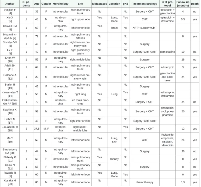
With regard to follow-up (range 0–36 months), no data were collected in five patients. In the remaining cases, a cumulative OS of 9.17 ± 3.77 (CI 95% OS: 9.71-24.49) months with a mortality rate of 58.82% (n.10/17) was observed. The 1-year OS was 53.6% (S.E. 0133), whereas the 2- and 3-year OS was, 26.8% (S.E. 0.128) and 0%, respectively (Figure 1).
Clustering the population according to the morphological type, no significant statistical differences in survival were found (Log Rank Mantel-Cox, p = 0.754). In particular, the actuarial median OS for intravascular patients was 14.12 ± 4.15 months, for intrabronchial 12.17 ± 1.31 mo and for intrapulmonary 19.50 ± 7.13 mo (Figure 2).
In our experience, from 1998 to 2016, only a 78-years old male patient with a T3N1M0 intrapulmonary leiomyosarcoma was admitted to our Department. He underwent upper pulmonary bilobectomy with partial thoracectomy with reconstruction (Figure 3,4) and subsequent adjuvant chemoradiotherapy. Ten months after surgery, the patient was still alive but a recurrence on the residual right lung was diagnosed.
dISCuSSIon
Primary Pulmonary Leiomyosarcoma (PPL) is an extremely rare and highly-aggressive mesenchymal tumor arising from the smooth muscle cells of the bronchial or pulmonary vessel walls and accounts for less than 0.5% of all lung cancers [1]. Nath, et al. [3] recently
AbStRACt
Not with standing advances in clinical and surgical oncology, primary pulmonary leiomyosarcoma remains a highly aggressive tumor with poor prognosis. It is a rare tumor, whose pathophysiology and the promoting genetic mechanisms of these tumors are still unclear. Aim of this work is a complete and exhaustive review on the morphological characteristics, clinical findings and current therapeutic strategies according to recent reports present in the literature.
Table 1: Primary pulmonary leiomyosarcoma cases published from 2010 to 2016. Demographic, clinical and therapeutic findings. Author N.
pa-tients Age Gender Morphology Site Metastases Location pN2 Treatment strategy
CHT pro-tocol Follow-up (months) Death Vakilian F [4] 1 35 F intravascular main pulmonary trunk No No Surgery + CHT docetaxel + gemcitabine 4 no Xie X [5] 1 48 M
intrabron-chial right upper lobe
Yes Lung, Bone Yes CHT epirubicin + ifosfamide 0,5 yes Colwell EM [6] 1 69 F
intrapulmo-nary left inferior lobe Yes Brain No XRT+ surgery+CHT Muganlins-kaya N [7] 1 72 F intravascular main pulmonary arteries No No 0 yes Srividya VV [8] 1 49 F intravascular
right inferior
pul-mony vein No No Surgery
Lv Y [9] 1 42 M intravascular right pulmonary artery No No Surgery+CHT+XRT gemcitabine 10 no Shen W [10] 1 52 F
intrapulmo-nary right-middle lobe
No No Surgery 28 no Adeli SH [11] 1 64 F intravascular main pulmonary trunk No No
Surgery + CHT adriamycin 3 yes
Galeone A
[12] 1 29 M intravascular
right inferior pul-mony vein No No Surgery+CHT+XRT gemcitabine and pacli-taxel 24 yes Staller B [13] 1 43 F intravascular main pulmonary trunk No No Surgery Kanematsu T [14] 1 56 M
intrapulmo-nary right lung Yes Lung
Yes CHT adriamycin, ifosfamide Falkenstern-Ge RF [15] 1 70 M intrabron-chial
left main bron-chus No No Surgery + CHT 24 no Kashima K [16] 1 53 M intravascular main pulmonary trunk No No Surgery + CHT pirarubicin, cyclophos-phamide 20 yes Luthra M [17] 1 43 F
intrapulmo-nary right inferior lobe
No No Surgery+CHT+XRT Elouazzani H [18] 2 37,5 M, F intrabron-chial right upper-middle lobe No Yes Surgery + CHT 12 yes Ma Y [19] 1 62 M
intrapulmo-nary left inferior lobe
Yes Lung, Skin No CHT Ifosfamide, etoposide, cisplatin, idarubicin 24 yes Sardenberg RA [20] 1 44 M
intrapulmo-nary left inferior lobe
No No Surgery 36 no Flaherty G [21] 1 69 F intravascular main pulmonary trunk Yes Kidney No 0 yes Colak N [22] 1 58 F intravascular main pulmonary trunk No No surgery 8 no Rozada R [1] 1 60 M
intrapulmo-nary left inferior lobe
Yes Lung,
Bone Yes 0 yes
Kosaka M
[23] 1 80 M
intrapulmo-nary left inferior lobe
No No
chemotherapy 1,5 yes
M: male; F: female; CHT: chemotherapy; XRT: radiotherapy; pN2: intrathoracic N2 (mediastinal) lymph node metastases.
reviewed the literature and reported 127 PPL cases. It can be classified as intraluminal or intrabronchial, intrapulmonary and intravascular according to its gross appearance [2]. The intrapulmonary type is the most common, while the intravascular one arises from pulmonary vessel intimae causing obstructions, stenoses or recurrent pulmonary embolisms [24]. Moreover, this latter is a very rare and highly aggressive neoplasm [25] with an incidence of 0.001-0.3% [26].
PPLs usually appear as a solitary pulmonary mass in middle-aged patients. Intravascular forms can engage the right ventricle outflow tract, the main pulmonary trunk or one of the pulmonary branches with an earlier onset and women are doubly affected than men [27].
The underlying pathophysiology and the promoting genetic mechanisms of these tumors are still unclear. However, recognized risk factors include radiation therapy, chemotherapy (e.g. cyclophosphamide, nitrosoureas), and environmental and
occupational exposures [28]. Patients may present with nonspecific signs and symptoms similar to those observed in other lung cancers, including fever, cough, hemoptysis, dyspnea and chest pain [29], although the constellation of symptoms can vary according to the neoplastic morphological pattern. In particular, in the case of endobronchial lesions, patients may complain of obstructive respiratory symptoms [30], on the other hand, in the case of vascular tumors, signs and symptoms of hemodynamic mismatch associated with respiratory failure are predominant, frequently leading to misdiagnosed cases of pulmonary thromboembolism [31,32].
It is difficult to distinguish PPLs from other lung cancers, pulmonary tuberculosis, granulomatosis and mediastinal tumors [33]. Physical examination, ECG usually does not reveal abnormalities and chest X-ray is often aspecific. For these reasons imaging must be completed by chest CT. Masses obstructing the
Table 2: Bivariate analysis between independent factors and survival.
OS1 Mortality rate
p p
Agea 0.200 0.354
Gender (male vs female)b 0.206 0.458
Morphology (intravascular vs intrabronchial vs intrapulmonary)b 0.669 0.919
Metastases (Yes vs No)b 0.291 0.082
pN22 (Yes vs No)b 0.210 0.156 acontinuous variable; ; bcategorical variable
1Overall Survival; 2pathological mediastinal (N2) lymph nodes Statistical significance: p < 0.05
Figure 1: General population (N. 17) cumulative overall survival according to Kaplan and Meier’s method.
main pulmonary vessels can be associated with both cardiomegaly and radiological signs of peripheral hypoperfusion [34]. Radio graphically, PPL appears as a well-defined mass with heterogeneous density, small calcifications or diffuse ischemic areas. It is generally a hilar neoplasm, but peripheral tumors with or without chest wall involvement has been described. Moreover, radiological appearances can also include findings such as obstructive atelectasis, mediastinal shift or pneumothorax [30]. On the other hand, in presence of a suspected intravascular type, a wall eclipsing sign on a pulmonary CT angiogram is suggestive of pathogenicity [35]. While ventilation/ perfusion scans make difficult to differentiate it between intravascular lesions and pulmonary embolism, a fluorine-18 fluorodeoxyglucose positron emission tomography scan has shown radiopharmaceutical uptake only by malignant tumors [36]. Moreover, magnetic resonance imaging could also differentiate a mass from a thrombus based on signal homogeneity [37].
Finally, Endobronchial Ultrasound-Guided Transbronchial Needle Aspiration (EBUS-TBNA) should be considered as an effective tool to diagnose intrabronchial neoplasms in selected cases [38].
The definitive diagnosis is mainly histological. Macroscopically, PPL presents with a roughly pearly surface; whereas, microscopically, the majority of these tumors presents a fascicular proliferation of spindle cells with a moderate amount of eosinophilic cytoplasm and inconspicuous nucleoli. Pulmonary leiomyosarcoma can show a broad spectrum of differentiation. Low grade FNCLCC stage I (French Federation Nationale des Centres de LutteContre le Cancer) tumors are characterized by fascicular arrangement, low mitotic rate (<3 per 10 HPF), the absence of cellular atypia, necrosis or hemorrhage and by a negative reaction to cytokeratin and epithelial markers (AE1/AE3, KL1, CK7, CK20, CK5/6). Ki-67 is less than 5%. Intermediate grade tumors are characterized by an increased cellularity with mild to moderate nuclear atypia and a mild mitotic
activity (3-8 mitoses per 10 HPF). Finally, high-grade tumors show a marked cellularity with nuclear pleomorphism and atypia, high mitotic activity (>8 mitoses per 10 HPF) and abundant areas of hemorrhage and necrosis [39]. Immunohistochemical positivity to actin, SMA, h-caldesmone and desmin can assume a smooth muscle neoplasm origin. Negativity to CD99, EMA and S100 allows you to exclude neoplasms such as Ewing’s sarcoma, undifferentiated tumors of epithelial origin and tumors of the nervous tissue [40]. Early stage pulmonary leiomyosarcomas are amenable for surgical resection (lobectomy, pneumonectomy, sleeve lobectomies, carinal resections with reconstruction according to Grillo or Brochart and endarterectomies) [41], which has to be considered the treatment of choice [42]. As reported by Tanaka, et al. [43], in a fascinating earlier review on 47 patients with primary pulmonary artery sarcoma, local recurrence after surgery significantly affects overall survival. For these reason the author proposed wider and careful tumor isolation with reconstruction with prostheses or grafts. Lymph node resections are generally not necessary because primary leiomyosarcomas rarely show lymph node involvement. If a radical resection is performed, the 5-years survival rate can achieve 50% for initial stage diseases, but long-survival case have been reported [12,22,35].
Prognostic factors include tumor’s dimension, bronchial and thoracic wall invasion. In addition, tumor cell necrosis, high cellularity, mitotic count above four and nuclear atypia are all predictive of more aggressive behavior and therefore increased risk of recurrence [44].
The role of adjuvant treatments is still debated; radio chemotherapy is recommended in cases of incomplete resection (R1 or R2) or recurrences [45], but the limited experiences in the treatment of these tumors makes it difficult to assess the relative importance of surgical resection and of adjuvant strategies, though Head HD et al are in favor of adjuvant protocols with encouraging results [46]. However, the therapeutic efficacy is controversial. In the majority of reports, adjuvant therapies have been largely ineffective making them to be considered unsensitive neoplasms. Sardenberg, et al. [47] reported a classic fractioning 50-60 Gy radiation protocol with good local field control.
With regard to conventional or intensive chemotherapy, standard-dose ifosfamide, doxorubicin and high-standard-dose ifosfamide have been described. Trabectedin and dacarbazine as 2nd-line options have proved effectiveness in leiomyosarcoma [48]. However, response rates are usually less than 20%. On the other hand, adriamycin-based protocols have achieved response rate in only 16-27% [49].
ConCLuSIon
In conclusion, PPL is a rare malignancy, difficult to differentiate from other lung cancers. Characterized by high biological aggressiveness and high resistance to conventional therapies, early detection and surgical therapy in selected cases can improve its poor and dismal prognosis. A better molecular characterization would definitely be crucial. Furthermore, the creation of multicentre and international databases in order to gather more experience and develop appropriate guidelines would be beneficial.
REfEREnCES
1. Rozada R, Vila A, Sosa L. Primary leiomyosarcoma of the lung. Arch Bronco neumol. 2010; 46: 338-339.
2. Yu H, Ren H, Miao Q, Wang Z, Zhang Z, Xu L. Pulmonary leiomyosarcoma. Chin Med Sci J. 1997; 12: 129-131.
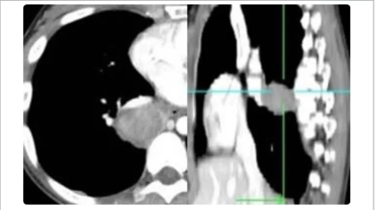
Figure 3: Chest CT in a patient with PPL arising from the right inferior lobe.
Figure 4: a) Surgical specimen: Pulmonary right bilobectomy for PPL; b-c) toracectomy and reconstruction with titanium struts and meshes.
3. Nath D, Arava S, Joshi P, Madan K, Mathur S. Primary pulmonary leiomyosarcoma of lung: An unusual entity with brief review. Indian J PatholMicrobiol. 2015; 58: 338-340.
4. Vakilian F, Shabestari MM, Poorzand H, Teshnizi MA, Allahyari A, Memar B. Primary Pulmonary Valve Leiomyosarcoma in a 35-Year-Old Woman. Tex Heart Inst J. 2016; 43: 84-87.
5. Xie X, Chen Y, Ding C, Yu X, Zou L, Xu B, et al. Primary pulmonary leiomyosarcoma: A case report. Oncol Lett. 2016; 11: 1807-1810.
6. Colwell EM, Algahim MF, Rao A, Gasparri MG. Primary pulmonary leiomyosarcoma with invasion of the pulmonary vein-A case report. Int J Surg Case Rep. 2016; 19: 103-105.
7. Muganlinskaya N, Guzman A, Dahagam C, Selinger SR. When a pulmonary embolism is not a pulmonary embolism: a rare case of primary pulmonary leiomyosarcoma. J Community Hosp Intern Med Perspect. 2015; 5: 29624. 8. Srividya VV, Sailendra V. An Extemly Rare Case of Left Atrium and Right
Pulmonary Vein Leiomyosarcoma. J ClinDiagn Res. 2015; 9: ED18-20. 9. Lv Y, Wang F, Qian W, Sun G. Treatment of a recurrent pulmonary artery
leiomyosarcoma with a combination of surgery, chemotherapy, and radiotherapy: A case report and literature review. Oncol Lett. 2015; 9: 1545-1548.
10. Shen W, Chen J, Wei S, Wang X, Li X, Zhou Q. Primary pulmonary leiomyosarcoma. J Chin Med Assoc. 2014; 77: 49-51.
11. Adeli SH, Nemati B, Jandaghi M, Riahi MM, Hosseinzadeh F, Salarvand F. Pulmonary Hypertension due to a Pulmonary Artery Leiomyosarcoma: A Case Report. Case Rep Pulmonol. 2013; 2013: 160619.
12. Galeone A, Validire P, Debrosse D, Folliguet T, Laborde F. Leiomyosarcoma of the right inferior pulmonary vein: 2 years survival with multimodality therapy. GenThoracCardiovascSurg. 2013; 61: 534-537.
13. Staller B, Schneider R, Grawe H, Beck FJ, Mayer E, Sigmund M. Progredient “pulmonary thromboembolism” in spite of sufficient anticoagulant therapy?. DtschMedWochenschr. 2013; 138: 638-641.
14. Kanematsu T, Toyoda Y, Goto H, Abe S, Kawakita N, Sakiyama S, Nishioka Y, Sone S. A case of primary pulmonary leiomyosarcoma with multiple nodular lesions in the lungs. NihonKokyukiGakkaiZasshi. 2011; 49: 167-171. 15. Falkenstern-Ge RF, Friedel G, Bode-Erdmann S, Ott G, Mentzel T, Kohlhäufl M, et al. Pulmonary leiomyosarcoma mimicking glomus tumor at first biopsy and surgically treated with isolated left main bronchus resection: rare clinical documentation. Ir J Med Sci. 2013; 182: 735-738.
16. Kashima K, Yamashita E, Mataki H, Yotsumoto G, Nomoto M, Sonoda M, Hanada S. Primary leiomyosarcoma of the pulmonary artery: a case of a 20-month survivor after incomplete surgical resection. InternMed. 2012; 51: 75-78.
17. Luthra M, Khan H, Suhail MF, Avadhani V. Primary pulmonary leiomyosarcoma — a case report. Arch Bronco neumol. 2012; 48: 476-478.
18. Elouazzani H, Zouaidia F, Jahid A, Bernoussi Z, Mahassini N. Primary endobronchial leiomyosarcoma of the lung: clinical, gross and microscopic findings of two cases. J ClinImaging Sci. 2012; 2: 35.
19. Ma Y, Chen BB, Xu XP, Lin GW, Ji Y, Akesu S, Zen H. Therapy-related acute myeloid leukemia in a primary pulmonary leiomyosarcoma patient with skin metastasis. Chin J Cancer Res. 2011; 23: 236-238.
20. Sardenberg RA, Cangnaci Neto R1, Cavalcanti F2, Younes RN3. High-grade primary pulmonary leiomyosarcoma. Einstein (Sao Paulo). 2011; 9: 523-526. 21. Flaherty G, McCarthy P, Mortimer G. Pulmonary artery leiomyosarcoma: an
unusual cause of shortness of breath. Ir J Med Sci. 2011; 180: 275-278. 22. Colak N, Nazli Y, Alpay MF, Haltas H, Cakir O. Surgical treatment of
pulmonary artery leiomyosarcoma: a good survival without adjuvant therapy. AnnThoracSurg. 2011; 92: 2252-2254.
23. Kosaka M, Chiaki T, Yokoyama T, Koizumi T, Shinohara N, Kubo K. A case of primary pulmonary leiomyosarcoma showing rapid growth and fatal outcome. Nihon Kokyuki Gakkai Zasshi. 2010; 48: 729-733.
24. Pain JA, Sayer RE. Primary leiomyosarcoma of the pulmonary artery. Eur J RespirDis. 1984; 65: 139-143.
25. Dumont P, Diot P, Aupart MR, Toumieux B. Leiomyosarcoma of the pulmonary artery. The Annals of Thoracic Surgery. 1998; 66: 2089–2091. 26. Bleisch VR, Kraus FT. Polypoid sarcoma of the pulmonary trunk: analysis
of the literature and report of a case with leptomeric organelles and ultrastructural features of rhabdomyosarcoma. Cancer. 1980; 46: 314–324 27. Delany SG, Doyle TCA, Bunton RW, Hung NA, Joblin LU, Taylor DR.
Pulmonary artery sarcoma mimicking pulmonary embolism. Chest. 1993; 103: 1631–1633.
28. Govindan R, Subramanian J. The Washington Manual of Oncology. Philadelphia, PA: Lippincott Williams & Wilkins; 2008. p: 363-376.
29. Petrov DB, Vlassov VI, Kalaydjiev GT, Plochev MA, Obretenov ED, Stanoev VI, Danon SE. Primary pulmonary sarcomas and carcinosarcomas - postoperative results and comparative survival analysis. Eur J Cardiothorac Surg. 2003; 23: 461-466.
30. Lee SH, Shim JJ, Shin JS, Baek MJ, Choi YH, Kim MK, et al. Primary endobronchial leiomyosarcoma. Diagnosis following expectoration of tumor fragment. Respiration. 2001; 68: 99-102.
31. Grazioli V, Vistarini N, Morsolini M, Klersy C, Orlandoni G, Dore R, et al. Surgical treatment of primary pulmonary artery sarcoma. J Thorac Cardiovasc Surg. 2014; 148: 113-118.
32. El-Sayed Ahmed MM, Aftab M, Al-Najjar RM, de la Cruz KI, Benjamin RS, Hallman CH. Pulmonary artery sarcoma mimicking pulmonary embolism. Tex Heart Inst J. 2014; 41: 515–517.
33. Yamasaki M, Sumi Y, Sakakibara Y, Tamaoka M, Miyazaki Y, Arai H, et al. Pulmonary Artery Leiomyosarcoma Diagnosed without Delay. Case Rep Oncol. 2011; 4: 287-298.
34. Moffat RE, Chang CH, Slaven JE. Roentgen considerations in primary pulmonary artery sarcoma. Radiology. 1972; 104: 283–288.
35. Li B, Zhang Y, Cai L, Hou J, Shi H. Primary pulmonary artery sarcoma differentiated from pulmonary thromboembolism by ventilation-perfusion scan. Long survival of the patient. Hell J Nucl Med. 2015; 18: 166-168. 36. Kessler A, Son H. Pulmonary artery angiosarcoma on 18F-FDG PET/CT
masquerading as pulmonary embolism. Clin Nucl Med. 2015; 40: 82-84. 37. Sun J, Tanga H, Lua C, Chen Y. Syncope caused by pulmonary artery
intima sarcoma: A cardiac magnetic resonance imaging-based differentiating diagnosis. Eur J Cardiothorac Surg. 2014; 46: 503.
38. Modi K, Dhillon S, Kumar A, Ylagan L, Harris K. Leiomyosarcoma of the pulmonary artery diagnosed by endobronchial ultrasound-guided transbronchial needle aspiration. Endosc Ultrasound. 2014; 3: 249-251. 39. Moran CA, Suster S. Tumors of the lung and pleura. Diagnostic Histopathology
of Tumors; Churchill Livingstone: New York; 2007. p: 193-194.
40. Arnold LM 3rd, Burman SD, O-Yurvati AH. Diagnosis and management of primary pulmonary leiomyosarcoma. J AmOsteopathAssoc. 2010; 110: 244-246.
41. Ming-Ching L, Chung-Ping H, Jiun-Yi H. Surgical treatment of endobronchial leiomyosarcoma with right main bronchus total obstruction: A case report. Ann Thorac Cardiovasc Surg. 2008; 14: 105-108.
42. Mazzucco A, Luciani GB, Bertolini P, Faggian G, Morando G, Ghimenton C. Primary leiomyosarcoma of the pulmonary artery: diagnostic and surgical implications. Ann Thorac Surg. 1994; 57: 222-225.
43. Tanaka I, Masuda R, Inoue M, Kasahara D, Furuhata Y, Shimizu S, et al. Primary pulmonary-artery sarcoma. Report of a case with complete resection and graft replacement, and review of 47 surgically treated cases reported in the literature. Thorac Cardiovasc Surg. 1994; 42: 64-68.
44. Dubose JJ, Sutherland MJ. Primary pulmonary spindle cell
neoplasm. CurrSurg. 2005; 62: 341-343.
45. Corpa-Rodríguez ME, Mayoralas-Alises S, García-Sánchez J, Gil-Alonso JL, Díaz-Agero P, Casillas-Pajuelo M. Postoperative course in 7 cases of primary sarcoma of the lung. Arch Bronconeumol. 2005; 41: 634-637.
46. Head HD, Flam MS, John MJ, Lipnik SS, Slater DL, Stewart RD. Long-term palliation of pulmonary artery sarcoma by radical excision and adjuvant therapy. Ann Thorac Surg. 1992; 53: 332-334.
primary pulmonary leiomyosarcoma. Einstein (Sao Paulo). 2011; 9: 523-526. 48. Casali PG, Jost L, Sleijfer S, Verweij J, Blay JY. Soft tissue sarcomas: ESMO clinical recommendations for diagnosis, treatment and follow-up. ESMO Guidelines Working Group. Ann Oncol. 2008;19 Suppl 2: ii89-93.
49. Lee MC, Hsu CP, Hsia JY. Surgical treatment of endobronchial leiomyosarcoma with right main bronchus total obstruction: a case report. Ann Thorac Cardiovasc Surg. 2008; 14: 105-108.