http://www.sciencepublishinggroup.com/j/frontiers doi: 10.11648/j.frontiers.20210101.11
Research/Technical Note
Protein-enriched Platelet-Rich Plasma (PEF
PRP
) a New
Products for Tissue Regeneration Developed Through the
Ultrafiltration of PRP - Preclinical Study
Laura Mazzucco
1, *, Valeria Balbo
1, Simona Martinotti
2, Elia Ranzato
2, Mauro Patrone
2,
Marcello Manfredi
2, Roberto Guaschino
11
Transfusion Medicine, “SS. Antonio e Biagio e Cesare Arrigo” Hospital, Alessandria, Italy
2
Department of Science and Technology Innovation, University of Piemonte Orientale, Alessandria, Italy
Email address:
*
Corresponding author
To cite this article:
Laura Mazzucco, Valeria Balbo, Simona Martinotti, Elia Ranzato, Mauro Patrone, Marcello Manfredi, Roberto Guaschino. Protein-enriched Platelet-Rich Plasma (PEFPRP) a New Products for Tissue Regeneration Developed Through the Ultrafiltration of PRP - Preclinical Study.
Frontiers. Vol. 1, No. 1, 2021, pp. 1-6. doi: 10.11648/j.frontiers.20210101.11
Received: December 28, 2020; Accepted: January 8, 2021; Published: January 22, 2021
Abstract:
Background: Platelet-Rich Plasma (PRP) is a blood component used for the biological treatment in many fields of regenerative medicine. The term PRP is currently applied to numerous blood components with different cellular and protein compositions. The optimal platelet concentration and the best technique for preparing PRP have not yet been defined and it is, therefore, important to understand the specific biological roles of the individual components. The aqueous part of PRP is plasma, which is an acellular component with containing proteins that are important for tissue regeneration. Objective: This preclinical study evaluated the biological characteristics and effects on proliferation (in an in vitro model) of a blood component Protein-Enriched Filtered PRP (PEFPRP) obtained through the ultrafiltration of low-concentration PRP and compared these effects with those of a standard PRP and other blood components preparation. Method: PEFPRP is a plasma enriched obtained by ultrafiltration of a plasma with low concentration platelets and its effects have been compared with those of a standard PRP and other blood components preparation. Result and Conclusion: PEFPRP provides a high concentration of proteins which have an important accessory function in in-vitro proliferation and could be highly significant in-vivo, accelerating tissue regeneration.Keywords: Platelet-rich Plasma, Plasma Proteins, Ultrafiltration, Growth Factors
1. Introduction
It has been 10 years since we published "Not every PRP-gel is born equal. Evaluation of growth factor (GF) availability for tissues through four PRP-gel preparations: Fibrinet®, RegenPRP-KIT®, Plateltex® and one manual procedure" [1] one of the first works comparing products obtained through different methods of preparing Platelet-Rich Plasma (PRP). Since then the world of non-transfusion blood components has changed greatly and many different preparation techniques have been introduced, which have led to similar but also very different blood components [2]. Our work concluded that even similar methods of preparing PRP resulted in different GF recovery and release kinetics; although it is unclear whether
these observable differences are important for clinical management, these findings do highlight the importance of the method of preparation used, since the very different technical characteristics (disparity of tubes, centrifugation force, gel-inducing enzyme) have variable effects on the final product and on the release kinetics of platelet GF and other molecules [3-5]. The purpose of PRP-based therapies is to promote tissue regeneration with a "platelet molecule load", so different release characteristics can alter the final clinical outcome. During our initial studies, we hypothesized that the diversity in transfer of GF from PRP prepared in different ways could be interesting if exploited for specific treatments depending on the complexities of the lesions or the pathologies and their consequent greater or lesser requirement
for regenerative stimuli to accelerate the healing process [6-8]. This approach of "tailor-made" treatment for a specific pathology is currently a new concept, shared by many authors, and is undoubtedly related to the availability of a broad variety of blood components for non-transfusion use which differ in cell composition, plasma quantity and fibrin content [9, 10]. In 1998 Marx et al. described PRP as a "simple" concentrate of platelets re-suspended in plasma [11] but we now have a multitude of PRP that contain different concentrations of platelets as well as fibrin and/or leucocytes [12]. The regenerative action of PRP is generally linked to the secretoma of the cellular component (platelets and leucocytes), which consists primarily of platelet GF and pro- and anti-inflammatory molecules of both platelet and leucocyte origin and is discharged into the plasma component [13]. The aqueous, liquid part of PRP is therefore the plasma, which does not convey cells, but does contain important proteins such as fibrinogen, fibronectin, vitronectin, thrombospondin, gelsolin and many others with adhesive, chemotactic, immunoregulatory and also anti-microbial properties, which are important for extracellular matrix reconstruction and healing [14, 15]. Having a blood component for non-transfusion use, protein-enriched filtered PRP (PEFPRP), which in addition to being a concentrate of cellular factors also provides a plasma protein concentrate at the site of the lesion, could be advantageous to deliver not only pro-angiogenic, proliferative and immunoregulatory factors, but also basic molecules for the reconstruction of the extracellular matrix and thus help the restoration of the natural scaffold necessary for the formation of new tissue [16]. PEFPRP is a conceptually new product obtained through the filtration of low-concentration PRP [17, 18]. The main objective of this work is to assess the biological characteristics of PEFPRP and compare its proliferative activity with that of conventional PRP and other blood components, using cultured fibroblasts as an in vitro model.
2. Material and Methods
2.1. Blood Collection
Blood was obtained from ten healthy donors, compatible group (A and 0), who gave prior consent to the use of their biological material for research purposes. From each donor, 14 mL of blood were collected into ACD- A and 5 mL were collected without anticoagulant. The blood was prepared in two different groups: 140 mL in ACD-A for PRP and PEFPRP, 50 mL for SERUM
2.2. PRP and PEFPRP
Of the 140 mL of blood collected into ACD-A, 110 mL were distributed into two specific devices (55 mL for each) and automatically separated (CPunT system; Eltek Group SpA, Hone, Italy) to obtain 40 mL of PRP (PRP 0.4) at a platelet concentration of 0.4 x 10⁶/µL (±5%) (ABX Micro ES 60; Horiba Medical Group, Montpellier, France). The PRP 0.4 was divided into two parts. One half, 20 mL, was subjected to
a second centrifugation (1200 g for 10’ - Heraeus Megafuge 16; ThermoFisher Scientific, Dreieich, Germany) in order to prepare aliquots of platelets at different concentrations: 0.6 (PRP 0.6), 0.8 (PRP 0.8), 1.0 (PRP 1.0), 1.2 (PRP 1.2), 1.4 (PRP 1.4), 1.6 (PRP 1.6), 1.8 (PRP 1.8) and 2.0 (PRP 2.0) x 106 platelets/µL. The second part, 20 mL, was ultrafiltrated for PEFPRP. To obtain the PEFPRP we used a protein and blood component concentrator, PROSMARTTM (Medica Group SpA, Medolla, Italy). This medical device (CE) exploits thin MediSulfone® fibres (average fibre cut-off 15 kDa, average porosity 5 nm) to make a platelet concentrate (about 4- to 5-fold the basal value) rich in proteins. Following the ultrafiltration step, platelets were morphologically intact and not aggregated (as determined by visual examination with a Leica DFC 295 microscope, Wetzlar, Germany) and functional (as determined by a Platelet Function Analyser; Siemens, Munich, Germany). The ultrafiltration/concentration of blood components was performed according to the manufacturer's instructions. Proteins and platelets are concentrated by the effect of removing part of the aqueous component of plasma. The concentrated product (about 8 mL) is recovered by suction through the same input path to the filter.
2.3. Other Blood Components
50 mL of whole blood without anticoagulant were centrifuged to obtain SERUM and 30 mL of blood collected into ACD-A were used to produce plasma (PL). PL was separated into two lots: 10 mL were ultrafiltrated to obtain protein-enriched filtered plasma (PEFPL) and the remaining 5 mL were stored untreated. Using an ADVIA 2400 (Siemens, Munich, Germany), total proteins were assayed in all products subjected to ultrafiltration (before and after) and also post-filtration, on the liquid (water) removed.
2.4. Identification of Proteins
The samples were then subjected to denaturation with trifluoroethanol, reduction with dithiothreitol 200 mM, alkylation with iodoacetamide 200 mM and complete protein digestion with Trypsin/Lys-C (Promega, Madison, WI, USA). The peptide digests were desalted on a Discovery® DSC-18 solid phase extraction (SPE) 96-well plate (25 mg/well) (Sigma-Aldrich Inc., St. Louis, MO, USA). The samples were spiked with 2 µL of stable isotope-labelled peptide standard (DPEVRPTSAVAA Val- 13C515N1 at V10; Cellmano Biotech Ltd., Anhui, China) before liquid chromatography with tandem mass spectrometry (LC-MS/MS) using a micro-liquid LC system (Eksigent Technologies, Dublin, USA). For identification purposes the samples were subjected to data-dependent acquisition: the MS analysis was performed using a mass range of 100-1500 Da (time-of-flight scan with an accumulation time of 0.25 s), followed by a MS/MS product ion scan from 200 to 1250 Da (accumulation time of 5.0 ms) with the abundance threshold set at 30 cps (35 candidate ions can be monitored during each cycle). Two data-dependent and three data-independent acquisitions were performed.
2.5. Cell Culture and Medium
For growth evaluation, human primary fibroblasts (Normal Human Dermal Fibroblast adult skin, NHDF; Lonza, Walkersville, MD, USA) were seeded (5x10³ cells) in 96-well culture plates in a complete medium (Invitrogen Corporation, Carlsbad, CA, USA). The following day, the cells were starved in FBS (Foetal Bovine Serum)-free Dulbecco’s modified Eagle’s medium for 72 h and then incubated with 10% blood component preparations: PL, PEFPL, PRP (from 0.4 to 2.0), PEFPRP, and SERUM. Cell proliferation was determined at 6 days after treatment with a colorimetric method, using an MTS Assay Kit (CellTiter 96 Aqueous One Solution cell proliferation assay, Promega, San Luis Obispo, CA, USA) and the contents of the plates were read at 490 nm with a microplate reader (Microplate Reader - BioTeck Instruments, Winooski, WT, USA).
2.6. Determination of Growth Factor Concentrations
The concentrations of vascular endothelial growth factor (VEGF), epidermal growth factor (EGF), and platelet-derived growth factor (PDGF-bb) were measured in PL, PEFPL, PRP, PEFPRP, SERUM using immunoassays (Quantikine® ELISA kits, R&D Systems, Abingdon, UK) and each sample was treated in according to the manufacturer’s protocol.
2.7. Protein Database Search and Statistical Analysis
The data-dependent acquisition files were searched using Protein Pilot software v. 4.2 (Sciex, Concord, Canada) and Mascot v. 2.4 (Matrix Science Inc., Boston, MA, USA). The UniProt Swiss-Prot reviewed database containing human proteins (version 2015.07.07, containing 42,131 sequence entries) was used and a target-decoy database search was performed. The false discovery rate was set at 1%. Data were analysed with Graph Pad Prism version 8.3.1 (Graph Pad Software Inc., San Diego, CA, USA). One-way analysis of variance (ANOVA) was used to analyse the effect of the different blood components.
3. Results
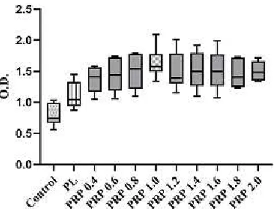
We first analysed the impact of PRP-released factors with different concentrations of platelets (Figure 1) on the proliferation of human fibroblasts PRP [19, 20], ss as standard protocol (see other items), with platelet counts from 0.4 x 10⁶ to 2.0 x 10⁶/mL was applied onto cultured human fibroblasts in FBS-free medium (10% vol/vol in cell culture medium); the increase in proliferation was compared to that of cells cultured in 10% FBS (Control) and PL at 6 day [21].
Figure 1. Figure illustrating the manufacturing process and sample preparation from whole-blood. PL: plasma; PRP: Platelet-Rich Plasma; PEFPL: Protein-Enriched Filtered Plasma; PEFPRP: Protein-Enriched Filtered PRP.
Figure 2. Effect of Platelet-Rich Plasma (PRP) with increasing platelet concentration (10% vol/vol as a supplement in culture medium) at 6 days of culture. Experiments were performed in 5 times and data (O. D.: optical density) representative of a minimum of 5 independent experiments are shown.
As expected, all PRP conditions induced an increase of fibroblast growth which was comparable to that measured in 10% of other blood components (Figure 2). At 6 days there were differences between all PRP conditions and other blood components: PRP increased NHDF proliferation by 1.4- to 2.2-fold, compared to the Control and PL. The number of the platelets in PRP induces increase of proliferation from 0.4 to 1.0, but from 1.2 to 2.0 the proliferation activity increased less and a plateau effects is observed. Then best condition was 10% PRP 1.0, which produced a 2.2-fold increase in fibroblast proliferation.
Next we analysed the impact of the best PRP (PRP 1.0) and other blood components on human fibroblasts, to further investigate whether PEFPRP was better than PRP 1.0 at promoting growth. Fibroblast growth was detected and quantified at 6 days of incubation with culture medium with different blood components (10% vol/vol in cell culture
BLOOD SAMPLE pool (10 donors) SERUM PEFPRP 1,0 PEFPL PL PRP 2,0 PPP PEFPPP PRP 1,8 PRP 1,6 PRP 1,4 PRP 0,6 PRP 0,8 PRP 1,0 PRP 1,2 PRP 0,4
medium); PRP 1.0, PEFPRP and SERUM increased fibroblast growth by 1.2- and 1.7-fold compared to the Control [22, 23] (Figure 3). The best result (1.7-fold increase in proliferation) was obtained with culture medium with 10% PEFPRP.
Figure 3. Comparison between the effect of different blood components, added as a supplement in culture medium, on proliferation of human fibroblasts at 6 days; “Control” is fibroblast proliferation in the standard condition (medium with 10% foetal bovine serum). All evaluations were performed 8 times with 8 independent experiments.
Since ultrafiltration increases the concentration of proteins present in PRP and PL, we next measured the total amount of proteins present before and after filtration [24] (Table 1).
Table 1. Quantification of total proteins (g/dL) in Plasma (PL) and Platelet-Rich Plasma (PRP 0.4) before and after ultrafiltration. The increment of total proteins in PEF is at least double the value at baseline.
PL PEFPL PRP 0.4 PEFPRP
g/dL 6.1±0.1 15.7±1.2 6±0.2 18±1.4
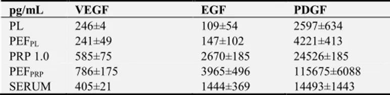
The concentrations of plasma proteins in the PEF products (PEFFL and PEFPRP) were higher (2.5- to 3-fold) than in their corresponding unfiltered products (PL and PRP 0.4). We also evaluated whether the concentration of some GF varied depending on the biological preparation; to do this, we measured the amounts of VEGF, EGF, and PDGF-bb in PL, PEFPL, PRP, PEFPRP and SERUM [25] (Table 2). The amounts of VEGF, EGF and PDGF-bb were significantly increased in PEFPRP compared to the amounts in other blood components and were higher in PRP and SERUM than in PL and PEFPL, (suggesting that most derived from platelets): it is, therefore, very interesting that PEFPRP contains all the GF present in the starting blood component (PRP 0.4) [26, 27].
Table 2. Mean concentrations (pg/mL)±standard deviation of vascular endothelial growth factor (VEGF), epidermal growth factor (EGF), and platelet-derived growth factor (PDGF-bb) detected in the different blood components studied. All assays were performed in triplicate.
pg/mL VEGF EGF PDGF PL 246±4 109±54 2597±634 PEFPL 241±49 147±102 4221±413 PRP 1.0 585±75 2670±185 24526±185 PEFPRP 786±175 3965±496 115675±6088 SERUM 405±21 1444±369 14493±1443
A label-free proteomic approach using sequential window acquisition of all theoretical mass spectra (SWATH)-MS technology was employed in order to quantify the relative
abundance of the proteins in each sample [28, 29]. The statistical analysis showed that 30 proteins were enriched in the PEFPRP with respect to PRP. Table 3 lists these proteins, classified based on the biological process in which they are involved.
Table 3. Modulated proteins in PEFPRP prepared with the immunodepletion of abundant proteins. The modulated proteins were classified in order to highlight the biological processes and the protein classes enriched during the preparation of PEFPRP. Cytoskeletal organisation, regulation of proteolysis and cellular response to cytokine stimulus are the activities upregulated and most represented in this new blood component.
Pro-inflammation CBG, GSTO1
Anti-inflammation CO5, AACT, GSTO1, CBPN Wound healing
ACTB, ACTG, FCN3, TNL1, HSP7C, ACTC, SPTC2, CBPN, GELS, ENOA, VTNC, ACTC, COF1, CAP1, ALDOA PROF1, PEDF, FCN3, AACT
Clot stabilisation FA12, FIBG, F13, FIBB
Anti-microbial PRGP2
4. Discussion
Platelets, blood components and their derivatives have numerous advantages as biological products for the treatment of many pathologies. The advantages of their therapeutic use include the fact that they are autologous products, easily prepared, cause minimal complications, and have a broad range of potential actions. Cells (platelets and leucocytes) and the liquid portion (plasma) of PRP and other products contemporaneously release many proteins and factors with different anabolic and catabolic functions that reduce inflammation and promote angiogenesis and tissue regeneration. The right number of platelets in PRP or platelet derivatives has been debated for many years: the platelet concentration must be at least three or four times higher the baseline value in order to provide a ‘therapeutic dose’ of platelet factors [30, 31]. We observed the effect of different PRP on a fibroblast culture model. PRP with different concentrations of platelets generally had an incremental effect on fibroblast proliferation, although this positive effect was not immediate, showing a biphasic trend: in fact, after having been starved of FBS, the stimulation of fibroblast proliferation at 6 days was evident. In vitro experiments have shown that proliferation of human fibroblast is enhanced by addition of PRP at different concentration to the culture medium, but it would seem that the effect is not directly related to platelets numbers, indeed after PRP 1.0 the variances between the different PRP concentration was minimal. The amounts of bioactive molecules released by platelets are roughly proportional to the platelet concentration in the preparation and the effect on cell growth of different platelet concentrations in PRP (according to previously published data by several authors) is not linear [32]. For this study we chose the best PRP concentration (PRP 1.0) and evaluated the effect of PEFPRP at the same platelet concentration. The technique of ultrafiltration of low-concentration PRP (PRP 0.4) is a new possible alternative to centrifugation to increase the concentration of platelets and, compared to this latter, is less aggressive to the platelets, better preserving their structure and function. In addition our results of the analysis of the eluate,
PEFPRP, demonstrated that other important biomolecules are concentrated, such as total plasma proteins and GF. Platelet derivatives are used to provide GF concentrates that are able to induce and accelerate the healing process. However, together with GF, PRP contains hundreds of plasma proteins that are more abundant. Much research has been conducted to study platelets and derivatives, but only a few have adopted a proteomic approach to characterise PRP [33]. The highest concentration of proteins was found in PEFPRP as this product contains plasma and platelet molecules. To investigate the role of these proteins in tissue regeneration we also applied proteomic techniques to PEFPRP to analyse the structural and functional pathways of the proteins. PEFPRP contained 30 upregulated proteins grouped into those involved in cytoskeleton organisation, regulation of proteolysis and cellular response to cytokine stimulus. To classify these proteins by function we defined keywords, focusing on the tissue healing process and identified subgroups classified by their functions: pro-inflammatory, anti-inflammatory, wound healing, clot stabilisation, anti-microbial and other plasma components such as those of the complement system, cell-matrix adhesion and immunoglobulins [34, 35]. Our results suggest that the plasma protein concentrate in PEF had an important role: gene ontology classification showed that there is an enrichment of several biological processes such as the immune system and biological adhesion, but also of enzyme modulator and signalling proteins. Key components for the regeneration or replacement of tissue are GF, such as VEGF, EGF and PDGF-bb, which provide vital stimuli for cell recruitment, proliferation, morphogenesis and differentiation [36, 37]. In accordance with previous studies we found that specific GF were present in variable concentrations depending on the biological preparation. Interestingly we observed that the amounts of GF were greater in cellular blood components, SERUM, PRP and PEFPRP and, in particular, in PRP and PEFPRP because these have higher platelet concentrations and in PEFPRP because of the ultrafiltration process. This method of concentrating PRP is better than centrifugation because the resulting eluate contains all the plasma proteins and platelets. In brief, we have provided further evidence that PEFPRP can exert a proliferative stimulus on fibroblasts, increasing the number of the cells in culture, and thereby contribute to improving tissue regeneration. The combination of plasma proteins and platelet proteins, both concentrated, may facilitate tissue regeneration. In fact, our results support the hypothesis of a beneficial action not only of GF, but of plasma proteins too, which are present in larger quantities in PEFPRP. The clot stabilisation and cell-matrix adhesion proteins (fibrinogen, fibrin, fibronectin, and thrombin) contribute to creating the three-dimensional environment that is a critical condition for cell-cell and protein-protein interactions as well as for tissue regeneration and healing [38].
5. Conclusion
PEFPRP produced by ultrafiltration of PRP triggers greater
cell growth than any other product tested. The probable less stress caused to the platelets during filtration produces a notable increase in the available fraction of GF. Tests performed cannot establish the real role of the high concentrations of proteins obtained by filtration; however, the plasma proteins, which increase moderately and probably have an accessory function in in vitro proliferation, may be highly relevant in vivo, in which the interactions between numerous plasma proteins, cells and the extracellular matrix of the tissues to be repaired play an important role in accelerating tissue regeneration.
Disclosure
The authors declare that they have no competing interests.
Acknowledgements
The authors would like to thank Michela Coppo, Melissa Tomasi and Letizia Bocchi for scientific and technical support.
References
[1] Mazzucco L, Balbo V, Cattana E, et al. Not every PRP-gel is born equal. Evaluation of growth factor availability for tissues through four PRP-gel preparations: Fibrinet®, RegenPRP-Kit®, Plateltex® and one manual procedure. Vox Sang 2009; 97: 110-8.
[2] Mazzucco L, Balbo V, Guaschino R. "Reasonable compromise" to define the quality standards of platelet concentrate for non-transfusion use (CPunT). Transfus Apher Sci 2012; 47: 207-211.
[3] Tsay RC, Vo J, Burke A, et al. Differential growth factor retention by platelet-rich plasma composites. J Oral Maxillofac Surg 2005; 63: 521-528.
[4] Han J, Meng HX, Tang JM, et al. The effect of different platelet-rich plasma concentrations on proliferation and differentiation of human periodontal ligament cells in vitro. Cell Prolif 2007; 40: 241-252.
[5] Russell RP, Apostolakos J, Hirose T, et al. Variability of platelet-rich plasma preparations. Sports Med Arthrosc Rev 2013; 21: 186-190.
[6] Anitua A, Alkhraisat G, Orive G, et al. Perspectives and challenges in regenerative medicine using plasma rich in growth factors. J Control Release 2012; 157: 29-38.
[7] Dhillon RS, Schwarz EM, Maloney MD. Platelet-rich plasma therapy-future or trend? Arthritis Re Ther 2012; 14: 219. [8] Anitua E, Troya M, Zalduendo M, et al. Personalized
plasma-based medicine to treat age-related diseases. Materials Science and Engineering 2016; 74: 459-464.
[9] Jain KK. Role of biological therapies in the development of personalized medicine. Expert Opin Biolo Ther 2012; 12: 1-5. [10] Milano G, Sanchet M, Jo CH, et al. Platelet-rich plasma in
orthopaedic sports medicine: state of the art. BMJ 2019; 4: 188-195.
[11] Marx RE, Carlson ER, Eichstaedt RM, et al. Platelet-rich plasma: growth factor enhancement for bone grafts. Oral Surg Oral Med Oral Pathol Oral Radiol Endod 1998; 85: 638-646. [12] Dohan Ehrenfest DM, Bielecki T, Mishra A, et al. In search of a
consensus terminology in the field of platelet concentrates for surgical use: platelet-rich plasma (PRP), platelet-rich fibrin (PRF), fibrin gel polymerization and leukocytes. Curr Pharm Biotechnol 2012; 13: 1131-1137.
[13] Nurden AT. The biology of the platelet with special reference to inflammation, wound healing and immunity. Front Biosci 2018; 23: 726-751.
[14] Karimi K, Rockwell H. The benefits of platelet-rich fibrin. Facial Plast Surg Clin North Am 2019; 27: 331-340.
[15] Zimmermann R, Arnold D, Strasser E, et al. Sample preparation technique and white cell content influence the detectable levels of growth factors in platelet concentrates. Vox Sang 2003; 85: 283-289.
[16] Mazzucco L, Borzini P, Gope R. Platelet-derived factors involved in tissue repair-from signal to function. Transfus Med Rev 2010; 24: 218-234.
[17] Gura V, Ronco C, Nalesso F, et al. A wearable hemofilter for continuous ambulatory ultrafiltration. Kidney Int 2008; 4: 497-502.
[18] Ronco C. Hemodialfiltration: technical and clinical Issues. Blood Purif 2015; 40: 2-11
[19] Noh KC, Liu XN, Zhuan Z, et al. Leukocyte-poor platelet-rich plasma-derived growth factors enhance human fibroblast proliferation in vitro. Clean Orthop Surg 2018; 10: 240-247 [20] Van der Bijl I, Vlig M, Middelkoop E, et al. Allogeneic
platelet-rich plasma (PRP) is superior to platelets or plasma alone in stimulating fibroblast proliferation and migration, angiogenesis, and chemotaxis as relevant processes for wound healing. Transfusion 2019; 59: 3492-3500.
[21] Muraglia A, Nguyen VT, Nardini M, et al. Culture medium supplements derived from human platelet and plasma: cell commitment and proliferation support. Front Bioeng Biotechnol 2017; 20: 55-66.
[22] Hung Y, Elder MJ, Rawstron JA, et al. A retrospective crossover study of autologous and allogeneic serum eye drops for the management of ocular surface disease. Transfus Med 2019; 29: 69-71.
[23] Kawase T, Nagata M, Okuda K, et al. Platelet-rich fibrin extract: a promising fetal bovine serum alternative in explant cultures of human periosteal sheets for regenerative therapy. Int J Mol Sci 2019; 20: 1053.
[24] Zernii EY, Baksheeva VE, Iani EV, et al. Therapeutic proteins for treatment of corneal epithelial detects. Curr Med Chem 2019; 26: 517-545.
[25] Lee K, Silva EA, Mooney DJ. Growth factor delivery-based tissue engineering: general approaches and a review of recent developments. J R Soc Interface 2011; 8: 153-170.
[26] Qiao J, An N, Ouyang X. Quantification of growth factors in different platelet concentrates. Platelets 2017; 28: 774-778. [27] Ziegler CG, Sloun RV, Gonzalez S, et al. Characterization of
growth factors, cytokines, and chemokines in bone marrow concentrate and platelet-rich plasma. Am J Sport Med 2019; 47: 2174-2187
[28] Brandi J, Manfredi M, Speziali G, et al. Proteomic approaches to decipher cancer cell secretome. Semin Cell Dev Biol 2018; 78: 93-101.
[29] Cipriani V, Ranzato E, Balbo V, et al. Long-term effect of platelet lysate on primary fibroblasts highlighted with a proteomic approach. J Tissue Eng Regen Med 2009; 3: 531-538.
[30] Andia I, Maffulli N. Blood-derived products for tissue repair/regeneration. Int J Mol Sci 2019; 20: 4581.
[31] Kim SJ, Davis RP, Jenne CN. Platelets as modulators of inflammation. Semin Thromb Hemost 2018; 44: 91-101. [32] Borzini P, Mazzucco L. Platelet gels and releasates. Curr Opin
Hematol 2005; 12: 473-479.
[33] Lee HW, Choi KH, Kim JY, et al. Proteomic classification and identification of proteins related to tissue healing of platelet-rich plasma. Clin Orthop Surg 2020; 12: 120-129. [34] Golebiewska EM, Poole AW. Platelet secretion: from
haemostasis to wound healing and beyond. Blood 2015; 29: 153-162.
[35] Olczyk P, Mencner L, Komosinska-Vassev K. The role of the extracellular matrix components in cutaneous wound healing. Biomed Res Int 2014; 11: 1-8.
[36] Moss AJ, Scharma S, Brindle NP. Rational design and protein engineering of growth factors for regenerative medicine and tissue engineering. Biochem Soc Trans 2009; 37: 717-721. [37] Sundman EA, Cole BJ, Fortier LA. Growth factor and catabolic
cytokine concentrations are influenced by the cellular composition of platelet-rich plasma. Am J Sports Med 2011; 39: 2135-2140.
[38] Bretschneider H, Quade M, Lode A, et al. Characterization of naturally occurring bioactive factor mixtures for bone regeneration. Int J Mol Sci 2020; 21: 1412-1427.