Neurobiology of Disease
Activation of PPAR
␥ Attenuates the Expression of Physical and
Affective Nicotine Withdrawal Symptoms through Mechanisms
Involving Amygdala and Hippocampus Neurotransmission
Esi Domi,
1,3Francesca Felicia Caputi,
2Patrizia Romualdi,
2Ana Domi,
1Giulia Scuppa,
1Sanzio Candeletti,
2Alison Atkins,
3X
Markus Heilig,
3Gregory Demopulos,
4George Gaitanaris,
4Roberto Ciccocioppo,
1*
and
X
Massimo Ubaldi
1*
1School of Pharmacy, Pharmacology Unit, University of Camerino, Camerino 62032, Italy,2Department of Pharmacy and Biotechnology, Alma Mater Studiorum-University of Bologna, Bologna, 40126 Italy,3Center for Social and Affective Neuroscience, IKE, Linko¨ping University, Linko¨ping, 58183, Sweden, and4Omeros Corporation, Seattle, Washington 98101
An isoform of peroxisome proliferator-activated receptors (PPARs), PPAR
␥, is the receptor for the thiazolidinedione class of
anti-diabetic medications including pioglitazone. Neuroanatomical data indicate PPAR
␥ localization in brain areas involved in drug
addic-tion. Preclinical and clinical data have shown that pioglitazone reduces alcohol and opioid self-administration, relapse to drug seeking,
and plays a role in emotional responses. Here, we investigated the behavioral effect of PPAR
␥ manipulation on nicotine withdrawal in
male Wistar rats and in male mice with neuron-specific PPAR
␥ deletion (PPAR␥
(⫺/⫺)) and their littermate wild-type (PPAR
␥
(⫹/⫹))
controls. Real-time quantitative RT-PCR and RNAscope
in situ hybridization assays were used for assessing the levels of expression and
cell-type localization of PPAR
␥ during nicotine withdrawal. Brain site-specific microinjections of the PPAR␥ agonist pioglitazone were
performed to explore the role of this system on nicotine withdrawal at a neurocircuitry level. Results showed that activation of PPAR
␥ by
pioglitazone abolished the expression of somatic and affective nicotine withdrawal signs in rats and in (PPAR
␥
(⫹/⫹)) mice. This effect was
blocked by the PPAR
␥ antagonist GW9662. During early withdrawal and protracted abstinence, the expression of PPAR␥ increased in
GABAergic and glutamatergic cells of the amygdala and hippocampus, respectively. Hippocampal microinjections of pioglitazone
re-duced the expression of the physical signs of withdrawal, whereas excessive anxiety associated with protracted abstinence was prevented
by pioglitazone microinjection into the amygdala. Our results demonstrate the implication of the neuronal PPAR
␥innicotinewithdrawal
and indicates that activation of PPAR
␥ may offer an interesting strategy for smoking cessation.
Key words: addiction; nicotine; pioglitazone; PPAR
␥; withdrawal
Introduction
Tobacco use is one of the leading causes of preventable disease
and death worldwide, killing
⬎7 million people each year (
World
Health Organization, 2017
). Nicotine, the major psychoactive
compound in tobacco, exerts its addictive properties by affecting
Received Aug. 6, 2019; accepted Sept. 18, 2019.
Author contributions: E.D., P.R., S.C., M.H., R.C., and M.U. designed research; E.D., F.F.C., A.D., G.S., A.A., and M.U. performed research; E.D., F.F.C., A.D., G.S., S.C., A.A., G.D., G.G., R.C., and M.U. analyzed data; E.D., F.F.C., and M.U. wrote the paper.
This work was supported by a fellowship (to E.D.) from the Italian Society of Pharmacology, and by NIH Grants RO1 AA017447 and AA014351, and Grant PRIN 2017 (2017SXEXT5) to R.C. We thank Dr. Kevin Niswender at Van-derbild University (Nashville, TN) for providing the PPAR␥engineeredmouselines,SerenaStopponiforanimalcare supervision, and Alfredo Fiorelli for technical support.
Significance Statement
Smoking cessation leads the occurrence of physical and affective withdrawal symptoms representing a major burden to quit
tobacco use. Here, we show that activation of PPAR
␥ prevents the expression of both somatic and affective signs of nicotine
withdrawal. At molecular levels results show that PPAR
␥ expression increases in GABAergic cells in the hippocampus and in
GABA- and glutamate-positive cells in the basolateral amygdala. Hippocampal microinjections of pioglitazone reduce the
insur-gence of the physical withdrawal signs, whereas anxiety linked to protracted abstinence is attenuated by pioglitazone injected into
the amygdala. Our results demonstrate the implication of neuronal PPAR
␥ in nicotine withdrawal and suggest that PPAR␥
agonism may represent a promising treatment to aid smoking cessation.
neuronal circuits that control reward, motivation, and habit
pro-cesses (
De Biasi and Dani, 2011
).
Smoking cessation leads to the insurgence of the physical and
affective withdrawal symptoms, the major burden to quit tobacco
use (
Grunberg, 2007
). Anxiety, depression, and impaired
mem-ory represent the major negative affective disturbances in
nico-tine withdrawal (
Hughes et al., 1986
;
Caan et al., 1996
;
Jorenby et
al., 1996
;
Kenny et al., 2001
). Insurgence of the physical and
affective withdrawal symptoms can be also be observed in rats
and mice upon cessation of nicotine when administered via
os-motic minipumps or chronic injections (
Shiffman and Jarvik,
1976
;
Rossetti et al., 1999
;
Watkins et al., 2000
;
Malin et al., 2006
).
Recently peroxisome proliferator-activated receptors (PPARs)
have been proposed as new therapeutic targets in drug addiction
including nicotine dependence (
Le Foll et al., 2013
). PPARs are a
family of nuclear receptor proteins that regulate gene expression
as ligand-activated transcription factors (
Michalik et al., 2006
).
Although PPARs were first identified in peripheral tissue, there is
now evidence of their abundant distribution in the brain (
Sarruf
et al., 2009
;
Schnegg and Robbins, 2011
). Among the three
iso-forms (␣, ␦, and ␥), PPAR␥ has the highest level of expression in
the CNS. Initially, PPAR
␥ expression in the brain was suggested
mainly in astrocytes, and glial cells (
Sarruf et al., 2009
). Recent
studies, however, suggest that PPAR
␥ expression in the brain
occurs almost exclusively in neurons and astrocytes (
Warden et
al., 2016
). PPAR
␥ agonists have been shown to modulate genes
linked to synaptic transmission and neuronal function in regions
such as the amygdala (AMY) and hippocampus (HIPP;
Searcy et
al., 2012
;
Ferguson et al., 2014
). However, despite the evidence of
a role of PPAR
␥ in drug addiction, the neuronal contribution
underlying its effects has not been yet explored. We previously
showed that pioglitazone, a selective PPAR
␥ agonist, was highly
effective in reducing behavioral responses to stress and
prevent-ing stress-induced relapse to alcohol and opioid seekprevent-ing in rats
(
Stopponi et al., 2011
;
Domi et al., 2016
;
de Guglielmo et al.,
2017
). Pioglitazone was also shown to reduce heroin craving and
anxiety in humans (
Jones et al., 2018
), and most important, a
recent clinical study reported that pioglitazone attenuates
mea-sures of nicotine craving (
Jones et al., 2017
).
Here, we explored the role of PPAR
␥ in nicotine withdrawal to
better determine the potential of this receptor system as a
treat-ment target for nicotine abuse. To this end, we assessed the
effi-cacy of pioglitazone on the expression of the physical and
affective withdrawal symptoms in a rat model of nicotine
depen-dence induced by application of nicotine patches (
Cippitelli et al.,
2011
). To address the functional contribution of neuronal
PPAR␥ in nicotine addiction we used mice with a neuronal
dele-tion of PPAR
␥ (PPAR␥
(⫺/⫺)) in a model of nicotine dependence
using chronic subcutaneous injections of nicotine. The specificity
of the effects of pioglitazone was confirmed by using the selective
PPAR␥ antagonist GW9662. Based on our previous findings on
the role of amygdaloid PPAR
␥ on anxiety, and the anatomical
distribution of PPAR␥ in limbic structures involved in nicotine
addiction we investigated the regional, temporal, and cell-specific
changes in PPAR
␥ expression occurring in AMY and HIPP
dur-ing nicotine withdrawal in PPAR␥
(⫹/⫹)mice.
Materials and Methods
Subjects. Male Wistar rats (275–325 g at the beginning of the
experi-ments) were single housed and kept on a reverse 12 h light/dark cycle (lights on 20:00 – 08:00 h). Male mice with neuronal-specific PPAR␥ deletion (PPAR␥(⫺/⫺)) and their littermate wild-type (PPAR␥(⫹/⫹); 20 –25 g,⬃8 weeks of age at the beginning of the experiments), were housed in groups of four to five and kept in a normal light/dark cycle (lights on 08:00 –20:00 h). Experiments were conducted in sepa-rate groups of animals and during the dark phase of the light/dark cycle except for the physical spontaneous nicotine withdrawal assessment. Upon arrival, animals were weighed and handled daily. To obtain the conditional inactivation of PPAR␥ in neuronal cells transgenic mice ex-pressing the Cre recombinase under the control of rat Nestin (Nes) pro-moter were bred to homozygous PPAR␥ loxP/loxP mice. The resulting heterozygous F1 offspring (PPAR␥⫹/loxP) were either positive or nega-tive for Nes–Cre. From mating of PPAR␥⫹/loxP with PPAR␥⫹/loxP-Nes–Cre mice, F2 generation of the desired genotypes [PPAR␥ loxP/ loxPNes–Cre (PPAR␥NestinCre) and PPAR␥ loxP/loxP] were obtained, which were then intercrossed to obtain F3 generation. PPAR␥ loxP/loxP mice were used as control littermates (PPAR␥(⫹/⫹)) for conditional PPAR␥ mice (PPAR␥(⫺/⫺)). The mice used for this study, were bred at the School of Pharmacy of the University of Camerino and originally provided by Dr. K. Niswender (Vanderbilt University, Nashville, NT) were on a C57BL/6J background (Jones et al., 2002). All animals were housed at constant temperature (20 –22°C) and humidity (45–55°). Ad
libitum food and water were provided for the entire duration of the
experiments.
All procedures followed the EU Directive for Care and Use of Labora-tory Animals and were approved by the Ethical Committee of the Uni-versity of Camerino.
Drugs. Nicotine patches (NIQUITIN CQ Step 1,21 mg/d, Glaxo) were
used for the induction of nicotine dependence in rats (Cippitelli et al., 2011). (-)-Nicotine hydrogen tartrate salt (Sigma-Aldrich) was dissolved in sterile saline and administered by subcutaneous injections in mice (10 ml/kg). Pioglitazone (Actos, Takeda Pharmaceuticals) was suspended in distilled water and administered orally (1 ml/kg or 10 ml/kg) in rats and mice, respectively. GW9662 (Tocris Bioscience) was dissolved in 5% DMSO, 5% Tween 20, and 90% distilled water and administered intra-peritoneally (1 ml/kg or 10 ml/kg), in rats and mice, respectively. For brain site-specific microinjections, pioglitazone was purchased from Molcan and dissolved in 15% DMSO, 10% Cremophor and the final volume was adjusted adding sterile saline. Pioglitazone (5g/0.6 l) was administered in a volume of 0.3l per site. Drugs were prepared immediately before administration.
Nicotine dependence and spontaneous nicotine withdrawal. Rats was
thoroughly shaved on the back, depilated with a depilatory lotion, and cleansed with water as previously described (Cippitelli et al., 2011). Patches were divided into four equal parts so that 5.2 mg/rat/d of nicotine was administered by patch applied to the shaved region. Pieces of flexible fabric Band-Aid and waterproof tape were used to wrap the nicotine patch and to improve its adherence to the rat’s back. Control rats were shaved and depilated, but only the Band-Aid and waterproof tape were placed on their backs. Comparable doses were previously shown to produce sufficiently high blood nicotine and cotinine levels to elicit the occurrence of reliable nicotine abstinence symptoms (Cippitelli et al., 2011). This application procedure was repeated for 7 consecutive days and on Day 8, transdermal patches were removed to induce spontaneous nicotine withdrawal. To assess the physical withdrawal signs, 16 h after removal of the transdermal nicotine patches, rats were placed into trans-parent cylinders and the frequency of nicotine abstinence signs was counted for 10 min by an observer blind to treatment condition. The most frequently observed categories included teeth-chattering/chews, writhes/gasps, wet dog shakes/tremors, and yawns. For statistical analysis withdrawal signs were compounded and the total score was evaluated as
G.D. is the Chairman and CEO of Omeros, G.G. is Chief Scientific Officer of Omeros, and R.C. is the inventor on several patent applications relating to the therapeutic use of PPAR␥ agonists in addiction. Omeros, through agree-ments with the University of Camerino and with R.C., exclusively controls the intellectual property rights directed to R.C.’s inventions related to the use of PPAR␥ receptor agonists for the treatment of addiction and addictive behav-iors. Under these agreements, R.C. may be entitled to receive payments and royalties from Omeros. The remaining authors declare no competing financial interests.
*R.C. and M.U. contributed equally to this work.
Correspondence should be addressed to Roberto Ciccocioppo at [email protected]. https://doi.org/10.1523/JNEUROSCI.1922-19.2019
Copyright © 2019 the authors
previously described (Cippitelli et al., 2011). Previous research, reported that a similar dose of nicotine (5.0 mg/kg) induced blood nicotine levels averaged 88.5⫾ 21.5 ng/ml and blood cotinine levels averaged 647.6 ⫾ 123.2 ng/ml after administration with the same procedure (Slawecki and Ehlers, 2002).
In mice nicotine dependence was induced by four daily subcutaneous injections of 2 mg/kg for 8 consecutive days. On Day 9 and 20 h after the last nicotine injection, mice were placed into transparent cylinders (di-ameter 28.5 cm) and then observed for 30 min for the occurrence of the following signs: paw tremors, chewing, genital licks, scratches, teeth chat-tering, head shakes, abdominal constrictions, and jumps. All signs were compounded and the total score was evaluated (Kenny and Markou, 2001).
Elevated plus maze. The elevated plus maze (EPM) test was performed
6 d following the removal of nicotine patches in rats and 6 d after the last nicotine injections in mice. This time point corresponds to protracted abstinence during which affective withdrawal signs (i.e., anxiety) are exhibited. Each trial lasted 5 min and animals were recorded using an EthoVision video tracking system (Noldus Information Technology). Percentage OAT⫽ (time in open arm/time in “open arm” ⫹ time in “closed arm”)⫻ 100 was considered an index of anxiety, whereas total number of entries was considered a locomotion index.
Real-time quantitative RT-PCR. Tissue samples were collected from
PPAR␥(⫹/⫹)mice 20 h and 6 d after last nicotine injection corresponding
to the early and late abstinence time points. Brains were rapidly removed and dissected to harvest HIPP and AMY. Tissues were immediately fro-zen in dry ice and stored at⫺80°C until RNA extraction. Total mRNA was isolated using TRIZOL reagent (Life Technologies) and its integrity was checked by gel electrophoresis. The amounts of RNA were deter-mined by measuring optical densities and only RNA samples with an OD260/OD280 ratio 1.8 –2.0 were used. Total RNA was reverse tran-scribed using random hexamers and MMLV Reverse Transcriptase (Life Technologies) in a final volume of 20l, according to the manufacturer’s instructions. Quantitative real-time PCR was performed using TaqMan Gene Expression Master Mix on an Applied Biosystems Step One System as previously reported (Caputi et al., 2015,2016). Relative expression of PPAR␥ gene transcripts (Mm 01184322_m1 FAM, Applied Biosystems) was calculated by Delta–Delta Ct (DDCt) method and converted to rel-ative expression ratio (2-DDCt) for statistical analysis (Livak and Schmittgen, 2001). Data were normalized to the endogenous reference gene glyceraldehyde-3-phosphate dehydrogenase (GAPDH) expres-sion (Mm 99999915_g1 VIC, Applied Biosystems). Data are ex-pressed as mean⫾ SEM of six samples/group (each sample run in triplicate). After PCR, a dissociation curve (melting curve) was con-structed in the range of 60 –95°C to evaluate the specificity of the amplification products (Lyon, 2001). Results are represented as fold-change in mRNA levels.
RNAscope in situ hybridization assay. We performed RNA in situ
hybrid-ization in HIPP and AMY for PPAR␥ (accession number NM_011146.3, target nucleotide region 170 –1490), Slc17a7 (VGlut1, accession number NM_182993.2, target nucleotide region 464 –1415), and Gad1 (GAD67, accession number NM_008077.4, target nucleotide region 62–3113) mRNAs as described previously (Rubio et al., 2015). Brains were re-moved and quickly frozen in isopentane on dry ice and stored at⫺80°C. Brains slices (20m) were collected approximately at the bregma level ⫺1.7 mm (Paxinos and Franklin, 2003) were mounted directly onto Superfrost Plus microscope slides (Fisher Scientific) and stored at⫺80°C until the in situ hybridization was performed. In situ hybridization was performed according to the RNAscope Fluorescent Multiplex Kit User Manual (Advanced Cell Diagnostics). Briefly, the slides were transferred to slide racks and fixed by immersion in 10% neutral buffered formalin for 15 min at 4°C, and then dehydrated in a series of 50, 70, and 100% ethanol at room temperature. A hydrophobic pen was used to create a barrier around each brain section. Sections were incubated for 30 min at room temperature with the protease pretreatment solution from the RNAscope Fluorescent Multiplex Kit (Advanced Cell Diagnostics) and then washed in PBS and incubated with the target probes for 2 h at 40°C using the HybEZ Hybridization System (Advanced Cell Diagnostics). Sections were then incubated with a series of four amplifier probes each
for Steps 1 and 3, 30 min each for Steps 2 and 4) at 40°C, fluorescently labeled in Step 4 with AlexaFluor 488 (green), Atto 550 (red), and Atto 647 (far red) to visualize probes in each of the three channels in different colors, washing with the wash buffer provided with the kit between each step. Finally, sections were briefly incubated with DAPI to visualize nu-clei in blue. Slides were coverslipped, air dried, and stored at 4°C.
Tissue sections were examined with a confocal microscope (Zeiss LSM 700) at 40⫻ magnification to determine colocalization and to create images for quantification.
Tile scan images to visualize the entire brain section were taken with a fluorescent microscope (Leica, DMi8) at 10⫻ magnification. For quan-tification of each PPAR␥, GAD67, and VGlut1-positive cells (n ⫽ 5/per group, 2 sections) we counted the total pixels of the fluorescent signal (fluorescent “dots”), which represent a single molecule of mRNA above the sensitivity threshold of the assay, using ImageJ software (Rubio et al., 2015). RNAscope in situ hybridization analysis revealed measurable tran-script of PPAR␥ expression in GABAergic (GAD67-positive) and glutama-tergic (VGlut1-positive) cells of the HIPP, basolateral amygdala (BLA), and central amygdala (CeA) in PPAR␥(⫹/⫹) mice (n⫽ 5/group).
Intracranial surgery and histological analysis. PPAR␥(⫹/⫹)mice were anesthetized by intramuscular injection of 100 –150l of a tiletamine chlorohydrate (58.17 mg/10 ml) and zolazepam chlorohydrate (57.5 mg/10 ml) and placed into a stereotaxic frame. The skull was exposed and stainless steel guide cannulae (diameter: 0.35 mm; length: 7 mm) were bilaterally implanted to reach the AMY or the HIPP (Paxinos and Frank-lin, 2003). Mice were bilaterally implanted to reach AMY or HIPP using the following coordinates: (1) AMY: 1.4 mm caudal from the bregma, ⫾3.0 mm mediolateral and ⫺3.9 mm ventral from the dura; and (2) dorsal HIPP: 1.7 posterior to bregma,⫾ 1.5 mm mediolateral, and 1.3 mm ventral to skull surface (Paxinos and Franklin, 2003). The guide cannulae were fixed to the skull with dental cement and two anchoring screws.
The rate of injection was precisely controlled by an infusion pump (SPLab02, Baoding Shenchen Precision Pump Co.LTD). The stainless-steel injector protruding beyond the cannula tip 1.00 mm and 0.5 mm for the AMY and the HIPP, respectively, was allowed to remain in the brain 2 min/site before being retracted. Pioglitazone was administered via a 10l Hamilton syringe at a rate of 0.25 l/min. At completion of the experiments, to verify the cannula placement, mice were lightly anes-thetized with isoflurane and 0.3l/site Malachite green solution was injected into the area. After the animals were killed, the ink diffusion into the region was histologically evaluated.
The injection sites were confirmed for both AMY and HIPP by com-parison with plates taken from a mouse brain atlas (Paxinos and Frank-lin, 2003). Histological analysis confirmed correct bilateral injections into the AMY in 39 mice and in the HIPP in 43 mice. Only these mice were used for statistical analysis (seeFig. 5). Behavioral tests were initi-ated following full recovery (5– 6 d after surgery).
Statistical analysis. Behavioral experiments were analyzed by mean of
one- or two-way ANOVA. Differences were considered significant if p⬍ 0.05. Post hoc comparisons were performed by Newman–Keuls or Dun-nett tests, when appropriate. Data were first examined for significant violations to the assumption of homogeneity of variances using Bar-tlett’s test. When deviation from homogeneity of variances was detected (RNAscope ISH assay), nonparametric Kruskall–Wallis analysis was performed followed by multiple comparisons between the independent groups.
Results
Systemic pioglitazone reduced the expression of the physical
and affective nicotine withdrawal signs in rats
Physical withdrawal signs: rats (n
⫽ 27) received nicotine patches
and were treated with pioglitazone (0, 15, or 30 mg/kg) 12 and 1 h
before assessing physical nicotine withdrawal. Nicotine
with-drawal increased the insurgence of the physical signs that were
reduced by pioglitazone treatment.
One-way ANOVA showed a significant overall treatment
ef-fect (F
(3,23)⫽ 77.7; p ⫽ 0);
2⫽ 0.77⫽). Post hoc analysis showed
a significant expression of physical nicotine withdrawal signs in
rats subjected to nicotine and treated with vehicle versus control
( p
⬍ 0.001). Withdrawal score was significantly reduced
follow-ing administration of pioglitazone 15 mg/kg ( p
⬍ 0.01) and 30
mg/kg ( p
⬍ 0.001) versus vehicle (
Fig. 1
A).
Affective withdrawal signs
EPM test was performed 6 d after the removal of nicotine patches
and pioglitazone (0, 15, or 30 mg/kg) was given twice 12 and 1 h
before the test.
Nicotine withdrawal decreased the percentage time in the
open arms and this anxiogenic-like behavior was reversed by
pioglitazone.
Overall ANOVA of the percentage time spent in open arms
showed a significant effect of treatment (F
(3,23)⫽ 7.3; p ⫽ 0.001);
2⫽ 0.48. Post hoc comparisons showed a significant increase of
anxiety-like behavior in nicotine-treated rats versus control ( p
⬍
0.05). Pioglitazone significantly attenuated anxiety-like behavior
(15 mg/kg: p
⬍ 0.001; 30 mg/kg: p ⬍ 0.05;
Fig. 1
B).
No significant main effect of treatment was observed in the
total number of entries in the EPM (F
(3,23)⫽ 0.1; p ⫽ 0.93;
Fig. 1
C).
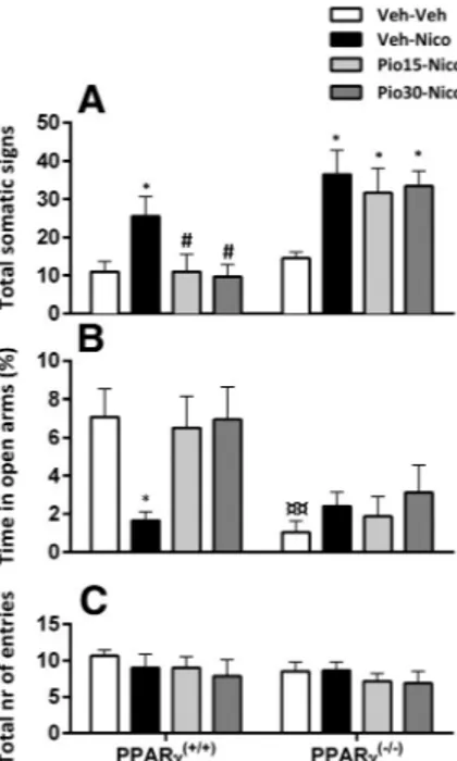
Systemic pioglitazone reduced the expression of the physical
and affective nicotine withdrawal signs in PPAR
␥
(ⴙ/ⴙ)but
not in PPAR
␥
(ⴚ/ⴚ)mice
Physical withdrawal signs
PPAR
␥
(⫹/⫹)and PPAR
␥
(⫺/⫺)mice were divided into four
groups (n
⫽ 6–8/group). Nicotine withdrawal induced a
sub-stantial increase of the physical withdrawal signs in PPAR
␥
(⫹/⫹)and PPAR␥
(⫺/⫺)mice and pioglitazone reduced the expression
only in the PPAR
␥
(⫹/⫹)genotype. Factorial ANOVA showed a
significant effect of genotype (F
(1,54)⫽ 25.4; p ⫽ 0.001);
2⫽ 0.48,
treatment (F
(3,54)⫽ 5.9; p ⫽ 0.001);
2⫽ 0.24 and interaction
genotype
⫻ treatment (F
(3,54)⫽ 2.77; p ⫽ 0.04);
2⫽ 0.13. Post
hoc analysis showed a significant expression of physical
with-drawal signs in PPAR␥
(⫹/⫹)( p
⬍ 0.05) and PPAR␥
(⫺/⫺)( p
⬍
0.01) in nicotine-treated mice versus control. Pioglitazone 15 and
30 mg/kg significantly reduced physical withdrawal signs in
PPAR
␥
(⫹/⫹)mice ( p
⬍ 0.05;
Fig. 2
A).
Affective withdrawal signs
PPAR
␥
(⫺/⫺)spent less time in the open arms of the EPM
com-pared with PPAR␥
(⫹/⫹)mice. Nicotine withdrawal decreased the
percentage time in the open arms in PPAR
␥
(⫹/⫹) mice and this
effect was reversed by pioglitazone.
Factorial ANOVA showed a significant genotype effect (F
(1,54)⫽
16.5; p
⫽ 0);
2⫽ 0.23, no effect of treatment (F
(3,54)
⫽ 2.26;
p
⫽ 0.09) but a significant interaction genotype ⫻ treatment
(F
(3,54)⫽ 3.15; p ⫽ 0.03);
2⫽ 0.15. Post hoc analysis showed a
higher basal anxiety-like behavior in PPAR
␥
(⫺/⫺)compared with
PPAR␥
(⫹/⫹)( p
⬍ 0.01). Nicotine withdrawal induced a
signifi-cant decrease of percentage of time in the open arms in
PPAR␥
(⫹/⫹)( p
⬍ 0.05;
Fig. 2
A). Pioglitazone, 15 and 30 mg/kg
significantly reversed nicotine withdrawal-induced anxiety ( p
⬍
0.05) in PPAR␥
(⫹/⫹)mice. In PPAR␥
(⫺/⫺)mice anxiety-like
re-sponse was significantly higher in all groups compared with
PPAR␥
(⫹/⫹)mice ( p
⬍ 0.05) and was not affected by nicotine or
pioglitazone. (
Fig. 2
B). The total number of entries did not differ
between genotypes and was not affected by the treatment.
Facto-rial ANOVA did not show a significant effect of genotype (F
(1,54)⫽
1.24; p
⫽ 0.26), treatment (F
(3,54)⫽ 0.62; p ⫽ 0.6), or interaction
genotype
⫻ treatment in the total number of entries (F
(1,54)⫽ 0.2;
p
⫽ 0.89;
Fig. 2
C).
Figure 1. Pioglitazone (Pio) on nicotine (Nico) withdrawal-induced physical symptoms and anxiety-like behavior. A, Nicotine cessation elicited a robust score of the physical withdrawal signs (***p⬍ 0.001) reduced by Pio (15 and 30 mg/kg;##p⬍ 0.01,###p⬍
0.001). B, Percentage time in open arms in the EPM. Nicotine induced an anxiogenic effect (*p⬍ 0.05) that was blocked by Pio 15 and 30 mg/kg (###p⬍ 0.001,#p⬍ 0.05). C, Total
number of entries. Data are expressed as mean⫾ SEM values. Veh, Vehicle.
Figure 2. Effects of pioglitazone (Pio) on nicotine (Nico) withdrawal-induced physical signs and anxiety-like behavior in PPAR␥(⫹/⫹)and PPAR␥(⫺/⫺)mice. A, Nicotine in-duced significant expression of physical withdrawal symptoms (*p⬍ 0.05) prevented by Pio in PPAR␥(⫹/⫹)(#p⬍0.05).B,Percentagetimeinopenarms.Nicotineinducedananxiogenic
effect in PPAR␥(⫹/⫹)mice (*p⬍ 0.05), prevented by Pio 15 and 30 mg/kg (#p⬍ 0.05).
PPAR␥(⫺/⫺)mice exhibited a higher anxiety baseline compared with PPAR␥(⫹/⫹)mice (¤¤p⬍ 0.01). C, Total number of entries. Data are expressed as mean ⫾ SEM values. Veh, Vehicle.
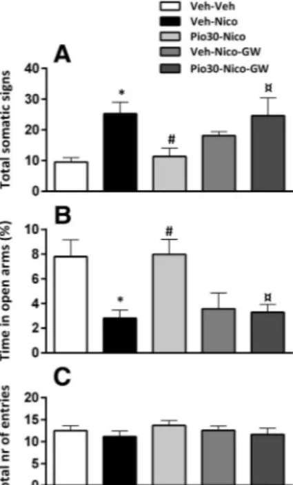
Systemic GW9662 blocked the effect of pioglitazone on the
physical and affective nicotine withdrawal signs in wild-type
PPAR
␥
(ⴙ/ⴙ)mice
A new cohort of PPAR
␥
(⫹/⫹)mice (n
⫽ 51), divided into five
groups (n
⫽ 8–12/group) was tested to verify the specificity of
pioglitazone on PPAR
␥ receptors by pretreatment with the
selec-tive PPAR␥ antagonist, GW9662. Nicotine withdrawal induced
an increase of the physical signs that were reduced by
pioglita-zone. The effect of pioglitazone was prevented by GW9662.
Physical withdrawal signs
ANOVA showed a significant main effect of pioglitazone
treat-ment (F
(1,46)⫽ 3.6; p ⫽ 0.012);
2⫽ 0.23. Post hoc analysis
confirmed a significant expression of the physical withdrawal
signs in nicotine-treated mice versus control ( p
⬍ 0.05).
Piogli-tazone 30 mg/kg significantly reduced the expression of the
phys-ical withdrawal score ( p
⬍ 0.05). GW9662 significantly blocked
the effect of pioglitazone ( p
⬍ 0.05;
Fig. 3
A).
Affective withdrawal signs
Nicotine withdrawal decreased the percentage time in the open
arms and this effect was reversed by pioglitazone. GW9662
pre-vented the effect of pioglitazone.
One-way ANOVA showed a significant main effect of
treat-ment (F
(1,46)⫽ 6.7; p ⫽ 0);
2⫽ 0.37 on anxiety-like behavior.
Post hoc analysis showed a significant reduction in percentage
time in open arms in nicotine exposed mice vs control ( p
⬍ 0.01).
Pioglitazone 30 mg/kg significantly reversed the anxiogenic
con-dition ( p
⬍ 0.01). GW9662 and nicotine-treated mice showed a
decrease in percentage spent in open arms compared with
vehicle-treated mice ( p
⬍ 0.05). The effect of pioglitazone was
completely blocked by GW9662 ( p
⬍ 0.01;
Fig. 3
B). Total
num-ber of entries did not differ between groups. ANOVA did not
show a significant effect of treatment in total number of entries
(F
(1,46)⫽ 0.7; p ⫽ 0.55;
Fig. 3
C).
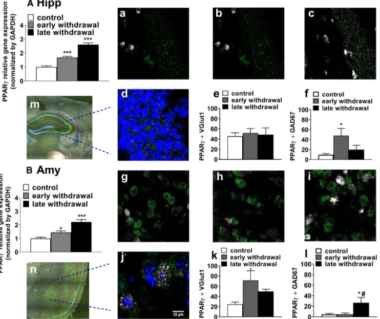
PPAR
␥ gene expression is increased in the early and late stage
of nicotine withdrawal
PPAR␥ mR⌵〈 levels were analyzed in HIPP and AMY in
PPAR
␥
(⫹/⫹)mice (n
⫽ 17). PPAR␥ expression increased during
the early and late nicotine withdrawal in both dorsal HIPP and
AMY. ANOVA showed a significant main difference of PPAR
␥
expression in both dorsal HIPP (F
(2,15)⫽ 66.56; p ⫽ 0);
2⫽ 0.89
and AMY (F
(2,14)⫽ 21.95; p ⫽ 0);
2⫽ 0.23. Compared with the
control group, Dunnett’s post hoc tests showed a significant
in-crease of PPAR
␥ mRNA levels at both 20 h and 6 d into nicotine
withdrawal ( p
⬍ 0.001) in the HIPP (
Fig. 4
A). In AMY, PPAR␥
mRNA levels were also significantly increased at 20 h ( p
⬍ 0.05)
and 6 d ( p
⬍ 0.001;
Fig. 4
B).
Nicotine withdrawal induced PPAR
␥ expression changes in
GABAergic and glutamatergic cells of HIPP and AMY
In dorsal HIPP nicotine withdrawal increased PPAR
␥ expression
in GAD67⫹ but not in VGlut1⫹ cells. Nonparametric analysis
showed an increase of PPAR
␥ in GAD67⫹ cells (
2⫽ 10.17, df ⫽
2, p
⫽ 0.006) and no significant difference in VGlut1⫹ cells (
2⫽
0.5, df
⫽ 2, p ⫽ 0.7). Multiple pairwise comparison between the
independent groups showed a significant increase of PPAR␥⫹/
GAD67
⫹ in early withdrawal compared with control (p ⬍ 0.05;
Fig. 4
f ). In the late nicotine withdrawal period PPAR␥⫹/
GAD67
⫹ cells did not differ compared with control or to the
early time point.
Bartlett’s test showed a significant violation assumption of
homogeneity of variances in PPAR␥ expression in VGlut1⫹ and
GAD67
⫹ cells in BLA in the early withdrawal phase (Bartlett’s
2⫽ 10.43, df ⫽ 2, p ⫽ 0.005;
2⫽ 10.92, df ⫽ 2, p ⫽ 0.004,
respectively). The nonparametric Kruskal–Wallis test showed a
significant difference between groups (
2⫽ 6.9, df ⫽ 2, p ⫽ 0.03;
2⫽ 8.6, df ⫽ 2, p ⫽ 0.01). Multiple comparisons between
groups showed a significant increase in PPAR␥⫹/VGlut1⫹ cells
compared with control ( p
⬍ 0.05;
Fig. 4
k). PPAR
␥ expression
increased significantly in GAD67⫹ cells of the BLA in the late
withdrawal group compared with control and early withdrawal
group ( p
⬍ 0.05;
Fig. 4
l ). Expression of PPAR␥ in VGlut1⫹ cells
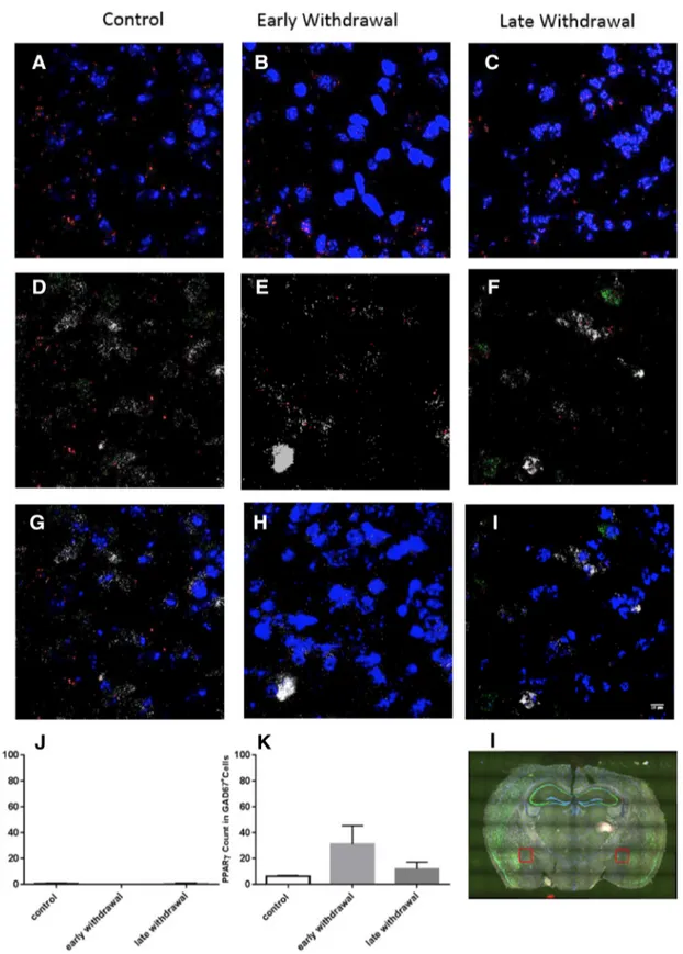
in the CeA was too low for statistical quantification, whereas the
expression of PPAR␥ was well quantifiable in GAD67⫹ cells
(Bartlett
⬘s
2⫽ 10.43, df ⫽ 2, p ⫽ 0.005) There was no significant
difference between experimental groups in PPAR␥⫹/GAD67⫹
cells of the CeA in both phases of nicotine withdrawal (
2⫽
2.142, df
⫽ 2, p ⫽ 0.34; see
Fig. 5
).
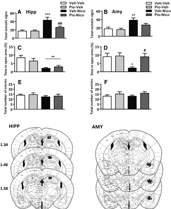
HIPP infusion of pioglitazone attenuated the expression of
the physical but not affective nicotine withdrawal signs in
PPAR
␥
(ⴙ/ⴙ)mice
Physical withdrawal signs
PPAR␥
(⫹/⫹)mice (n
⫽ 45) were subjected to chronic nicotine
or saline treatment followed by pioglitazone (0 or 0.3
l). Four
mice lost the cannula implant and were excluded from the
statistical analysis.
Two-way ANOVA showed a significant effect of nicotine
expo-sure (F
(1,37)⫽ 20.23; p ⫽ 0);
2⫽ 0.34, pioglitazone (F
(1,37)⫽ 7.34;
p
⫽ 0.016);
2⫽ 0.15, and a significant interaction nicotine ⫻
pi-oglitazone (F
(1,37)⫽ 5.48; p ⫽ 0.02);
2⫽ 0.13 in the physical
with-drawal score. Post hoc comparisons showed a significant increase of
the physical withdrawal signs in nicotine mice compared with
vehi-cles (p
⬍ 0.001) reduced by pioglitazone (p ⬍ 0.01;
Fig. 6
A).
Figure 3. Effects of pioglitazone (Pio), GW9662 and the combination on the expression of the physical withdrawal signs and anxiety-like behavior in PPAR␥(⫹/⫹)mice. A, Nicotine (Nico) induced a significant physical withdrawal score (*p⬍0.05)preventedbyPio(30mg/kg;
#p⬍ 0.05). The effect was blocked by GW9662 (¤p ⬍ 0.05). B, Nicotine reduced percentage
time in open arms (*p⬍ 0.05), effect reversed by Pio (30 mg/kg;#p⬍ 0.05) and blocked by
GW9662 (¤p⬍ 0.05). C, Total number of entries. Data are expressed as Mean ⫾ SEM values. Veh, Vehicle.
Affective withdrawal signs
Factorial ANOVA showed a significant effect of nicotine on the
percentage time spent in the open arms (F
(1,37)⫽ 19.06; p ⫽ 0);
2⫽ 0.3, no effect of pioglitazone (F
(1,37)
⫽ 0.24; p ⫽ 0.5) or
interaction nicotine
⫻ pioglitazone (F
(1,37)⫽ 1.60; p ⫽ 0.2).
Nicotine-treated mice spent less time in the open arms compared
with control ( p
⬍ 0.01;
Fig. 6
C). No difference was observed in
the total number of arm entries by nicotine (F
(1,37)⫽ 1.36; p ⫽
0.25), pioglitazone (F
(1,37)⫽ 0.1; p ⫽ 0.7), or their interaction
(F
(1,37)⫽ 0.01; p ⫽ 1.2;
Fig. 6
E).
Infusion of pioglitazone into the AMY attenuated the
expression of the affective but not physical nicotine
withdrawal signs in PPAR
␥
(ⴙ/ⴙ)mice
Physical withdrawal signs
PPAR
␥
(⫹/⫹)mice (n
⫽ 46) were subjected to chronic nicotine or
saline followed by administration of pioglitazone (0 or 0.3
l).
Mice that lost the cannula implant before assessing the affective
withdrawal signs (n
⫽ 2) and mice with wrong cannula
place-ment (n
⫽ 5) were excluded from the statistical analysis.
Two-way ANOVA showed a significant effect of nicotine
(F
(1,35)⫽ 13.53; p ⫽ 0);
2⫽ 0.25; but not a significant
pioglita-zone treatment (F
(1,35)⫽ 2.62; p ⫽ 0.15) or nicotine ⫻
pioglita-zone interaction (F
(1,35)⫽ 1.46; p ⫽ 0.19;
Fig. 6
B).
Affective withdrawal signs
Two-way ANOVA on percentage time spent in the open arms
showed a significant effect of nicotine (F
(1,35)⫽ 4.9; p ⫽
0.035);
2⫽ 0.13, pioglitazone (F
(1,35)
⫽ 4.34; p ⬍ 0.04);
2⫽
0.11 and interaction nicotine
⫻ pioglitazone (F
(1,35)⫽ 4.21;
p
⫽ 0.42);
2⫽ 0.1. Post hoc analysis showed a significant
reduction of the percentage of time spent in open arms in
nicotine mice versus control ( p
⬍ 0.05). Pioglitazone
in-Figure 4. PPAR␥ transcription levels in the (A, left) HIPP (B, left) AMY at 20 h and 6 d into nicotine withdrawal PPAR␥(⫹/⫹)mice. Nicotine withdrawal increased PPAR␥ mRNA levels after 20 h
and 6 d in HIPP (***p⬍ 0.001) and AMY (*p ⬍ 0.05, **p ⬍ 0.01). Representative images (40⫻ magnification) in the HIPP (top; a–c) and in the AMY (bottom; g–i) for PPAR␥ (red) ⫹ VGLUT1 (green) and GAD67 (white) in control, early, and late nicotine withdrawal. m, n, Coronal brain section of HIPP and BLA region from RNAscope ISH analysis. d, j, Merge of PPAR␥ (red) ⫹ GAD67 ⫹ VGLUT1 (green) and DAPI (blue) PPAR␥ counts in HIPP and AMY. e, f, PPAR␥ counts in VGlut1⫹ and GAD67 ⫹ cells in HIPP. Early nicotine withdrawal increased PPAR␥⫹ cells in GAD67 ⫹ cells (*p⬍0.05).k,IntheBLAearlynicotinewithdrawalincreasedPPAR␥⫹cellsinVGLUT1⫹cells(#p⬍0.05).l,PPAR␥expressioninGAD67⫹cellsincreasedinthelatewithdrawalphasecompared
with control and early withdrawal (#p⬍ 0.05). Data are expressed as mean ⫾ SEM values.
creased percentage time spent in the open arms compared the
nicotine-vehicle group ( p
⬍ 0.05;
Fig. 6
D). ANOVA did not
show a significant main effect of nicotine (F
(1,35)⫽ 1.52; p ⫽
0.24), pioglitazone (F
(1,35)⫽ 0.05; p ⫽ 0.86), or nicotine ⫻
pioglitazone interaction (F
(1,35)⫽ 0.19; p ⫽ 0.67) in total
number of entries (
Fig. 6
F ).
Discussion
Smoking cessation leads to aversive physical and affective
with-drawal symptoms that contribute to the maintenance of tobacco
use, and promote relapse (
Slawecki et al., 2003
;
Cippitelli et al.,
2011
;
Piper et al., 2011
). In the nicotine dependence models
Figure 5. PPAR␥transcriptionlevelsinCeA.Representativeimages(40⫻magnification)inCeAforPPAR␥(red)⫹GAD67(white)andDAPI(blue)incontrol,early,andlatenicotinewithdrawal.A–C,Merge ofPPAR␥(red)⫹DAPI(blue)counts.(D–F)representsPPAR␥⫹GAD67⫹cellsand(G–I)representsthemergeofPPAR␥⫹,GAD67⫹,andDAPIcells.J,PPAR␥countsinVGLUT1⫹cells,(K)PPAR␥counts in GAD67⫹cells,and(L)coronalbrainsectionillustratingCeAregionsampledforRNAscopeISHanalysis.Dataareexpressedasmean⫾SEMvalues.
used in our study, discontinuation from chronic nicotine
ex-posure elicited the expression of marked physical and affective
withdrawal signs (
Robinson et al., 1994
). PPAR
␥ activation by
pioglitazone attenuated the expression of the withdrawal
symptoms through neuronal mechanisms involving PPAR␥ in
HIPP and AMY.
Specifically, we found that pioglitazone reduced both the
neg-ative physical signs expressed in acute withdrawal and the
anx-iogenic response associated to the protracted withdrawal stage in
both rats and mice. Previous data have shown that PPAR␥
acti-vation does not affect locomotor activity in rodents, indicating
the specificity of its anxiolytic action (
Morgenweck et al., 2010
;
Sadaghiani et al., 2011
).
Here to characterize the involvement of PPAR
␥ in the
modu-lation of nicotine withdrawal, we compared the effect of chronic
nicotine exposure and pioglitazone treatment in mice with a
restricted deletion of neuronal PPAR
␥ (PPAR␥
(⫺/⫺)) with its
wild-type (PPAR
␥
(⫹/⫹)) counterpart. Spontaneous insurgence
of nicotine withdrawal induced a marked increase in the physical
withdrawal signs in both genotypes. Nicotine withdrawal also
produced an anxiogenic-like behavior in PPAR
␥
(⫹/⫹)mice,
whereas in PPAR
␥
(⫺/⫺)mice, anxiety level was already extremely
high under the basal condition, limiting the possibility to observe
the effect of nicotine withdrawal on this parameter. The
hyper-anxious phenotype of PPAR␥
(⫺/⫺)mice was described in an
ear-lier report in which we demonstrated that genetic deletion of the
neuronal PPAR␥ reduces resilience to environmental changes,
dampening the innate ability to adapt to stressful stimuli (
Domi
et al., 2016
). In subsequent experiments we found that
pioglita-zone ameliorated both physical and affective withdrawal signs in
Figure 6. Effect of pioglitazone (Pio) in HIPP and AMY (left, right, respectively) on physical and affective nicotine withdrawal signs in PPAR␥(⫹/⫹)mice. A, B, Nicotine (Nico) withdrawal induced
physical withdrawal signs (***p⬍ 0.001, **p ⬍ 0.01) prevented by Pio in HIPP (##p⬍ 0.01). C, D, Nicotine withdrawal decreased percentage open arm time (**p ⬍ 0.01, *p ⬍ 0.05) versus
control. D, Pio in the AMY reversed the anxiogenic behavior (#p⬍0.05).E,F,Totalnumberofarmentries.Dataareexpressedasmean⫾SEMvalues.Histologicalreconstructionsshowcorrect(filled
circles) injections into the AMY and dorsal HIPP, taken fromPaxinos and Franklin (2003). Veh, Vehicle.
PPAR␥
(⫹/⫹)but not in the PPAR␥
(⫺/⫺)mice. Moreover, the effect
of pioglitazone in PPAR
␥
(⫹/⫹)mice was completely blocked by
prior administration of the selective PPAR
␥ antagonist GW9662.
Altogether, these findings provide robust evidence of the
spec-ificity of neuronal PPAR␥ activation as a mechanism to
ame-liorate symptoms associated with nicotine withdrawal and
demonstrate that the effect of pioglitazone is mediated by this
nuclear receptor.
Neuroanatomical evidence of PPAR␥ expression in AMY,
HIPP, and other brain regions involved in the regulation of
emo-tion and motivaemo-tion have been previously described (
Moreno et
al., 2004
;
Inestrosa et al., 2005
;
Gofflot et al., 2007
;
de Guglielmo
et al., 2015
). Cortical and amygdalar PPAR
␥ activation has been
linked to the attenuation of the negative effects induced by acute
and chronic stress exposure (
García-Bueno et al., 2005
;
Domi et
al., 2016
) and specific hippocampal PPAR␥ activation appears to
play a role in attenuating the cognitive deficits caused by alcohol
intoxication (
Cippitelli et al., 2017
).
We decided to ascertain the impact of nicotine withdrawal on
PPAR␥ expression and distribution in both HIPP and AMY
during early (20 h) and protracted (6 d) nicotine withdrawal in
PPAR
␥
(⫹/⫹)mice. Nicotine withdrawal was associated with a
significant increase of PPAR
␥ mRNA levels in both regions in the
examined time points.
It is well known that expression and transcriptional activity of
PPAR␥ is affected by the cross talk with several kinases,
phospha-tases (i.e., ERK- and p38-MAPK, PKA, PKC GSK3;
Rochette-Egly, 2003
) and cAMP response element binding protein (CREB;
Liu et al., 2016
). All these intracellular signaling elements are also
known for their role in mediating nicotine effects (
Brunzell et al.,
2003
;
Burns and Vanden Heuvel, 2007
;
Michalak and Biala, 2017
;
Wang et al., 2017
). Notably,
Fisher et al. (2017)
highlighted
dis-tinct roles of CREB within the HIPP in mediating nicotine
with-drawal phenotypes in animals chronically treated with nicotine
and undergoing 24 h into the withdrawal phase. Hence, we can
speculate that the increase in PPAR␥ gene expression observed
into early or late withdrawal in the AMY and HIPP may represent
an adaptive response counteracting the physical and emotional
state associated with nicotine withdrawal, similarly to increased
PPAR␥ levels in stress conditions previously described (
García-Bueno et al., 2008
).
Nicotine exerts its pharmacological effects through activation
of the nicotinic acetylcholine receptors which in turn activate
differ-ent neurotransmitter systems, including glutamate and GABA, in
various brain regions that can regulate not only the reinforcing
properties of nicotine but also modulate the negative state
asso-ciated with drug withdrawal (
Picciotto and Corrigall, 2002
;
Had-jiconstantinou and Neff, 2011
). Notably, PPAR
␥ activation in the
AMY induces significant changes in the expression of several
genes linked to the GABAergic and glutamatergic transmission,
emphasizing the hypothetical role of PPARs in the modulation of
these two neurotransmitters in this region (
Ferguson et al., 2014
).
Nicotine withdrawal was associated with enhanced PPAR
␥
mRNA expression in AMY and HIPP; therefore, we decided to
investigate whether these changes occurred in GABAergic or in
glutamatergic neurons. Results showed a site-specific and
time-dependent increase in the expression of PPAR␥ in both
GABAe-rgic and glutamateGABAe-rgic cells of AMY and HIPP. Specifically, in the
early phase of nicotine withdrawal PPAR
␥ mRNA was increased
in glutamatergic neurons of the BLA and in GABAergic neurons
of the HIPP. During protracted abstinence PPAR␥ transcript was
increased in GABAergic neurons only of the BLA.
Marked biochemical and intracellular signaling alteration have
been described in AMY and HIPP neurotransmission during
nico-tine withdrawal (
Pandey et al., 2001
;
Tzavara et al., 2002
;
Fisher et
al., 2017
). In particular, electrophysiological experiments
dem-onstrated that unpleasant withdrawal symptoms are linked to
profound alteration in glutamate and GABA neurotransmission
in HIPP (
Yamazaki et al., 2006
).
In this framework, it has been shown that nicotine exposure
causes the downregulation of mGluR
2/3resulting in the
impair-ment of the negative feedback control on glutamatergic terminals
in several cortical and limbic brain sites (
Liechti et al., 2007
).
Accord-ingly, the metabotropic glutamate receptor agonist, LY354740,
de-creases glutamate levels and reduces physical nicotine withdrawal
symptoms in the rat (
Helton et al., 1997
;
Cartmell and Schoepp,
2000
). Considering the coexpression of PPAR
␥ with
VGLUT-positive cells, which mostly colocalize with mGlu
2/3(
Di Prisco
et al., 2016
), one possibility is that the increase of PPAR␥ in
the AMY might counteract the expression of nicotine
with-drawal syndrome through the modulation of glutamatergic
transmission.
The role of GABA in nicotine withdrawal is not entirely clear
(
D’Souza and Markou, 2013
). However, recent evidence
sug-gested that GABAB1 subunit of the GABAB receptor is involved
in the regulation of behavioral alterations induced by nicotine
withdrawal. In particular, the severity of physical withdrawal signs
and anxiety was markedly reduced in mice with GABAB1 deletion
compared with wild-type littermates (
Varani et al., 2015
). Hence,
the possibility that the activation of PPAR
␥ may counteract the
expression of nicotine withdrawal symptoms through the
mod-ulation of GABA transmission in HIPP or AMY also exists and
requires further investigation.
To further understand the role of PPAR␥ receptors in the
modulation of physical and affective nicotine withdrawal signs,
we performed microinjection studies to selectively deliver
piogli-tazone in the dorsal HIPP and the AMY.
Results showed that intra-amygdalar injections of
pioglita-zone abolished the anxiogenic response linked to protracted
nicotine abstinence but did not significantly modify the
ex-pression of the physical withdrawal signs. On the other hand,
pioglitazone microinjected into the HIPP attenuated the
ex-pression of the physical but not the affective withdrawal signs.
Overall, these results suggest a brain region-dependent role of
PPAR␥ in the modulation of physical and affective aspects of
nicotine withdrawal.
Based on expression data, at the mechanistic level it is
tempting to speculate that in the HIPP expression of the
phys-ical withdrawal may involve PPAR
␥-mediated adaptations in
the GABAergic transmission. Whereas, in AMY PPAR
␥ appears
to interact with glutamatergic transmission to control the
expres-sion of physical signs of nicotine withdrawal and with GABA to
regulate affective responses linked to protracted abstinence.
Be-cause of the close apposition and the small size of the basolateral
and central portion of the AMY, it was not possible to target
specifically these two subregions with pioglitazone
microinjec-tions. However, nicotine withdrawal increased PPAR
␥
expres-sion only in the BLA (in both GABAergic and glutamatergic
neurons) but did not alter the expression of PPAR␥ in the CeA
neurons, suggesting that the effect of pioglitazone may probably
involve the BLA.
The negative condition associated with nicotine withdrawal
is one of the major factors that maintain tobacco use and that
promotes resumption of smoking in patients attempting to quit
smoking (
D’Souza and Markou, 2011
). Anxiolytic and
antide-pressant drugs have been proposed to ameliorate nicotine
with-drawal symptoms and are used to aid smoking cessation (
Hughes
et al., 2000
,
2014
). Preclinical research has shown that activation
of PPAR␥ may result in marked anxiolytic and antidepressant
effects, in part because of the receptor’s ability to modulate AMY
neurotransmission and in part through inhibition of microglia
function (
Sadaghiani et al., 2011
;
Domi et al., 2016
;
Zhao et al.,
2016
;
Guo et al., 2017
). These data have been confirmed in
clin-ical trials that demonstrated the efficacy of pioglitazone as mood
stabilizer and in the treatment of unremitted depression (
Kemp
et al., 2012
;
Sepanjnia et al., 2012
;
Zeinoddini et al., 2015
;
Colle et
al., 2017
). Preclinical experiments demonstrated that
pioglita-zone attenuated alcohol, opioid, cocaine consumption, and
rein-statement of drug seeking in rodents (
Stopponi et al., 2013
;
de
Guglielmo et al., 2017
;
Miller et al., 2018
). Of note pilot clinical
experiments confirmed that pioglitazone attenuates craving
in-tensity and improves brain white matter integrity in cocaine-use
disorder patients (
Schmitz et al., 2017
), and importantly, in a
recent clinical trial pioglitazone reduced nicotine craving in
heavy smokers (
Jones et al., 2017
). In contrast with our data on
nicotine, in a recent small randomized clinical trial, pioglitazone
was unable to prevent the expression of opioid withdrawal
symp-toms pointing to the possibility that the effects on withdrawal are
substance dependent (
Schroeder et al., 2018
).
Together, the results of our study shed new light on
neurobi-ological mechanisms responsible for the effect of PPAR
␥ agonists
on drug abuse and support the possibility of using these
com-pounds as adjunct treatments for smoking cessation. In this
re-gard, it is important to highlight that clinical studies revealed the
efficacy of PPAR
␥ agonists in treatment of chronic obstructive
pulmonary disease, a pathological condition that is largely due to
smoking (
Lakshmi et al., 2017
).
References
Brunzell DH, Russell DS, Picciotto MR (2003) In vivo nicotine treatment regulates mesocorticolimbic CREB and ERK signaling in C57Bl/6J mice. J Neurochem 84:1431–1441.
Burns KA, Vanden Heuvel JP (2007) Modulation of PPAR activity via phos-phorylation. Biochim Biophys Acta 1771:952–960.
Caan B, Coates A, Schaefer C, Finkler L, Sternfeld B, Corbett K (1996) Women gain weight 1 year after smoking cessation while dietary intake temporarily increases. J Am Diet Assoc 96:1150 –1155.
Caputi FF, Carretta D, Lattanzio F, Palmisano M, Candeletti S, Romualdi P (2015) Proteasome subunit and opioid receptor gene expression down-regulation induced by paraquat and maneb in human neuroblastoma SH-SY5Y cells. Environ Toxicol Pharmacol 40:895–900.
Caputi FF, Palmisano M, Carboni L, Candeletti S, Romualdi P (2016) Opi-oid gene expression changes and post-translational histone modifications at promoter regions in the rat nucleus accumbens after acute and repeated 3,4-methylenedioxy-methamphetamine (MDMA) exposure. Pharmacol Res 114:209 –218.
Cartmell J, Schoepp DD (2000) Regulation of neurotransmitter release by metabotropic glutamate receptors. J Neurochem 75:889 –907.
Cippitelli A, Astarita G, Duranti A, Caprioli G, Ubaldi M, Stopponi S, Kallupi M, Sagratini G, Rodrìguez de Fonseca F, Piomelli D, Ciccocioppo R (2011) Endocannabinoid regulation of acute and protracted nicotine withdrawal: effect of FAAH inhibition. PLoS One 6:e28142.
Cippitelli A, Domi E, Ubaldi M, Douglas JC, Li HW, Demopulos G, Gaitanaris G, Roberto M, Drew PD, Kane CJM, Ciccocioppo R (2017) Protection against alcohol-induced neuronal and cognitive damage by the PPAR␥ receptor agonist pioglitazone. Brain Behav Immun 64:320 –329. Colle R, de Larminat D, Rotenberg S, Hozer F, Hardy P, Verstuyft C, Fe`ve B,
Corruble E (2017) Pioglitazone could induce remission in major de-pression: a meta-analysis. Neuropsychiatr Dis Treat 13:9 –16.
De Biasi M, Dani JA (2011) Reward, addiction, withdrawal to nicotine. Annu Rev Neurosci 34:105–130.
de Guglielmo G, Melis M, De Luca MA, Kallupi M, Li HW, Niswender K, Giordano A, Senzacqua M, Somaini L, Cippitelli A, Gaitanaris G, De-mopulos G, Damadzic R, Tapocik J, Heilig M, Ciccocioppo R (2015) PPAR␥ activation attenuates opioid consumption and modulates mesolim-bic dopamine transmission. Neuropsychopharmacology 40:927–937. de Guglielmo G, Kallupi M, Scuppa G, Demopulos G, Gaitanaris G,
Cicco-cioppo R (2017) Pioglitazone attenuates the opioid withdrawal and vul-nerability to relapse to heroin seeking in rodents. Psychopharmacology 234:223–234.
Di Prisco S, Merega E, Bonfiglio T, Olivero G, Cervetto C, Grilli M, Usai C, Marchi M, Pittaluga A (2016) Presynaptic, release-regulating mGlu2-preferring and mGlu3-mGlu2-preferring autoreceptors in CNS: pharmacological profiles and functional roles in demyelinating disease. Br J Pharmacol 173:1465–1477.
Domi E, Uhrig S, Soverchia L, Spanagel R, Hansson AC, Barbier E, Heilig M, Ciccocioppo R, Ubaldi M (2016) Genetic deletion of neuronal PPAR␥ enhances the emotional response to acute stress and exacerbates anxiety: an effect reversed by rescue of amygdala PPAR␥ function. J Neurosci 36:12611–12623.
D’Souza MS, Markou A (2011) Neuronal mechanisms underlying develop-ment of nicotine dependence: implications for novel smoking-cessation treatments. Addict Sci Clin Pract 6:4 –16.
D’Souza MS, Markou A (2013) The “stop” and “go” of nicotine depen-dence: role of GABA and glutamate. Cold Spring Harb Perspect Med 3:a012146.
Ferguson LB, Most D, Blednov YA, Harris RA (2014) PPAR agonists regu-late brain gene expression: relationship to their effects on ethanol con-sumption. Neuropharmacology 86:397– 407.
Fisher ML, LeMalefant RM, Zhou L, Huang G, Turner JR (2017) Distinct roles of CREB within the ventral and dorsal hippocampus in mediating nicotine withdrawal phenotypes. Neuropsychopharmacology 42:1599 – 1609.
García-Bueno B, Madrigal JL, Lizasoain I, Moro MA, Lorenzo P, Leza JC (2005) Peroxisome proliferator-activated receptor gamma activation de-creases neuroinflammation in brain after stress in rats. Biol Psychiatry 57:885– 894.
García-Bueno B, Madrigal JL, Pe´rez-Nievas BG, Leza JC (2008) Stress media-tors regulate brain prostaglandin synthesis and peroxisome proliferator-activated receptor-gamma activation after stress in rats. Endocrinology 149: 1969 –1978.
Gofflot F, Chartoire N, Vasseur L, Heikkinen S, Dembele D, Le Merrer J, Auwerx J (2007) Systematic gene expression mapping clusters nuclear receptors according to their function in the brain. Cell 131:405– 418. Grunberg NE (2007) A neurobiological basis for nicotine withdrawal. Proc
Natl Acad Sci U S A 104:17901–17902.
Guo M, Li C, Lei Y, Xu S, Zhao D, Lu XY (2017) Role of the adipose PPAR ␥-adiponectin axis in susceptibility to stress and depression/anxiety-related behaviors. Mol Psychiatry 22:1056 –1068.
Hadjiconstantinou M, Neff NH (2011) Nicotine and endogenous opioids: neurochemical and pharmacological evidence. Neuropharmacology 60: 1209 –1220.
Helton DR, Tizzano JP, Monn JA, Schoepp DD, Kallman MJ (1997) LY354740: a metabotropic glutamate receptor agonist which ameliorates symptoms of nicotine withdrawal in rats. Neuropharmacology 36:1511– 1516.
Hughes JR, Hatsukami DK, Skoog KP (1986) Physical dependence on nic-otine in gum: a placebo substitution trial. JAMA 255:3277–3279. Hughes JR, Stead LF, Lancaster T (2000) Anxiolytics and antidepressants for
smoking cessation. Cochrane Database Syst Rev 2:CD000031.
Hughes JR, Stead LF, Hartmann-Boyce J, Cahill K, Lancaster T (2014) Antide-pressants for smoking cessation. Cochrane Database Syst Rev 1:CD000031. Inestrosa NC, Godoy JA, Quintanilla RA, Koenig CS, Bronfman M (2005)
Peroxisome proliferator-activated receptor gamma is expressed in hip-pocampal neurons and its activation prevents beta-amyloid neurodegen-eration: role of Wnt signaling. Exp Cell Res 304:91–104.
Jones JD, Comer SD, Metz VE, Manubay JM, Mogali S, Ciccocioppo R, Mar-tinez S, Mumtaz M, Bisaga A (2017) Pioglitazone, a PPAR␥ agonist, Domi et al.• Activation of PPAR␥ on Nicotine Withdrawal J. Neurosci., December 4, 2019•39(49):9864 –9875 • 9873
reduces nicotine craving in humans, with marginal effects on abuse po-tential. Pharmacol Biochem Behav 163:90 –100.
Jones JD, Bisaga A, Metz VE, Manubay JM, Mogali S, Ciccocioppo R, Madera G, Doernberg M, Comer SD (2018) The PPAR␥ agonist pioglitazone fails to alter the abuse potential of heroin, but does reduce heroin craving and anxiety. J Psychoactive Drugs 50:390 – 401.
Jones JR, Shelton KD, Guan Y, Breyer MD, Magnuson MA (2002) Genera-tion and funcGenera-tional confirmaGenera-tion of a condiGenera-tional null PPAR␥ allele in mice. Genesis 32:134 –137.
Jorenby DE, Hatsukami DK, Smith SS, Fiore MC, Allen S, Jensen J, Baker TB (1996) Characterization of tobacco withdrawal symptoms: transder-mal nicotine reduces hunger and weight gain. Psychopharmacology 128:130 –138.
Kemp DE, Ismail-Beigi F, Ganocy SJ, Conroy C, Gao K, Obral S, Fein E, Findling RL, Calabrese JR (2012) Use of insulin sensitizers for the treatment of major depressive disorder: a pilot study of pioglitazone for major depression accompanied by abdominal obesity. J Affect Dis-ord 136:1164 –1173.
Kenny PJ, Markou A (2001) Neurobiology of the nicotine withdrawal syn-drome. Pharmacol Biochem Behav 70:531–549.
Kenny PJ, File SE, Rattray M (2001) Nicotine regulates 5-HT1Areceptor gene
expression in the cerebral cortex and dorsal hippocampus. Eur J Neurosci 13:1267–1271.
Lakshmi SP, Reddy AT, Reddy RC (2017) Emerging pharmaceutical thera-pies for COPD. Int J Chron Obstruct Pulmon Dis 12:2141–2156. Le Foll B, Di Ciano P, Panlilio LV, Goldberg SR, Ciccocioppo R (2013)
Peroxisome proliferator-activated receptor (PPAR) agonists as promising new medications for drug addiction: preclinical evidence. Curr Drug Tar-gets 14:768 –776.
Liechti ME, Lhuillier L, Kaupmann K, Markou A (2007) Metabotropic glu-tamate 2/3 receptors in the ventral tegmental area and the nucleus accum-bens shell are involved in behaviors relating to nicotine dependence. J Neurosci 27:9077–9085.
Liu YH, Tsai YS, Lin SC, Liao NS, Jan MS, Liang CT, Hsu SW, Chen WC, Sung JM, Maeda N, Tsai PJ (2016) Quantitative PPAR␥ expression affects the balance between tolerance and immunity. Sci Rep 6:26646.
Livak KJ, Schmittgen TD (2001) Analysis of relative gene expression data using real-time quantitative PCR and the 2(-Delta Delta C(T)). Methods 25:402– 408.
Lyon E (2001) Mutation detection using fluorescent hybridization probes and melting curve analysis. Expert Rev Mol Diagn 1:92–101.
Malin DH, Lake JR, Smith TD, Khambati HN, Meyers-Paal RL, Montellano AL, Jennings RE, Erwin DS, Presley SE, Perales BA (2006) Bupropion attenuates nicotine abstinence syndrome in the rat. Psychopharmacology 184:494 –503.
Michalak A, Biala G (2017) Calcium homeostasis and protein kinase/phos-phatase balance participate in nicotine-induced memory improvement in passive avoidance task in mice. Behav Brain Res 317:27–36.
Michalik L, Auwerx J, Berger JP, Chatterjee VK, Glass CK, Gonzalez FJ, Grimaldi PA, Kadowaki T, Lazar MA, O’Rahilly S, Palmer CN, Plutzky J, Reddy JK, Spiegelman BM, Staels B, Wahli W (2006) International union of pharmacology: LXI. Peroxisome proliferator-activated recep-tors. Pharmacol Rev 58:726 –741.
Miller WR, Fox RG, Stutz SJ, Lane SD, Denner L, Cunningham KA, Dineley KT (2018) PPAR␥ agonism attenuates cocaine cue reactivity. Addict Biol 23:55– 68.
Moreno S, Farioli-Vecchioli S, Ceru` MP (2004) Immunolocalization of per-oxisome proliferator-activated receptors and retinoid X receptors in the adult rat CNS. Neuroscience 123:131–145.
Morgenweck J, Abdel-Aleem OS, McNamara KC, Donahue RR, Badr MZ, Taylor BK (2010) Activation of peroxisome proliferator-activated re-ceptor gamma in brain inhibits inflammatory pain, dorsal horn expres-sion of Fos, and local edema. Neuropharmacology 58:337–345. Pandey SC, Roy A, Xu T, Mittal N (2001) Effects of protracted nicotine
exposure and withdrawal on the expression and phosphorylation of the CREB gene transcription factor in rat brain. J Neurochem 77:943– 952.
Paxinos G, Franklin KBJ (2003) The mouse brain in stereotaxic coordinates, Ed 2. San Diego: Academic.
Picciotto MR, Corrigall WA (2002) Neuronal systems underlying behaviors related to nicotine addiction: neural circuits and molecular genetics. J Neurosci 22:3338 –3341.
Piper ME, Cook JW, Schlam TR, Jorenby DE, Baker TB (2011) Anxiety diagnoses in smokers seeking cessation treatment: relations with tobacco dependence, withdrawal, outcome and response to treatment. Addiction 106:418 – 427.
Robinson SF, Pauly JR, Marks MJ, Collins AC (1994) An analysis of re-sponse to nicotine infusion using an automated radiotelemetry system. Psychopharmacology 115:115–120.
Rochette-Egly C (2003) Nuclear receptors: integration of multiple signal-ling pathways through phosphorylation. Cell Signal 15:355–366. Rossetti ZL, Isola D, De Vry J, Fadda F (1999) Effects of nimodipine on
extracellular dopamine levels in the rat nucleus accumbens in ethanol withdrawal. Neuropharmacology 38:1361–1369.
Rubio FJ, Liu QR, Li X, Cruz FC, Lea˜o RM, Warren BL, Kambhampati S, Babin KR, McPherson KB, Cimbro R, Bossert JM, Shaham Y, Hope BT (2015) Context-induced reinstatement of methamphetamine seeking is associated with unique molecular alterations in Fos-expressing dorsolat-eral striatum neurons. J Neurosci 35:5625–5639.
Sadaghiani MS, Javadi-Paydar M, Gharedaghi MH, Fard YY, Dehpour AR (2011) Antidepressant-like effect of pioglitazone in the forced swimming test in mice: the role of PPAR-gamma receptor and nitric oxide pathway. Behav Brain Res 224:336 –343.
Sarruf DA, Yu F, Nguyen HT, Williams DL, Printz RL, Niswender KD, Schwartz MW (2009) Expression of peroxisome proliferator-activated receptor-gamma in key neuronal subsets regulating glucose metabolism and energy homeostasis. Endocrinology 150:707–712.
Schmitz JM, Green CE, Hasan KM, Vincent J, Suchting R, Weaver MF, Moeller FG, Narayana PA, Cunningham KA, Dineley KT, Lane SD (2017) PPAR-gamma agonist pioglitazone modifies craving intensity and brain white matter integrity in patients with primary cocaine use disorder: a double-blind randomized controlled pilot trial. Addiction 112:1861–1868.
Schnegg CI, Robbins ME (2011) Neuroprotective mechanisms of PPAR␦: modulation of oxidative stress and inflammatory processes. PPAR Res 2011:373560.
Schroeder JR, Phillips KA, Epstein DH, Jobes ML, Furnari MA, Kennedy AP, Heilig M, Preston KL (2018) Assessment of pioglitazone and proinflam-matory cytokines during buprenorphine taper in patients with opioid use disorder. Psychopharmacology 235:2957–2966.
Searcy JL, Phelps JT, Pancani T, Kadish I, Popovic J, Anderson KL, Beckett TL, Murphy MP, Chen KC, Blalock EM, Landfield PW, Porter NM, Thibault O (2012) Long-term pioglitazone treatment improves learning and at-tenuates pathological markers in a mouse model of Alzheimer’s disease. J Alzheimers Dis 30:943–961.
Sepanjnia K, Modabbernia A, Ashrafi M, Modabbernia MJ, Akhondzadeh S (2012) Pioglitazone adjunctive therapy for moderate-to-severe major depressive disorder: randomized double-blind placebo-controlled trial. Neuropsychopharmacology 37:2093–2100.
Shiffman SM, Jarvik ME (1976) Smoking withdrawal symptoms in two weeks of abstinence. Psychopharmacology 50:35–39.
Slawecki CJ, Ehlers CL (2002) Lasting effects of adolescent nicotine expo-sure on the electroencephalogram, event related potentials, and locomo-tor activity in the rat. Brain Res Dev Brain Res 138:15–25.
Slawecki CJ, Gilder A, Roth J, Ehlers CL (2003) Increased anxiety-like behavior in adult rats exposed to nicotine as adolescents. Pharmacol Biochem Behav 75:355–361.
Stopponi S, Somaini L, Cippitelli A, Cannella N, Braconi S, Kallupi M, Rug-geri B, Heilig M, Demopulos G, Gaitanaris G, Massi M, Ciccocioppo R (2011) Activation of nuclear PPARgamma receptors by the antidiabetic agent pioglitazone suppresses alcohol drinking and relapse to alcohol seeking. Biol Psychiatry 69:642– 649.
Stopponi S, de Guglielmo G, Somaini L, Cippitelli A, Cannella N, Kallupi M, Ubaldi M, Heilig M, Demopulos G, Gaitanaris G, Ciccocioppo R (2013) Activation of PPAR␥ by pioglitazone potentiates the effects of naltrexone on alcohol drinking and relapse in msP rats. Alcohol Clin Exp Res 37: 1351–1360.
Tzavara ET, Monory K, Hanoune J, Nomikos GG (2002) Nicotine with-drawal syndrome: behavioural distress and selective up-regulation of the cyclic AMP pathway in the amygdala. Eur J Neurosci 16:149 –153. Varani AP, Pedro´n VT, Machado LM, Antonelli MC, Bettler B, Balerio GN
(2015) Lack of GABAB receptors modifies behavioural and biochemical alterations induced by precipitated nicotine withdrawal. Neuropharma-cology 90:90 –101.