UNIVERSITA’ DEGLI STUDI DI FOGGIA
Facoltà di Medicina e Chirurgia
Dipartimento di Medicina Clinica e Sperimentale Direttore: Prof. Lorenzo Lo Muzio
Tesi di Dottorato in
Medicina sperimentale e rigenerativa
PROGNOSTIC RELEVANCE OF COMPREHENSIVE
NON-INVASIVE IMAGING APPROACH IN A DIABETIC AND NON-DIABETIC ASYMPTOMATIC POPULATION
Relatore: Prof. Gaetano Serviddio
Dottorando: Dott. Andrea Igoren Guaricci
Index
PREFACE 4
Cap.1 Atherosclerotic Disease 7
1.1 Introduction 7
1.2 Anatomo-pathological features 7
1.3 Subclinical atherosclerosis 8
Cap.2 Coronary Artery Disease 10
2.1 Cardiovascular risk factors 10
2.2 Stratification of cardiovascular risk 11
2.3 Clinical manifestations 12
Cap.3 Diabetic Disease 14
3.1 Introduction 14
3.2 Diabetes Mellitus as coronary risk equivalent 15
3.3 Standard diagnostic approach to asymptomatic diabetic patient 17
Cap.4 Carotid Ultrasound 20
4.1 Introduction 20
4.2 Supra-aortic trunk and intima-media thickness 20
4.3 Clinical relevance of intima-media thickness 21
4.4 Carotid stenosis and plaque at risk 22
4.5 Correlation between carotid and coronary artery disease 23
Cap.5 Coronary Computed Tomography Angiography 26
5.1 The development of the technique 26
5.2 Coronary motion 28
5.2.1 Respiratory motion 29
5.2.2 Cardiac motion 29
5.3 Retrospective e prospective gating 30
5.4 Imaging of coronary plaque 31
5.5 Diagnostic accuracy 32
5.6 Calcium scoring of non-diabetic and diabetic asymptomatic patient 5.7 CCTA prognostic value of non-diabetic and diabetic asymptomatic patient
33 34
Cap.6 Experimental study 38
6.1 Aims 38
6.2 Material and methods 38
6.2.2 Carotid Ultrasound 39
6.2.3 CCTA protocol and images analysis 40
6.2.4 Follow up 43
6.3 Statistical analysis 44
6.4 Results 45
Cap.7 Final considerations and conclusion 55
Preface
Cardiovascular diseases are the leading cause of death in Europe and the United States. In Western Europe the incidence of myocardial infarction is 300 per 100,000 inhabitants and in Italy the figure is roughly comparable. Coronary Artery Disease (CAD), a pathology that underlies myocardial infarction, is caused by atherosclerotic disease, whose genesis is associated with numerous risk factors such as arterial hypertension, dyslipidemia, obesity, smoking and genetic predisposition. Despite the direct role of the aforementioned risk factors in the progression of atherosclerotic disease, the weight of each of them in CAD determinism is not yet quantifiable. Certainly however, the simultaneous presence of more than one cardiovascular risk factor increases exponentially the possibility of having cardiovascular events in the future.
The use of "risk cards", such as the Framingham Risk Score (FRS), allows to stratify the population, distinguishing within it three groups of risk, low, intermediate and high, to develop disease in the future. Nevertheless, this distinction allows a very rough estimate of the real cardiovascular risk of each subject. For example, most of the subjects fall in the intermediate risk group but, despite this, the fate of each will be different from each other; some of them will never develop a cardiac event while others will be affected early. Here the need to reclassify those subjects, placing them in a higher or lower risk group.
In the last twenty years the WHO and the scientific community have shown great interest in the prevention of cardiovascular diseases in which primary prevention is distinguished, which seeks to reduce the incidence of the first cardiovascular event by intervening on known risk factors, and the secondary prevention, which instead aims to reduce the incidence of subsequent events. Certainly, the results obtained recently are comforting for what regards secondary prevention but concretely less brilliant in the field
of primary prevention. In fact, in spite of the considerable advances in diagnostic techniques, as cardiovascular ultrasound, radiographic and nuclear methods, currently one of the most pressing problems remains the accurate and non-invasive identification of patients with significant CAD, of those who are really likely to face events in the short term, as well as the choice of the most effective therapeutic strategy in this area. This evidence justifies the unlikely need to overcome the limits of the main risk charts with the aim of tailoring the exact risk of future cardiovascular events on each subject in a sartorial manner.
In this context, a particular meaning is attributed to the diabetic disease. Diabetes mellitus (DM) is projected to spread more to the future, increasing from the current 415 million worldwide to more than 642 million. The prognostic importance of DM on cardiovascular outcomes is likewise well known. As consequence of previous studies showing the high incidence of CAD in diabetic patients and the relatively poor outcome compared to non-diabetic populations, DM is considered as CAD equivalent which means to label the diabetic patients as asymptomatic individuals at high cardiovascular risk. Lessons learned from the analysis of prognostic studies over the past decade have challenged this dogma and now support the idea that diabetic population is not uniformly distributed in the highest risk box. Detecting CAD in asymptomatic high-risk individuals is controversial and, what is more, in patients with diabetes is challenging, and this is why the reliability of traditional cardiac stress tests for detecting myocardial ischemia is limited.
Over the last few years, carotid disease, evaluated using ultrasonographic or tomographic methods, has been compared for diagnostic and prognostic purposes with the coronary one by traditional coronary angiography. Unfortunately, the latter technique is not able to accurately evaluate coronary subclinical atherosclerosis and this probably has the effect of underestimating the correlations between the two anatomical sites. Cardiac computed tomography angiography (CCTA) represents an emerging non-invasive technique able to explore the atherosclerotic involvement of the coronary arteries and, thus, to distinguish different risk categories tailoring this evaluation on each patient.
This study sought to evaluate to prognostic role of CCTA in asymptomatic type-2 diabetic patient as compared to a nondiabetic population. Moreover, it assesses the adding prognostic value of FRS, carotid artery disease and coronary artery calcium score (CACS) in diabetics in comparison to a non-diabetic population.
Chapter 1
Atherosclerotic disease
1.1 Introduction
Atherosclerosis is a chronic inflammatory disease predominantly affecting the elastic arteries (the aorta, the carotid arteries and the iliac arteries) and the large and medium-caliber muscular arteries (the coronary arteries and the poplite arteries). It is characterized by the accumulation of lipids in the intimal layer which, through a series of cell-mediated responses, leads to the formation of atherosclerotic plaque 1.
Atherosclerotic disease is the basis of a wide range of diseases with very different clinical presentation such as ischemic cardiopathy, stroke and peripheral vasculopathy.
Despite extensive knowledge on the topic, some important aspects of the evolution of these diseases are still unclear or unknown. Atherosclerotic lesions derive from a series of highly specific cellular and molecular responses, which together can configure the picture of an inflammatory disease, characterized by a monocyte-macrophage infiltrate and by the proliferation of smooth muscle cells.
Numerous prospective studies, conducted on well-defined population groups, have identified numerous risk factors, some constitutional, such as age and gender, others acquired and therefore potentially modifiable.
1.2 Anatomo-pathological features
An authoritative classification of arterial lesions is that proposed by Stary and identifies the evolutionary stages of the disease: a) isolated foam cells; b) lipid striations; c) promenomes; d) atheromas; e) fibroateromes; d) complicated injury 2.
Initially, atherogenesis is characterized by an adaptation process known as positive remodeling (positive remodeling), due to the fact that plaque increases externally to the vascular lumen without producing significant stenosis; only later the accumulation of lipids in the context of the arterial wall has a negative effect on the vascular caliber 3.
A feature of the most advanced lesion, fibroateroma, is given by the presence of a fibrous cap that covers the lesion on its luminal side. The fibrous cap is made up of smooth muscle cells inside a dense matrix containing collagen and capillaries. Microscopic hemorrhages are sometimes present in correspondence of neovascularized areas.
While the presence of isolated macrophages or lipid striae is demonstrable from the earliest years of life, atheroma appears in the age of puberty. The fibroateroma is identifiable from the third decade of life and increases slowly over time, sometimes assuming instability characteristics with intraplate haemorrhage, rupture, superimposed thrombosis.
1.3 Subclinical atherosclerosis
Subclinical atherosclerosis is that phase of the atherosclerotic process, not yet clinically manifest, but present from a biomolecular point of view, which can be identified by non-invasive instrumental investigations and specific markers. It is therefore presumable that patients with positivity of the markers of subclinical atherosclerosis are more likely to become ill with cardiovascular disease.
In is well known that endothelial dysfunction represents one of the earliest alterations of atherosclerotic disease. It is identified with a lower vessel capacity to modify its caliber in response to exogenous and endogenous stimuli. One of the methods for estimating the presence of endothelial dysfunction relies on the measurement of brachial artery capacity to dilate in response to an increase in blood flow and more precisely "shear stress" (reactive hyperemia), which is the main stimulus for release of nitric oxide
by endothelial cells. Normal flow-mediated nitric oxide dilatation requires the presence of intact endothelium. Endothelial dysfunction constitutes the functional prelude to the histo-pathological alterations of the vessel wall and the consequent clinical manifestations, including cardio-vascular and peripheral accidents 4,5.
The coronary Calcium Score, ie the amount of calcium deposits at the coronary level, is assessed by electron beam computed tomography (EBCT) and multislice spiral computed tomography (MSCT). The volume and density (expressed in Hunsfield units) of the calcifications themselves is graduated using a score scale according to Agatston et al
6.Absolute calcium score values> 400 or above the 75th percentile for age and gender
identify subjects with advanced coronary disease and a high risk of coronary events. Furthermore, the calcium score has a high negative predictive power: in other words, the absence of coronary calcifications makes the presence of steno-occlusive coronary artery atherosclerosis very unlikely and it poses for a low risk of short and medium term events
7,8.
The increase in the intima-media thickness of the common carotid arteries (IMT) is a fundamental marker for assessing the onset and progression of atherosclerotic disease and above all it is an indirect index of atherosclerotic involvement in other districts such as the coronary one and of the lower limbs. Clinical studies have shown a strong correlation between IMT, carotid plaque, and cardiovascular risk factors, and between IMT and cardio and cerebro-vascular events. The carotid disease could be used to identify those at higher risk and to intervene with a more aggressive therapeutic attitude 9.
Chapter 2
Coronary Artery Disease
2.1 Cardiovascular risk factors
Cardiovascular diseases and, in particular, CAD have a multifactorial etiology and therefore more risk factors contribute simultaneously and synergistically to their development. Risk factors are represented by those conditions that increase the probability of the onset of the disease and are traditionally divided into modifiable, with lifestyle changes and drug therapy, and not modifiable.
The most important non-modifiable risk factors are:
- age: the risk increases progressively with age, especially after age 65, since atherosclerotic disease is a chronic-degenerative disease. In particular, with increasing age, the activation of the renin-angiotensin-aldosterone system increases and the production of toxic oxygen radicals that favor endothelial dysfunction and the triggering of apoptotic phenomena;
- sex: men are more at risk than women, but in women the risk increases significantly after menopause, due to the lack of estrogen leading to unfavorable changes in lipid molecules;
- genetic factors and familiarity: several epidemiological studies have shown a family predisposition to coronary disease that would be determined by the interaction between polygenic inheritance and environmental factors. A person is considered at risk in which a first-degree family member presented a coronary event at an age <55 years in case of male gender and <65 years in case of female gender.
The main risk factors that can be modified are obesity, a sedentary lifestyle, arterial hypertension, dyslipidemia, insulin resistance / diabetes and a tabagic habit.
To the classic risk factors, today we have added "emerging" risk factors, which include conditions such as abdominal adiposity, inflammation, psycho-physical stress and depression, which have a significant impact on cardiovascular risk.
2.2 Stratification of cardiovascular risk
Since the 1980s, numerous risk stratification algorithms based on predetermined criteria have been developed in the USA, but none of these has substantially distinguished itself from others in achieving the intended purpose.
International guidelines state that the risk of Coronary Heart Disease (CHD) is determined to best address preventive treatment, and the FRS is the most frequently suggested tool for this purpose10.
The FRS is based on the measurement of blood pressure, total cholesterolemia, HDL cholesterolemia and the comparison of smoking habit, with which it is possible to assess whether an asymptomatic individual has a risk ranging from 0 to 20 % of having a myocardial infarction in the next ten years.
With the Framingham risk tables, three risk subgroups can be identified: high, intermediate and low. Asymptomatic patients who have a risk at 10 years> 20% are high-risk individuals, those with a high-risk between 10 and 20% are intermediate-high-risk individuals and those at risk <10% are at low risk.
High risk patients have the absolute risk of CHD equivalent to the risk of patients who have already had clinical manifestations of CHD and therefore should be treated as such. In the high-risk category, patients with DM and peripheral obliterative arteriopathy are also included.
In the intermediate risk group, the highest number of patients is collected and among these there are both patients who will develop an acute coronary syndrome and patients who never know who are affected. Therefore, the need arises to more accurately stratify patients belonging to this group, who must be considered potentially candidates for advanced risk assessment.
The accurate stratification of the risk profile is essential to identify the most suitable clinical-therapeutic attitude, in order to effectively prevent injurious, often fatal, cardio- and cerebrovascular events.
Intense research was performed to identify new risk factors that could improve the accuracy of the prognosis.
A cardiovascular risk chart has been developed by an italian research group that belongs to the “Progetto CUORE”. To formulate this risk chart, only Italian data were used, derived from different longitudinal studies started in the mid-80s and 90s whose risk factors were collected in a standardized way. The paper estimates the risk of developing a first fatal cardio- or cerebrovascular event in a population aged 40 to 69 years. Among the elements taken into consideration for the risk assessment, besides age, there are sex, systolic pressure values, total cholesterolemia, the presence of smoke and diabetes mellitus. For the estimation of the risk in symptomatic patients, appropriate scores are used such as Morise and Diamond and Forrester 11.
2.3 Clinical manifestations
CAD is a degenerative-inflammatory process that begins with endothelial dysfunction and, through the formation of atheromatous plaque, in the course of a few years, can induce the appearance of different clinical manifestations. Different clinical scenario can be distinguished: stable or chronic angina, acute coronary syndromes (acute myocardial infarction not associated with ST-segment elevation, NSTEMI, acute myocardial infarction with persistent ST segment elevation, STEMI, unstable angina), sudden cardiac death and silent ischemia.
The clinical manifestations of ischemic heart disease are a pathophysiological continuum based on the primary reduction of the coronary flow with consequent myocardial ischemia linked to a myocardial oxygen demand higher than the possibility of
contribution by the stenotic vessel (angina with fixed threshold). Otherwise, acute myocardial ischaemia occurs as a result of an occlusive or subocclusive thrombotic process established at the level of a vulnerable atheromatous plaque. Although this mechanism is the most common, there are also other conditions, less frequent, able to determine an acute reduction of coronary flow: coronary spasm, ischemia secondary to tachycardia, anemia, thyrotoxicosis, hypotension, anoxia and coronary embolism 12.
Chapter 3
Diabetic disease
3.1 Introduction
Diabetes mellitus (DM) is a major public health problem, the incidence of which seems to be drastically increased and will grow in the next years 13,14. Many studies in the
literature showed a clear correlation between DM and risk of CHD 15-17. Moreover,
comparing with matched nondiabetic individuals, patients with diabetes has a higher prevalence, extent, and severity of CAD18. On the basis of these considerations and on
the beneficial removal of the risk factors on progression of atherosclerotic disease, early detection of diabetic patients at increased risk of adverse cardiac events is crucial.
CCTA is an emerging non-invasive technique for the evaluation of coronary stenosis and for the characterization of the atherosclerotic plaques 19-21. However,
although the diagnostic accuracy and prognostic value of CCTA has been largely proved in symptomatic low-intermediate patients 22-25, its role in the asymptomatic and diabetic
individuals is still widely debated 26,27. The American Diabetes Association and American
Heart Association recently issued a joint statement that urges the identification of asymptomatic patients with subclinical CAD in whom more aggressive lifestyle or treatment changes would allow prevention of progression of the disease and reduce future clinical events 28.
3.2 Diabetes Mellitus as coronary risk equivalent
CAD represents the main cause of mortality and morbidity in patients affected by DM, which was considered as a "coronary risk" equivalent 29,30. The validated correlation
between DM and increased risk of CAD sparked a vivacious debate in the scientific community about the appropriateness of considering the diabetics as patients affected by CAD by default. The consideration of the DM as a "coronary equivalent" however, has a remarkable role because it implies a very aggressive treatment with a significant health care costs, possible lack of patient’s compliance and risk of adverse effects.
In order to weigh up the risk of CAD in the diabetic population compared to non-diabetic population, Haffner et al.31 compared the incidence of AMI in 1373 diabetic
patients and 1059 non-diabetic subjects in the Finnish population and followed them up to 7 years. The study showed that previous AMI had a substantial role in determination of second AMI, stroke and cardiovascular death. AMI incidence in non-diabetic population was 18.8% in the population with prior AMI, conversely 3.5% in non-diabetic population without prior AMI. In parallel, in diabetes group, the incidence of AMI was 45.0% in the population with prior AMI and 20.2% in the population without prior AMI. On the other hand, non-diabetic patients without previous AMI showed a better survival. The substantial novelty of the study is to have found a similar incidence of cardiovascular events in the group of 890 patients with DM without prior AMI and in the group of 69 patients without DM but with prior AMI, in a follow-up of 7 years. This correlation is unchanged even after adjustment for demographic variables (age and gender) and other cardiovascular risk factors (smoking, hypertension, lipid profile). On the basis of these results, the authors affirmed the negative impact of DM on coronary perfusion because diabetic patients with no known history of CAD presented the same risk of cardiovascular death than patients without DM but with prior AMI. For this reason, Haffner considered DM as an equivalent of CAD, implying an increase of 20% in the 10 years-cardiovascular risk of adverse events. This result suggests and encourages the treatment of all diabetic patients, as if they were really affected by known CAD 32. Although recent studies
confirmed and supported the consideration of “coronary risk equivalency” 33,34, other
evidences seem to reconsider this assumption, suggesting the identification of different classes of risk 35,36. A meta-analysis published in 2009 37 compared the total risk of
coronary events in diabetic patients without previous AMI and non-diabetic patients with previous AMI. This meta-analysis evaluated 13 studies including 45.108 patients with a mean follow-up of 13.4 years and a mean age of the enrolled subjects between 25 and 84 years. 2603 CHD events were found in diabetic population with no previous AMI; on the other hand 3927 events were recorded in the non-diabetic population with prior AMI. This work showed that diabetic patients without previous AMI presented a 43% lower risk of developing coronary events compared with non-diabetic patients with prior AMI (summary odds ratio 0.56, 95% confidence interval 0.53-0.60). This result suggests that, although DM is an important risk factor for the development of cardiovascular adverse events, it can’t be considered as a "coronary risk equivalent". Recently, Rana et al 38 have studied a
large cohort of 1.586.081 adults, afferent to the Kaiser Permanente Northern California health care system, aged between 30 and 90 years with a 10-year follow-up. The study compared the risk of adverse cardiac events in the population divided into 4 groups according to the presence of DM and coronary heart disease (CHD). The study confirmed that the sole presence of prior CHD is associated with an almost twofold increased risk of CHD, compared to the presence of sole DM (12.2 vs 22.5 per 1000 person-years) suggesting that the DM is an additional risk factor rather than a trigger in the progression of CAD. Remarkably, only when diabetes was present for more than 10 years, the risk of future CHD for patients with diabetes was similar to those with previous CHD. Of note, although the Adult Treatment Panel (ATP) III guidelines in 2001 recommended lifestyle and therapeutic primary prevention in diabetics, subsequent ACC/AHA American guidelines on the individual risk assessment reduced the DM role in the progression of atherosclerotic disease, on the basis of these new scientific evidences in the literature 39.
Therefore, there is no scientific evidence to support an aggressive therapeutic strategy with statins and aspirin in all patients with DM, but only in diabetic patients at high risk, in order to reduce cardiovascular mortality. On the basis of this important meta-analysis, it is crucial and essential to identify diabetic patients at high risk of adverse coronary events, worthy of an adequate aggressive therapy with statins and aspirin.
3.3. Standard diagnostic approach to asymptomatic diabetic patient
In view of the high prevalence of CAD and the not negligible autopsy rates of silent coronary ischemia in diabetic patients due to prevalent neuropathy 40 non-invasive stress
imaging could be useful in the prognostic stratification of asymptomatic diabetic patients in order to minimize vascular consequences of chronic hyperglycemia and optimize therapeutic approach.
Detecting CAD in patients with diabetes is challenging 41. The involvement of small
vessels due to metabolic abnormalities and the diffuse nature of the disease limit the reliability of cardiac stress tests for detecting myocardial ischemia, further worsened by the comorbidities. In addition, the silent fashion of CAD due to the high threshold for pain reduces the sensitivity of clinical risk assessment 42.
Exercise electrocardiography (EKG) is the most used non-invasive technique for the diagnostic and prognostic evaluation of non-diabetic patients with known or suspected CAD. Unfortunately, the accuracy of exercise EKG is reduced in the diabetic population. The poor response in terms of pressure and heart rate increasing during exercise, the high incidence of silent myocardial ischemia and microvascular disease, the alterations of impulse conduction due to visceral neuropathy, the presence of baseline ST-segment abnormalities, left ventricular hypertrophy and the impaired exercise capacity due to peripheral vascular disease, limited the diagnostic and prognostic value of exercise EKG. Given the limited accuracy of exercise EKG stress in diabetic patients, myocardial stress imaging tests have been proposed also thanks to their ability to identify changes in regional contractility depending on the location and size of the ischemic area. In asymptomatic diabetic patients, the sensitivity and specificity of stress echocardiography in the diagnosis of CAD is reported to be 81% and 85%, respectively 43,44.
Stress echocardiography allows to identify, in the absence of signs of stress ischemia, patients at low risk of developing adverse cardiac events 45,46. Two studies
identification of CAD, sustaining a good efficacy of stress echocardiography in identifying low-risk diabetic patients to develop cardiac events 47,48. Few years later, Kamalesh et al 49 studied the incidence of adverse cardiac events in a diabetic and non-diabetic
population with absence of signs of inducible ischemia as assessed by stress echocardiography and followed up for 25 ± 7 months. The study showed that diabetic patients, compared to non-diabetic ones, had a higher incidence of cardiac events (19% vs 9.7%, p = 0.03), a worse event-free survival (p = 0.03), and a greater number of non-fatal MI (6.7% vs 1.4%, p <0.05). The study revealed also that the history of CAD was the only predictor of adverse cardiac events (R = 0.18, p <0.05). On the basis of these results, Kamalesh concluded that diabetic patients with negative stress echocardiogram have more risk for adverse cardiac events compared to non-diabetic patients. The cause of this substantial difference could be explained by the greater tendency of diabetic patients to have distal CAD, slightly detectable by stress echocardiography. Moreover, diabetic patient presents an alteration of the coagulation pattern, intense platelet activity and reduced fibrinolysis which, together with the recognized autonomic dysfunction, most frequently predispose to coronary occlusion 50. On the same line, Cortigiani et al. 51
showed that a negative, non-ischemic stress test in the diabetic population, particularly in the subset of patients aged>65 years, is associated with an increased risk of developing adverse cardiac events when compared to non-diabetic subjects of the same age. Confirming these results, other studies showed an annual incidence of adverse cardiac events in diabetic patients with normal stress test equal to 3-6%, about twice respect of non-diabetic patients with normal stress test 52,53. On the other hand, some large studies
assessed the prognostic value of single-photon emission computed tomography imaging in patients with DM and, importantly, also in this scenario the event rate was higher compared with the control population, even in presence of a normal scan 54.
On the basis of these considerations, although exercise stress testing and myocardial perfusion imaging remain an important technique for risk assessment and prognosis of CAD in asymptomatic diabetic patients, presence of confounding factors,
such as autonomic dysfunction, multivessel disease, EKG abnormalities and interpretative difficulties, peripheral artery disease and the need of poly-pharmaco therapy, could compromise the diagnostic efficacy explaining why their role remain controversial 55.
Chapter 4
Carotid Ultrasound
4.1 Introduction
Vascular ultrasound is a non-invasive method of investigation that allows the visualization of the anatomical structure of the peripheral vessels (vessel wall and lumen) and at the same time the study of the endoluminal blood flow, thus obtaining anatomical and hemodynamic information.
Therefore, the echo-color-Doppler ultrasound is the referred technique to evaluate, in vivo and in a non-invasively, the changes in the arterial wall of the large vessels induced by atherosclerosis.
4.2 Supraortic trunks and intima-media thickness
The ultrasound technique allows the measurement of the carotid IMT 56. The
intima-media thickening is mainly represented by the hypertrophy of the medium layer, often due to hypertension, while atherosclerosis is generated by a process that mainly concerns the intima. The carotid plaque, is probably a late phase of the process of atherogenesis, related to inflammation, endothelial dysfunction, oxidative stress and the proliferation of smooth muscle cells 57.
Carotid atherosclerosis occurs more frequently in the carotid bulb and is not easily visualized with the two-dimensional ultrasonography technique.
In contrast, intimal thickening in response to age and high flow, as in hypertension, develops in the proximal segments of the common carotid artery, near the bifurcation; it is more easily identifiable through the B mode technique, compared to atherosclerosis 58.
The analysis of the mean intimal thickness in B mode involves the measurement of the common carotid, the bulb, the bifurcation and the internal carotid, with a normal average intimal thickness <0.9 mm. The atheromatous plaque is defined as a focal structure with a thickness of 0.5 mm or equal to 50% of the contiguous walls or an IMT> 1.5 mm measured from the media-adventitia interface to the intimate-lumen interface 59.
4.3 Clinical relevance of intima-media thickness
The analysis of the IMT is a simple and reproducible method for detecting and measuring the evolution of atheromatous disease.
Morphologically there are many similarities in the development of carotid and coronary atherosclerosis and there are numerous studies that correlate carotid atherosclerosis to cardiovascular risk. Epidemiological studies have correlated an increased carotid IMT to the presence of atherosclerotic lesions in other vascular districts, in particular to coronary lesions.
Extremely important information is derived from 3 prospective epidemiological studies (CHS, Rotterdam and Aric), which have shown that subjects with high values of carotid IMT have a higher probability of incurring time in a cardio or cerebrovascular event.
In the CHS (Cardiovascular Health study) we have seen how the thickness of the IMT in the internal carotid is a predictor of ischemic cardiopathy or atherosclerotic disease in apparently healthy subjects over 65 years of age 60.
In the Rotterdam study in a large cohort of 7983 subjects over 55 years of age, the increase in IMT in the common carotid was associated with an increased risk of stroke and acute myocardial infarction61.
In the ARIC (Atherosclerotic Risk in the Communities) study in which 7865 women and 6349 men aged between 45 and 65 were enrolled, the increase in mean-intimal thickness was associated with an increased incidence of stroke62.
Recent studies have shown an 11% increase in the risk of myocardial infarction for each increase in the IMT of 0.1 mm and that a median-intimal thickness> 1 mm is associated with a doubled heart attack risk over the following 3 years. Thus, IMT reflects the atherosclerotic burden of the organism and is a strong predictor of cardiovascular events. Linear regression analysis also demonstrated a significant association between IMT and left ventricle mass and between decreased IMT and hypocholesterolemic therapy.
Based on these data from the numerous studies, the European societies of Cardiology and Hypertension (ESC-ESH), have included in the guidelines the evaluation of IMT as a marker of organ damage. The American Heart Association recommends measuring the IMT in individuals over the age of 45 with an intermediate risk for cardiovascular risk assessment.
4.4 Carotid stenosis and plaques at risk
In the 50s and 60s, after correlation between cerebral ischemia and carotid atheroma was established, the researchers' attention was directed to the study of lesions that cause flow reduction. However, since the '70s, after the publication of the Framingham Study, it was realized that most of the cerebral ischemia was of a thromboembolic nature and therefore not only the lesions causing the flow reductions but also and especially the potentially embolic ones were to be found.
In the last decade, it has become progressively clear that the most important mechanism responsible for acute cardio and cerebrovascular events is the rupture of an atherosclerotic plaque with overlapping of a thrombotic formation. Anatomo-pathological studies have shown that the risk of rupture is related to the type of plaque rather than its size. Determinants of carotid plaque vulnerability to rupture are similar to those responsible for coronary instability:
1) size and composition of the lipid nucleus,
2) activity of inflammatory cells inside the fibrous cap, 3) thickness and collagen content of the cap.
Unstable plaques have a large lipid core, a thin fibrous cap, high density of macrophages and a few smooth muscle cells 63,64.
Numerous imaging methods, invasive and non-invasive, have been used to try to define the characteristics of atherosclerotic plaques responsible for instability 65. Among
these, high-resolution B-mode ultrasonography has been widely used at the carotid level as it allows not only an assessment of luminal impairment, but also a characterization of the size, morphology and surface of atherosclerotic plaque. The criteria with which the wall lesions are characterized, under the morphological profile, are the echogenicity and ecostructure, the characteristics of the endoluminal surface and the extent of the stenosis.
4.5 Correlation between carotid and coronary atherosclerotic disease
Although atherosclerosis is considered to be a widespread, or at least multifocal, process, and although both carotid ultrasonography and calcium scoring (CCS) can identify subclinical atherosclerosis, IMT and CCS are modestly correlated in the single individual.
The available studies on the prognostic potency of one or the other index in predicting future cardiovascular events are not univocal. Newman et al. they found that in adults over 70, IMT and CCS had the same ability to predict cardiovascular disease, whereas CCS would more likely predict stroke. Folsom et al. reported the results of the MESA study by giving the CCS a better ability to predict cardiovascular events than IMT
Despite the importance of medium-intimal thickening, the presence of atheromatous carotid disease seems to assume great importance in predicting cardiovascular events and, in particular, it is the plaque area and its volume (plaque burder) and its composition.
In a clinical study, Spence et al. demonstrated how the total plaque area is a strong predictor of cardiovascular syndromes. After adjustment for cardiovascular risk factors and antihypertensive and hypolipidemic therapy, over a 5-year period patients in the first quartile per plaque area had a 3.4-fold higher risk of stroke, death, or myocardial infarction than those in the last quartile 66. A recent study by Steinvil et al demonstrated
how tight the link between carotid atherosclerosis and coronary artery disease is. In a population of 1405 subjects, in fact, 42% had no carotid atherosclerotic plaques, 12.8% had moderate carotid atherosclerosis, and 4.6% had severe. A clinically significant carotid atheromasia (> 50%) was evident in 5.9% of the patients with non-obstructive coronary artery disease, 6.6% of the patients with single vessel disease, 13% of the patients with bivessel disease, 17.8% of the patients with trivessel disease, and in 31.3% of the patients with obstruction of the left main. The independent predictors of carotid atheromasia or total occlusion of the carotid artery were the presence of left main or 3-vessel obstruction, advanced age, stroke history, smoking, and DM. This study demonstrates that the presence of carotid stenosis, mainly of the internal carotid artery, is directly related to coronary artery disease.
Therefore, the clinical significance of the concomitant presence of carotid atherosclerosis and coronary disease lies in the prognostic implications of both
symptomatic patients eligible for revascularization and the entire population of asymptomatic individuals.
Chapter 5
Coronary Computed Tomography Angiography
5.1 The development of the technique
The first non-invasive X-ray method used for the study of epicardial coronary arteries was EBCT (Electron Beam Computed Tomography), introduced more than twenty-five years ago, aiming to estimate the presence of coronary calcifications, a surrogate marker of coronary atherosclerosis.
Thanks to the spiral technology, thanks to the advancement of the patient carrier bed during the scan and subsequent helical acquisition, it was possible to acquire the data in a volumetric way, opening new doors to the use of computerized tomography in clinical practice. Notwithstanding, it was the innovation of the multislice or multidetector CT, associated with the spiral technique, to put under the eyes of everyone the diagnostic potential of CT. The idea was to increase the number of "row" of detectors in order to cover a greater body volume in a short time, leading to a radical speeding up of the scanning time, allowing, at the same time, thinner layer thicknesses.
In recent years, Multilayer Computed Tomography (MDCT) has evolved to become a promising cardiology imaging methodology
The relatively late application of the tomographic techniques at the heart is due to numerous factors, among which the difficulty of studying an organ with its own movement, as well as secondary to respiratory excursions, stands out. This technological
challenge was won through the acquisition of images in a synchronized manner to the cardiac cycle, together with a phase of respiratory apnea. In addition to this, the heart is an organ characterized by medium-small caliber arteries, which imposes a high spatial resolution to obtain images of adequate quality.
Sixteen-slice detector systems allow the acquisition of 16 contiguous rows of detectors and therefore make it possible to acquire a larger portion of the chest at the same time. This entails the additional advantage of drastically reducing the time of the examination, in relation to the fact that the patient must maintain an inspiratory apnea. Therefore, the 16-slice CT devices have been able to provide cardiac and coronary images of sufficient quality 67,68. The image detail, the spatial resolution, is therefore fundamental and is
determined by the width of the detector table. No less important is the time necessary to acquire a single image that, generally, is equal to the time taken by the tube-detector system to perform 180 ° around the patient, and is called temporal resolution.
In order to improve the diagnostic accuracy, improving the temporal and spatial resolution, the technological development has led to the creation of increasingly advanced machines, including the 64-slice CT equipment, which today are the most widespread in the area. The recent 64-slice generation, in addition to reduced tube rotation time (0.33 sec), has significantly obtained an increased spatial resolution. This parameter can be obtained by setting volumetric acquisitions with very thin layer collimations (1.25 mm) and with increments of no more than 50% (0.65 mm) between one layer and another. This overlap is essential in post-processing, in order to obtain almost isotropic voxels, equal to about 0.4-0.5 mm3.
The implementation of the equipment has led, therefore, to a substantial increase in spatial and temporal resolutions, allowing the achievement of adequate image quality 69,70.
With the improvement of spatial resolution:
increases the ability to visualize smaller diameter vessels (eg distal coronary branches); increases the ability to quantify coronary calcium;
allows to reduce the blooming effect of the stents and therefore to improve the visualization inside the stent; It is a multifactorial artifact linked to interpolation, to the partial volume effect and to the beam hardening, which makes these structures appear much larger than they actually are (a sort of magnifying effect).
allows to better define the presence of coronary plaques and to evaluate their characteristics (i.e. volume, attenuation).
With the improvement of the temporal resolution:
increases the ability to "fix" images in ever tighter time in the cardiac cycle; allows to find additional reconstruction windows within the cardiac cycle;
increases the performance of the system when the left ventricular function must be evaluated;
reduces the scan time and therefore the inspiratory apnea time
It is important to highlight that industrial development is implementing cardiac CCTA technology with different strategies. A first type of approach is to increase the volume coverage (ie the number of layers), which is about 15x15x15 cm, in order to make cardiac imaging even less vulnerable to changes in heart rate and arrhythmias. These are systems with 256 and 320 layers with collimation thickness of about 0.5 mm whose wide coverage along the z axis (the longitudinal axis of the patient) can allow the acquisition of the whole heart in a single cardiac cycle. A second type of approach is to combine two X-ray tubes and the two detector systems in a single gantry, arranged at an angle of 90 °. With this approach (dual-source CT), only a quarter of the gantry is required to acquire data. In this case, the temporal resolution is twice that of single-tube scanners, which substantially reduces movement artifacts 71.
The success of CCTA is largely determined by how effectively it is possible to eliminate movement artifacts. Image definition is related to cardiac motion and temporal resolution. Thus, CCTA requires the elimination of movement (cardiac and respiratory motion) which degrade the image quality causing artifacts.
5.2.1 Respiratory motion
The heart is located above the diaphragm, which has a piston movement during the respiratory cycle. The dominant component of the coronary movement due to respiration is a linear translation along the caudo-cranial direction in the order of about 10 mm. Finally, there is an expansion along the caudo-cranial direction due to the fact that the roots of the coronary arteries are dislocated less than the distal tracts of the arteries towards the tip of the heart. In order to suppress the artifacts of resipratory movement, the technique of "Breath - hold" (apnea) in the meso-teleinspiratory phase is used. It is important to underline that during the apnea phase the diaphragm is not completely stopped but it performs a slow involuntary sliding movement in the caudo-cranial direction which is greater the greater the apnea time.
5.2.2 Cardiac motion
The heart has a highly variable pattern of movement, more pronounced during systole and the first phase of diastole, but little present during meso and telediastole periods. Therefore, this last period of the cardiac cycle turns out to be the best for the purposes of the CTCA. Furthermore, the diastolic phase is characterized by the greater coronary blood flow and, therefore, the most suitable for the acquisition of images. The rest period, or period of minimal cardiac movement (change in position less than one millimeter), varies substantially from patient to patient and is not determined solely by the length of the cardiac cycle. The resting period measured by a group of patients ranged from 66 to 333 msec for the left coronary artery and from 66 to 200 msec for the right coronary
artery. It is important to note that the range of transverse movement of the right coronary artery is greater than that of its contralateral.
5.3 Retrospective and prospective gating
The acquisition of image data in the AC-TCMS scan is continuous within the cardiac cycle; for this reason the data corresponding to the phase in which the cardiac movement is minimal must be extracted retrospectively to minimize the "blurring" and movement artifacts. This process is called cardiac gating and is accomplished by recording the ECG throughout the scan 72. Triggering with the electrocardiogram (ECG-triggering)
synchronizes data acquisition to the cardiac cycle, and allows a better definition of the cardiac and surrounding structures. Once acquired, the data can be reconstructed with retrospective gating at any stage of the cardiac cycle, moving the starting point of the image reconstruction relative to the R wave. It has been mentioned that the most favorable phase of the cardiac cycle is generally from meso- to telediastole just before the P wave (so-called isovolumetric filling phase). The time window of data reconstruction in the diastolic phase, in particular, is expressed as a percentage of the interval between two contiguous R waves (typically between 60% and 80% of the RR interval), ie as distance in msec from the next wave R (typically 350-400 msec). The acquired data are subsequently reconstructed with the use of convolution filters. The field of view should be as small as possible including the whole heart, so as to fully exploit the matrix of the image that is constant (512 x 512 pixels). The convolution filter should be halfway between noise and image quality. Coronary imaging filters are generally used for coronary imaging. When the coronary arteries are very calcific or there are stents, the higher convolution filters, even if they increase the noise of the image, usually increase the visualization of the vessel wall or of the structure of the stent and of the lumen inside it.
For coronary CT, reconstruction increments of 0.4 mm and 0.6 mm collimation (0.4 mm3 isotropic voxel) are generally used. Usually, multiple reconstructions are performed in different time windows, and the physician / operator then chooses the dataset where motion artifacts are minimized, paying particular attention to the visualization of the right coronary artery 73,74.
Differently from the retrospective gating in which data are acquired continuously, the concept of perspective gating is based on sequential acquisition, ie only during the phase of interest of the cardiac cycle (i.e. diastole). The prospective triggering can be performed effectively in patients with low heart rate (i.e. <65b / m) and regulate and it is possible to reconstruct only the images from the acquired datasets (i.e. telediastole). The great advantage of this technique is the significant reduction of the radiogenic dose below 5 mSv.
5.4 Imaging of coronary plaque
The CTCA differs from conventional coronary artery as it is able to directly visualize the vascular wall in addition to coronary lumen. The CTCA is able to highlight the presence of atheromatous coronary plaques and describe its location and extension. Moreover, it is possible to classify the plaques in calcifications, non calcifications (fibro-lipidic) and mixed, thanks to the different density (attenuation) expressed in Hounsfield Units. The peculiarity of MSCT is, therefore, the possibility of non-invasively evaluating the atheromatous plaques present independently of the degree of stenosis they determine. It is known that during the initial phase of atherosclerotic plaque formation there is a compensatory dilation of the vessel lumen defined as "positive remodeling" even in the absence of a significant reduction of the coronary caliber 75. A similar
alteration of the vessel wall is one of the characteristics of the plaques at risk of rupture and its identification seems to assume great importance.
The gold standard method for classifying coronary plaque based on the different composition is Intra Vascular Ultra Sound (IVUS). When CTCA images are compared with the aforementioned technique, a good correlation is observed. However the CTCA tends to underestimate the volume of non-calcified plaque due to the limited spatial resolution and tends to overestimate the size of the predominantly calcified plaque due to the effects of blooming and partial volume.
The identification of the "vulnerable" plaque is currently a subject of great interest and the CTCA could be useful as a non-invasive method for the identification of at-risk coronary plaques and consequently for the stratification of the risk of acute coronary events of the patient.
5.5 Diagnostic accuracy
The current generation of scanners, ie the 64 layers, has a high diagnostic accuracy in the identification of obstructive CAD when compared with percutaneous conventional coronary angiography (CAG) in selected patient populations. Therefore, CTCA is used as a diagnostic imaging modality in patients with known or suspected CAD 76,77. The
validation literature on the diagnostic accuracy of the CTCA compared with the CAG is extensive. Numerous reports have been published on this accuracy for the identification / exclusion of obstructive stenosis (> 50%) of the coronary artery demonstrating a high performance of CTCA with sensitivity values of 90% (85% -94%), specificity of the 89% (83% -94%), positive predictive value of 76% (44% -93%) and negative predictive value always constantly high and close to 99% (95% -100%); this last value represents the real strength of the method and indicates that CTCA is very accurate and reliable in identifying patients who do not have significant coronary stenosis and therefore do not need further investigation. The positive predictive value, on the other hand, is still lower due to the still
not optimal temporal and spatial resolution which makes the method vulnerable to artifacts 78.
It is important to underline that the diagnostic accuracy of coronary CT scans can not yet be expressed in absolute terms as for conventional coronary angiography. In the case of coronary CT it depends on: 1) the type of technology available, 2) the experience of the operator and 3) the characteristics of the patient to be examined. In fact, the predictability largely depends on the population under examination and this is higher if the subjects undergoing coronary CT scan are at low-intermediate coronary risk 79-82.
5.6 Calcium scoring of non-diabetic and diabetic asymptomatic patient
Coronary artery calcium score (CACS) is widely considered a marker of subclinical atherosclerosis, validated in asymptomatic patients 83. Extent of CACS, in fact, well
correlates with the vascular atherosclerotic involvement and the probability of adverse cardiac events in the general population 84-86. Although the latest european guidelines on
cardiovascular prevention suggested evaluation of the CACS only in diabetic patients with high or very high cardiovascular risk (SCORE> 5% and SCORE> 10%), the latest american guidelines for risk stratification in patients with CAD recommended an " appropriate " use of CACS and CCTA in asymptomatic patients with high global risk 87,88.
Type 2 DM patients have higher values of CACS when compared with the general population 89. The mechanisms responsible of the extensive intracoronary calcium
accumulation in diabetic patients is multifactorial and not completely understood. Previous studies revealed that the increased production of advanced glycation end-products induces the overexpression of genes and enzymes involved in active calcification of the coronary plaque 90. CACS has been proposed as a first-line test for
CAD in patients with diabetes since it was widely demonstrated its higher capability respect to conventional cardio-vascular risk factors for predicting silent myocardial
ischaemia and short-term outcome 91,92. Numerous studies showed that higher values of
CACS in diabetic patients with metabolic syndrome are closely associated with increased prevalence of ischemia, adverse cardiac events, AMI, and mortality 93-96.
Notwithstanding, a significant percentage of patients with DM have very low or CACS zero, with a better long-term prognosis, revealing that DM is not an equivalent of coronary risk. Raggi P et al. documented a high proportion of asymptomatic patients with DM (39%) with CACS <10 97. In this study the authors confirmed a significant correlation
between CACS and DM (p = 0.00001), indicating that each increase of CACS correlates with an increase in mortality in diabetic and non-diabetic patients. However, diabetic patients without known CAD showed a similar survival to patients without DM and intracoronary calcium (98.8% and 99.4% respectively, p: 0.5). The results of other studies show the same trend 98,99.
5.7 CCTA prognostic value of non-diabetic and diabetic asymptomatic patient
Recently, CCTA has emerged as a reliable noninvasive imaging tool for the identification of CAD 100,104. Since is first steps, the technique has been characterized by a
very high negative predictive value whereas the positive predictive value has been growing progressively, mainly according to the improving of many technical aspects 105.
The sub-optimal positive predictive value and specificity of CCTA in assessing the coronary stenosis degree is mostly due to the “blooming” artifacts secondary to the presence of wall calcifications. In particular, the coronary arteries in diabetic subject are characteristically “small and calcific” and this explain why the specificity of CCTA in this specific subset of patients may be particularly low. At the same time, the technological innovation has been taking a giant step towards the artefacts reduction by acting different strategies regarding throughout the process, from the premedication of the patient before scanning to the acquisition and analysis of the images 106-110. Of note, a new important
and very attractive tool able to evaluate the functional value of a single stenosis, the fractional flow reserve CT (FFRCT) is not influenced by the presence of calcifications and
thus is particularly reliable in diabetic population 111-115.Among all others, the employment
of high definition techniques allows high values of specificity and diagnostic accuracy (close to 90% and 95-98% respectively) 116.
Pivotal information are obtained by CCTA and specifically that obstructive and non-obstructive CAD are characterized by a higher prevalence in the diabetic population compared to normoglycemic patients and that a different plaque composition does exist
117-119. Among patients with DM, non-obstructive and obstructive CAD according to CCTA
are associated with higher rates of all-cause mortality and major adverse cardiovascular events at follow up, and this risk is significantly higher than in nondiabetic subjects 120-122.
Despite this, current european guidelines do not advise CCTA for risk assessment and suggest other noninvasive testing methods (nuclear imaging, echocardiography, and carotid ultrasound) in high-risk diabetic patients 123. Conversely, the latest american
guidelines for detection and risk assessment of stable CAD state that calcium scoring and CCTA use “may be appropriate” in asymptomatic patients with high global risk124.
The fulcrum of non-invasive coronary assessment in diabetic population consists in its prognostic value. Numerous efforts have been made so far in order to add useful information upon this debated topic.
Min JK et al.125 evaluated the prognostic value of CCTA in a population of 400
asymptomatic diabetic patients without known history of CAD. This study showed that, after adjustment for CAD risk factors, the maximum stenosis, the number of coronary arteries involved and the segment stenosis score are associated with increased risk of developing adverse cardiac events and had incremental power for predicting cardiac events over conventional risk factors. Moreover, the study revealed that CCTA confers incremental risk prediction, discrimination and reclassification over CACS. Based on these results, CCTA seems to be very useful in risk stratification of asymptomatic diabetic
patients at higher risk of developing adverse cardiac events. Halon et al.126examined the
added value of CCTA over clinical risk scores United Kingdom Prospective Diabetes Study (UKPDS), and coronary artery calcium in a population based cohort of 630 asymptomatic type 2 diabetics with no history of CAD assessed for coronary heart disease related events over 6.60.6 years. Discrimination of all events was improved by addition of total plaque burden to the clinical risk and CACS combined and further improved by addition of an angiographic score.
Van Werkhoven JM et al. confirmed the usefulness of CCTA in prognostic stratification of diabetic patients (N = 313) with known or suspected CAD compared to non-diabetic patients (N = 303). Authors found that DM (P <0.001) and evidence of obstructive CAD (> 50% coronary stenosis) (P <0.001) were independent predictors of outcome. In particular and similarly to other evidences the presence of obstructive CAD is an important predictor of survival both in diabetic patients than in non-diabetic patients127,128. Conversely, absence of atherosclerosis in CCTA is associated with an
excellent (100%) disease-free survival at a mean follow-up of 20 ± 5.4 months, confirming the known high predictive value of CT both in diabetic and in non-diabetic patients129,130.
Furthermore, the study conducted by Kim JJ et al.131 demonstrated that the duration
of DM is associated significantly with the extent and the severity of CAD. Patients with a longer history of DM had higher levels of CACS, atheroma burden obstructive score, segment involvement score and segment stenosis score (P <0.001 for all). In addition, the severity of coronary stenosis clearly increases the incidence of adverse cardiac events, independently of other cardiovascular risk factors. On the basis of these considerations, authors suggest the introduction of CCTA screening in all patients with a history of DM > 10 years.
On the contrary, the study of Muhlestein JB et al. revealed that the use of CCTA as screening of asymptomatic diabetic patients did not reduce the incidence of mortality from
all causes and non-fatal myocardial infarction. However, the value of this result could be resized taking into consideration the low incidence of adverse cardiac events, in the study which reduces the statistical difference between the two groups132. Other than
unpowered, the study was biased by the fact that adequate care targets for risk factor reduction in most of patients assigned to receive aggressive therapy in CCTA group were not achieved. Moreover, the control group without CCTA scanning, also received good preventive medical treatment so that differences in therapy between the screened and non-screened groups were subtle.
Recently, Kang et al. confirmed the prognostic value in long term of CCTA in a population of asymptomatic diabetics133. This study analyzed clinical outcome of 591
asymptomatic patients with type 2 DM undergoing CCTA showing that the survival free of cardiac events was 99.3 ± 0.7% in patients with normal coronary arteries, 96.7 ± 1.2% in those with non-obstructive CAD, and 86.2 ± 3.0% in those with obstructive CAD (log- rank p <0.001). The present study confirmed that asymptomatic diabetic patients with normal coronary arteries or with non-obstructive CAD have an excellent clinical outcome even after five years, conversely to patients with obstructive CAD. An overview is given by a recent meta-analysis based on eight studies with a total of 6225 participants (56% male with average age of 61 years) and a mean follow-up of 20 to 66 months that evaluated the prognostic efficacy of CCTA in diabetic patients134.This meta-analysis concluded that
CCTA is critical in identifying diabetic patients at high risk of CAD to be addressed to an aggressive modification of risk factors, glycemic control and optimized medical therapy.
Chapter 6
Experimental study
6.1 Aims of the study
The purposes of our study were the following:
1. to explore the presence and features of atherosclerotic disease at carotid and coronary level by employing the ultrasonographic technique and CCTA, respectively, in a wide cohort of asymptomatic patients;
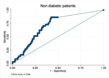
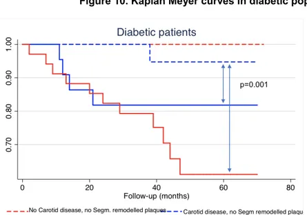
2. to asses the incremental prognostic value of FRS, carotid artery disease, CACS, and CAD in diabetic in comparison to non-diabetic subjects.
6.2 Material and methods
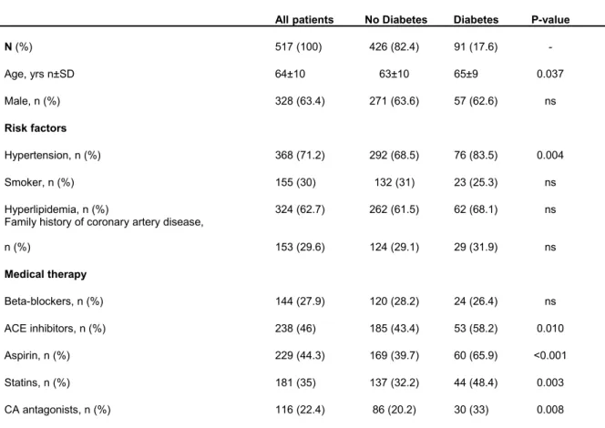
6.2.1. Study population and enrollment
A total of 691 consecutive patients asymptomatic for chest pain and referred for carotid ultrasound (CUS) assessment were enrolled between February 2004 and June 2010. Exclusion criteria included previous history of cardiovascular disease (n.51), contraindication to CCTA (n.102) and inadequate heart rate (>75bpm) (n.21). Accordingly, 517 patients were included in the study and underwent structured clinical interview, cardiovascular risk factors collection, 10-year cardiovascular risk calculation according to FRS CUS, CACS and CCTA within 2 weeks from the enrolment. Written
informed consent was obtained from all patients, and the institutional ethics committee approved the study protocol.
6.2.2 Carotid Ultrasound
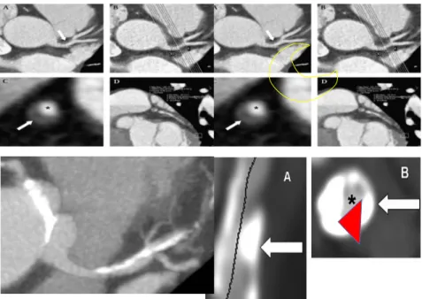
All patients underwent CUS evaluation using Hawk 2002 gray-scale ultrasound with a special multi-frequency linear probe (V-K Medical Company, Denmark). Two experienced operators, blinded to the clinical, laboratory and radiological findings performed independently the measurements. Longitudinal and transverse views of the left and right carotid arteries were obtained. Carotid artery disease was defined as the presence of increased carotid intima-media thickness (CIMT) and/or carotid plaque (Figure 1). Measurements were obtained on the far wall of the distal common carotid artery according to the Mannheim consensus135,136. According to the current statement, carotid
plaque was defined as a focal structure encroaching on the arterial lumen of at least 0.5 mm or 50% of the surrounding CIMT value or demonstrating a thickness of ≥1.5 mm, as measured from the media-adventitia interface to the intima-lumen interface.
A. Right Common Carotid Artery (CCA) intima-media thickness (IMT). IMT is measured as the distance between lumen-intima (light blu line) and media-adventitia (orange line) interfaces. The curly brackets show CCA (b, near wall; c, far wall); a shows the vein wall. B. Right Common Carotid Artery (CCA) plaque (double head arrow shows maximum diameter). Vertical red and green lines indicate the plaque along the near wall (b); c, CCA far wall; a, vein wall.
6.2.3 CCTA protocol and images analysis
CCTA was performed with a 64-slice scanner (Discovery HD 750 Medical System, GE Healthcare, Milwaukee, WI). At first, images were acquired for the calculation of calcium score (CACS) and then contrast-enhanced CCTA was performed. Retrospective and prospective electrocardiographic triggering (SnapShot Pulse, GE Healthcare, Milwaukee, WI) was used with variable padding, tube voltage and tube current as previously described137-139. An 80-100 ml amount of non-ionic contrast agent (Iomeron 400 mgI/ml,
Bracco, Milan, Italy) was administered intravenously at a rate of 5 ml/s, followed by 50 mL of saline. The dose length product (DLP) was recorded140. An approximation of the
effective radiation dose (ED) was obtained with the equation ED=k x DLP (k=0.017 x mGy-1 x cm-1 for the chest).
The CCTA images were transferred to a remote dedicated workstation (GE Healthcare, Milwaukee, WI). Two experienced operators evaluated the datasets (both with ≥8 years of clinical experience in CCTA performance and analysis.) blinded to all patient information and consensus agreement was achieved by a third reader (D.A. with equal years of experience) in case of any disagreement. Coronary calcium was identified on axial images as a dense area on the coronary artery pathway characterized by attenuation ≥130 Hounsfield Units (HU). The overall CACS was calculated in each patient based on the Agatston algorithm.
For the analysis of the coronary arteries all conventional modes of visualization and reconstruction were used: axial images, multiplanar reconstructions (MPR), multiplanar curved (c-MPR), maximum intensity projections (MIP), volume rendering reconstructions (VRT). The thickness of the MIP images was between 5 and 8 mm, reducing it to 3 mm in the case of extensive coronary calcifications.
The coronary arteries were divided into 15 segments according to the modified classification of the American Heart Association. the intermediate branch, when present, has been added to the classification with the number 16 (Figure 2)141. All segments were
considered in the analysis. First, each segment was classified as interpretable or not. Subsequently, the interpretable segments were evaluated for the presence of any atherosclerotic plaque using axial images and multiplanar curved reconstructions. Coronary plaques have been defined as> 1 mm structures within or adjacent to the coronary lumen, which can be clearly distinguished from the vessel lumen and surrounding pericardium tissue. Each plaque has been assigned in a segment
Figure 2. Coronary segmentation
RCA: right coronary artery; LCA: left coronary artery; LM: left main; LAD: left anterior descending; CX: circumflex artery.