1
UNIVERSITÀ DEGLI STUDI DI FOGGIA
D
OTTORATO DIR
ICERCA IN“HEALTH FOOD INNOVATION AND MANAGEMENT- I.M.A.E.V.” (XXXI CICLO)
C
OORDINATORE:
PROF.
MATTEO
A.
DEL
NOBILE
TRANSGLUTAMINASE,
NUTRITION AND HUMAN HEALTH
Dottorando:
GIORDANO DEBORAH
Tutor:
SABATO D’AURIA
Co-tutor:
ANGELO FACCHIANO
2
Abstract
Background: transglutaminases (TGase) are a class of enzymes widely spread in eukaryotic and prokaryotic organisms. Enzymes of this family catalyze post-translational modifications in many proteins by acyl transfer reactions, deamidation and crosslinking (polymerisation) between protein intra- or inter-chain glutamine (acyl donor) and lysine (acyl acceptor) peptide residues. Due to its facility of expression and purification, the only TGase enzyme widely used for industrial applications is the microbial TGase extracted from Streptomyces mobaraensis (MTGase). Nowadays the MTGase is commercially available and widely used in biopolymers industry, in cosmetics, in clinical applications, in wool textiles, and above all in the food processing industry. Its ability to catalyze crosslinks on many different protein substrates is increasingly used not only for sausage, ham and cheese production but, very recently, also for flour detoxification, as a possible alternative therapy to the gluten free diet.
It follows that nowadays the industrial applications of MTGase have increased, covering more and more fields producing a very active scientific research about this topic aimed at attempt to meet specific industrial needs, as the implementation of more efficient system for MTGase production, the research of alternative sources of microbial TGase, and safe source of recombinant enzymes.
Aims of the doctorate project: the main aim of the project is the identification of novel forms of microbial TGases that could become an alternative to that in use. A depth screening of known sequences has been performed, with the aim of obtaining a classification of microbial TGases for their similarity to known forms. To select the best candidates to be active forms under appropriate conditions, molecular modelling and molecular simulations have been performed on selected sequences. To test the enzymatic activity, experimental assays have been performed with a novel form, and another novel form has been expressed.
Results: the present work proposes at first an analysis, lacking so far, of the wide microbial transglutaminase world, developing the first classification of the microbial TGase based on their sequence features and their specific predicted secondary structures.
In order to classify and analyze the structural features of all the sequences annotated as having a TGase core computational techniques involving sequence analyses, comparative studies, building of phylogenetic trees, homology models and molecular dynamic simulations have been used. From this approach, a preliminary classification of these sequences was done by dividing them in five main groups. Each group has been investigated from the sequence point of view to analyze the presence of specific motifs. For three of this five groups, also the secondary structures have been investigated and, from this analysis, features specific for each group have
3 been detected. Moreover, two novel forms of microbial TGase (mTGase) have been investigated in the detail: K. albida mTGase and the hypothetical mTGase from SaNDy (organism not disclosed for patent opportunity). Molecular dynamics simulations and active site pocket analyses have been performed for the first, in comparison with MTGase. For the second, instead, experimental technique has been used to purify the hypothetical enzyme in order to test it on food related substrates. Experimental assays on both the proteins are still ongoing, to find the best enzymatic activity conditions and the best substrates of reaction. The molecular dynamic simulations performed on K. albida mTGase have suggested some explanations to the higher specificity of this enzyme than MTGase, experimentally demonstrated by Steffen et colleague, and several indications to change the activity conditions used to test it. Moreover, the substrates screening has allowed to find novel possible substrates, on which this enzyme could be employed for the allergenicity reduction. On the other hand, the enzyme extracted from SaNDy, showing a higher similarity with MTGase, could be less selective thanK. albida mTGase for specific substrates, so it could be possible its application also on the gliadin substrate, but to prove it further experiments are necessary.
Note: the present PhD work has been mainly performed in the Bioinformatics Laboratory at the CNR of Avellino under Dr. Facchiano’s supervision, however all the MD simulations have been performed at the Biochemistry Department of the University of Zurich, in the computational and structural biology laboratory under the supervision of Prof. A. Caflisch and his research group (compulsory abroad training period). Experimental activity assays on gliadin substrate have been performed by the spectrometry mass CeSMA-ProBio lab at the CNR of Avellino; and the hypothetical mTGase from SaNDy was instead cloned, expressed and purified in collaboration with the Laboratory for Molecular Sensing at the CNR of Avellino.
4
Index
LIST OF ABBREVIATIONS ... 1
INTRODUCTION ... 2
1.Transglutaminase enzymes ... 2
1.1 The human transglutaminases (hTGases)... 4
1.1.a The human tissue transglutaminase: structure, activity and allosteric regulation. 8 1.2 Microbial transglutaminase ... 11
1.2.a Streptomyces mobaraensis Transglutaminase (MTGase) ... 13
1.2.b Bacillus subtilis Transglutaminase (TGl) ... 15
1.2.c Chryseobacterium sp. Transglutaminase ... 17
1.2.d Kutzneria albida Transglutaminase (KalbTGase) ... 18
2. Application of microbial Transglutaminase in the industrial fields ... 21
2.1 MTGase enzymatic effects exploiting by the food-processing industry ... 22
2.2 MTGase, functional food and allergy prevention ... 25
2.3 Microbial Transglutaminase and innovative treatment for Celiac Disease ... 27
2.3.a The Celiac Disease and the gluten free diet ... 27
2.3.b The gluten free diet, its customers and its economic impact ... 30
2.3.c Risks and benefits of a gluten free diet ... 32
2.3.d Wheat flour detoxification: its results and its limits ... 36
AIM OF THE PROJECT ... 40
3. MTGase a wide topic for a wide scientific research: the aim of this project ... 40
MATERIALS AND METHODS ... 41
4. Sequence and structural analyses ... 41
4.1 Databases searches ... 41
4.2 Sequence alignments ... 44
4.3 Sequence clustering by phylogenetic tree constructions ... 45
4.3.a Workflow of the clustering procedure ... 45
4.3.b Methods applied for phylogenetic tree constructions ... 46
4.4 Motif researches ... 47
4.5 The source organism as selection criterion for proteins to model ... 48
4.6 Models building ... 48
4.7 Models validation ... 49
4.8 Secondary structure analysis ... 50
4.9 Analysis of the catalytic site pocket ... 51
5.0 Molecular dynamics (MD) simulations ... 51
5
5.2 MD analyses ... 54
5.2.a RMSD and RMSF analysis ... 54
5.2.b PCA ... 55
5.2.c SAPPHYRE PLOT ... 56
5.2.d Volume Analysis (fpocket ed Md pocket) ... 56
6.0 Experimental tests ... 58
6.1 KalbTGase: mass spectrometry assays ... 58
6.2.a Clone and Expression of the protein ... 58
6.2.b Purification ... 60
RESULTS AND DISCUSSION ... 62
7. Sequence analyses ... 62
7.1 Databases searches and sequence alignments results ... 62
7.1a Streptomyces Mobaraensis TGase (MTGase) as query ... 62
7.1b hTGase2 as query ... 64
7.1c Hypothetical transglutaminase dissimilar to both hTGase2 and MTGase ... 65
7.2 Sequence clustering ... 65
7.3 Motif research ... 68
7.3.a Motif research in the 1st group ... 68
7.3.b Motif research in the 2nd group ... 69
7.3.c Motif research in the 3rd group ... 71
7.3.d Motif research in the 4th group ... 73
7.3.e Motif research in the 5th group ... 74
7.3.f Motif research in the group Va ... 77
8. Structural analyses ... 80
8.1 Protein 3D models and their validation ... 80
8.2 Secondary structure analysis ... 84
8.3 Analysis of the catalytic site pocket ... 87
9. Molecular dynamics (MD) simulations ... 88
9.1 Kutzneria Albida mTGase: model validation ... 89
9.2 KalbTGase and MTGase: MD simulations at 300K ... 92
9.3 KalbTGase and MTGase: MD simulations at 335K ... 101
9.4 KalbTGase and MTGase: MD simulations at 355K ... 107
9.5 KalbTGase and MTGase: volume pocket analysis ... 118
10. Experimental tests ... 122
10.1 KalbTGase: mass spectrometry assays ... 122
6
10.2.a SandyTGase: protein expression results ... 127
10.2.b SandyTGase: purification results ... 128
CONCLUSION AND FUTURE WORKS ... 131
11.Work summary, conclusions and related future works. ... 131
Site-ography ... 134
7 Index of figures
Fig.1: Transglutaminase catalyze post-translational reaction….……….3
Fig.2: Structure of the human tissue transglutaminase………...…..8
Fig.3: The reaction mechanism of hTGase2………...…….9
Fig.4: Open and closed conformation of the hTGase2……….………..…10
Fig.5:Transamidation active site of hTGase2 in the closed conformation………....10
Fig.6: Structure of the Streptomyces mobaraensis transglutaminase………...13
Fig.7: Structure of the Streptomyces mobaraensis zymogen transglutaminase……….…14
Fig.8: Structure of the Bacillus subtilis transglutaminase………...…….…16
Fig.9: Structure of the Kutzneria albida transglutaminase………...…….…19
Fig.10: Workflow of the clustering procedure………...45
Fig.11: Phylogenetic tree showing sequence clustering….………...67
Fig.12: Motifs found in the 1st group of sequences………..…..69
Fig.13: Motifs found in the 2nd group of sequences………...………....……..69
Fig.14: Sequence motif shared by all the sequences belonging to the 2nd group………...70
Fig.15: Motifs found in the 2nd group of sequences………...…..70
Fig.16: The 2nd group branch……….71
Fig.19: Motifs found in the 4th group of sequences………73
Fig.20: Phylogenetic trees showing sequence clustering in the 5th group………..74
Fig.21: Motifs found in the 5th group of sequences………75
Fig.22 Motifs found in the 5th group of sequences labeled by the red circle………...76
Fig.23: Sequence motifs detected in TGases from Firmicutes of group Va………...77
Fig.24: Motifs detected by the analysis of selected Firmicutes sequences from group Va and group V. ……….…………...79
Fig.25: model of the hypothetical mTGase from K. albida. ……….……….…...84
Fig.26: Secondary structure topology of protein belonging to the group II. ……..…………...85
Fig.28: Secondary structure topology of protein belonging to the group III. ……..…………..85
Fig.27: Secondary structure topology of protein belonging to the group V………..………...86
Fig.29: RMSD of MD simulation on mTGase from K. albida. ………..…….…..90
Fig.30: Structural evolution of mTGase model from K. albida after the MD simulation..…...90
Fig.31: Superimposition of Kalb mTGase model and its real crystal structure…………..……91
Fig.32: RMSD plot of all the five different simulations of KalbTGase performed at 300K... ………...92
Fig.33: RMSF plot of the last 100ns of MD simulation for KalbTGase at 300K…………...93
Fig.34: Flexible regions of KalbTGase……….………93
Fig.35: KalbTGase regions used for the construction of thr Sapphire plot………..…..94
Fig.36: Sapphire plot of KalbTGase MD simulations at 300K………...94
Fig.37: Sapphire plot with dhiedral angles annotation of the catalytic residues (KalbTGase MD at 300K)……….………....95
Fig.38: Structural analysis of the Sapphire plot (KalbTGase-300K)…...……….96
Fig.39: RMSD plot of all the five different simulations of MTGase performed at 300K….…..97
Fig.40: RMSF plot of the last 100ns of MD simulation for MTGase at 300K………….……...98
Fig.41: Flexible regions of MTGase………..98
8
Fig.43: Sapphire plot of MTGase MD simulations at 300K……….……….99
Fig.44 Structural analysis of the Sapphire plot (MTGase-300K)……….100
Fig.45: Comparison between RMSD plot of KalbTGase MD simulation at 300K and at 335K… ……….101
Fig.46: RMSF plot of the last 100ns of MD simulation for KalbTGase at 335K………...101
Fig.47: Flexible regions of KalbTGase (MD simulation at 335K)...………..……….102
Fig.48: Sapphire plot of KalbTGase MD simulations at 335K…....…...……..………....103
Fig.49: Structural analysis of the Sapphire plot (KalbTGase-335K). …………...…………...104
Fig.50: Comparison between RMSD plot of MTGase MD simulation at 300K and at 335K… ………..………...104
Fig.51: RMSF plot of the last 100ns of MD simulation for MTGase at 335K…………...…105
Fig.52: Flexible regions of MTGase (MD simulation at 335K)……….105
Fig.53: Sapphire plot of MTGase MD simulations at 335K……….………..106
Fig.54: Structural analysis of the Sapphire plot (MTGase-335K)………...107
Fig.55: Comparison between RMSD plot of KalbTGase MD simulations………...108
Fig.56: RMSF plot of the last 100ns of MD simulation for KalbTGase at 355K…………...109
Fig.57: Flexible regions of KalbTGase (MD simulation at 355K). ……….…...109
Fig.58: Sapphire plot of KalbTGase MD simulations at 355K……….……..110
Fig. 59: Structural analysis of the Sapphire plot (KalbTGase-335K). ……….……..111
Fig.60: Comparison between RMSD plot of MTGase MD simulation at 300K and at 355K… ………..…...112
Fig.61: Comparison between RMSD plot of MTGase MD simulations………….………....113
Fig.62 RMSF plot of the last 100ns of MD simulation for MTGase at 355K…….…...……113
Fig.63: Flexible regions of MTGase (MD simulation at 355K)………...………….……….114
Fig.64: Sapphire plot of MTGase MD simulations at 355K………...……....115
Fig.65: Structural analysis of the Sapphire plot (MTGase-355K). ……….……...116
Fig.66: RMSD comparison between MTGase and KalbTGase MD simulations…….……..118
Fig.67: Volume comparison between KalbTGase and MTGase active sites…….………….120
Fig.68: Volume plot variations at different temperature………..………121
Fig.69: Mass spectra of the gliadin peptide 56-68 and its reaction mTGase……..……..…....124
Fig.70: BL21DE3 growth curve……….………….127
Fig.71: SDS PAGE results: tests for IPTG concentration to use………...……...128
Fig.72: SDS PAGE results: tests for temperature of induction to use……….…...…….129
Fig.73: SDS PAGE results: protein purification by osmotic shock……….………...129
Fig.75: SDS PAGE results: protein purification by anion exchange chromatography…...130
Index of tables
Table.1: property of the prediction of the stability index calculated for each sequence…...80Table.2: property of the best models of proteins belonging to group III……….………....82
Table.3: property of the best models of proteins belonging to group II……….…...82
Table.4: property of the best models of proteins belonging to group V……….…...83
Table.5: structural features of the putative catalytic site of modelled TGases, compared to those of related templates……….….…..88
Table.6: pocket volume analysis on MD simulations at 300K………...119
1
LIST OF ABBREVIATIONS
AOECS DH EFSA EmA ESPAGHAN FXIII GDP Gln hTGase1 hTGase2 hTGase3 hTGase3 hTGase5 hTGase6 hTGase7 hTGases IPTG iTCLs KalbTGase K-C2H5 K-CH3 K-gliadins LGG Lys MD mTGase MTGase NCGS OD PDB QMEAN QPS SandyTGase SPF SPI T1D TAP TAPI TGase TGase3 TGase4 TGlAssociation Of European Coeliac Societies Dermatitis Herpetiformis
European Food Safety Authority Anti-Endomysial Antibody
European Society for Pediatric Gastroenterology Hepatology and Nutrition Plasma Human Transglutaminase/Factor XIII
Guanosine diphosphate Glutamine
Human Keratinocyte Transglutaminase/Transglutaminase type 1 Human Tissue Transglutaminase/Transglutaminase type 2 Human Epidermal Transglutaminase/transglutaminase type 3 Human Prostate Transglutaminase/transglutaminase type 4 Human Transglutaminase type 5
Human Transglutaminase type 6
Human Transglutaminase type 7/ hTGase Z Human Transglutaminases
Isopropyl β-D-1-thiogalactopyranoside Gliadin-Specific Intestinal T-Cell Lines Kutzneria Albida Transglutaminase Lysine Ethyl Ester
Lysine Methyl Ester
Insoluble-Transamidated gliadin Lactobacillus Rhamnosus GG Lysine
Molecular Dynamic
Microbial Transglutaminase
Streptomyces mobaraensis Transglutaminase Non-Celiac Gluten Sensitivity
Optical Density Protein Data Bank
Quality Model Energy Analysis Qualified Presumption of Safety
hypothetical mTGase sequence from SaNDy (organism not disclosed for patent opportunity)
Soluble Protein Fraction Soy Protein Isolate Type 1 Diabetes
Transglutaminase Activating Proteases
Transglutaminase Activating Proteases Inhibitor Transglutaminase
Mammalian Epidermal Transglutaminase Mammalian Prostate Transglutaminase Bacillus Subtilis Transglutaminase
2
INTRODUCTION
1.Transglutaminase enzymes
Transglutaminases, also named protein-glutamine gamma-glutamyltransferase (TGases; EC 2.3.2.13) are a class of enzyme widely spread in nature,being found in mammalian, vertebrates, invertebrates, mollusks, plants, and microorganisms (Martins et al., 2014).
TGases are enzymes that catalyze post-translational modification of proteins by acyl transfer reactions, deamidation and cross-link formation:
Acyl transfer reactions: occur through the transfer of γ-carboxamide groups of glutamine residues (acyl donor) in proteins/peptides to a variety of primary amines (acyl acceptor) (Fig.1a).
Deamidation:when lysine residues, free lysine or primary amines are absent from the reaction system, water becomes the acyl acceptor and hydrolytic deamidation of the glutamine residues occurs, transforming them into glutamic acid (Fig.1b). This reaction alters the protein charge, which also leads to changes in protein solubility (Kuraishi et al., 2001).
Inter- or intramolecular cross-linking (polymerisation): occurs through acyl-transfer between γ-carboxyamide groups of glutamine residues (acyl donor) and Ɛ-amino groups of lysine peptide residues (acyl acceptor) (Fig.1c) (Carvajal et al., 2011). These isopeptide bonds form a stable protein network that affects its solubility and therefore other functional properties, such as gelation, emulsification, foaming formation and viscosity, resulting from the production of changes in the hydrophobicity of the protein surface. Obtaining a stable protein network is very important, for example, in the formation of gels (Calmolezi Gaspar et al., 2014).
Among the three possible reactions, the hydrolytic deamidation is much slower than the linkage to primary amines and the formation of cross-links in the presence of accessible ε-amino side-chains from protein-bound lysine, respectively, moreover, in protein-containing food systems, the crosslinking reaction proceeds prior to the other reactions (Dube et al., 2007; Kuraishi et al., 2001).
The results of TGase activity are the modification of the protein conformation and other more extensive conformation changes due to bonding between the same protein and between different proteins, forming high molecular weight conjugates (Carvajal et al., 2011).
There are many examples of the TGase deamidation activity in nature. Among the most studied is the human tissue transglutaminase (hTGase2) deamidation activity on the gliadin peptides, which induces the trigger of the coeliac disease. Noteworthy is also the selective
3 deamidation of Gln53 and Gln61 of the Rho family GTPase RhoA and Ras, respectively, by Escherichia coli cytotoxic factor and Bordetella bronchiseptica necrotoxin, which are considered functional relatives of TGase (Lorand and Graham, 2010).
Fig1: Transglutaminase catalyze post-translational reaction. TGase can catalyze a: Acyl transfer reactions: incorporation of an amine (H2NR) into the Gln residue of a protein (blue rectangle) b: Deamidation c: Protein cross-link by forming a NƐ(γ-glutamyl)lysine isopeptide bridge between the deprotonated Lys residue of one protein (purple ellipse) and the Gln residue of another (blue rectangle) (image from Lorand and Graham, 2010).
With regard to TGase-catalyzed amide incorporation and the cross-links formation, there is a wide range of examples: processes of crosslinking are involved in the clotting reactions, in the dimerization of interleukin-2, in the inactivation of the hepatitis C virus core protein, which can be modified by both amine incorporation and crosslinking by hTGase2. Further example of TGase amide incorporation could be represented by the conjugation to polyamines by γ-glutamyl linkages of proteins in the chloroplast of Helianthus tuberosus and the soybean storage protein glycinin (Lorand and Graham, 2010).
However, among these reactions, from the industrial point of view, only the cross-linking is of interest in modification of the techno-functional properties of proteins.
Furthermore, the reaction between the γ-carboxylamide groups and primary amines is a promising tool to improve the nutritional value of vegetable proteins by fortification with amino acids (Dube et al., 2007).
4 More in general, it is possible to say that the functional roles of the TGase are very different. In multicellular organisms TGase reactions involved manly the transamidation of glutamine residues to lysine residues and the resulting side-chain to side-chain isopeptide bonds add strength to tissues and increase their resistance to degradation (Griffin et al, 2002).
In higher organisms, transglutaminases play important roles in diverse biological functions by selectively cross-linking proteins. Among the members are factor XIIIa, which stabilizes fibrin clots; keratinocyte transglutaminase and epidermal transglutaminase, which cross-link proteins on the outer surface of the squamous epithelium; and transglutaminase 2 (hTGase2), which shapes the extracellular matrix and promotes cell adhesion and motility (Santhi et al., 2017). In sea urchin and fish, TGases are necessary to the assembly and the elevation of the fertilization envelope (Ha and Iuchi, 1998)
In bacteria the functions of this enzyme are still unknown, even if some studies suggest that microbial transglutaminase (mTGase) could have a role in sporulation, being required for the cross-linking of a specific coat protein (Zilhão et al., 2005) or in the inhibition of several proteins during the development of aerial hyphae (Santhi et al., 2017).
In plants, TGases and their functionality have been less studied than in humans and animals (Carvajal et al. 2011). However, their physiological role in plants appears to be related to photosynthesis, fertilization, response to abiotic and biotic stresses, senescence, and programmed cell death (Martins et al. 2014).
In the following paragraphs the human and microbial TGases already known will be analyzed more in the details.
1.1 The human transglutaminases (hTGases)
Mammalian transglutaminase can be classified into immunogenically and genetically distinct types. Nine hTGase enzymes are present in humans; among them, eight are catalytically active and one, named the erythrocyte membrane protein band 4.2, is inactive. As mentioned in the previous paragraph, these proteins serve as scaffolds, maintain membrane integrity, regulate cell adhesion, and modulate signal transduction (Eckert. et al., 2014). Although the primary sequence of these enzyme shows differences, all share an identical not contiguous amino acid triad at the active site, i.e. Cysteine, Histidine, Aspartate.
Therefore, according to their physiological role and/or their organ-specific location hTGases are described as hTGase type 1, type 2, type 3, type 4, type 5, type 6, type 7, XIII factor and band 4.2. (Eckert et al., 2014).
5 HTGase type1 (hTGase1): it is the keratinocyte hTGase; it is expressed in the stratified squamous epithelia of the skin and upper digestive tract and in the lower female genital tract, is activated by proteolytic cleavage, increased Ca2+ level, and interaction with tazarotene-induced gene 3 (Eckert et al., 2014). HTGase1 together with hTGase 3 plays essential roles in epidermal keratinization. TGase 1 predominantly serves as a membrane-bound TGase isozyme (Thacher and Rice 1985), whose role is associated mainly with generation of the cross-linked cell envelope in epidermal keratinocytes. Mutations of the gene encoding membrane-bound TGase 1 elicit an autosomal recessive skin disorder known as lamellar ichthyosis, which results from an aberrant stratum corneum with the lipid and cornified envelopes being seriously injured (Terazawa et al., 2015).
HTGase type2 (hTGase2): it is the tissue transglutaminase; it is a multifunctional, Ca2+ dependent enzyme, which has been involved in the regulation of cell growth, differentiation, and apoptosis (Peng et al., 1999). In addition to the transamidation reaction, this enzyme displays GTPase, ATPase, protein kinase, and protein disulfide isomerase activity. It interacts with phospholipase Cδ1, β-integrins, fibronectin, osteonectin, RhoA, multilineage kinases, retinoblastoma protein, PTEN, and IκBα (Eckert et al., 2014).
HTGase2 has been localized mainly in the cytosol, however, a detectable hTGase2 expression, both as a cross-linking enzyme and a G-protein, has been reported in the nucleus where it may be actively transported into via binding to an importin-α3/Qip-1 family protein (Peng et al., 1999). Moreover, hTGase2 is also localized in plasma (10-15%) and the nuclear membranes (5%); it is secreted by unknown mechanism from the cell, where it localizes to the cell surface and the extracellular matrix (Lorand and Graham, 2010).
Retinoic acid, vitamin D, TGF-β1, IL-6, tumor necrosis factor (TNF), NF-κB, epidermal growth factor (EGF), phorbol ester, oxidative stress, and Hox-A7 induce hTGase2 expression. HTGase2 dysfunction contributes to celiac disease, neurodegenerative disorders, and cataract formation (Eckert et al., 2014).
HTGase type3 (hTGase3): it is the epidermal hTGase; it is present in hair follicles, epidermis, and brain. Like TG2, TG3 binds to and hydrolyzes GTP. It catalyzes the crosslinking of trichohyalin and keratin intermediate filaments to harden the inner root sheath of a hair follicle, which is critical for hair fiber morphogenesis (Eckert et al., 2014).
Moreover, the enzyme is thought to be critically involved in the cross-linking of structural proteins and in the formation of the cornified cell envelope, thereby contributing to rigid structures that play vital roles in shape determination and/or barrier functions (Lee et al., 1996).
6 TGase3 knockout mice show impaired hair development and increased fragility of isolated corneocytes of the skin barrier (Bognar et al., 2014).
HTGase type4 (hTGase4): it is the prostate hTGase; it is present in the prostate gland, prostatic fluids, and seminal plasma (Eckert et al., 2014). TGase4 knockout mice show defects in copulatory plug formation resulting in a fertility reduction (Dean 2013). In rats, TGase4 participates in masking the antigenicity of the male gamete via incorporating seminal protein, such as uteroglobin, or polyamines into sperm cell surfaces (Cho et al., 2010).
In contrast to rodent, the exact function of TGase4 in humans is not known, but some recent reports suggest that this enzyme showed prostate-restricted expression pattern (Dubbink et al., 1999). These results raise a possibility to use this enzyme as a novel target for prostate-related diseases, particularly in prostate cancer, due to the observation of a link between increased expression of hTGase4 and promotion of an aggressive prostate cancer phenotype (Cho et al., 2010; Eckert et al., 2014)
HTGase type5 (hTGase5): transglutaminase 5 is mainly expressed in foreskin keratinocytes, epithelial barrier lining, and skeletal muscle. hTGase5 crosslinks loricrin, involucrin, and SPR3 in epidermis and contributes to hyperkeratosis in ichthyosis and psoriasis patients. hTGase5 inactivating mutations result in skin peeling syndrome (Eckert et al., 2014). Studies observed that hTGase 5 contributes, as a secondary effect, to the hyperkeratotic phenotype in ichthyosis (both vulgaris and lamellar) and in psoriasis. Moreover, in patients affected by Darier's disease, an autosomal dominant skin disorder characterized by loss of adhesion between epidermal cells (acantholysis) and abnormal keratinization, hTGase5 expression, as well as hTGase3, is completely missregulated, being overexpressed or totally absent in different areas of the same lesion (Candi et al., 2002).
HTGase type6 (hTGase6): hTGase6 expression is localized in the human testes and lungs, and in the brain of mice (Eckert et al., 2014). Analysis of its temporal and spatial pattern of induction in mouse development indicates an association with neurogenesis.Autoantibodies to hTGase6 were identified in immune-mediated ataxia in patients with gluten sensitivity, suggesting a critical role for hTGase6 in cortical and cerebellar neurons. Moreover, hTGase6 is also expressed in human carcinoma cells with neuronal characteristics. Molecular modelling of hTGase6 indicates that this enzyme could have Ca2+ and GDP-binding sites related to those of hTGase3 and hTGase2, respectively (Thomas et al., 2011).
HTGase type7 (hTGase7): hTGase 7, also known as hTGase Z, is widespread expressed in different tissues (Grenard et al., 2001). One report suggested a correlation between hTGase7
7 transcript levels and cancer cells (Eckert et al., 2014). Significantly increased levels of transcripts of hTGases7, together with higher levels of hTGase4 and significantly lower levels of FXIII, were seen in tumor tissues; instead, lowest levels of hTGases-7 and 3 were seen in patients with metastatic disease. Moreover, breast cancer displays an aberrant expression of TGases, wherein the levels of TGases 7, 2 and 3 have a relationship with node involvement and patient outcome (Jiang et al., 2003).
XIII Factor (FXIII): Plasma hTGase (FXIII) is found in cells of osteoblast lineage, in plasma, platelets, macrophages, astrocytes, dermal dendritic cells, chondrocytes, synovial fluid, the placenta, the eyes, and the heart. It is an important component of the blood coagulation cascade because it is the last zymogen activated in the blood coagulation cascade. In the presence of Ca2+, the enzyme catalyzes crosslinking of fibrin molecules to stabilize fibrin clots. FXIII also plays a role in inflammation and bone synthesis (Eckert et al., 2014). Moreover, this enzyme catalyzed also the crosslinking of the Angiotensin II receptor type 1, resulting in enhanced monocyte adhesiveness of hypertensive patients and thereby may sustain the process of atherogenesis by chronic sensitization of circulating monocytes (AbdAlla et al, 2004).
FXIII exists as the A2 homodimer in many cells (such as platelets, macrophages, astrocytes and chondrocytes) and as A2B2 heterotetramer in blood plasma. The active site is located in the A subunits, and the B subunits serve as a carrier protein protecting the A subunits in the circulation and contributing to the binding of the complex to fibrinogen; moreover B subunits act as a brake on FXIII activation (Lorand and Graham, 2010). FXIII deficiency is an extremely rare autosomal recessive disorder characterized by a lifelong bleeding tendency, including recurrent pregnancy loss, umbilical cord bleeding, and intracranial hemorrhage. Life-threatening bleeding episodes can be reduced significantly with timely diagnosis of FXIII deficiency and appropriate prophylaxis utilizing blood-derived components such as fresh frozen plasma and FXIII concentrate, or recombinant FXIII (Dorgalaleh et al. 2016).
Band 4.2: Band 4.2 is a unique hTGase that lacks catalytic activity. The Cysteine-Alanine substitution in band 4.2 of the cysteine corresponding to the active site in the other forms is responsible for the lack of enzymatic activity.
Band 4.2 is mainly present in erythrocytes, bone marrow, fetal liver, and spleen.Band 4.2 is a major component of the erythrocyte membrane cytoskeleton and plays an important role in maintenance of membrane integrity and regulation of cell stability (Eckert et al., 2014). Actually, Band 4.2 is a major membrane-associated protein of 72kDa and is present at≈200,000 copies per cell. It is clearly important for normal erythrocyte function since patients whose
8 erythrocytes are deficient in or lack band 4.2 are anemic due to accelerated erythrocyte destruction and have abnormally shaped possibly fragile, erythrocytes (Korsgren and Cohen, 1991). Band 4.2 null mice show alterations in red blood cell function, including spherocytosis and altered ion transport.
From this brief overview is possible to conclude that most tissues express multiple TGase forms and share common substrates. This may explain why TGase family members can compensate for the loss of an individual enzyme. Studies on TGase2 gene knockout mouse indicated tissue-specific mechanisms of compensation for the loss of TG2, including transcriptional compensation in heart and liver versus functional compensation in aorta, kidney and skeletal/cartilagenous tissues. On the contrary, no compensation has been detected in skeletal muscle, suggesting a limited role for the TGase2-mediated transamidation in normal development of this tissue (Deasey and Nurminskaya, 2013).
Of the eight catalytic hTGases, hTGase2 has been the most comprehensively studied due to its ubiquitous expression in multiple cell types, that has allowed to obtain a wider knowledge about the hTGase world and their regulation and activities.
1.1.a The human tissue transglutaminase: structure, activity and allosteric regulation. The multifunctionality of hTGase2 is dependent on its structural features. HTGase2 is composed of four domains: a N-terminal β-sandwich domain (aa 1–140), the catalytic core (aa 141–460) and two C-terminal β-barrel domains (aa 461–586 and 587–687) (Fig.2a). The site of transamidating activity is composed of the catalytic triad of cysteine proteases (Fig.2b): cysteine 277 (C277), histidine 335 (H335) and aspartate 358 (D358).
Fig.2: Structure of the human tissue transglutaminase. HTGase2 PDB code 4PYG shows A: 4 domains: N-terminal β-sandwich domain in blue, the catalytic core in green (with the catalytic triad highlighted in red) and the C-terminal β-barrel-1 and β-barrel-2 domains in yellow and orange, respectively; B: the catalytic triad is composed of C277, H335 and D358.
9 The enzyme is regulated by redox potential, Ca2+ and GDP, particularly Ca2+ induces enzyme activation, instead GDP induces enzyme inhibition.
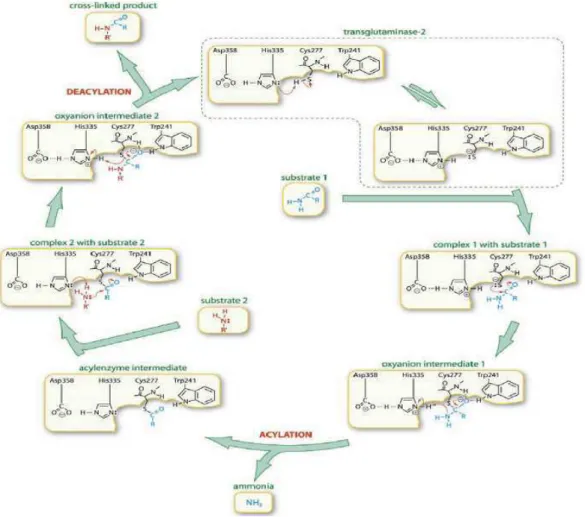
The mechanism for hTGase2-catalysed transamidation (Fig.3) is proposed on the basis of papain-reaction mechanism; due to their similarities in the catalytic triad and reaction mechanism. The reaction primarily involves the exchange of primary amines for ammonia at the γ-carboxamide group of glutamine residues, in the presence of Ca2+. The binding of Ca2+ is vital to the cross-link formation because it initiates a conformational change that exposes a cysteine residue in the active site domain; the cysteine reacts with the glutamine substrate, resulting to the formation of an acyl-enzyme intermediate and release of ammonia. The subsequent reaction between the acyl-enzyme complex and a primary amine results to the formation of γ-glutamyl-amino cross-link, and concomitant release of the enzyme (Onyekachi and Coussons, 2014).
10 If the Ca2+ is important to induce the conformational change that exposes the catalytic cysteine residue, the GDP molecule, bounded at the hTGase2, blocks the access to the active site (Fig.4).
Fig.4: Open and closed conformation of the hTGase2.The N-terminal β-sandwich is shown in blue ribbons (N), the catalytic domain (Core) in green ribbons, and the C-terminal β-barrels (β1 and β2) in yellow and red ribbons, respectively. A: Closed conformation: GDP-bound hTGase2. B: Open conformation: hTGase2 inhibited with the active-site inhibitor Ac-P(DON)LPF-NH2,which mimics the acyl-donor substrate. Simplified cartoons are included for clarity (Pinkas et al., 2007).
In the GDP-bound form of hTGase2, access to the transamidation active site is blocked by a loop connecting the third and fourth β-strands, as well as by a loop connecting the fifth and sixth β-strands of the first β-barrel domain (Fig.5). Tyr-516, which is conserved in hTGases and located in the first loop, forms a hydrogen bond with Cys-277 (Fig.5). Transamidation activity requires an accessible Cys-277, and Tyr-516 (Y516) with its associated loop from the first β-barrel domain must move to make the active site accessible to substrates (Liu et al., 2002).
Fig.5:Transamidation active site of hTGase2 in the closed conformation. Highlighted by red ribbons the catalytic core with the catalytic triad in ball and stick (C277–H335–D358) by the blue ribbons the first β-barrel domain with Y516 (showed in ball and stick) relative to the guanine nucleotide-binding site. Y516 points toward C277, the catalytic nucleophile, in the active site.
11 The GDP molecule engages both the first and last β-strands of the first β-barrel domain, which should maintain the inactive state by stabilizing the loops that block the access to the catalytic domain.
ATP and GDP were found to bind the same nucleotide-binding pocket. It is a perfect example of a tertiary pocket, with contributing residues from the catalytic domain barrel-1 and β-barrel-2 domains. It is composed of at least ten residues, many of which are involved in both adenine and guanine nucleotide binding; however, Ser482 and Arg580 were found to be involved only in guanine, not adenine, nucleotide binding (Gundemir et al., 2012).
If the GDP binding pocket has been resolved, the Ca2+ binding sites are still unknown, so they have been proposed comparing hTGase2 structure with FXIII structure, whose Ca2+ pockets have been demonstrated. A putative Ca2+ binding site, homologous to the one of FXIII, is distorted in hTGase2, with the largest difference occurring in the proximity of Ser-419 (equivalent to Ala-457 in FXIII), due to the bound nucleotide. Ca2+ binding, by altering the position of the Ile-416–Ser-419 peptide, would eliminate the stabilizing effects of these peptides on the nucleotide-binding site and could thereby weaken nucleotide binding, as has been observed experimentally. In consequence of this weakening nucleotide binding; the protein has a conformational change, which involves the substrate binding and the related displacement of the hydrogen bonded Tyrosine, making the active site accessible (Liu et al., 2002; Fesus and Piacentini 2002).
Actually, in addition to the catalytic triad, two conserved tryptophan residues (W241 and W332), located at the opposite sides of the “catalytic tunnel”, are critical for the transamidating activity, since these residues stabilize the enzyme-thiol intermediate that forms during catalysis. A threonine residue (T360) at the entrance of the catalytic tunnel controls the entry of the acyl-acceptors for the second step of the catalysis. The mutation of this residue increases the preference for deamination over transamidation. Another important residue in the catalytic site is the tyrosine residue at the position 516 (Y516). Actually, a hydrogen bond forms between C277 and Y516 in the closed conformation of hTGase2, which is believed to further stabilize the closed conformation and keep the enzyme inactive (Gundemir et al., 2012).
1.2 Microbial transglutaminase
TGases have been found in prokaryotes and eukaryotes, including guinea pig, and were first extracted from the liver of this animal in 1973 (Folk and Chung 1973). This TGase was the only form arriving at the market by the end of the 80s, not arousing much interest from the industrial point of view, since it was very expensive. In addition, as the other mammalian TGases, it was
12
a Ca2+-dependent enzyme, which led to the precipitation of proteins of some foods containing
casein, soybean globulin, or myosin (Martins et al., 2014).
However, TGases are present also in bacteria, and peculiar forms of this enzyme have been widely characterized. In 1989, a Ca2+-independent microbial TGase from Streptomyces
mobaraensis (MTGase) was extracted by Ando et al. Purification was rather easy, and the production of MTG has been established commercially and applied in research and biotechnology fields (Dube et al., 2007; CalmoleziGaspar and de Góes-Favoni, 2015).
This enzyme is characterized by the same triad that appears in the mammalian TGase with a different order, i.e. Cysteine, Aspartate, Histidine. Another relevant difference concerns the structural organization as a single domain in the bacterium enzyme, while in eukaryotes, four structural domains are observed. Nowadays mTGase by Streptomyces mobaraensis is commercially available and widely used in biopolymers industry, cosmetics, clinical applications, wool textiles, and food processing industry (Carvajal et al. 2011). Since the early 1990s, many mTGase-producing strains have been found, and production processes have been optimized. In the meantime, novel bacterial forms of the enzyme have been investigated. From a screening of microorganisms that produce MTGase, an actinomycetes strain, T-2, has been isolated from soil and identified as Actinomadura sp. (Kim et al., 2000). A protein with the deamidation activity typical of TGases has been isolated from Chryseobacterium sp. (Yamaguchi et al., 2001), but it lacks other typical activities of TGases and has been defined as a protein-glutaminase, a different enzyme class (EC 3.5.1). Transglutaminase activity in vitro has been demonstrated for the periplasmic portion of a protein from Pseudomonas aeruginosa, named TgpA (Milani et al., 2012). A small microbial transglutaminase from Bacillus Subtilis works through a unique partially redundant catalytic dyad formed by Cysteine and Glutamine residues, with a Histidine residue that also plays a role in the function of the enzyme, that is reduced but not eliminated by its mutation (Fernandes et al., 2015). TGase from Kutzneria albida (KalbTGase) has been characterized for 3D structure and specificity of substrate recognition, for potential applications in highly site-specific labelling (Steffen et al., 2017). Therefore, investigations on microbial TGases during the recent years suggest evolutive differences in specificity and catalytic mechanisms within the world of microbial TGases, with many aspects to be deciphered, too. The large application of genome sequencing in the last years increased the availability of sequences and make it possible now a large-scale comparison, in order to implement more efficient system for mTGase production and to obtain mTGase from other microorganisms for an industrial utilization.
13 1.2.a Streptomyces mobaraensis Transglutaminase (MTGase)
Ando et al. in 1989 isolated about 5,000 strains from soil, collected from a variety of locations, and investigated hydroxamate-forming activity, found strong enzyme activity in an actinomycete strain that seemed to be Streptoverticillium S-SI12, later classified as Streptomcyes mobaraensis.
According to Ando et al. reports, MTG is a calcium independent enzyme with a molecular weight of 40,000 Da, isoelectric point about 8.9 and optimal pH is from 6 to 7 with the reaction time of 10 min at 37°C. This was an important finding for the utilization of MTG, a low cost and easy to purify enzyme, in food processing. Actually, this enzyme is able to cross-link most food proteins including the meat proteins through Glutamine-Lysine bond (Santhi et al., 2017) The amino acid sequence is very different from that of mammalian TGases, therefore, no sequence homology is detectable. Moreover, MTGase exhibits a unique 3D structure compared to the mammalian TGase. Actually, despite the hTGase2 composed of four domains, this enzyme consists in a unique catalytic domain, containing a central 8-stranded β-sheet surrounded by 11 α-helices (Fig.6A). Both the human transglutaminases and mTGase contain a Cysteine-Histidine-Aspartate catalytic triad (Fig.6B); however, the structural orientation differs. Relative to the active site cysteine, the position of Histidine and Aspartate are reversed in the two enzymes. The active site is a 16 Å deep cleft created by two protruding loops, with the catalytic cysteine located at the bottom of the cleft (Strop, 2014).
A B
Fig6: Structure of the Streptomyces mobaraensis transglutaminase. MTGase PDB code 3IU0 shows A| the catalytic core composed of 8 central β-strands (highlighted in yellow ribbons) surrounded by 11 α-helices (highlighted in red ribbons), with the catalytic triad highlighted in blue sticks B| the catalytic triad is composed of C110, D301 and H320.
14
The mechanism of human transglutaminases is thought to be similar to a number of cysteine proteases where the first step consists of deprotonation of the active site cysteine thiol by nearby histidine. In MTGase, it was proposed that catalytic Aspartate plays a similar role as the catalytic histidine in hTGase2. The critical catalytic role of Aspartate in MTGase was also confirmed by alanine mutagenesis where the activity of mutant was reduced to background levels. Furthermore, mutagenesis of MTGase catalytic Histidine reduced the catalytic activity only by 50%, suggesting that this residue does not play a critical role (Kashiwagi et al., 2002).
The enzyme is produced as a zymogen, where the N-terminus folds into a helical structure that occupies the active site and blocks substrate access (Fig.7). The zymogen is activated by the cleavage and dissociation of the N-terminal helical structure by endogenous metalloprotease and tripeptidyl aminopeptidase (Strop, 2014).
Fig.7: Structure of the Streptomyces mobaraensis zymogen transglutaminase. The N-terminus folds into a helical structure (cyan ribbons) that occupies the active site and blocks substrate access.
The discovery of transglutaminase zymogen (pro-transglutaminase) has revealed the activation mechanism of transglutaminase from Streptomyces. In 1998, Pasternack and colleagues found that MTGase was secreted as a pro-transglutaminase and could be activated by several exogenous proteases, such as bovine trypsin, intestinal chymotrypsin.
Subsequently a metalloprotease was isolated from Streptomyces mobaraensis as an endogenous transglutaminase-activating protease (Zotzel et al., 2003). Recent studies suggest that Streptomyces pro-transglutaminases have a conserved amino acid sequence preceding the N-terminal domain of transglutaminase, which contains cleavage sites for both serine protease and metalloprotease, indicating that activation of pro-transglutaminase is not a specific process (Zhang et al., 2008).
15 Further researches have proven that transglutaminase activation process is inhibited by a transglutaminase activating protease inhibitor (TAPI), that it is a member of the Streptomyces subtilisin inhibitor family. TAPI possess surface activity, therefore these molecules are distributed mostly at the air-liquid interface, allowing the existence of enough free transglutaminase-activating proteases (TAP) molecules in the submerged liquid. This is what makes TAP able to perform their function to activate pro-transglutaminase (Zhang et al., 2009). Metalloprotease, serine protease and Streptomyces subtilisin inhibitor protein, the key factors involved in the activation process of transglutaminase, are all under regulation by the A-factor, a microbial hormone controlling the differentiation of Streptomyces (Kato et al., 2002). Actually, MTGase results secreted and activated during differentiation rather than during nutrition growth, suggesting a strategical role in this phase (Zhang et al., 2009).
A better comprehension of the activation mechanisms and the biological role of the MTGase is crucial from the industrial point of view. So far, based on the activation mechanism of pro-transglutaminase in Streptomyces, novel strategies have been developed to improve the production: i.e. the use of molecules as the cetyltrimethyl ammonium bromide to remove the inhibition of TAPI or the addition of protease in the prophase of fermentation. Moreover, because transglutaminase secretion is associated with differentiation of Streptomcyes, appropriate feeding strategy are under assessment in order to enhance transglutaminase production (Zhang et al., 2009).
1.2.b Bacillus subtilis Transglutaminase (TGl)
TGl is a transglutaminase extracted from the bacterium Bacillus subtilis. It is produced during sporulation and cross-links the surface of the highly resilient spore.
TGl is the smallest transglutaminase characterized to date, with a molecular weight of 38KDa. It is a single-domain protein, is produced in active form and no factor as Ca2+ or GTP is known to control its activity (Fernandes et al, 2015).
Bacillus spores are known to be resistant to a variety of environmental stresses including heat, organic solvents, ultraviolet radiation, X-rays, hydrogen peroxide and lysozyme. This resistance is due to the presence of a coat, which consists of various polypeptides. The structure of the spore coat is similar to that of keratin and some of these proteins are insoluble even under extreme conditions (Kobayashi et al, 1996). Since often, in a large variety of animals, insoluble structural proteins have Ɛ-(γ-Glu) Lys crosslinks, these bonds have been researched also in the Bacillus subtilis spore coat. The results of these studies showed the detection of Ɛ-(γ-Glu) Lys crosslinks in both spore coat fraction and spore coat proteins, isolated from disrupted spores,
16 and a significant TGase activity in sporulating cells at 6 or 10h after the beginning of the stationary phase (Kobayashi et al, 1996; Zilhão et al., 2005).
Activity assays performed on Bacillus subtilis TGase demonstrated that this enzyme presents optimal temperature and pH values of respectively 60 °C and 8.2 (Soares de Barros et al, 2003) and is able to catalyze cross-links; in particular, the gel forming reaction of αs-casein and BSA by the formation of Ɛ-(γ-Glu)Lys isopeptides (Suzuki et al., 2000).
Crystalized in 2015 by Fernandes C. G. et al., the enzyme exhibits the NlpC/P60 fold at its catalytic core, suggesting to be structurally related to a group of bacterial cell wall endopeptidase. NlpC/P60 domain is the current closest representative of the minimal ancestral structural unit of the thiol protease fold, it takes its name from the bacterial NlpC/P60 cell wall endopeptidases, which show a catalytic triad composed of a Cysteine, Histidine, and a third polar residue.
Moreover, the detected structure of TGl shows a unique, partially redundant, catalytic dyad, where the catalytic Cys116 is insulated within a hydrophobic tunnel that traverses the molecule from side to side (Fernandes et al., 2015).
In general, the molecule comprises three β-sheets (two-, four-, and six-β-stranded) and 10 helices, of which three are 310-helices (Fig.8A). Cys116, essential for the activity of TGl both in vivo and in vitro, is located at the N-terminus of a long helix, α6.
8A 8B
Fig.8: Structure of the Bacillus subtilis transglutaminase. TGl PDB code 4PA5 shows A: the catalytic core composed of 12 β-stranded (highlighted in yellow ribbons) and 10 α-helices (highlighted in red ribbons), with the catalytic residues highlighted in sticks; B: the catalytic residues C116 in cyan, E115 and E187 in blue; H200 is highlighted in green sticks.
The superimposition of the catalytic Cysteine in TGl (Cys116), MTGase (Cys101), and TGase 3 (Cys272) places His200 of TGl in the same relative position of His320 in MTGase, raising
17 the possibility that His200 is also not essential for catalysis. Furthermore, it also suggests that Glu187 in TGl could be a catalytic residue: not only because it occupies the position equivalent to His330 in TGase 3 or Asp301 in MTGase, but also because the nearest side-chain atoms of Glu187 are close (∼4.3 Å) to the Sγ atom of the catalytic Cys116 residue. However, despite of that, from the purification and the activity, testing in parallel with the wild-type enzyme, of the enzyme bearing single Alanine substitutions of residues Cys116 (TglC116A), Glu187 (TglE187A), and His200 (TglH200A), was detected that unlike Cys116, Glu187 and His200 are not essential for catalysis. TglH200A and TglE187A, in fact, still retains considerable activity in both amine incorporation and crosslinking assays, raising the possibility that another residue, near the catalytic Cys116, can compensate for the absence of Glu187, which, for its interactions, appears to be a catalytic residue serving the role of the proton acceptor for Cys116. Actually, it was found a second acidic residue, Glu115, in the close vicinity of Cys116, which can substitute for Glu187 and whose substitution (E115A) abrogates enzyme activity.
However, if the role of Glu 187 seems to be the primary proton acceptor for Cys116, His200, located at the back entrance of the catalytic tunnel, may be involved in substrate recognition, contributing in that way to the overall activity of the enzyme. In particular, strong positive correlations between the His200-Glu187 and Cys116-Glu115 interactions have been found. The interaction between His200 and Glu187 weakens the Glu187-Cys116 interaction, so that Cys116 turns to the free Glu115. Similarly, the Cys116-Glu187 and His200-Glu115 pairs are positively correlated. It follows that the His200-Glu115 interaction directs the catalytic Cys116 to the alternative Glu187. These observations suggest that TGl uses a catalytic dyad formed by either Cys116 and Glu187 or Cys116 and Glu115. (Fernandes et al, 2015).
1.2.c Chryseobacterium sp. Transglutaminase
In 2001 Yamaguchi and colleagues published the discovery of a novel protein-deamidating enzyme, purified to homogeneity from nonpathogenic bacterium Chryseobacterium proteolyticum and the cloning of the gene encoding it in E. coli.
The enzyme is a monomer with a pI of 10.0, a measured Mr of ≈ 20000 and a calculated Mr of 19860. It is an extracellular enzyme, expressed as a prepro-protein with a putative signal peptide of 21 amino acids and a pro-sequence of 114 amino acids. The amino-acid sequence has no obvious homology to any sequence included in public database.
In their studies, the researchers performed extensive comparison with MTGase in order to examine the putative transglutaminase activity of this enzyme. In particular, the purified enzyme was compared with MTGase in terms of hydroxamate-formation between
18 benzyloxycarbonyl-Gln-Gly and hydroxylamine, deamidation of benzyloxycarbonyl-Gln-Gly, and amine-incorporation into casein. The results showed that the protein-deamidating enzyme lacked transglutaminase activity in terms of hydroxamate formation between benzyloxycarbonyl-Gln-Gly and hydroxylamine, or monodansylcadaverine (a fluorescent primary amine) incorporation into casein, however showed also an initial rate for deamidation of the purified enzyme equal to 25.01 μmol∙min-1∙ mg-1.
To determine which amino acids in protein are deamidated by the enzyme, Yamaguchi and collegues incubated oxidized insulin A and B chains with the enzyme, monitoring the released ammonia during the reactions and determining the complete amino-acid sequence of the deamidated A chain. The results showed that the enzyme deamidates the two glutaminyl residues (Gln5 and Gln15) in the oxidized insulin A chain; moreover, it was also observed that it deamidates both casein and the oxidized insulin B chain (long chain peptide) with higher catalytic efficiencies (kcat /Km) than with short peptides.
The enzyme is active against several proteins as milk caseins and insoluble wheat gluten, whereas the activity against bovine serum albumin and hen egg ovalbumin is very poor. It does not deamidate asparaginyl residues in peptides, free glutamine or other amides.
Due to these results and the absence of any proof of its cross-links activity, the enzyme was named protein-glutaminase (EC 3.5.1).
Additionally, Yamagouchi studies demonstrate that the enzyme shows the highest activity at pH 5.0 and more than 90% of the remaining activity at pH 5± 8.7 after incubation of the enzyme in the buffers at various pHs for 18 h. The optimal temperature for the activity is between 50 and 60°C when the activity is assayed in sodium phosphate buffers for 10 min at various temperatures. The heat stability studies indicate the protein-glutaminase retains more than 93% of its activity after incubation at up to 55°C for 60 min. The enzyme loses 21% and 90% of its activity after incubation at 60°C for 10 min and 60 min, respectively (Yamaguchi et al, 2001). So far, the structure of this enzyme is still unresolved and, due to its particular amino acids composition, very different from the others whose structure is already known, no models are available.
1.2.d Kutzneria albida Transglutaminase (KalbTGase)
In 2017, it was published by W. Steffen et al., from Roche Diagnostics GmbH, the functional and structural characterization of a novel microbial transglutaminase extracted from Kutzneria Albida, (KalbTG), which exhibited no cross-reactivity with known MTG substrates or commonly used target proteins, such as antibodies. KalbTGase was produced in Escherichia coli as soluble and active enzyme in the presence of its natural inhibitor ammonium to prevent
19 potentially toxic cross-linking activity. The crystal structure of KalbTG revealed a conserved core similar to other mTGases but very short surface loops, making it one of the smallest mTGases characterized to date. KalbTGase and MTGase show 30% of sequence similarity with a distinct conservation of the active site residues Cysteine-Aspartate-Histidine. The K.albida gene product is significantly smaller than MTGase, amounting to a calculated molecular mass of 30.1kDa and a molecular mass of 26.4kDa in the active form, even smaller by 2kDa than the structurally unrelated TGl. Because MTGase is produced as inactive proenzyme and processed by extracellular proteases to yield the 38kDa active form, it has been assumed a similar activation mechanism also for KalbTGase. From the resolved structure, it is possible to observe the catalytic Cys82-Asp211-His224 triad located at the bottom of the active site groove (Fig.9B). The groove is wide enough to be covered by a kinked helical pro-peptide in the unprocessed enzyme, similar to what has been observed with the MTGase zymogen. As in all other TGase structures, the catalytic Cys82 in KalbTGase is located at the N terminus of an α-helix, which reduces its pKa and increases its nucleophilicity for attack on the substrate glutamine. Of note, the catalytic Cys82 in KalbTGase is embedded 1.7Å deeper in the active cleft than its MTGase counterpart. More in general, from the superimposition of KalbTGase structure with the MTGase structure is possible to see a similar disc-shaped core structure (root mean square deviation of 1.5Å) of a central β-sheet with flanking α-helices and a surface depression forming the active site cleft (Fig.9A). However, KalbTGase is more compact, having much shorter surface loops.
A B
Fig.9: Structure of the Kutzneria albida transglutaminase. KalbTGase PDB code 5M6Q shows A: the catalytic core composed of a central β-sheet(highlighted in yellow ribbons) with flanking α-helices (highlighted in red ribbons), and a surface depression forming the active site cleft with the catalytic residues highlighted in blue sticks B: the catalytic residues Cys82-Asp211-His224 highlighted in blue sticks are numerated with a difference of 36, corresponding to the first 36 amino acids not crystalized because considered to be the pro-peptide part.
20
In order to test KalbTGase cross-links activity, to find the subtrates that could react with this enzyme and compare its specificity with MTGase, Steffen et colleagues synthetized arrays with millions of spatially addressable peptides using a light-directed, digitally controlled process and developed methods for in situ analysis of enzyme activity and substrate specificity for both KalbTGase and MTGase. More in the details, after they confirmed that the mature KalbTGase possess basic microbial transglutaminase activity of at least 1.65 units/mg, they searched for specific recognition motifs by assaying KalbTGase with the NimbleGen peptide array technology. The turnover of the transamidation reaction between 1.4 million unique5-mer peptides and biotinylated amine donor N-(biotinyl)cadaverine used as a substitute for a Lysine substrate was quantified via fluorescence measurement of CyTM5–streptavidin binding. The experiments were performed on two arrays in parallel, and the sequences of the peptides with the highest turnovers were determined. The 9 best peptides were resynthesized and tested for KalbTGase activity in a stand-alone glutamate dehydrogenase (GLDH)-coupled assay. From these studies, it was possible to detect the best Glutamine 5-mer substrates: YRYRQ and RYRQR, with turnover rates of 3.52 ± 0.08 pmol of NADH/s and 3.60 ± 0.12 pmol of NADH/s, respectively.
A second round of maturation on the array was performed and the best substrate found was then resynthesized as biotinylated peptide to act as acyl donor for the discovery of optimized Lysine recognition motifs back on the 5-mer peptide array. Six of the best Lysine peptides from the array were resynthesized and tested in the GLDH-coupled assay, now using a peptide containing the optimized Glutamine recognition sequence YRYRQ as acyl donor. In this way, it was possible to detect also the best Lysine 5-mer substrates: RYESK, the sequence with the highest turnover (4.47 ± 0.16 pmol NADH/s) in the GLDH assay.
Further analyses have also demonstrated that KalbTGase shows poor or undetectable turnover with substrates recognized by conventional MTGase. Moreover, the top KalbTGase Glutamine substrates can be found in the midfield of the signal distribution on the array performed with MTGase, and, vice versa, the best-performing MTGase Glutamine substrates exhibit only signal close to background level on the KalbTGase array.
These results suggest that KalbTGase is a more selective enzyme than MTGase, which displays, instead, a broad substrate specificity for both acyl donor and alkyl amine groups.
Furthermore, the enzyme requires no additives, works well in standard buffers, such as Tris, MOPS, or PBS and is strongly inhibited by the addition of NH4+, the product of the
21
transglutaminase reaction, making this enzyme very promising for the biotechnology industry, particularly for site-specific coupling applications.
2. Application of microbial Transglutaminase in the industrial fields
As mentioned in the previous paragraphs, until the discovery by Ando et al. in 1989 of the Ca2+ independent MTGase, an application of TGase enzyme in the industrial fields was not possible, due to the relatively small quantities obtained, the extensive separation and purification steps required, and the costs involved. However, after 1989, thanks to the rather easy purification of the enzyme extracted from Streptomyces mobaraensis via traditional fermentation, the production of MTGase has been established commercially.
Protein cross-linking catalyzed by MTGase has attracted the greatest interest, finding its application in food and industrial processes (Camposa et al., 2013).Due to its effects on the physical and chemical properties of proteins, MTGase has many biotechnological applications particularly in the food processing industry, in medicine, and in cosmetics (Martins et al., 2014).
Although, it is associated predominantly to food industry, as a food additive (texturing agent), it is also applied in wool textiles and biopolymers (Carvajal et al. 2011). Moreover, the interest in these enzymes is also focused on several biological processes (blood clotting, wound healing, epidermal keratinization, curing membranes) and clinical applications such as neurodegenerative diseases and blood coagulation disorders, bone tissue healing processes and cell differentiation processes (Calmolezi Gaspar et al., 2015).
As regards the food processing industry, today, MTGase is mainly used in meat, fish, dairy, and baking industries. In the meat and fish industries, the main applications of MTGase are to alter mechanical properties of meat and as a bonding agent. Altering mechanical meat properties and meat bonding is used in production of sausages, improving texture and allowing utilization of lower quality meat. In dairy applications, MTGase modulates texture, structure, curd yield, and consistency of yogurts, ice cream, milk, and cheese. In baking, MTGase is used for improving flour properties such as elasticity and dough resilience, bread texture and volume, and pasta texture (Strop, 2014).
In research and biotechnology field MTGase is used for the construction of Protein-DNA conjugates, protein-polymer conjugates, full-length IgG conjugates, radioimmunoconjugates and antibody drug conjugates. This latter application has great therapeutic potentiality, above all on the treatment of cancer and has been one motivation to develop an enzymatic method for site-specific antibody drug conjugation using MTGase.
More in the details, in the case of protein-DNA conjugates, a successful strategy has been the employment of the approach that uses a synthetic nucleotide analogue, Z-QG-dUTP, that is
22 incorporated into DNA via PCR reaction, resulting in multiple sites of attachment on DNA. Z-QG-labeled DNA is conjugated to multiple alkaline phosphatases and used in hybridization experiments, resulting in comparable detection sensitivity to digoxigenin labeled probes. In the case of protein-polymers conjugates, instead, a successful strategy is represented by MTGase-based site-specific PEGylation of pharmaceutical proteins. In most cases, the number of reactive glutamines is typically low in comparison to the total number of surface-exposed glutamines and, in several cases, a single glutamine is identified resulting in site-specific conjugates. A good example of MTGase-based PEGylation is its application in human growth hormone where two glutamine residues are identified as major conjugation sites (Q40 and Q141) (Strop, 2014).
Even if the amount of lysine and glutamine residues available may be limited, especially on the surface of wool fibres, MTGase is industrially a very important enzyme in textile industry for wool fabrication, since it recovers wool damaged by chemicals and protease. In addition, it helps to incorporate amines, proteins, and antimicrobials to wool to bring desired properties in wool.
MTGase is also used in the treatment of silk to solve the problem of the poor elastic recovery. Both solo MTGase treatment and treatment with MTGase followed by hydrogen peroxide, protease and ultrasonic exhibit that MTGase can improve the crease resistance of silk fabric. Moreover, it enhances its tensile breaking strength or amends damage in the tensile breaking strength caused by pretreatments (Tesfaw and Assefa, 2014).
2.1 MTGase enzymatic effects exploiting by the food-processing industry
The major interest and the resulting widest fields of application of the MTGase is in the food-processing industry.
The functional properties of proteins are the physicochemical properties that affect their behavior in food systems, depending on the conditions of preparation, processing, storage and consumption, contributing to the quality and sensory characteristics of foods. Among these properties, MTGase acts in principle on solubility, gelation, emulsification and foam formation, water-holding capacity and viscosity.
More in the details, deamidation promoted by the action of MTGase can increase the solubility of proteins and thus their ability to stabilize emulsions and foams. Proteins reach in glutamine and asparagine can be deamidated into glutamic and aspartic acid respectively. The resulting deamidated proteins present a lower isoelectric point, which increases the protein solubility in the majority of slightly acid food systems. Cross-links activity instead enables the obtainment of highly elastic and irreversible gels in different substrates, even at relatively low protein