Cronicon
O P E N A C C E S SEC MICROBIOLOGY
Review Article
In Vitro Reconstructed Human Epithelial Models for Microbial Infection
Research: Why Do We Need them?
Andrea Chiesa
1, Rita Sorrentino
1,2, Diletta Francesca Squarzanti
1, Andrea Cochis², Lia Rimondini
2,3and Barbara
Azzimonti
1,3*
1Department of Health Sciences, Laboratory of Applied Microbiology, University of Piemonte Orientale (UPO), School of Medicine, Via
Solaroli 17, Novara, Italy
2Department of Health Sciences, Laboratory of Biomedical Materials, University of Piemonte Orientale (UPO), School of Medicine, Via
Solaroli 17, Novara, Italy
3Consorzio Interuniversitario Nazionale per la Scienza e Tecnologia dei Materiali (INSTM), Firenze, Italy
*Corresponding Author: Barbara Azzimonti, Department of Health Sciences, Laboratory of Applied Microbiology, University of Piemonte Orientale (UPO), School of Medicine and Consorzio Interuniversitario Nazionale per la Scienza e Tecnologia dei Materiali (INSTM), Firenze, Italy.
Received: May 15, 2017; Published: May 30, 2017
Abstract
In the last 50 years, the Replacement, Reduction and Refinement principles have become a framework for conducting high quality academic, pre-clinical, clinical and industrial research experimentation studies, in order to respond to the European Union legisla-tive demand of alternalegisla-tives to animal-based experimentation, often difficult to translate to human applications, expensive and not ethically approved.
Thanks to the improvement of cellular isolation protocols, culture and co-culture conditions, together with the increased clinical demand, several novel in vitro three-dimensional tissue engineered human epithelial models, able to create sophisticate pre-clinical tests and produce results more reliable than the traditional bi-dimensional flat cell culture systems, have been developing also for microbial infection research purposes.
Keywords: Three-Dimensional Tissue Engineered Human Epithelial Models; Microbiology; Alternative Methods; Pre-Clinical Testing
Abbreviations
3D: Three-dimensional; MDR: Multi Drug Resistant; ISH: In Situ Hybridization; IF: Immunofluorescence; PCR: Polymerase Chain Reaction; HSV: Human Herpes Viruses; VZV: Varicella Zoster Viruses; LPS: Lipopolysaccharide; MRSA: Methicillin Resistant S. aureus.
Introduction
Three-dimensional in vitro epithelial models
Three-dimensional (3D) in vitro epithelial models are the first successful example for the in vitro study of tissue interactions; they are engineered tissue equivalents able to allow human keratinocytes to stratify onto a fibroblast repopulated dermal substitute and fully differentiate in defined media, at the air-liquid interface, into a squamous epithelium in about two weeks via the production of differen-tiation-specific cytokeratins [1].
These systems, structurally and functionally similar to in vivo human epithelia, are opening new in vitro frontiers for the comprehen-sion of a plethora of topics of microbiological interest, starting from the complex physiological processes at the basis of epithelia
homeo-stasis, bacteria skin/mucosa colonization/infection, replicative program of epitheliotropic viruses [2,3] such as human Papilloma-, Pox-, Adeno-, Parvo- and Herpes-Viruses, antimicrobial strategies, wound healing and anti-microbial tissue regeneration applications.
Bacteria-host epithelia colonization and infection
The skin and mucosae are the coating epithelia that delimit the whole human body. Normally these physical barriers are densely colonized by bacteria, fungi and viruses, together constituting the epithelia specific active microbiota, that characterizes each individual [4]. As known, the endogenous microbial flora has a very important role for human health, not only because it protects from infections, but also for its involvement in many autoimmune diseases. The distinction between the behavior of resident and transient microbiota is at the basis of many medical research.
To this regard, since it is important to elucidate pathogen-host interactions of multi drug resistant (MDR) bacteria that cause frequent hospitalized patients invasion and spreading, De Breij A., et al. [5] studied A. baumannii skin surface colonization and its capability to form biofilm by using an air-liquid interface growing 3D human epidermal skin equivalent.
Conversely Bermudez-Brito M., et al [6] tried to reproduce host-microbiome interactions by co-culturing probiotics, dendritic cells and intestinal epithelial cells in a 3D reconstructed model, and thus to deep the knowledge on the complex relations between the intestinal epithelium and bacteria on the lumen and between the epithelium and the immune cells on the opposite side.
On the other hand, Popov L., et al. [7] created a 3D epithelium to gain information on the different behavior of Staphylococcus aureus, which is able to act as both a member of the skin microbiota and as a pathogen, and to analyse its interactions, mediated by factors pro-duced by the bacterium itself, with the multilayer skin tissue.
Finally, Pinnock A., et al. [8] compared the different response induced by Porphyromonas gingivalis, known periodontitis pathogen that leads to tissue destruction and tooth loss, when in vitro cultured to infect monolayer or 3D oral mucosa models.
Epitheliotropic viruses-host infection
3D epithelial models represent an essential tool also for investigating the growth of viruses that are able to selectively infect epithelia, virus–host cell interactions, genetic analysis of structural or regulatory viral proteins and for the assessment of antiviral agents.
They started to be appreciated for the study of human Papillomaviruses replication, since their life cycle, strictly linked to the epithe-lium stratification and differentiation processes [2,3], can be detected by directly evaluating viral DNA and oncoproteins presence via in situ hybridization (ISH) and immunofluorescence (IF) analysis.
In the last years such models have been used also for the study of other viruses that target epithelial cells, such as Pox-, Adeno-, Parvo- and Herpes-Viruses, at least during a part of their replicative cycle.
To this regard Andrei G., et al. [9] reproduced in the reconstructed squamous epithelium the typical cytopathic changes promoted by human herpes viruses in vivo, such as ballooning and multinucleate cells, while Rayaija J., et al. [10] evaluated the innate immune respons-es to Adenovirus infection in a 3D human cornea culture system by including both human cells and extracellular matrix components.
Epithelia and antimicrobial drugs
94
PCR assays respectively. For example, Andrei G., et al. [11] evaluated and quantified the antiviral activity of a reference agent, such as acy-clovir, against wild-type and mutants of Human Herpes Viruses (HSV) and Varicella Zoster Viruses (VZV) in the 3D cultures.
Conversely Bedran TB., et al. [12] produced a 3D co-culture epithelial model that was stimulated with Aggregatibacter actinomycetem-comitans lipopolysaccharide (LPS) to investigate the anti-inflammatory properties, in single or in combination, of anti-microbial peptides.
Infected reconstructed epithelia healing and regenerative medicine
Three-dimensional reconstructed epithelial models are an attractive tool also to study healing processes for regenerative medicine purposes; in fact, they own in vivo-like characteristics such as morphology, cell cycle and proteins or genes patterns.
As a first evidence of bacteria biofilm role in influencing skin fate, Holland., et al. [13] infected reconstructed models with S. aureus and S. epidermidis biofilm, showing a clear up-regulation of several patterns related to skin defense factors (toll-like receptor 2, b defensin 4, properdin, PTX3), pro-inflammatory cytokines (TNF-a, IL-1 a, IL-1 b, IL-17C, IL-20, IL-23A) and chemokines (IL-8, CCL4, CCL5, CCL20, and CCL27).
Moving to an example of infected wounded 3D model, Haisma., et al. [14] performed a thermal wound onto a reconstructed model us-ing liquid nitrogen; histological analysis confirmed that the simulated woundus-ing process resulted in a reproducible wound distus-inguished by dead epidermis separating from the dermis followed by re-epithelization.
When Methicillin Resistant S. aureus (MRSA) bacteria were introduced in the wounded model, a significant difference was noticed in several genes expression in comparison with the uninfected controls models; in fact, IL-6 and IL-8 were clearly up-regulated as well as IL-1b, Toll-like receptor 2. Moreover, as expected the presence of bacteria biofilm led the healing reconstructed model to up-regulate the expression of typical antimicrobial peptides - defensin-2,-3 and RNAse 7 [14]. Accordingly, thanks to the relevant similarities between the natural and the reconstructed epithelia, it was demonstrated as the latter represent a suitable tool to investigate healing-involved processes of biofilm contaminated tissues.
Another example of 3D reconstructed epithelia applications is represented by the possibility to deliver bioactive cyto-compatible substances with antibacterial effect by novel biocompatible and tissue- engineered implantable scaffolds used for regenerative purposes. These models are able to culture cells onto biocompatible scaffolds within a microfluidic system that favour epithelia differentiation.
Appropriate Headings
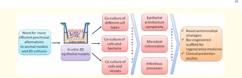
The European Union demands new valid alternatives to animal-based experimentation and to bi-dimensional flat cell culture systems. 3D in vitro epithelial models are opening new in vitro frontiers for the comprehension of a plethora of topics of microbiological inter-est, such as epithelia homeostasis, bacteria skin/mucosa colonization/infection, replicative program of epitheliotropic viruses, antimicro-bial strategies, wound healing and anti-microantimicro-bial tissue regeneration applications.
Figure 1: Schematic representation of 3D epithelial models applications.
Discussion and Conclusion
There is a general interest towards in vitro reconstructed 3D epithelial models and their applicability in in vitro studies, especially for pre-clinical purposes.
In fact, they are demonstrating to be suitable also for several microbiological applications, starting from their capability to allow the bacteria to grow and form biofilm on the cornifying layers, to invade the epithelial strata or to evaluate keratinocytes activation, prolifera-tion or differentiaprolifera-tion and proinflammatory cytokines release.
Although each 3D epithelial culture needs particular optimization protocols which seems to be time consuming, they really represent the best system to resemble the in vivo situation.
Moreover, since they can be realized with patients derived human cells, they can allow to get closer to a personalized medicine and in fact, since they can be treated with antimicrobial agents, they can allow to test the same drugs delivered to the corresponding patient. Overall, these models can be functionally validated by analysing their morphology, viability, proliferation, cell death and tumor invasion and thus used to predict the clinical responses.
Further experiments will provide insight into the many poorly understood molecular interactions that occur between commensal or pathogenic bacteria and the surface and all the strata of the stratified human epithelial tissues.
Conflict of Interest
The author declares no conflicts of interest.
Funding
Andrea Chiesa, Diletta Francesca Squarzanti and Barbara Azzimonti were supported by “Fondazione Cariplo 2013” (prot. 2013-0954). The providers of funds played no role in study design, data collection and analysis, decision to publish or preparation of the manuscript.
Bibliography
96
Volume 8 Issue 2 May 2017
© All rights are reserved by Barbara Azzimonti., et al.
3. Azzimonti B., et al. “The epithelial-mesenchymal transition induced by keratinocyte growth conditions is overcome by E6 and E7 from HPV16, but not HPV8 and HPV38: characterization of global transcription profiles”. Virology 388.2 (2009): 260-269.
4. Egert M and Simmering R. “The Microbiota of the Human Skin”. Advances in Experimental Medicine and Biology 902 (2016): 61-81. 5. de Breij A., et al. “Three-dimensional human skin equivalent as a tool to study Acinetobacter baumannii colonization”. Antimicrobial
Agents and Chemotherapy 56.5 (2012): 2459-2464.
6. Bermudez-Brito M., et al. “In vitro cell and tissue models for studying host-microbe interactions”. British Journal of Nutrition 109.2 (2013): S27-S34.
7. Popov L., et al. “Three-Dimensional Human Skin Models to Understand Staphylococcus aureus Skin Colonization and Infection”. Frontiers in Immunology 5 (2014): 41.
8. Pinnock A., et al. “Characterisation and optimisation of organotypic oral mucosal models to study Porphyromonas gingivalis inva-sion”. Microbes and Infection 16.4 (2014): 310-319.
9. Andrei G., et al. “Epithelial raft cultures for investigations of virus growth, pathogenesis and efficacy of antiviral agents”. Antiviral Research 85.3 (2010): 431-449.
10. Rajaiya J., et al. “Novel model of innate immunity in corneal infection”. In Vitro Cellular and Developmental Biology - Animal 51.8 (2015): 827-834.
11. Andrei G., et al. “Organotypic epithelial raft cultures as a model for evaluating compounds against alphaherpesviruses”. Antimicrobial Agents and Chemotherapy 49.11 (2005): 4671-4680.
12. Bedran TB., et al. “Synergistic anti-inflammatory activity of the antimicrobial peptides human beta-defensin-3 (hBD-3) and cathe-licidin (LL-37) in a three-dimensional co-culture model of gingival epithelial cells and fibroblasts”. PLoS One 9.9 (2014): e106766. 13. Holland DB., et al. “Differential innate immune responses of a living skin equivalent model colonized by Staphylococcus epidermidis
or Staphylococcus aureus”. FEMS Microbiology Letters 290.2 (2009): 149-155.
14. Haisma EM., et al. “Inflammatory and antimicrobial responses to methicillin-resistant Staphylococcus aureus in an in vitro wound infection model”. PLoS ONE 8.12 (2013): e82800.