Abstract - Osteoporosis and avascular necrosis
(AVN) are long-lasting and debilitating
complica-tions of hematopoietic stem cell transplantation
(HSCT).
We describe the magnitude of bone loss, AVN
and impairment in osteogenic cell compartment
fol-lowing autologous (auto) and allogeneic (allo)
HSCT, through the retrospective bone damage
re-valuation of 100 (50 auto- and 50 allo-HSCT)
long-term survivors up to 15 years after transplant.
Cur-rent treatment options for the management of these
complications are also outlined.
We found that
auto- and allo-HSCT recipients
show accelerated bone mineral loss and
micro-architectural deterioration during the first years after
transplant. Bone mass density (BMD) at the lumbar
spine,
but not at the femur neck, may improve in
some patients after HSCT, suggesting more
pro-longed bone damage in cortical bone. Phalangeal
BMD values remained low for even more years,
suggesting persistent bone micro-architectural
altera-tions after transplant. The incidence of AVN was
higher in allo-HSCT recipients compared to
auto-HSCT recipients. Steroid treatment length, but not
its cumulative dose was associated with a higher
in-cidence of bone loss. Allo-HSCT recipients affected
by chronic graft versus host disease seem to be at
greater
risk of continuous bone loss and AVN
devel-opment. Reduced BMD and higher incidence of
AVN was partly related to a reduced regenerating
capacity of the normal marrow osteogenic cell
com-partment.
Our results suggest that all patients after
auto-HSCT and allo-auto-HSCT should be evaluated for their
bone status and treated with anti-resorptive therapy
as soon
as abnormalities are detected.
Keywords - osteoporosis, hemopoietic stem
cell transplantation, bisphosphonates
I. INTRODUCTION
Patients with several malignant and non-malignant
acute lymphoblastic leukemia in children, lymphoma, myeloma, major thalassemia and severe hemophilia are at risk for bone damage[1-15]. Moreover, various hemato-logical related therapies, including chemotherapy, gluco-corticoids, hormonal agents and newer targeted therapies may affect bone health resulting in long-term skeletal dis-orders, particularly osteopenia, osteoporosis, fractures and avascular necrosis (AVN) [16-23].
In the last four decades the number of autologous (au-to) and allogeneic (allo) hematopoietic stem cell trans-plantation (HSCT) in the treatment of malignant and non-malignant hematological diseases has grown together with a progressive increase of long-term survivors due to a de-crease of transplant-related mortality.
With the number of long-term survivors after HSCT pro-gressively increasing, early and late complications of this procedure have gained much more attention. Osteoporo-sis, fractures and AVN leading to pain and disability rep-resent some of more frequent complications which may worsen the quality of life of long-term survivors after HSCT [24].
The aim of this review is to describe the magnitude of bone loss and AVN following HSCT and their current management, as well as to examine the role of marrow microenvironment in the development of bone loss after HSCT.
II. METHODOLOGY
Subjects
A retrospective cohort of 100 patients who had un-dergone auto- (n=50) or allo-HSCT (n=50) for a hemato-logical malignancy, and had survived free of disease for one or more years post-HSCT, was evaluated in this study. Clinical features of the patients and underlying dis-eases are summarized in Table 1. Their age at transplanta-tion ranged between 20 and 52 years (median, 32) and their post-HSCT follow-up lasted from 1 to 15 years (me-dian, 6). Primary diseases were acute (n=44) or chronic (n=13) myeloid leukemia, Hodgkin disease (n=17), non-Hodgkin lymphoma (n=12), and multiple myeloma (n=14). Auto- and allo-HSCT groups were similar for age, body mass index (BMI), and time elapsed from transplant.
Measurement of bone mass density
Bone mass density (BMD) was measured by dual
en-Accelerated Bone Mass Senescence After Hematopoietic Stem Cell
Transplantation
Serio B1, Pezzullo L1, Fontana R1, Annunziata S1, Rosamilio R1, Sessa M1, Giudice V1, Ferrara I1, Rocco M1, De Rosa G2, Ricci P2, Tauchmanovà L1, Montuori N3, Selleri C1.
1
Hematology and Bone Marrow Transplant Center, Department of Medicine and Surgery, University of Salerno, Italy; 2 Hematology, Federico II University of Napoli, Italy;
3Department of Translational Medicine and Pediatrics, Federico II University of Napoli, Italy ([email protected])
trasonometry (P-QUS), as previously described [25]. BMD was expressed as Z scores, that assess standard de-viation (SD) of patients’ BMD compared with normal values from age and sex-matched controls, respectively. Osteopenia and osteoporosis were defined by a Z score below –1 and –2.5 SD, respectively [26, 27].
Diagnosis of AVN suspected cases was documented by magnetic resonance imaging (MRI).
Measurement of osteogenic precursors
The marrow compartment of osteogenic precursors was evaluated in 35 auto- and allo-HSCT recipients by growing colony-forming units-osteogenic cells (CFU-O), defined by their ability to express alkaline phosphates ac-tivity from enriched mesenchymal stem cells, as previous-ly described [28].
Statistical analysis
Statistical analysis of the data (expressed as mean ± SD and ± SEM as appropriate throughout the text and in figures) was performed using paired students t-tests for non-parametric variables. Statistical significance was con-sidered for p < 0.05.
III. RESULTS
Bone mineral density in transplanted patients
At the time of testing (mean follow-up after HSCT: 65 months; range: 1-15 years), lumbar spine (Z score mean value: -0.4 and -0.9 in auto- and in allo-HSCT recipients, respectively; p<0.05), femoral neck (0.6 and -1.4 in auto- and in allo-HSCT recipients, respectively;
p<0.05) and phalanges (-1 and -1.5 in auto- and in
allo-HSCT recipients, respectively; p<0.05) BMD were signif-icantly reduced in comparison with BMD of 100 healthy controls (p<0.001 in all examined sites) (Figure 1).
By DEXA and P-QUS, we could not document sig-nificant correlation between BMD reduction and age at the time of HSCT, cumulative dose of prednisone and cy-closporine A, length of steroid treatment and follow-up after BMT (Table 1). Development of acute and chronic graft versus-host disease (GVHD), requiring long lasting high dose steroid treatment, was associated with a more severe BMD reduction in all bone sites (<0.05).
TABLE I
PATIENTS CHARACTERISTICS
Hematopoietic stem cell transplant Controls Allogeneic Autologous Number 50 50 100 Age (years) 32±12* 41±12 36±13 Female/Male 23/27 21/29 43/57 Diagnosis: AML CML HD NHL MM 30 13 2 2 3 14 - 15 10 11 Follow-up (months) 17.5±17 16.9±11 - BMI (kg/m2) 25±5.7 26±4.8 25.3±5 Conditioning BUCY2/ BEAM/TTCY MEL BUCY2/ BEAM/TTCY MEL - Steroid dose (g/m2) duration (months) CsA dose (g/m2) duration (months) 7.6±3.4 11.7±9.3°* 14±10 8±8 7.8±3.5 6.9±6 - - - - - Abbreviations: AML, Acute Myeloid Leukemia; CML, Chronic Myeloid Leukemia; HD, Hodgkin’ Disease; NHL, Non Hodgkin Lymphoma; MM, Multiple Myeloma; BMI, Body Mass Index; BU-CY2, Busulfan – Cyclophosphamide; BEAM, Carmustine - Etoposide – Cytarabine - Melphalan; TTCY, ThioTepa – Cyclophosphamide; MEL, Melphalan; CsA, Cyclosporine A. Symbols: °, expressed as prednisone equiva-lents.;*, p<0.05 vs autologous stem cell transplantation.
Fig. 1. Bone mass evaluation in transplanted patients versus normal controls.
Mean values of Z score (expressed as mean ± SD) at lumbar spine (L1-L4), femoral neck and phalanges, in
allogeneic and autologous hemopoietic stem cell transplantation (auto- and allo-HSCT) recipients. Each column represents the mean values of Z scores, vertical bars represent the standard error of mean.
Auto- and allo- HSCT recipients who were evaluated > 3 years after transplant had significantly higher (p< 0.001) lumbar BMD than patients evaluated < 3 years. On the other hand, no difference was seen in HSCT recipients evaluated before or after 3 years since transplant at the femoral neck and phalanges (data not shown). Phalangeal BMD values remained low even after more than 6 years (data not shown).
Avascular necrosis in transplanted patients
Eight patients developed AVN, 1 to 15 years (median, 28) following HSCT: 6 (12%) after allo-HSCT and 2 (4%) after auto-HSCT (p<0.05). A total of 15 joints were affected and all patients had femoral head involvement, which required surgical management in 4 of them. All but one patient affected by AVN in the allogeneic setting were suffering from chronic extensive GVHD; its exacer-bation or recurrence was documented in 3/6 patients shortly (1-6 months) before the AVN diagnosis. Concern-ing possible risk factors, AVN was related to allogeneic type of transplant, chronic extensive GVHD, longer ster-oid treatment and higher cumulative sterster-oid dose (data not shown).
Fig. 2. Number of colony forming units-osteogenic progenitors (CFU-O) in hematopoietic stem cell transplantation (HSCT)
recipi-ents and normal controls.
Each orange and red dot represents an auto- and allo-HSCT recipient. Horizontal bars represent mean values, vertical bars represent the
stand-ard error of mean.
Osteogenic compartment in transplanted patients
The marrow compartment of osteogenic precursors, measured as CFU-O cells, was decreased 2 to 3- fold in auto- and allo-transplanted recipients compared to normal donors (30±6 and 21±5 in auto- and allo-HSCT recipients, respectively, vs 55±4/105 cells plated in normal controls; all p<0.001) (Figure 2). Analyzing the effect of the time elapsed since HSCT on marrow CFU-O numbers, we found the marrow CFU-O compartment markedly deplet-ed during the first years after transplant (data not shown). Between 6 and 15 years, the mean marrow CFU-O cell number tended to increase (13 ±3 vs 33 ±5 before and
af-served in normal controls. CFU-O growth correlated sig-nificantly with lumbar, femoral and phalangeal bone loss (all p<0.01). Chronic GVHD was correlated with a lower number of CFU-O colonies in vitro (p<0.01).
Finally, almost all transplanted patients who had AVN showed a number of CFU-O below that observed in transplanted patients without this complication (CFU-O: 24.5±3 vs 12.4±4 in HSCT patients without and with os-teonecrosis, respectively; p<0.05) (Figure 3).
Fig. 3. Number of colony-forming units-osteogeneic progenitors (CFU-O) in auto- (orange dot) and allo (red dot) hemopoietic stem cell transplantation (HSCT) recipients with and without
osteonecro-sis.
Each dot represents a subject studied. Horizontal bars represent mean values, vertical bars represent the standard error of mean.
IV. DISCUSSION
Osteopenia and osteoporosis are relatively common early complications of auto- and allo-HSCT. They are at-tributable to the influence of multiple factors including myeloablative conditioning regimens, huge cytokine re-lease at the time of transplant, altered kidney, liver and bowel function resulting in reduced intake and altered me-tabolism of calcium and vitamin D, frequent gonadal fail-ure and, in allogeneic HSCT setting, long-lasting high-dose steroids and cyclosporin-A [29, 30]. In the current study, we retrospectively followed 100 consecutive pa-tients who had undergone auto- and allo-HSCT and sur-vived one or more years. As already suggested by our and other studies, we documented a marked decrease in BMD after auto- and allo-HSCT both at the lumbar spine (25%) and even more at the femoral neck (50%). We have con-firmed that a significant decrease in BMD at lumbar and femoral neck level appears early after transplant and seems to continue over the first 3 years with no further deterioration afterwards.
Although bone density is among the strongest predic-tors of the mechanical behavior of trabecular bone, the whole bone strength is determined also by bone quality. Apart from bone mineralization, bone architecture, turno-ver, and damage accumulation also account for bone
qual-influences trabecular
bone strength.
Early bone loss may consist of both demineralization and architectural damage with associated organic matrix deficit. DEXA measures bone density and mineralization but does not provide in-formation on architectural damage and bone in-formation [31]. High-resolution computed tomography and MRI al-low for three-dimensional assessment of trabecular struc-ture, but their use in the routine clinical practice for diag-nosis and follow-up of bone damage is limited, being too costly and time-consuming. Ultrasonographic evaluation by phalangeal QUS is a safe and less expensive proce-dure, which permits to assess more physical properties of bone tissue and to account for more structural changes than DEXA [32]. Phalangeal QUS allows to evaluate bone density and elasticity, trabecular orientation and cor-tical-to-trabecular ratio, all of which are influenced by mineral content and organic matrix. In HSCT recipients, we have further confirmed by DEXA that, while mineral-ization seems to improve at trabecular rich sites such as lumbar spine, no improvement was detected at cortical bone such as femoral neck; in addition, no improvement was revealed by phalangeal QUS even several year after transplant.Allo- and auto-HSCT recipients were mostly pooled together in several clinical studies on bone complications after HSCT, whereas there are considerable differences between these two settings. The differences consist in a higher grade of immunologic derangement and more pro-longed use of immunosuppressive treatments needed to avoid development of acute and chronic GVHD in the al-logeneic setting [27]. Apart from women which may ex-perience ovarian failure after auto- and allo-HSCT, in the setting of allogeneic HSCT, we have also documented that an important high risk factor is the development of chronic GVHD requiring prolonged high-dose of steroids [33-37].
Avascular necrosis (AVN) has been described in 3-41% of patients who had received an organ transplant, with femoral head being the prevalent localization [38]. AVN is the result of multiple triggering factors such as metabolic disorders, local vascular damage with transient or permanent loss of blood supply to the bone, increased intra-osseous pressure and mechanical stress leading to demineralization, and death of trabecular bone and col-lapse.
The process is mostly progressive, resulting in joint destruction within three to five years if left untreated. AVN occurred in 8 of 100 long-term survivors (8%), within 3-15 years (median, 3.6) after-HSCT, all but two of them having received an allo-HSCT. Surgical treatment was required in half of HSCT patients because of func-tional limitations. A significant statistical association was found between AVN occurrence, allogeneic type of trans-plant, presence and grade of GVHD, steroid treatment length, and cumulative dose [17, 39]. In addition, we have recently reported a close relationship between AVN oc-currence and flare up of chronic GVHD [38].
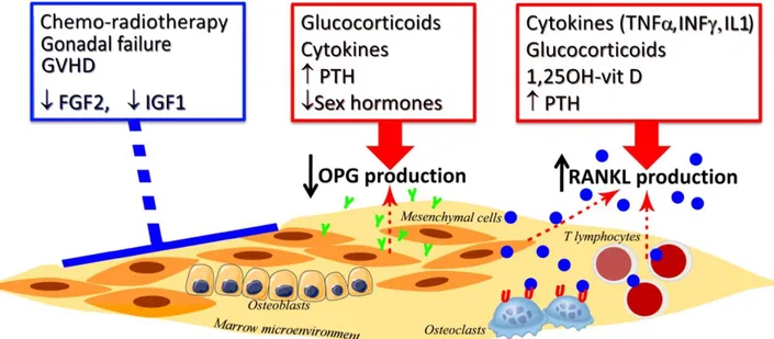
Bone remodeling, specifically the balance between formation and resorption, is the biologic process that me-diates architectural changes influencing the whole bone strength. We have documented a marked and permanent quantitative and qualitative defect in the marrow osteo-genic cell compartment, suggesting that inability to re-store a normal number of osteoblastic precursors in the bone microenvironment may account at least in part for severe bone damage after auto- and allo-HSCT. The re-duced repopulating capacity of osteoblast precursors after HSCT is likely related to the effects of chemothera-py/radiotherapy,to the concomitant endocrine disorders, immunosuppressive treatments,and alteration in the bal-ance of cytokines and growth factors. According to the above described long-lasting deficit of osteogenic progen-itors, we have recently documented within the bone mar-row microenvironment of these HSCT recipients low os-teoprotegerin (OPG) levels, exacerbated by a high ratio for receptor activator for nuclear kappa B ligand (RANKL)/OPG favouring bone resorption after HSCT (Figure 4) [40].
Few data are available so far on the treatment of bone loss in HSCT recipients. Early diagnosis of osteoporosis or early bone senescence in this particular population is a major aim to promptly start appropriate supportive measures, such as lifestyle modification, calcium and vit-amin D supplementation or bisphosphonates. However, there is sufficient evidence that common preventive measures for bone loss, such as calcium and vitamin D supplements and sex steroid replacement, are ineffective in HSCT recipients. Bisphosphonates are currently the most effective inhibitors of osteoclastic bone resorption; they directly impair the ability of osteoclasts to adhere to the bone surface, and inhibit osteoclast activity by de-creasing osteoclast progenitor development and recruit-ment and by promoting osteoclast apoptosis. In addition, recent evidences have been accumulated on the anabolic bone effects of bisphosphonates, enhancing osteoblast proliferation, and preventing osteoblast apoptosis [41-43]. Oral bisphosphonates, widely used for treating osteoporo-sis, have been shown to improve bone BMD and decrease the rate of fractures in various patient populations [44]. However, the use of these drugs in the clinical routine of HSCT recipients is limited by their poor gastrointestinal tolerance, variable oral bioavailability and long-term compliance. We have previously reported that zoledronic acid, given as three consecutive monthly doses of 4 mg each every years, is able to obtain a rapid and measurable increase of bone mass. Given the persistent bone loss at trabecular and cortical rich skeletal sites in HSCT recipi-ents, zoledronic acid may be considered the treatment of choice in the near-transplant period for the prevention of bone loss. We also documented that beneficial effects on lumbar and femoral BMD of zoledronic acid treatment in HSCT patients were associated with a significant im-provement of the osteogenic progenitors [28].
Fig. 4. Critical factors involved in bone remodeling after hemopoietic stem cell transplantation.
Chemotherapy, radiotherapy, gonadal failure, acute and chronic graft versus host disease (GVHD) and growth factors (such as fibroblast growth factor-2 -FGFfactor-2-, insulin growth factor-1 -IGF1-) may affect directly bone marrow microenvironment altering the capacity to synthesize osteoprotegerin (OPG) and receptor activator for nuclear kappa-B ligand (RANKL). In particular, high dose of steroids, cytokines, high levels of parathyroid hormone (PTH) and low levels of sexual hormones decrease OPG production by osteoblastic progenitors, whereas pro-inflammatory cytokines (such as
tumor-necrosis factor-α -TNFα-, interleukin-1 -IL1- and interferon-γ -IFNγ -), long-lasting glucocorticoids, 1,25OH-vitamin D and PTH increase RANKL production by osteoblasts and T lymphocytes. All of these marrow microenvironment changes may increase RANKL/OPG ratio stimulating
osteoclas-tic activity and bone resorption. Symbols: Y, OPG; blue O , RANKL; U, RANK. In addition, we have recently documented in a small
co-hort of HSCT recipients that 1-year treatment with risedronate increases lumbar spine BMD and prevents bone loss at the femoral neck [44]. Clinical experience with the human monoclonal antibody anti-RANKL (deno-sumab), which is documented to be effective in non meta-static prostate and breast cancer as well as in postmeno-pausal osteoporosis, as well as with the parathyroid hor-mone derived peptides, with horhor-mone analogues and se-lective estrogen receptor modulators, is still limited in HSCT recipients [30, 45, 46].
V. CONCLUSIONS
In this study, we have confirmed that an accelerated and persistent multifactorial bone loss occurs in long-term survivors after HSCT, being more severe in the allogeneic setting. Moreover, osteoporosis is more frequent than AVN in patients after allo-HSCT. Several new risk fac-tors have been identified and includea dysregulation of the immune system occurring during acute and chronic GVHD and their treatments, persistently reduced regener-ating capacity of normal osteogenic cell compartment, and osteoclastic activation by alterations in the RANKL/ OPG balance.
DEXA bone density testing remains an accurate method to detect bone loss in HSCT recipients. Since phalangeal QUS is a safe and easy method, it should be more frequently used for early recognition and monitoring of BMD reduction as well as for decision-making in transplanted patients who may need specific therapy. Oral
treatments able to prevent and treat accelerated bone mass senescence after transplant. The choice between oral or intravenous bisphosphonate therapy in patients who had undergone HSCT should be made on the basis of individ-ual clinical conditions, including presence, grading and localization of GVHD as well as prevalent site of bone loss. Oral risedronate and intermittent short course of zoledronic acid were easily manageable and effective in increasing densitometric values at trabecular skeletal sites. Zoledronic acid was the only therapy that improved also femoral BMD. At least part of these effects may be related to the bisphosphonates’ ability to partly restore the persistent post-transplant reduction of osteogenic progeni-tors. Despite the evidence of onlypartial effectiveness of preventive anti-resorptive treatments,it is reasonable to start administration of these drugs beforeor at the time of HSCT in all patients, as soonas bone abnormalities are detected, and continue them at least for thefirst years after transplant.
ACKNOWLEDGMENT
We are grateful to our research nursing staff (Pelle-grino D, Cuffa B and Pennino M) for their excellent care and professionalism.
REFERENCES
[1] McClune B, Majhail NS, Flowers ME. Bone loss and
trans-[2] Tauchmanovà L, Colao A, Lombardi G, Rotoli B, Selleri C. Bone loss and its management in long-term survivors from allogeneic stem cell transplantation. J Clin Endo-crinol Metab. 2007;92(12):4536-45.
[3] Ghosh K, Shetty S. Bone health in persons with
haemo-philia: a review. Eur J Haematol 2012;89(2):95-102.
[4] McClune BL, Polgreen LE, Burmeister LA, Blaes AH,
Mulrooney DA, Burns LJ, Majhail NS. Screening, preven-tion and management of osteoporosis and bone loss in adult and pediatric hematopoietic cell transplant recipients. Bone Marrow Transplant 2011;46(1):1-9.
[5] Follin C, Link K, Wiebe T, Moëll C, Björk J, Erfurth EM.
Bone loss after childhood acute lymphoblastic leukaemia: an observational study with and without GH therapy. Eur J Endocrinol 2011;164(5):695-703.
[6] Van Waas M, Neggers SJ, Te Winkel ML, Beishuizen A,
Pieters R, van den Heuvel-Eibrink MM. Endocrine late sequelae in long-term survivors of childhood non-Hodgkin lymphoma. Ann Oncol 2012;23(6):1626-1632.
[7] Alos N, Grant RM, Ramsay T, Halton J, Cummings EA,
Miettunen PM, Abish S, Atkinson S, Barr R, Cabral DA, Cairney E, Couch R, Dix DB, Fernandez CV, Hay J, Is-raels S, Laverdière C, Lentle B, Lewis V, Matzinger M, Rodd C, Shenouda N, Stein R, Stephure D, Taback S, Wilson B, Williams K, Rauch F, Siminoski K, Ward LM. High incidence of vertebral fractures in children with acute lymphoblastic leukemia 12 months after the initia-tion of therapy. J Clin Oncol 2012;30(22):2760-2767.
[8] Rayar MS, Nayiager T, Webber CE, Barr RD, Athale UH.
Predictors of bony morbidity in children with acute lym-phoblastic leukemia. Pediatr Blood Cancer 2012;59(1):77-82.
[9] Muszynska-Roslan K, Panasiuk A, Latoch E,
Krawczuk-Rybak M, Konstantynowicz J. Little evidence of low bone mass in acute lymphoblastic leukemia survivors. J Clin Densitom 2012;15(1):108-115.
[10] C.H.Buckle, E.De Leenheer, M.A.Lawson, K.Yong, N.
Rabin, M.Perry, K.Vanderkerken, P.I.Croucher. Soluble
rank ligand produced by myeloma cells causes generalised bone loss in multiple myeloma. PLoS One “in press” 2012.
[11] Skordis N, Toumba M. Bone disease in thalassaemia ma-jor: recent advances in pathogenesis and clinical aspects. Pediatr Endocrinol Rev 2011;8(2):300-306.
[12] Forni GL, Perrotta S, Giusti A, Quarta G, Pitrolo L, Cap-pellini MD, D'Ascola DG, Borgna Pignatti C, Rigano P, Filosa A, Iolascon G, Nobili B, Baldini M, Rosa A, Pinto V, Palummeri E. Neridronate improves bone mineral den-sity and reduces back pain in β-thalassaemia patients with osteoporosis: results from a phase 2, randomized, parallel-arm, open-label study. Br J Haematol 2012;158(2):274-282.
[13] Tauchmanovà L, Selleri C, De Rosa G, Esposito M, Di Somma C, Orio F, Palomba S, Lombardi G, Rotoli B, Co-lao A. Endocrine disorders during the first year after au-tologous stem-cell transplant. Am J Med 2005;118(6):664-670.
[14] Tauchmanovà L, Selleri C, Rosa GD, Pagano L, Orio F, Lombardi G, Rotoli B, Colao A. High prevalence of endo-crine dysfunction in long-term survivors after allogeneic bone marrow transplantation for hematologic diseases. Cancer 2002;95(5):1076-1084.
[15] Tauchmanovà L, Colao A, Selleri C, De Rosa G, Rotoli B. Thyroid dysfunction after autologous hematopoietic stem cell transplant. Am J Med 2006;119(6):5-6.
[16] Unal A, Kocyigit I, Sipahioglu MH, Tokgoz B, Kavun-cuoglu F, Oymak O, Utas C. Loss of bone mineral density
in renal transplantation recipients. Transplant Proc 2010;42(9):3550-3553.
[17] McAvoy S, Baker KS, Mulrooney D, Blaes A, Arora M,
Burns LJ, Majhail NS. Corticosteroid dose as a risk factor for avascular necrosis of the bone after hematopoietic cell
transplantation. Biol Blood Marrow Transplant
2010;16(9):1231-1236.
[18] Selleri C, Serio B, Risitano AM. Novel immunosuppres-sive strategies for bone marrow failure syndromes: a focus on alemtuzumab. Mini Rev Med Chem 2011;11:536-543. [19] Tauchmanovà L, Selleri C, De Rosa G, Sammartino A, Di
Carlo C, Musella T, Martorelli C, Lombardi G, Rotoli B, Nappi C, Colao A. Estrogen-progestin therapy in women after stem cell transplant: our experience and literature re-view. Menopause 2007;14(2):320-330.
[20] Tauchmanovà L, Selleri C, Di Carlo C, De Rosa G, Biful-co G, Sammartino A, Lombardi G, Colao A, Rotoli B, Nappi C. Estrogen-progestogen induced hematocolpomet-ra following allogeneic stem cell thematocolpomet-ransplant. Gynecol On-col 2004;93(1):112-115.
[21] Vandyke K, Fitter S, Drew J, Fukumoto S, Schultz CG, Sims NA, Yeung DT, Hughes TP, Zannettino AC. Pro-spective Histomorphometric and DXA Evaluation of Bone Remodeling in Imatinib-Treated CML Patients: Evidence for Site-Specific Skeletal Effects. J Clin Endocrinol Metab “in press” 2012.
[22] Serio B, Rosamilio R, Giudice V, Pepe S, Zeppa P, Espos-ito S, Pezzullo L, Rocco M, Montuori N, Selleri C. Low-dose valgancyclovir as cytomegalovirus reactivation prophylaxis in allogeneic haematopoietic stem cell trans-plantation. Infez Med 2012;1(20):26-34.
[23] Risitano AM, Notaro R, Pascariello C, Sica M, Del Vec-chio L, Horvath CJ, Fridkis-Hareli M, Selleri C, Lindorfer MA, Taylor RP, Luzzatto L, Holers VM. The complement receptor 2/factor H fusion protein TT30 protects paroxys-mal nocturnal hemoglobinuria erythrocytes from comple-ment-mediated hemolysis and C3 fragment. Blood 2012;119(26):6307-6316.
[24] Ebeling PR. Approach to the patient with
transplantation-related bone loss. J Clin Endocrinol Metab
2009;94(5):1483-1490.
[25] Albanese CV, De Terlizzi F, Passariello R. Quantitative ultrasound of the phalanges and DXA of the lumbar spine and proximal femur in evaluating the risk of osteoporotic vertebral fracture in postmenopausal women. Radiol Med 2011;116(1):92-101.
[26] Serio B, Rosamilio R, Giudice V, Zeppa P, Esposito S, Fontana R, Annunziata S, Selleri C. Successful manage-ment of pulmonary mucormycosis with liposomal ampho-tericin B and surgery treatment: a case report. Infez Med 2012;1(20):43-47.
[27] Czerwiński E, Badurski JE, Marcinowska-Suchowierska E, Osieleniec J. Current understanding of osteoporosis ac-cording to the position of the World Health Organization (WHO) and International Osteoporosis Foundation. Ortop Traumatol Rehabil 2007;9(4):337-356.
[28] Tauchmanovà L, Ricci P, Serio B, Lombardi G, Colao A, Rotoli B, Selleri C. Short-term zoledronic acid treatment increases bone mineral density and marrow clonogenic fi-broblast progenitors after allogeneic stem cell transplanta-tion. J Clin Endocrinol Metab 2005;90(2):627-634. [29] Weilbaecher KN. Mechanisms of osteoporosis after
hema-topoietic cell transplantation. Biol Blood Marrow Trans-plant 2000;6(2A):165-174.
[30] Hautmann AH, Elad S, Lawitschka A, Greinix H, Bertz H, Halter J, Faraci M, Hofbauer LC, Lee S, Wolff D, Holler E. Metabolic bone diseases in patients after allogeneic
hematopoietic stem cell transplantation: report from the Consensus Conference on Clinical Practice in chronic graft-versus-host disease. Transpl Int 2011;24(9):867-879.
[31] Karaguzel G, Holick MF. Diagnosis and treatment of
os-teopenia. Rev Endocr Metab Disord 2010;11(4):237-251.
[32] Tauchmanovà L, Serio B, Del Puente A, Risitano AM, Esposito A, De Rosa G, Lombardi G, Colao A, Rotoli B, Selleri C. Long-lasting bone damage detected by dual-energy x-ray absorptiometry, phalangeal osteosonogram-metry, and in vitro growth of marrow stromal cells after allogeneic stem cell transplantation. J Clin Endocrinol Metab 2002;87(11):5058-5065.
[33] Blazar BR, Murphy WJ, Abedi M. Advances in graft-versus-host disease biology and therapy. Nat Rev Immu-nol 2012;12(6):443-458.
[34] Tauchmanovà L, Matarese G, Carella C, De Rosa G, Serio B, Ricci P, Lombardi G, Rotoli B, Colao A, Selleri C. High serum leptin in patients with chronic graft-versus-host disease after hematopoietic stem cell transplantation. Transplantation 2004;78(9):1376-1383.
[35] Tauchmanovà L, Selleri C, De Rosa G, Esposito M, Orio F Jr, Palomba S, Bifulco G, Nappi C, Lombardi G, Rotoli B, Colao A. Gonadal status in reproductive age women af-ter haematopoietic stem cell transplantation for haemato-logical malignancies. Hum Reprod 2003;18(7):1410-1416.
[36] Tauchmanovà L, De Simone G, Musella T, Orio F, Ricci P, Nappi C, Lombardi G, Colao A, Rotoli B, Selleri C. Ef-fects of various antireabsorptive treatments on bone min-eral density in hypogonadal young women after allogeneic stem cell transplantation. Bone Marrow Transplant 2006;37(1):81-88.
[37] Tauchmanovà L, Alviggi C, Foresta C, Strina I, Garolla A, Colao A, Lombardi G, De Placido G, Rotoli B, Selleri C. Cryptozoospermia with normal testicular function after al-logeneic stem cell transplantation: a case report. Hum Re-prod 2007;22(2):495-499.
[38] Tauchmanovà L, De Rosa G, Serio B, Fazioli F, Mainolfi C, Lombardi G, Colao A, Salvatore M, Rotoli B, Selleri C. Avascular necrosis in long-term survivors after allogeneic or autologous stem cell transplantation: a single center ex-perience and a review. Cancer 2003;97(10):2453-2461. [39] Campbell S, Sun CL, Kurian S, Francisco L, Carter A,
Kulkarni S, Parker P, Karanes C, Forman SJ, Bhatia S. Predictors of Avascular Necrosis of Bone in Long-term
Survivors of Hematopoietic Cell Transplantation. Cancer
2009;115(18):4127–4135.
[40] Ricci P, Tauchmanova L, Risitano AM, Carella C, Maz-ziotti G, Lombardi G, Colao A, Rotoli B, Selleri C. Imbal-ance of the osteoprotegerin/RANKL ratio in bone marrow microenvironment after allogeneic hemopoietic stem cell transplantation. Transplantation 2006;82(11):1449-1456. [41] Drake MT, Clarke BL, Khosla S. Bisphosphonates:
mech-anism of action and role in clinical practice. Mayo Clin Proc 2008;83(9):1032-1045.
[42] Kimmel DB. Mechanism of action, pharmacokinetic and pharmacodynamic profile, and clinical applications of
ni-trogen-containing bisphosphonates. J Dent Res
2007;86(11):1022-1033.
[43] Kavanagh KL, Guo K, Dunford JE, Wu X, Knapp S, Ebetino FH, Rogers MJ, Russell RG, Oppermann U. The molecular mechanism of nitrogen-containing bisphospho-nates as antiosteoporosis drugs. Proc Natl Acad Sci USA 2006;103(20):7829-7834.
[44] Tauchmanovà L, Selleri C, Esposito M, Di Somma C, Orio F Jr, Bifulco G, Palomba S, Lombardi G, Rotoli B, Colao A. Beneficial treatment with risedronate in long-term survivors after allogeneic stem cell transplantation
for hematological malignancies. Osteoporos Int
2003;14(12):1013-1019.
[45] Yee AJ, Raje NS. Denosumab, a RANK ligand inhibitor, for the management of bone loss in cancer patients. Clin Interv Aging 2012;7(9):331-338.
[46] Cummings SR, San Martin J, McClung MR, Siris ES, Eastell R, Reid IR, Delmas P, Zoog HB, Austin M, Wang A, Kutilek S, Adami S, Zanchetta J, Libanati C, Siddhanti S, Christiansen C, for the FREEDOM Trial, Denosumab for prevention of Fractures in Postmenopausal Women