Research Article
Acute Footshock Stress Induces Time-Dependent
Modifications of AMPA/NMDA Protein Expression and
AMPA Phosphorylation
Daniela Bonini,
1Cristina Mora,
1Paolo Tornese,
2Nathalie Sala,
2Alice Filippini,
1Luca La Via,
1Marco Milanese,
3Stefano Calza,
4Gianbattista Bonanno,
3Giorgio Racagni,
2Massimo Gennarelli,
1,5Maurizio Popoli,
2Laura Musazzi,
2and Alessandro Barbon
11Biology and Genetic Division, Department of Molecular and Translational Medicine, University of Brescia, 25123 Brescia, Italy
2Laboratorio di Neuropsicofarmacologia e Neurogenomica Funzionale, Dipartimento di Scienze Farmacologiche e Biomolecolari and
CEND, Universit`a degli Studi di Milano, 20133 Milano, Italy
3Department of Pharmacy, Unit of Pharmacology and Toxicology and Center of Excellence for Biomedical Research,
University of Genoa, 16148 Genoa, Italy
4Unit of Biostatistics and Biomathematics, Department of Molecular and Translational Medicine, University of Brescia, Brescia, Italy
5Genetic Unit, IRCCS Centro San Giovanni di Dio Fatebenefratelli, 25125 Brescia, Italy
Correspondence should be addressed to Alessandro Barbon; [email protected] Received 25 September 2015; Revised 21 December 2015; Accepted 10 January 2016 Academic Editor: Zygmunt Galdzicki
Copyright © 2016 Daniela Bonini et al. This is an open access article distributed under the Creative Commons Attribution License, which permits unrestricted use, distribution, and reproduction in any medium, provided the original work is properly cited. Clinical studies on patients with stress-related neuropsychiatric disorders reported functional and morphological changes in brain areas where glutamatergic transmission is predominant, including frontal and prefrontal areas. In line with this evidence, several preclinical works suggest that glutamate receptors are targets of both rapid and long-lasting effects of stress. Here we found that acute footshock- (FS-) stress, although inducing no transcriptional and RNA editing alterations of ionotropic AMPA and NMDA glutamate receptor subunits, rapidly and transiently modulates their protein expression, phosphorylation, and localization at postsynaptic spines in prefrontal and frontal cortex. In total extract, FS-stress increased the phosphorylation levels of GluA1 AMPA subunit at Ser845immediately after stress and of GluA2 Ser8802 h after start of stress. At postsynaptic spines, stress induced a rapid decrease of GluA2 expression, together with an increase of its phosphorylation at Ser880, suggesting internalization of GluA2 AMPA containing receptors. GluN1 and GluN2A NMDA receptor subunits were found markedly upregulated in postsynaptic spines, 2 h after start of stress. These results suggest selected time-dependent changes in glutamatergic receptor subunits induced by acute stress, which may suggest early and transient enhancement of AMPA-mediated currents, followed by a transient activation of NMDA receptors.
1. Introduction
Stress can be defined as any condition that perturbs the phys-iological homeostasis [1]. A stressful event rapidly activates both the hypothalamic-pituitary-adrenocortical axis, leading to secretion of glucocorticoids (mainly cortisol in humans, corticosterone in rats), and the autonomic nervous system, which releases catecholamines (noradrenaline, adrenaline). The stress response is physiologically proadaptive, when
efficiently turned on and then shut off, but may became mal-adaptive, particularly in subjects with a genetic background of vulnerability or when the stressful stimulus is chronic or overwhelming [2, 3].
The prefrontal cortex (PFC), a region involved in work-ing memory, decision-makwork-ing, and behavioral flexibility, as well as in social interaction and emotional processing, is a main target of the stress hormones [4–6]. A large body of literature has consistently shown that the fast response to Volume 2016, Article ID 7267865, 10 pages
stress involves increased attention, vigilance, and improved PFC-mediated cognitive performance, mainly mediated by potentiation of glutamate transmission [7–9]. Indeed, acute stress and glucocorticoids rapidly modulate glutamate release and excitatory synaptic transmission in PFC [8, 10–12]. In particular, it has been shown that acute stress induces a rapid and transient enhancement of
N-methyl-D-aspartic acid- (NMDA-) and
𝛼-amino-3-hydroxy-5-methyl-4-isoxazolepropionic acid- (AMPA-) receptor-mediated cur-rents in PFC in juvenile rats, together with increasing the surface expression of AMPA and NMDA receptor subunits [10, 11].
Taken together, these data strongly suggest that the en-richment, localization, and posttranslational modifications, as well as posttranscriptional and translational regulations of glutamate receptors, may be involved in the neuronal response to behavioral stress.
In this study, we exposed adult male rats to acute foot-shock- (FS-) stress and investigated time-dependent mod-ifications of AMPA and NMDA receptor subunits mRNA and protein expression, RNA editing, and posttranslational regulation. The analyses have been performed in the pre-frontal and pre-frontal cortex (PFC/FC) at different time points (immediately after the 40 min of stress and 2 hours and 24 hours after stress start), to monitor the early and delayed effects of acute stress on the regulatory mechanisms of ionotropic glutamate receptors.
The results provided here indicate that exposure to acute stress causes transient and time-dependent subunit-specific changes in glutamate receptor, in line with previously observed adaptive modifications of excitatory synaptic trans-mission in the PFC/FC.
2. Materials and Methods
2.1. Footshock Stress Procedure. All experimental procedures
involving animals were performed in accordance with the European Community Council Directive 86/609/EEC and were approved by the Italian legislation on animal experi-mentation (Decreto Ministeriale 116/1992). Experiments were performed with adult male Sprague-Dawley rats (275–300 g). Rats were housed two per cage and maintained on a 12/12 h light/dark schedule (lights on at 7:00 am), in a temperature controlled facility with free access to food and water. The experiments were performed during the light phase (between 9:00 and 12:00 am), at least one week after arrival from the supplier (Charles River, Wilmington, MA, USA). The footshock- (FS-) stress protocol was performed essentially as previously reported (40 min FS-stress: 0.8 mA, 20 min total of actual shock with random intershock length between 2 and 8 sec) [8, 12, 13]. Sham-stressed rats (controls) were kept in the stress apparatus without delivering of shocks. Rats were killed by decapitation at different time points (10 rats/group): immediately after the stress session (𝑡 = 0) and 2 or 24 h after stress start. The 2 and 24 h groups were left undisturbed in their cages after the 40 min stress session. Sham-groups were prepared at each time point, as specific controls for respective stressed groups.
The whole frontal lobe, referred to as PFC/FC, was quickly dissected on ice and right and left hemiareas were randomly assigned to RNA extraction or postsynaptic spine membranes (triton insoluble fraction; TIF) purification.
Serum corticosterone levels were measured using a com-mercial kit (Corticosterone ELISA kit, Enzo Life Sciences, Farmingdale, NY, USA).
2.2. RNA Extraction and Retrotranscription. Samples from
PFC/FC of each animal were homogenized, and total RNA was extracted using TRIZOL reagent (Life Technologies, Milano, Italy). RNA was recovered by precipitation with isopropyl alcohol, washed with a 75% ethanol solution, and dissolved in RNase-free water. RNA quantification and quality controls were carried out using both spectrophoto-metric analysis and AGILENT Bioanalyzer 2100 lab-on-a-chip technology (AGILENT Technologies, Santa Clara, CA, USA). Reverse-transcription (RT) was done using Moloney murine leukemia virus-reverse transcriptase (MMLV-RT)
(Life Technologies). Briefly, 2.5𝜇g of total RNA from each
sample was mixed with 2.2𝜇L of 0.2 ng/𝜇L random hexamers,
10𝜇L of 5x buffer, 10 𝜇L of 2 mM dNTPs, 1 𝜇L of 1 mM DTT,
0.4𝜇L of 33 U/𝜇L RNaseout, and 2 𝜇L MMLV-RT (200 U/𝜇L)
in a final volume of 50𝜇L. The reaction mix was incubated at
37∘C for 2 h and the enzyme was then heat inactivated at 95∘
for 10 min.
2.3. Quantitative Real-Time PCR and RNA Editing Quantifica-tion. RNA expression pattern of the glutamate receptors was
analyzed by means of an Applied Biosystems 7500 Real-Time PCR system (Applied Biosystems, Foster City, CA, USA). PCR was carried out by using TaqMan Universal PCR Master Mix (Applied Biosystems). 25 ng of sample was used in each real-time PCR reaction (TaqMan Gene Expression Assay id probes: GluA1: Rn00709588 m1; GluA2: Rn00568514 m1; GluN1: RN01436038 m1; GluN2A: Rn00561341 m1; GluN2B: Rn00561352 m1, Applied Biosystems). The expression ratio of target genes in treated sample groups, compared to
control group, was calculated using theΔΔCt method and
H2AFZ, GAPDH, and PolII geometric mean as reference (ID H2AFZ TaqMan probe: Rn00821133 g1; ID GAPDH: Rn99999916 m1; ID PolII: Rn00580118 m1). Each individual determination was repeated in triplicate. The quantification for AMPA receptor subunits GluA2 Q/R and GluA2, GluA3, and GluA4 R/G editing levels were measured by sequence analysis as previously described [14, 15].
2.4. Protein Extracts and Western Blotting. PFC/FC were
homogenized in 0.32 M ice-cold sucrose containing 1 mM
HEPES, 1 mM MgCl2, 1 mM EDTA, 1 mM NaHCO3, and
0.1 mM PMSF, at pH 7.4, 2 mg/mL of protease inhibitor cock-tail (Thermo scientific, Rockford, IL, USA), and phosphatases
inhibitors (Sigma-Aldrich, Milan, Italy), pH 7.4. 200𝜇L of
homogenate was aliquoted and immediately frozen.
Triton-X-100 insoluble fractions (TIF) were purified as previously reported [12]. The homogenized tissue was
cen-trifuged at 1000×g for 10 min. The resulting supernatant (S1)
Table 1: Corticosterone serum levels.
𝑡 = 0 2 h 24 h
Control 60.32± 13.06 (𝑛 = 10) 11.85± 3.82 (𝑛 = 11) 21.53± 6.93 (𝑛 = 9) FS-stress 308.30± 23.30∗∗∗(𝑛 = 10) 57.20± 17.34∗∗∗(𝑛 = 11) 53.63± 13.41 (𝑛 = 9)
Data are expressed as ng/mL and reported as mean± SE.∗∗∗𝑝 < 0.001.
membrane fraction (P2 fraction). The pellet was resuspended
in 1 mM HEPES and centrifuged at 100,000×g for 1 h. The
pellet (P3) was resuspended in buffer containing 75 mM KCl
and 1% Triton-X-100 and centrifuged at 100,000×g for 1 h.
The supernatant was stored and referred to as Triton-X-100-soluble fraction (TSF) (S4). The final pellet (P4) was homogenized in 20 mM HEPES. Then, an equal volume of glycerol was added, and this fraction, referred to as TIF, was
stored at−80∘C until processing.
The BCA protein concentration assay (Sigma-Aldrich, St. Louis, MO, USA) was used for protein quantitation.
Before electrophoresis, each sample was incubated at 75∘C
for 10 min. Equal amounts of proteins were applied to precast SDS polyacrylamide gels (4–12% NuPAGEBis-Tris gels; Life Technologies, Milan, Italy), and proteins were electrophoret-ically transferred to a Hybond-P PVDF Transfer Membrane
(GE Healthcare Life Science), for 2 h at a 1 mA/cm2 of
membrane surface. Membranes were blocked for 60 min with 3–5% nonfat dry milk or 5% BSA in TBS-T (Tris-buffered saline with 0.2% Tween-20, Sigma-Aldrich, Milan,
Italy). Immunoblotting was carried out overnight at 4∘C with
specific antibodies against phosphoSer831-GluA1 (1 : 1000,
cod. ab109464, Abcam, Cambridge, UK), phosphoSer845
-GluA1 (1 : 1000, cod. ab3901, Abcam), and phosphoSer880
-GluA2 (1 : 1.000, cod. Ab52180, Abcam). Immunoblotting was also carried out on the same stripped membranes with anti-bodies against total GluA1 (1 : 200, cod. AGC004, Alomone Labs, Jerusalem, Israel) and GluA2 (1 : 2500, cod. AGC005, Alomone) in blocking buffer. Primary antibodies were used to detect GluN1 (1 : 500, cod. AB9864, Millipore, Biller-ica, MA, USA), GluN2A (1 : 500, AB1555P, Millipore), and GluN2B (1 : 500, cod. 454582, Calbiochem-Millipore). Mouse monoclonal anti-GAPDH (1 : 40.000, cod. Mab374, Milli-pore) or rabbit monoclonal anti-𝛽-Actin (1 : 3000, cod.04-1116, Millipore) were used as internal controls. Membranes were washed five times with TBS-Tween-20 0.2% and incu-bated for 1 h at room temperature with AP-conjugated secondary antibodies (Promega, Milan, Italy). Immunola-beled proteins were detected by incubation with Supersignal West Pico Chemiluminescent Substrate (Pierce, Rockford, IL, USA) or CDPStar (Roche Applied Science) detection reagents and then exposed to imaging film. Prestained Novex Sharp Protein Standards (Life Technologies) were used as molecular weight standards loaded on the same gel. The intensity of immunoreactive bands was analyzed with Image-Pro Plus. Data are presented as optical density ratios of the
investigated protein band normalized for GAPDH or𝛽-Act
bands in the same line and are expressed as percentage of controls. The levels of GluA phosphorylated subunits were
normalized to total GluA levels, based on previous reports [16, 17].
2.5. Data Analysis. All the analyses were carried out in
individual animals (independent determinations).
Preliminary data inspection showed a fairly constant coefficient of variation among groups, as well as a multiplica-tive effect on the mean. Therefore, we modeled data using a gamma regression model with log-link (via Generalized Linear Models, GLM [18]), with treatment (stress, control), time, and their interaction as predictors. Where needed, a robust Generalized Linear Model was used to account for potential outliers. Due to some additional heteroskedasticity in corticosterone levels between groups, tests were performed using “sandwich” robust standard error estimates. Data in the text are reported as estimated fold changes (FC) and 95% confidence intervals (CI 95%). The interaction between treatment and time (treatment-×-time) was considered the main effect of interest, as it indicates a differential effect on stressed versus control groups during time. Pairwise
con-trasts𝑝 values between groups were adjusted by Bonferroni
Post Hoc Test (reported as𝑝adj). Statistical significance was
assumed at𝑝 < 0.05.
For simplicity, data on graphs are represented as esti-mated group mean values + standard errors of the means (SEM). Stressed groups are represented as percentage of controls at each time point. Statistical analysis was carried out
by using𝑅 [19].
3. Results
3.1. Corticosterone Levels. To test the efficacy of the stress
protocol, we evaluated plasma corticosterone levels in all the animals. As expected, the FS-stress procedure markedly and transiently increased serum corticosterone levels as shown in Table 1.
We observed a significant increase in corticosterone levels in stressed animals sacrificed immediately after the stress
session (𝑡 = 0 FC = 5.11, CI 95% = 2.42–10.77, 𝑝adj< 0.001),
with a relatively slow decrease in the following time points
(2 h FC = 4.83, CI 95% = 2.19–10.64,𝑝adj < 0.001; 24 h FC =
2.49, CI 95% = 0.84–7.39,𝑝adj= 0.13).
3.2. Acute Stress Does Not Induce Any Alteration in Tran-scriptional Levels and Editing of Ionotropic Glutamate Recep-tors. mRNA expression analysis of glutamate receptor
sub-units showed no changes in transcript levels, at the three time points analyzed (see Supplemental Figure 1 in
Supplementary Material available online at http://dx.doi.org/ 10.1155/2016/7267865). Furthermore, no alterations were observed for Q/R and R/G editing sites of GluA2, GluA3, and GluA4 AMPA subunits (Supplemental Figure 2).
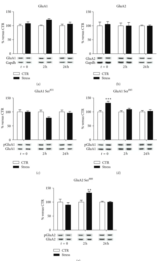
3.3. Modulation of AMPA Receptor Subunits Expression and Phosphorylation Induced by Acute Stress. To assess
time-dependent changes induced by acute stress in glutamate receptor subunits expression, Western blot analyses for AMPA and NMDA receptor subunits were performed on PFC/FC total homogenates and purified postsynaptic spine membranes (TIF) of rats subjected to acute FS-stress and sacrificed at the different time points.
In total PFC/FC homogenate, no significant effects of FS-stress were found on the total expression of GluA1 and GluA2 AMPA receptor subunits at different time points (GluA1:
interaction term,𝑝 = 0.47; GluA2: interaction term, 𝑝 =
0.94) (Figures 1(a) and 1(b), resp.), although a trend for increase could be observed for GluA1, 2 h after the stress
beginning (FC = 1.21,𝑝adj= 0.08).
No significant changes were also observed for GluA1
Ser831phosphorylation in FS-stress animals at different time
points (interaction term𝑝 = 0.15, Figure 1(c)), despite single
comparison at 2 hours after stress had a marginally significant
effect (FC = 0.78;𝑝 = 0.045). In contrast, we measured a
sig-nificant treatment-×-time interaction for GluA1 Ser845
phos-phorylation (𝑝 = 0.026, Figure 1(d)). In particular, exclu-sively immediately after the stress protocol, a marked
upreg-ulation of GluA1 Ser845phosphorylation was observed (FC =
1.32, CI 95% = 1.12–1.55,𝑝adj < 0.001), with no significant
variations at other time points (2 h FC = 1.09, CI 95% =
0.93–1.28,𝑝adj = 0.49; 24 h FC = 1.03, CI 95% = 0.87–1.21,
𝑝adj = 0.97). Moreover, we found a significant
treatment-×-time effect for GluA2 Ser880phosphorylation (𝑝 = 0.002;
Figure 1(e)): acute stress caused an increase in GluA2 Ser880
phosphorylation 2 h after its start (FC = 1.33, CI 95% =
1.09–1.62,𝑝adj = 0.0015), while no significant changes were
observed at the other time points (2 h FC = 0.90,𝑝adj= 0.62;
24 h FC = 1.00,𝑝adj= 0.99).
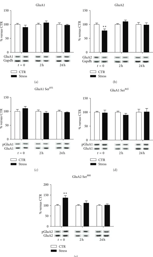
At postsynaptic membranes, no significant modifications
were observed for GluA1 (interaction term, 𝑝 = 0.32;
Figure 2(a)), while treatment-×-time interaction was signif-icant for GluA2 subunit (𝑝 = 0.013; Figure 2(b)), with a significant downregulation immediately after the stress
protocol (FC = 0.77, CI 95% = 0.63–0.95,𝑝adj = 0.01). No
significant modifications were found in GluA1
phosphory-lation at Ser831 (interaction term, 𝑝 = 0.27; Figure 2(c))
or Ser845 (interaction term 𝑝 = 0.76; Figure 2(d)). On the
contrary, we observed a significant treatment-×-time
inter-action for GluA2 at Ser880phosphorylation levels (𝑝 = 0.025;
Figure 2(e)), which were significantly increased immediately
after the stress protocol (FC = 1.37, CI 95% = 1.11–1.69,𝑝adj=
0.0013) and reduced at following time points (2 h FC = 1.12,
CI 95% = 0.91–1.38,𝑝adj = 0.49; 24 h FC = 1.02, CI 95% =
0.83–1.26,𝑝adj= 0.99).
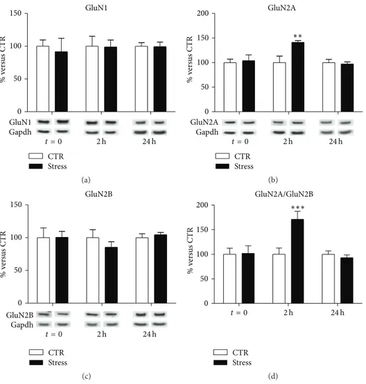
3.4. Acute Stress Induces Alterations in NMDA Receptor Sub-units Expression. In total PFC/FC homogenates, we found no
effect of stress on GluN1 subunit expression levels at different
time points (interaction term,𝑝 = 0.94, Figure 3(a)). Instead,
with regard to GluN2A, a significant treatment-×-time inter-action term was found (𝑝 = 0.022; Figure 3(b)). Indeed, total GluN2A expression levels were found increased in total homogenates of PFC/FC from FS-stress rats, selectively 2 h after the beginning of stress (FC = 1.41, CI 95% = 1.07–1.86,
𝑝adj= 0.008), and not at the other time points analyzed (𝑡 = 0
FC = 1.04,𝑝adj = 0.99; 24 h FC = 0.97, 𝑝adj = 0.99). With
regard to GluN2B subunit, a trend for decrease although not statistically significant was observed 2 h after the beginning
of stress (FC = 0.85,𝑝adj= 0.53; Figure 3(c)).
Given the key role of GluN2A/GluN2B ratio in regulating glutamatergic synapses activity [20], the ratio between the two subunits has also been calculated (Figure 3(d)). We found a significant treatment-×-time interaction effect (𝑝 = 0.003), with GluN2A/GluN2B ratio significantly higher in PFC/FC total homogenates from stressed rats sacrificed 2 h after the
stress beginning (FC = 1.71, CI 95% = 1.22–2.40,𝑝adj< 0.001),
and no significant changes in GluN2A/GluN2B ratio at other
time points (𝑡 = 0 FC = 1.02, 𝑝adj = 0.99; 24 h FC = 0.93,
𝑝adj= 0.94).
At postsynaptic membranes, we observed a significant treatment-×-time interaction (Robust GLM, 𝑝 = 0.005) for GluN1 expression levels (Figure 4(a)), which were signifi-cantly increased in FS-stressed rats sacrificed 2 h after the
beginning of stress (FC = 1.36, CI 95% = 1.05–1.68, 𝑝adj =
0.0013). A similar result was found for GluN2A subunit (Figure 4(b)). GluN2A protein expression level showed a significant stress-×-time interaction (Robust GLM, 𝑝 = 0.0009), with a marked increase 2 h after stress beginning
(FC = 1.50, CI 95% = 1.16–1.93, 𝑝adj = 0.0005). On the
contrary, no alterations were found either in postsynaptic
level of GluN2B (interaction term,𝑝 = 0.85; Figure 4(c))
or in GluN2A/GluN2B ratio (interaction term, 𝑝 = 0.39;
Figure 4(d)).
4. Discussion
We report here that acute footshock- (FS-) stress, although inducing no transcriptional or posttranscriptional alterations of ionotropic AMPA and NMDA glutamate receptor sub-units, modulates, in a time- and subunit-dependent way, their protein expression, phosphorylation, and localization at postsynaptic spines in PFC/FC of rats.
In particular, FS-stress rapidly increased phosphorylation
of GluA1, selectively at Ser845 (not at Ser831), and of GluA2
at Ser880in total homogenate, while reducing GluA2 levels,
together with increasing its phosphorylation at Ser880, in
postsynaptic spine membranes. Acute stress exerted no effect on GluA1 and GluA2 protein expression levels in total homogenate, as previously reported [10]. All the changes in AMPA receptor subunits expression and phosphorylation levels were selectively measured immediately after the 40 min
of stress session (except for increased GluA2 phospho-Ser880
0 50 100 150 GluA1 % v er sus CTR Gapdh GluA1 CTR Stress 24 h 2 h t = 0 (a) 0 50 100 150 GluA2 % v er sus CTR GapdhGluA2 CTR Stress 24 h 2 h t = 0 (b) 0 50 100 150 % v er sus CTR GluA1 pGluA1 CTR Stress 24 h 2 h t = 0 GluA1 Ser831 (c) 0 50 100 150 % v er sus CTR GluA1 pGluA1 CTR Stress 24 h 2 h t = 0 GluA1 Ser845 ∗∗∗ (d) 0 50 100 150 % v er sus CTR GluA2 pGluA2 CTR Stress 24 h 2 h t = 0 GluA2 Ser880 ∗∗ (e)
Figure 1: Time-dependent changes of protein expression levels of GluA1 (a), GluA2 (b), GluA1 phospho-Ser831(c), GluA1 phospho-Ser845(d), and GluA2 phospho-Ser880(e) in PFC/FC total homogenate of rats subjected to FS-stress and sacrificed immediately after stress and 2 h and 24 h from stress beginning. Data are represented as percentage of controls at each time point, as means± SEM (𝑛 = 8). Statistics: Generalized Linear Models (GLM) and Bonferroni Post Hoc Test (see Section 2 for details).∗∗𝑝 < 0.01;∗∗∗𝑝 < 0.001.
0 50 100 150 GluA1 GapdhGluA1 CTR Stress 24 h 2 h t = 0 % v er sus CTR (a) 0 50 100 150 GluA2 GapdhGluA2 CTR Stress 24 h 2 h t = 0 % v er sus CTR ∗∗ (b) 0 50 100 150 GluA1 pGluA1 CTR Stress 24 h 2 h t = 0 % v er sus CTR GluA1 Ser831 (c) 0 50 100 150 GluA1 pGluA1 CTR Stress 24 h 2 h t = 0 % v er sus CTR GluA1 Ser845 (d) 0 50 100 150 200 GluA2 pGluA2 % v er sus CTR CTR Stress 24 h 2 h t = 0 GluA2 Ser880 ∗∗ (e)
Figure 2: Time-dependent changes of protein expression levels of GluA1 (a), GluA2 (b), GluA1 phospho-Ser831(c), GluA1 phospho-Ser845 (d), and GluA2 phospho-Ser880(e) in PFC/FC postsynaptic spine membranes of rats subjected to FS-stress and sacrificed immediately after stress and 2 h and 24 h from stress beginning. Data are represented as percentage of controls at each time point, as means± SEM (𝑛 = 8). Statistics: Generalized Linear Models (GLM) and Bonferroni Post Hoc Test (see Section 2 for details).∗∗𝑝 < 0.01.
0 50 100 150 GluN1 Gapdh GluN1 CTR Stress 24 h 2 h t = 0 % v er sus CTR (a) 0 50 100 150 200 GluN2A Gapdh GluN2A CTR Stress 24 h 2 h t = 0 % v er sus CTR ∗∗ (b) 0 50 100 150 Gapdh GluN2B GluN2B CTR Stress 24 h 2 h t = 0 % v er sus CTR (c) 0 50 100 150 200 GluN2A/GluN2B CTR Stress 24 h 2 h t = 0 % v er sus CTR ∗∗∗ (d)
Figure 3: Time-dependent changes of protein expression levels of GluN1 (a), GluN2A (b), GluN2B (c), and GluN2A/GluN2B (d) in PFC/FC total homogenate of rats subjected to FS-stress and sacrificed immediately after stress and 2 h and 24 h from stress beginning. Data are represented as percentage of controls at each time point, as means± SEM (𝑛 = 8). Statistics: Generalized Linear Models (GLM) and Bonferroni Post Hoc Test (see Section 2 for details).∗∗𝑝 < 0.01;∗∗∗𝑝 < 0.001.
2 h after stress start), suggesting fast and transient modulation of AMPA receptor subunits at PFC/FC synapses induced by acute stress.
Phosphorylation of GluA1 at Ser831and Ser845 has been
shown to modulate potentiation of AMPA receptor-mediated synaptic currents and to be involved in both Long Term Potentiation (LTP) and Long Term Depression (LTD) [21].
In particular, phosphorylation at Ser845 increases the open
channel probability, and the peak amplitude of currents mediated by AMPA receptors [21]. Therefore, the increase of
GluA1 phosphorylation at Ser845rapidly induced by FS-stress
is in line with increased AMPA receptor currents.
Phosphorylation of GluA2 at Ser880was shown to affect its
association with PDZ domain-containing proteins, thereby modifying trafficking and redistribution of the subunit at synaptic sites, facilitating GluA2 internalization [22–25], and subsequent lysosomal degradation [26]. GluA2 is a critical subunit in determining the function of AMPA
receptors. Indeed, GluA2-containing AMPA receptors are
Ca2+-impermeable and have a relatively low single channel
conductance [27], while AMPA receptors lacking GluA2
subunit have a higher Ca2+ permeability and conductance
[28, 29]. Intriguingly, it was shown that homomeric GluA1 AMPA receptors are delivered to synapses after LTP induc-tion, whereas homomeric GluA2 or GluA3 AMPA receptors are constitutively inserted [30, 31].
Taken together, this body of evidence strongly suggests that acute FS-stress, increasing GluA1 phosphorylation at
Ser845 and reducing the levels of GluA2-containing AMPA
receptors at postsynaptic membranes, may rapidly and tran-siently activate AMPA receptor-mediated synaptic currents.
We have also found here that acute FS-stress markedly increased GluN2A expression levels and GluN2A/GluN2B ratio in PFC/FC homogenate and GluN1 and GluN2A pro-tein levels in postsynaptic spine membranes, 2 h after the stress session. Notably, no changes were detected in NMDA
0 50 100 150 200 GluN1 Gapdh GluN1 CTR Stress 24 h 2 h t = 0 ∗∗ % v er sus CTR (a) 0 50 100 150 200 GluN2A Gapdh GluN2A CTR Stress 24 h 2 h t = 0 ∗∗∗ % v er sus CTR (b) 0 50 100 150 GluN2B Gapdh GluN2B CTR Stress 24 h 2 h t = 0 % v er sus CTR 0 50 100 150 GluN2B Gapdh GluN2B CTR Stress 24 h 2 h t = 0 % v er sus CTR (c) 0 50 100 150 200 GluN2A/GluN2B CTR Stress 24 h 2 h t = 0 % v er sus CTR (d)
Figure 4: Time-dependent changes of protein expression levels of GluN1 (a), GluN2A (b), GluN2B (c), and GluN2A/GluN2B (d) in PFC/FC postsynaptic spine membranes of rats subjected to FS-stress and sacrificed immediately after stress and 2 h and 24 h from stress beginning. Data are represented as percentage of controls at each time point, as means± SEM (𝑛 = 8). Statistics: Generalized Linear Models (GLM) and Bonferroni Post Hoc Test (see Section 2 for details).∗∗𝑝 < 0.01;∗∗∗𝑝 < 0.001.
receptor subunits at other time points, suggesting a time-dependent modulation of GluN2A and GluN2B subunits induced by acute stress.
In the forebrain, NMDA receptors are composed of one GluN1 subunit and one or more GluN2A or GluN2B subunits, and the precise combination of subunits determines the functional properties of the receptor [32]. It is well known that NMDA receptor subunit composition changes during development: while GluN2B is abundant in the early postnatal brain, the level of GluN2A, characterized by faster rising and decay kinetics, increases progressively during development [33]. In the adult brain, GluN2A is enriched at synaptic sites, while GluN2B is mainly extrasynaptic [34], and the GluN2A/GluN2B ratio was shown to be dependent on neuronal activity [35]. In particular, since increased GluN2A/GluN2B ratio is related with increased synaptic stimulation and transmission, its dynamic regulation is a major determinant of synaptic plasticity [36]. In this con-text, the increase of GluN2A/GluN2B ratio in homogenate, together with enrichment of GluN1- and GluN2A-containing
NMDA receptors in postsynaptic spine membranes mea-sured 2 h, but not 24 h, after the start of FS-stress, is in line with a delayed and transient enhancement of NMDA receptor-mediated synaptic currents.
In line with our results, in previous studies it was shown that both acute stress in vivo and short-term incubation of PFC neurons with corticosterone in vitro increase AMPA and NMDA receptor-mediated synaptic transmission and expression levels at membranes [10, 37]. However, contrary to the fast and transient effect measured after FS-stress, acute forced-swim stress was shown to induce a long-lasting increase (from 1–4 h, to 24 h after stress) of AMPA and NMDA-mediated excitatory postsynaptic currents amplitude and surface expression [10]. The apparent discrepancy with our results may be dependent on a number of factors, including different types of stress used, different age of the rats (juvenile versus adult), time points analyzed, and measurement of glutamate receptor subunits expression in different compartments (total membrane fraction versus postsynaptic spine membranes). Moreover, although the
changes in AMPA and NMDA receptor subunits expres-sion and phosphorylation levels induced by FS-stress were found to be transient, it cannot be excluded that FS-stress may induce long-lasting alterations of synaptic transmission, mediated by other molecular mechanisms. Further studies are required to address this point.
In previous studies, we showed that FS-stress, together with enhancing depolarization-dependent release of endoge-nous glutamate, increases excitatory postsynaptic currents amplitude (measured immediately after the stress session) [8]. Acute FS-stress also strongly decreases synaptic facilita-tion and its calcium-dependence, in line with an increase in release probability. The results obtained in the present study strongly suggest that postsynaptic mechanisms may also be involved in the enhancement of glutamate transmission induced by FS-stress in PFC/FC. In addition, we have also shown recently, by using electron microscopy stereology, that the total number of nonperforated and axoshaft excitatory synapses in medial PFC is increased remarkably (over 40%) immediately after acute FS-stress [38], demonstrating that the early functional changes in glutamate transmission are accompanied by large-scale changes in brain architecture at a fast pace.
5. Conclusion
In this study, we reported a time-dependent modulation of both AMPA and NMDA receptor subunits expression and phosphorylation induced by acute FS-stress in PFC/FC.
Although further studies are warranted to dissect the time-dependent functional, molecular, and structural alter-ations induced by stress in PFC/FC, the present study may further support the evidence of an enhancement of gluta-matergic synaptic transmission as early response to acute stress.
Conflict of Interests
The authors declare that there is no conflict of interests regarding the publication of this paper.
Authors’ Contribution
Daniela Bonini and Cristina Mora equally contributed to this work. Laura Musazzi and Alessandro Barbon equally contributed to this work.
Acknowledgments
This work was supported by a grant from MIUR (PRIN 2012 prot.2012A9T2S9 004). The authors gratefully acknowledge Dr. Giulia Treccani for technical help.
References
[1] B. S. McEwen, “Allostasis and allostatic load: implications for neuropsychopharmacology,” Neuropsychopharmacology, vol. 22, no. 2, pp. 108–124, 2000.
[2] I. N. Karatsoreos and B. S. McEwen, “Psychobiological allosta-sis: resistance, resilience and vulnerability,” Trends in Cognitive
Sciences, vol. 15, no. 12, pp. 576–584, 2011.
[3] N. Sousa and O. F. X. Almeida, “Disconnection and recon-nection: the morphological basis of (mal)adaptation to stress,”
Trends in Neurosciences, vol. 35, no. 12, pp. 742–751, 2012.
[4] M. Popoli, Z. Yan, B. S. McEwen, and G. Sanacora, “The stressed synapse: the impact of stress and glucocorticoids on glutamate transmission,” Nature Reviews Neuroscience, vol. 13, no. 1, pp. 22–37, 2012.
[5] A. F. T. Arnsten, M. J. Wang, and C. D. Paspalas, “Neuromod-ulation of thought: flexibilities and vulnerabilities in prefrontal cortical network synapses,” Neuron, vol. 76, no. 1, pp. 223–239, 2012.
[6] B. S. McEwen and J. H. Morrison, “The brain on stress: vulnerability and plasticity of the prefrontal cortex over the life course,” Neuron, vol. 79, no. 1, pp. 16–29, 2013.
[7] B. S. McEwen, C. Nasca, and J. D. Gray, “Stress effects on neuronal structure: hippocampus, amygdala, and prefrontal cortex,” Neuropsychopharmacology, vol. 41, no. 1, pp. 3–23, 2015. [8] L. Musazzi, M. Milanese, P. Farisello et al., “Acute stress increases depolarization-evoked glutamate release in the rat prefrontal/frontal cortex: the dampening action of antidepres-sants,” PLoS ONE, vol. 5, no. 1, Article ID e8566, 2010. [9] R. S. Duman, “Pathophysiology of depression and innovative
treatments: remodeling glutamatergic synaptic connections,”
Dialogues in Clinical Neuroscience, vol. 16, no. 1, pp. 11–27, 2014.
[10] E. Y. Yuen, W. Liu, I. N. Karatsoreos, J. Feng, B. S. McEwen, and Z. Yan, “Acute stress enhances glutamatergic transmission in prefrontal cortex and facilitates working memory,” Proceedings
of the National Academy of Sciences of the United States of America, vol. 106, no. 33, pp. 14075–14079, 2009.
[11] E. Y. Yuen, J. Wei, W. Liu, P. Zhong, X. Li, and Z. Yan, “Repeated stress causes cognitive impairment by suppressing glutamate receptor expression and function in prefrontal cortex,” Neuron, vol. 73, no. 5, pp. 962–977, 2012.
[12] G. Treccani, L. Musazzi, C. Perego et al., “Stress and cor-ticosterone increase the readily releasable pool of glutamate vesicles in synaptic terminals of prefrontal and frontal cortex,”
Molecular Psychiatry, vol. 19, no. 4, pp. 433–443, 2014.
[13] G. Bonanno, R. Giambelli, L. Raiteri et al., “Chronic antide-pressants reduce depolarization-evoked glutamate release and protein interactions favoring formation of SNARE complex in hippocampus,” The Journal of Neuroscience, vol. 25, no. 13, pp. 3270–3279, 2005.
[14] A. Barbon, F. Fumagalli, L. Caracciolo et al., “Acute spinal cord injury persistently reduces R/G RNA editing of AMPA receptors,” Journal of Neurochemistry, vol. 114, no. 2, pp. 397– 407, 2010.
[15] D. Bonini, A. Filippini, L. La Via et al., “Chronic glutamate treatment selectively modulates AMPA RNA editing and ADAR expression and activity in primary cortical neurons,” RNA
Biology, vol. 12, no. 1, pp. 43–53, 2015.
[16] D. Caudal, B. P. Godsil, F. Mailliet, D. Bergerot, and T. M. Jay, “Acute stress induces contrasting changes in AMPA receptor subunit phosphorylation within the prefrontal cortex, amygdala and hippocampus,” PLoS ONE, vol. 5, no. 12, Article ID e15282, 2010.
[17] P. Svenningsson, H. Bateup, H. Qi et al., “Involvement of AMPA receptor phosphorylation in antidepressant actions with special reference to tianeptine,” European Journal of Neuroscience, vol. 26, no. 12, pp. 3509–3517, 2007.
[18] P. McCullagh and J. A. Nelder, Generalized Linear Models, Chapman & Hall/CRC, 2nd edition, 1989.
[19] R Development Core Team, R: A Language and Environment for
Statistical Computing, R Foundation for Statistical Computing,
Vienna, Austria, 2011.
[20] T. E. Bartlett and Y. T. Wang, “The intersections of NMDAR-dependent synaptic plasticity and cell survival,”
Neuropharma-cology, vol. 74, pp. 59–68, 2013.
[21] J. Q. Wang, A. Arora, L. Yang et al., “Phosphorylation of AMPA receptors: mechanisms and synaptic plasticity,” Molecular
Neu-robiology, vol. 32, no. 3, pp. 237–249, 2005.
[22] R. H. Scannevin and R. L. Huganir, “Postsynaptic organization and regulation of excitatory synapses,” Nature Reviews
Neuro-science, vol. 1, no. 2, pp. 133–141, 2000.
[23] S. Matsuda, S. Mikawa, and H. Hirai, “Phosphorylation of serine-880 in GluR2 by protein kinase C prevents its C terminus from binding with glutamate receptor-interacting protein,”
Journal of Neurochemistry, vol. 73, no. 4, pp. 1765–1768, 1999.
[24] W. Lu and K. W. Roche, “Posttranslational regulation of AMPA receptor trafficking and function,” Current Opinion in
Neurobi-ology, vol. 22, no. 3, pp. 470–479, 2012.
[25] H. J. Chung, J. Xia, R. H. Scannevin, X. Zhang, and R. L. Huganir, “Phosphorylation of the AMPA receptor subunit GluR2 differentially regulates its interaction with PDZ domain-containing proteins,” Journal of Neuroscience, vol. 20, no. 19, pp. 7258–7267, 2000.
[26] M. D. Ehlers, “Reinsertion or degradation of AMPA receptors determined by activity-dependent endocytic sorting,” Neuron, vol. 28, no. 2, pp. 511–525, 2000.
[27] J. T. R. Isaac, M. C. Ashby, and C. J. McBain, “The role of the GluR2 subunit in AMPA receptor function and synaptic plasticity,” Neuron, vol. 54, no. 6, pp. 859–871, 2007.
[28] H. Lomeli, J. Mosbacher, T. Melcher et al., “Control of kinetic properties of AMPA receptor channels by nuclear RNA editing,”
Science, vol. 266, no. 5191, pp. 1709–1713, 1994.
[29] G. T. Swanson, D. Feldmeyer, M. Kaneda, and S. G. Cull-Candy, “Effect of RNA editing and subunit co-assembly single-channel properties of recombinant kainate receptors,” The Journal of
Physiology, vol. 492, part 1, pp. 129–142, 1996.
[30] S.-H. Shi, Y. Hayashi, J. A. Esteban, and R. Malinow, “Subunit-specific rules governing AMPA receptor trafficking to synapses in hippocampal pyramidal neurons,” Cell, vol. 105, no. 3, pp. 331– 343, 2001.
[31] S. Bassani, A. Folci, J. Zapata, and M. Passafaro, “AMPAR trafficking in synapse maturation and plasticity,” Cellular and
Molecular Life Sciences, vol. 70, no. 23, pp. 4411–4430, 2013.
[32] A. Sanz-Clemente, R. A. Nicoll, and K. W. Roche, “Diversity in NMDA receptor composition: many regulators, many con-sequences,” Neuroscientist, vol. 19, no. 1, pp. 62–75, 2013. [33] C. Lohmann and H. W. Kessels, “The developmental stages of
synaptic plasticity,” Journal of Physiology, vol. 592, no. 1, pp. 13– 31, 2014.
[34] C. Bellone and R. A. Nicoll, “Rapid bidirectional switching of synaptic NMDA receptors,” Neuron, vol. 55, no. 5, pp. 779–785, 2007.
[35] K. Yashiro and B. D. Philpot, “Regulation of NMDA receptor subunit expression and its implications for LTD, LTP, and metaplasticity,” Neuropharmacology, vol. 55, no. 7, pp. 1081–1094, 2008.
[36] R. C. Malenka and M. F. Bear, “LTP and LTD: an embarrassment of riches,” Neuron, vol. 44, no. 1, pp. 5–21, 2004.
[37] E. Y. Yuen, W. Liu, I. N. Karatsoreos et al., “Mechanisms for acute stress-induced enhancement of glutamatergic transmis-sion and working memory,” Molecular Psychiatry, vol. 16, no. 2, pp. 156–170, 2011.
[38] N. Nava, G. Treccani, N. Liebenberg et al., “Chronic desipramine prevents acute stress-induced reorganization of medial prefrontal cortex architecture by blocking glutamate vesicle accumulation and excitatory synapse increase,”
International Journal of Neuropsychopharmacology, vol. 18, no.
Submit your manuscripts at
http://www.hindawi.com
Neurology
Research International Hindawi Publishing Corporation
http://www.hindawi.com Volume 2014
Alzheimer’s Disease
Hindawi Publishing Corporationhttp://www.hindawi.com Volume 2014
Scientifica
Hindawi Publishing Corporation
http://www.hindawi.com Volume 2014
Hindawi Publishing Corporation
http://www.hindawi.com Volume 2014
BioMed
Research International
Hindawi Publishing Corporation
http://www.hindawi.com Volume 2014
Research and Treatment
The Scientific
World Journal
Hindawi Publishing Corporation
http://www.hindawi.com Volume 2014
Hindawi Publishing Corporation
http://www.hindawi.com Volume 2014
Neural Plasticity
Hindawi Publishing Corporation
http://www.hindawi.com Volume 2014
Parkinson’s
Disease
Hindawi Publishing Corporationhttp://www.hindawi.com Volume 2014
Research and Treatment
Autism
Sleep Disorders
Hindawi Publishing Corporation
http://www.hindawi.com Volume 2014
Hindawi Publishing Corporation
http://www.hindawi.com Volume 2014
Neuroscience
Journal
Epilepsy Research and Treatment
Hindawi Publishing Corporation
http://www.hindawi.com Volume 2014
Hindawi Publishing Corporation
http://www.hindawi.com Volume 2014
Psychiatry
Journal
Hindawi Publishing Corporation
http://www.hindawi.com Volume 2014
Computational and Mathematical Methods in Medicine
and Treatment
Hindawi Publishing Corporation
http://www.hindawi.com Volume 2014
Hindawi Publishing Corporation
http://www.hindawi.com Volume 2014
Brain Science
International Journal ofStroke
Research and TreatmentHindawi Publishing Corporation
http://www.hindawi.com Volume 2014
Neurodegenerative
Diseases
Hindawi Publishing Corporation
http://www.hindawi.com Volume 2014
Journal of
Cardiovascular Psychiatry and Neurology
Hindawi Publishing Corporation