Abstract. –OBJECTIVE: Aim of this study is to evaluate the possibility of limb magnetic reso-nance lymphography (MRL) to differentiate lym-phatic vessels from pathological veins, collect a specimen of the identified lymphatic vessel dur-ing operations of super microsurgical lymphatic-venular anastomosis (s-LVA) and perform im-munohistochemical stainings to confirm the na-ture of the collected vessels.
PATIENTS AND METHODS: Twenty patients presenting lymphedema were enrolled in this study. Five patients reported lower limb lym-phedema and 15 patients reported upper limb lymphedema. All patients had the indication for s-LVA and underwent preoperative MRL imaging of the affected limb. A total of 57 lymphatic vesels were identified by MRL and used to guide s-LVA: all these vessels have also been used to perform an intraoperative biopsy for immunohis-tochemical evaluation.
RESULTS: A total of 53/57 vascular structures resulted compatible with lymphatic vessels at the immunohistochemical study performed with D2-40 antibody; 3/57 specimen showed the ab-sence of the D2-40 antibody. A significant asso-ciation was found between preoperative MRL and immunohistochemical marker D2-40 on col-lected specimen.
CONCLUSIONS: Most of the articles in the in-ternational literature report the concomitant presence of both lymphatic and venous vessels at MRL. However, no one in literature describes the possibility to differentiate venous vessels from lymphatic vessels, and this is a crucial is-sue for the correct evaluation of the lymphatic
Could MRI visualize the invisible? An Italian
single center study comparing magnetic
resonance lymphography (MRL), super
microsurgery and histology in
the identification of lymphatic vessels
P. GENNARO
1, A. BORGHINI
2, G. CHISCI
1, F.G. MAZZEI
3,
E. WEBER
2, E. TEDONE CLEMENTE
1, S. GUERRINI
4,
F. GENTILI
4, G. GABRIELE
1, C. UNGARI
5, M.A. MAZZEI
41Department of Biotechnology, Azienda Ospedaliero-Universitaria Senese, Siena, Italy 2Department of Molecular and Development Medicine, University of Siena, Siena, Italy 3Department of Diagnostic Imaging, Azienda Ospedaliero-Universitaria Senese, Siena, Italy 4Department of Medical, Surgical and Neuro Sciences, Diagnostic Imaging, Azienda
Ospedaliero-Universitaria Senese, Siena, Italy
5Department of Maxillo-Facial Sciences, “Sapienza” University of Rome, Rome, Italy
system in patients with limb lymphedema under-going a future surgical correction. In the present study, MRL allowed to identify active lymphatic vessels. MRL was predictive to determine preop-eratory lymphatic vessels and to perform suc-cessful s-LVA in lymphedema patients. This is the first study to prove the nature of the vessels identified at the preoperative MRL with immuno-histochemical stainings.
Key Words:
Lymphedema, MRL, D2-40.
Introduction
Lymphedema is a pathology characterized by a limb accumulation of subcutaneous protein-rich fluid due to a lymphatic system disorder. While primary lymphedema is characterized by congenital abnormalities, secondary lymphedema is caused in most cases by obstruction or stenosis of lymph vessels caused by surgical oncology1.
Lymphatic-venular anastomosis (LVA) is a microsurgical technique effective to improve limb circumference and to alleviate dermal scle-rosis2,3. Super microsurgical lymphatic-venular
anastomosis (s-LVA) is an advanced technique that allows to perform an anastomosis on even smaller vessels, and showed good results in lymphedema patients to reduce limb circumfer-ence and cellulitis4.
To perform LVA or s-LVA, the preoperative lymphatic mapping is necessary for surgical plan-ning: the ideal imaging technique to evaluate lym-phedema has been, for many years, lymphoscintig-raphy, using a radionuclide with various 99m
Tc-la-belled molecules5. Recently, Indocyanine Green
(ICG) lymphography has been introduced6.
Magnetic Resonance Lymphography (MRL) performed with injections of gadolinium-based contrast agent has been widely described in the literature with gadolinium contrast enhancement, injecting the contrast in the interdigital spaces. Its use is mainly related to the lymphedema and the imaging of the lymphatic system, and it is crucial to understand lymphatic vessels role in the disease development and to plan the opera-tion. The main difficulty of MRL imaging is the differential diagnosis between lymphatic vessels and venous vessels.
In the present paper, the authors have intended to verify the accuracy of preoperative MRL per-forming a biopsy of the lymphatic vessels for im-munohistochemical study (D2-40 antibody) dur-ing operations of s-LVA in lymphedema patients.
Patients and Methods
The present study included 20 female patients with a mean age of 57.6 years old affected by lymphedema (3 cases of primary lymphedema, 17 cases of secondary lymphedema). These pa-tients underwent to a MRL before operation of s-LVA. During the s-LVA operations specimen of the MRL-presumed lymphatic vessels were col-lected and sent to the laboratory for immunohis-tochemical stainings.
Magnetic Resonance Lymphography Before MRLA, interdigital injection of gadolinium-based contrast agent was performed using the most commercially available and wide-ly diffused paramagnetic contrast medium: Gadobenate Dimeglumine (Gd-BOPTA, Multi-hance, Bracco Imaging, Milan, Italy). The solu-tion to inject was prepared with 15 mL of gadobenate dimeglumine (that corresponds to one bottle) and 1.5 mL of lidocaine (1% solu-tion), then the mixed agent was injected intracu-taneously into the interdigital webs of the dorsal foot, with four injections for each limb. The vol-ume injected into each point was 0.7 to 0.8 mL.
All magnetic resonance (MR) exams were per-formed with a 1.5 Tesla MR unit (Signa Twin
Speed Hdxt; General Electric Healthcare, Mil-waukee, WI, USA), with a maximum gradient strength of 23 mT/m and a slew rate of 80 mT/m/ms (software release 15.0_0947A). Pa-tients were supine, feet first, with both legs on a ramp pillow in order to obtain a parallelism with the main magnetic field and to position them on the most homogeneous area of the B0. To obtain a large anatomical coverage and a good signal-to-noise ratio, a 7000-elements phased array pe-ripheral vascular receiving coil (Flow 7000) for the study of the lower extremity and a 8 channel Body Array for the upper extremity were used; both of them are built by USA Instrument. The fingers appeared from the holes of the coil, let-ting them to be out for an easy access during the injections of the contrast agent. To reduce the hy-perintensity artifacts we paid attention to avoid direct contact of the coil surface with the patient extremity, using some small pillows. Patients were instructed on the procedure in order to ob-tain complete collaboration.
After positioning a survey and calibration from all the stations, three for the lower extremi-ty (foot-ankle calf, calf knee, thigh hip) and two or three for the upper extremity (hand-wrist fore-arms, elbow arm shoulder) were performed. Sub-sequently, before the injection of the contrast agent, a coronal 3D SSFP Balanced (Fiesta, GE) ECG-triggered with spectral fat saturation (SPECtral Inversion At Lipidi, SPECIAL, GE) was acquired; the technical parameters used were: TR 4.0 ms, TE 1.9 ms, TI 90 ms, FOV 40 × 40 cm, Matrix 224 × 192, slice thickness 2.2 mm, NEX 0.53 (Half Fourier) and Bandwidth kHz. The ECG trigger was acquired with a Pe-ripheral Gating (PG, GE) and a Delay Time set for a systolic phase acquisition in order to obtain a no contrast venography (the high flow artery experienced some flow dephasing reducing their signal); we also obtained a good image to visual-ize the lymphoedema.
For dynamic MRL, 3D fast spoiled gradient-recalled echo T1-weighted images with a fat sat-uration technique (T1 high-resolution isotropic volume excitation) were acquired in every station at 5, 10, 15, 20, 25, 30, 35 and 40 minutes ap-proximately after contrast injection. The techni-cal parameters were: f TR/TE 5.0 ms/2.1 ms; TI 17 ms, flip angle 25 , FOV 480 × 480 mm, ma-trix 448 × 320, slice thickness 2.2 mm, NEX (signal average number) 2 and Bandwidth kHz, acquisition time 0 min 40 sec. The 3D MRL were then reconstructed from the post-contrast
serum albumin)7. The sections were then
incubat-ed 10 min in the dark with the secondary anti-body (biotinylated goat anti mouse IgG of the same kit). The reaction was revealed by a 10 min incubation with streptavidin horseradish peroxi-dase diluted in Tris Buffered Saline followed by a 2 min incubation with 3,3’-diaminobenzidine Substrate Kit for Peroxidase (VECTOR), which contains a nickel solution that converts the brown color characteristic of 3,3’-diaminobenzidine in black. Sections were counterstained with May-er’s hematoxylin and mounted with Eukitt (Sig-ma, St Louis, MO, USA).
Data Analysis
After data acquisition, image post-processing and subsequent analysis were performed by two experienced radiologists, who reached an agree-ment by consensus. At MRLA examination af-fected lymphatic vessels were distinguished from a vein because of their caliber (diameter) and their morphology.
Differences between groups were evaluated using chi-square test. Association between vari-ables were tested by univariate regression analy-sis. p-values < 0.05 were considered to be signifi-cant if not otherwise specified.
Results
No complications were observed after the ex-amination, in particular, no complications were observed during or after intracutaneous injec-tions of Gd-BOPTA. The lymphedema showed an epifascial distribution with a high signal in-tensity on Coronal 3D SSFP Balanced images. Affected lymphatic vessels were distinguished from veins because of their caliber and their morphology; in particular the diameter of the ectasic lymphatic vessels was smaller than the one of the adjacent vein and greater than the one of the lymphatic vessels of the unaffected extremity when visualized, whereas the mor-phology was tortuous (beaded appearance) in comparison with the morphology of the veins. The beaded appearance of the lymphatic vessels extending from the injection site was reliably detected 5-10 minutes after the injection and, in the majority of the cases, the lymphatic vessels could be detected, with the strongest enhance-ment at 35-40 minutes after contrast injection. Collateral vessels with dermal backflow (an area of progressive dispersion of the contrast coronal images at each time point using a MIP
technique. The exam ended acquiring, in every station, a coronal 3D Rapid Acquisition with Re-laxation Enhancement heavily T2-weighted with Driven Equilibrium, in order to reduce the acqui-sition time by reducing the Repetition Time with-out affect the images, (FRFSE T2, GE) with the following parameters: TR 2000 ms, TE 679 ms, FOV 48x48 cm, Matrix 320 × 256, Thickness 3.6 mm, NEX 1 and Bandwidth kHz; with these im-ages the lymphoedema is clearly visible and measurable. The examination time for one pa-tient was approximately 1.5 hours. No systemic or local complications were observed during or after the examination.
Image Interpretation
The images analysis was then performed using Multi Planar Reconstruction of the subtracted images and with Thin-Slab Maximum Intensity Projection in order to better visualize the lym-phatic vessels.
Super Microsurgery
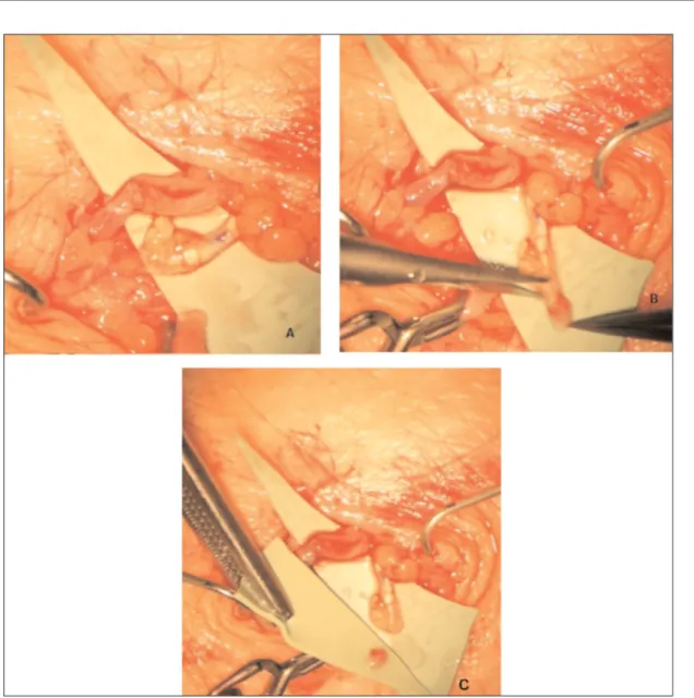
After the visualiation of the lymphatic vessels using MRLA, superficial veins mapping with Ac-cuvein device was performed to identify the mi-crosurgical sites. Under local anaesthesia with 1% xylocaine containing adrenalin a skin inci-sion was performed on each site, subcutaneous tissue was dissected under a high magnification operating microscope. Lymphatic collectors and adjacent subcutaneous veins were identified. Ves-sels were dissected and 4 or 5 11/0 endoluminal sutures with a 50-micron needle were performed between lymphatic and venous vessels (Figure 1). Afterwards, the skin was stitched up with continuous 6/0 resorbable microsutures.
Immunohistochemistry
Specimens were formalin fixed and paraffin embedded. Consecutive sections were obtained from each sample, dewaxed with xylene and re-hydrated in descending ethanol series. Endoge-nous peroxidases were blocked with 3% hydro-gen peroxide. Non-specific binding sites were blocked by a 5 min incubation with Blocking Reagent (IHC SelectTM kit, Millipore, Billerica, MA, USA) followed by a brief wash in PBS (Phosphate Buffered Saline). The sections were then incubated overnight with the primary anti-body D2-40, a monoclonal IgG1 specific for the lymphatic endothelium (Dako, Santa Clara, CA, USA, diluted 1:20 in PBS containing 1% bovine
medium between lymphatic vessels in the soft tissues), indicating proximal lymph flow ob-struction with alternate pathways of transport, were generally seen after 15-20 minutes (Figure 2). The mean diameter of a dilated lymphatic vessel was 2.20 ± 0.5 mm. The mean diameter of a venous vessel was 2.4 ± 0.2 mm.
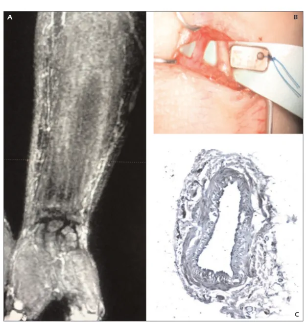
No complications were showed after s-LVA; mean hospitalization was 2 days. No correlation between age and results was found. A total of 57 (2.85 per patient mean) lymphatic vessels were identified at the preoperative MRL; among these 53/57 specimen collected of these vessels result-ed positive at the immunohistochemical marker D2-40 (Figure 2, Figure 3). A significant
associa-tion was found between preoperative MRI and immunohistochemical marker D2-40 on collect-ed specimen (Chi-square = 40.421, DF = 1, Sig-nificance level p < 0.0001, contingency coeffi-cient 0.644).
Discussion
Lymphoscintigraphy has been for many years the gold standard imaging in lymphoedema pa-tients; however, radiation exposure, long exami-nation time needed, side effects such as pul-monary embolism have limited its clinical applic-ability8.
Figure 1. Intraoperative image of the super microsurgical lymphatic-venular anostomosis (s-LVA) during the collection of biopsy specimen.
Lohrmann et al9-11in their papers reported the
visualization of venous vessels at MRL, as con-trast may be captured by both lymphatic and ve-nous capillaries: veve-nous vessels resulted to be contrast-enhanced faster than lymphatic vessels, which resulted slower.
At the Lymphoedema Mondial Congress in Rome in 2013, and at the International Lym-phoedema Congress in Genova in 2014, many criticisms have been raised against the use of the MRL and the potential discrimination between lymphatic and venous vessels.
Most of the articles present in the international literature report the concomitant presence of both
lymphatic and venous vessels in MRL. Lohrmann et al7and Ruehm et al12reported some suggestions
to differentiate lymphatic and venous vessels. However these criteria may be not enough, due to the concomitant dysfunction of deep and superfi-cial venous circulation, the identified structures may easily be dysfunctional venous vessels (closed or thrombosed veins, varicose veins or valvular insufficiency). Further resonance imaging of lymphatic vessels may be even more doubtful on nonedematous limbs9. The most of the
pub-lished papers suppose to identify the lymphatic vessels mostly due to their ectasic morphology, without any laboratory confirmation.
Figure 2.A, Preoperative magnetic resonance lymphography (MRL) of the affected limb. B, Intraoperative s-LVA image. C, Histological staining of the collected lymphatic vessel.
A B
MRL imaging is a relatively new technique used to map the lymphatic vessels injecting con-trast material, accompanied by high-resolution sequential 3D imaging of the affected organ. It is well known that the advantage of this technique is that it allows dynamic monitoring of the lym-phatic transit with simple and minimally invasive high-spatial- and high-temporal-resolution imag-ing techniques to visualize the lymphatic system. However, no one in literature describes the possi-bility to differentiate venous vessels from lym-phatic vessels and this is a crucial issue to cor-rectly evaluate the lymphatic system in patients with limb lymphedema undergoing future surgi-cal correction13,14.
S-LVA is a super microsurgical technique ef-fective to improve limb circumference, alleviate dermal sclerosis and reduce cellulitis in
lym-phoedema patients2,3,15: to perform s-LVA
preop-erative lymphatic mapping is necessary for surgi-cal planning.
Our criteria for differential diagnosis between venous and lymphatic vessels were mainly the caliber, the morphology and the beaded appear-ance of the vessels. The study of the unaffected limb has been essential for the imaging and dif-ferential diagnosis: diameter of the ectasic lym-phatic vessels was smaller than the adiacent vein and greater than the lymphatic vessels of the un-affected extremity. In comparison with others au-thors that referred their experience in MRL, their reported criteria to distinguish veins and lym-phatic vessels may be not effective, and especial-ly may not be effective on healthy limbs. Liu et al16 evidenced only 5/23 cases of
contrast-en-hanced lymphatic vessels with MRL.
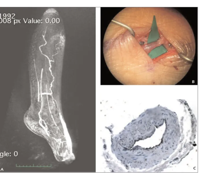
Figure 3. Another case with preoperative MRL of the affected limb.B, Intraoperative s-LVA image. C, Histological staining of the collected lymphatic vessel.
B
In this paper, we performed a biopsy to collect a specimen of the lymphatic vessels presumed at the MRL. Most of these specimen resulted to be lymphatic vessels thanks to immunohistochemi-cal stainings. For this reason, our criteria resulted to be significantly effective limited to the cases of this study: during s-LVA, we found a signifi-cant number of lymphatic vessels on the basis of preoperative MRL images. Mitsumori et al17
re-ported their experience of MRL on four consecu-tive patients. Mitsumori et al17 performed an
in-travenous systemic and subdermal injection of Gd-based MR contrast and supported the idea to identify all venous vessels that, with the techno-logical removal of these from the structures evi-denced with subdermal contrast, should leave on-ly the on-lymphatic vessels17. However, it is possible
that a thrombosed vein (e.g. a tied saphenous vein) could receive contrast from a cutaneous injection but not from systemic circulation. Further, no his-tological evidence of the identified vessels was re-ported.
MRL with the proper criteria is a feasible technique that studies the limb lymphatic system with the use of commercially available contrast agents. This technique offers a newer minimally invasive procedure to visualize the lymphatic system anatomy with the aim to guide surgical operation. However, further studies enrolling more patients should be performed to determine the kinetics of gadolinium transport in disease states with the high lymphatic flow.
Present diagnostic tools in order to quantify the lymphoedema are limb circumference, self-assessment questionnaires and water displace-ment18: MRL may find its clinical usefulness in
the follow-up of the s-LVA and to evaluate the flow into the anostomosis19. Further, screening of
oncological non-symptomatic patients could highlight possible latent lymphoedema and sug-gest conservative treatments or super microsurgi-cal noninvasive surgery: treatment in initial lym-phoedema has a significantly better prognosis than in the advanced ones20.
Conclusions
In the present work, MRL revealed to be pre-dictive to identify preoperatory lymphatic vessels and to perform successfully s-LVA in lym-phoedema patients. A significative number of lymphatic vessels were found during s-LVA on the basis of preoperatory MRL images. This is
the first single-center study that compares super microsurgery, magnetic resonance and immuno-histochemistry in order to perform differential di-agnosis between lymphatic and venous vessels: our significative results add credit to the imaging technique and let us define MRL as a possible standard diagnostic imaging technique for lym-phedema patients.
–––––––––––––––––-––––
Conflict of Interest
The Authors declare that there are no conflicts of interest.
References
1) OLSZEWSKIW. On the pathomechanism of develop-ment of postsurgical lymphedema. Lymphology 1973; 6: 35-51.
2) YAMAMOTOT, CHENWF, YAMAMOTON, YOSHIMATSUH, TASHIROK, KOSHIMAI. Technical simplification of the
supermicrosurgical side-to-end lymphaticovenular anastomosis using the parachute technique. Mi-crosurgery 2015; 35: 129-134.
3) GENNARO P, GABRIELEG, MIHARA M, KIKUCHIK, SALINI C, ABOHI, CASCINOF, CHISCIG, UNGARIC.
Suprami-crosurgical lymphatico-venular anastomosis (LVA) in treating lymphedema: 36-months preliminary report. Eur Rev Med Pharmacol Sci 2016; 20: 4642-4653.
4) GENNAROP, GABRIELEG, SALINIC, CHISCIG, CASCINOF, XU J-F, UNGARI C. Our supramicrosurgical experi-ence of lymphaticovenular anastomosis in lym-phedema patients to prevent cellulitis. Eur Rev Med Pharmacol Sci 2016; 21: 674-679.
5) SHERMANAI, TER-POGOSSIANM. Lymph-node
concen-tration of radioactive colloidal gold following inter-stitial injection. Cancer 1953; 6: 1238-1240. 6) MIHARA M, HARA H, KIKUCHIK, YAMAMOTO T, IIDA T,
NARUSHIMAM, ARAKI J, MURAIN, MITSUIK, GENNARO P, GABRIELE G, KOSHIMA I. Scarless lymphatic
ve-nous anastomosis for latent and early-stage lym-phoedema using indocyanine green lymphogra-phy and non-invasive instruments for visualising subcutaneous vein. J Plast Reconstr Aesthet Surg 2012; 65: 1551-1558.
7) KAHN HJ, MARKS A. A new monoclonal antibody,
D2-40, for detection of lymphatic invasion in primary tumors. Lab Invest 2002; 82: 1255-1257.
8) KOEHLERPR. Complications of lymphography. Lym-phology 1968; 1: 116-120.
9) LOHRMANN C, FOELDI E, SPECK O, LANGER M.
High-resolution MR lymphangiography in patients with primary and secondary lymphedema. AJR Am J Roentgenol 2006; 187: 556-561.
10) LOHRMANNC, FOELDIE, LANGERM. Indirect magnetic
lymphedema preliminary results in humans. Eur J Radiol 2006; 59: 401-406.
11) LOHRMANN C, FOELDI E, LANGER M. Assessment of
the lymphatic system in patients with diffuse lym-phangiomatosis by magnetic resonance imaging. Eur J Radiol 2011; 80: 576-581.
12) RUEHMSG, SCHROEDERT, DEBATINJF. Interstitial MR
lymphography with gadoterate meglumine: initial experience in humans. Radiology 2001; 220: 816-821.
13) KRISHNAMURTHYR, HERNANDEZA, KAVUKS, ANNAMA, PIMPALWAR S. Imaging the central conducting phatics: initial experience with dynamic MR lym-phangiography. Radiology 2015; 274: 871-878. 14) DORIY, ZVIMANMM, ITKINM. Dynamic
contrast-en-hanced MR lymphangiography: feasibility study in swine. Radiology 2014; 273: 410-416.
15) MIHARAM, HARAH, FURNISSD, NARUSHIMAM, IIDAT, KIKUCHI K, OHTSUH, GENNAROP, GABRIELEG, Murai
N. Lymphaticovenular anastomosis to prevent cel-lulitis associated with lymphoedema. Br J Surg 2014; 101: 1391-1396.
16) LIUNF, LUQ, JIANGZH, WANGCG, ZHOUJG.
Anatom-ic and functional evaluation of the lymphatAnatom-ics and lymph nodes in diagnosis of lymphatic circulation disorders with contrast magnetic resonance lym-phangiography. J Vasc Surg 2009; 49: 980-987. 17) MITSUMORILM, MCDONALD ES, WILSON GJ, NELIGAN
PC, MINOSHIMAS, MAKI JH. MR lymphangiography:
How I do it. J Magn Reson Imaging 2015; 42: 1465-1477.
18) SHAHC, VICINI FA. Breast cancer-related arm
lym-phedema: incidence rates, diagnostic techniques, optimal management and risk reduction strate-gies. Int J Radiat Oncol Biol Phys 2011; 81: 907-914.
19) GENNAROP, CHISCIG, MAZZEIF, GABRIELEG. Magnetic
resonance lymphangiography: How to prove it? J Magn Reson Imaging 2016; 44: 509-510.
20) ST O U T GE R G I C H NL, PFA L Z E R LA, MCGA R V E Y C, SPRINGERB, GERBERLH, SOBALLEP. Preoperative
as-sessment enables the early diagnosis and suc-cessful treatment of lymphedema. Cancer 2008; 112: 2809-2819.