Table of content
Chapter 1 Osteoarthritis ... 1 1.1. Pathology ... 1 1.2. Classification ... 4 1.3. Epidemiology ... 6 1.4. Risk factors ... 7 1.5. Sintomatology ... 91.6. Physiological modification of the OA joints ... 10
1.6.1. Articular cartilage ... 10
1.6.2. Subchondral bone ... 16
1.6.3. Synovial membrane ... 17
1.7. Molecules involved in OA pathophysiology ... 19
1.7.1. Interleukin-1 (IL-1) ... 19
1.7.2. Interleukin-6 (IL-6) ... 20
1.7.3 Tumor necrosis factor- (TNF- ) ... 20
1.7.4 Prostaglandin E2 (PGE2) ... 21
1.7.5 Fibroblast growth factor (FGF-2) ... 21
1.7.6 Insulin-like growth factor (IGF) ... 22
1.7.7 Hypoxia inducible factor (HIF) ... 22
Chapter 2 Therapeutic treatments for OA... 24
2.1 Non-pharmacological therapy of osteoarthritis ... 25
2.2 Pharmachological treatments of osteoarthritis ... 28
2.4 Surgery ... 32
Chapter 3 Hyaluronic acid (hyaluronan) ... 34
3.1 Biochemistry of hyaluronan ... 35
3.2 HA receptors ... 36
3.3 Mechanism of action ... 37
3.3.1 Condroprotective effects. ... 38
3.3.2 Effects of hyaluronan on the extracellular matrix. ... 38
3.4 Viscosupplementation ... 39
3.5 HYADD®4-G ... 40
Chapter 4 Growth hormone ... 43
4.1 Physiologic Effects of Growth Hormone ... 45
4.2 Secretion and regulation of Growth Hormone ... 47
4.3 The growth hormone receptor and signal transduction ... 48
4.4 Negative regulator of GH: Suppressor of Cytokine Signalling-2 (SOCS2) ... 51
4.5 GH and signalling molecules in normal and OA cartilage ... 53
4.6 GH in OA treatment ... 56
Chapter 5 Aim ... 58
Chapter 6 Materials and Methods ... 60
6.1 Patient characteristics ... 60
6.2 Immunoistochemical analysis ... 62
6.3 Cellular culture ... 63
6.3.1 Chondrocytes isolation ... 63
6.3.2 Synoviocytes isolation ... 64
6.4 Experimental culture conditions ... 64
6.5 Cellular morphology ... 65
6.6 Cellular viability...65
6.8 Cellular migration ... 67
6.9. Immunocychemical analysis ... 67
6.10 Molecular biology analysis ... 68
...68
6.10.2 Reverse transcription ... 68
6.10.3 Real-Time PCR ... 69
6.11. Soluble factors analysis ... 71
6.11.1 Multiplex system ... 71
6.11.2 Test ELISA : Enzyme-linked immunosorbent assays ... 73
6.12 Intracellular proteins analysis by Western Blotting ... 74
6.12.1 Protein extraction ... 74
6.12.2 Western Blot... 75
6.13 Sta ... 75
Chapter 7 Results ... 76
7.1 GHR and CD44 expression on cartilage biopsies and isolated chondrocytes ... 76
7.2 HYADD®4-G effect on chondrocytes ... 79
7.3 Morphology and viability of chondrocytes treated with hGH and HYADD®4-G ... 83
7.4 Evaluation of chondrocyte metabolic activity ... 87
7.5 Effect of hGH and HYADD®4-G on GHR and CD44 ... 89
7.6 Effects of hGH and HAD on chondrocyte markers ... 90
7.7 Release of IGF-1, FGF-2 and IL6 by treated chondrocytes ... 98
7.8 Evaluation of SOCS2 ... 101
Chapter 8 Discussion ... 104
References ... 107
1. Osteoarthritis
1.1 Pathology
Osteoarthritis (OA) is the most common form of arthritis and a major cause of pain and disability in older adults (over the age 65) (Blumenkrantz et al., 2004). The prevalence of OA in most joints is higher, before 50 years, in men than in women and after this age, women are more often affected than men. (Felson et al., 2000)
and it is characterized by joint pain and limited function of the articulation. This is a misnomer because OA is not simply a process of wear and tear but rather abnormal remodelling of joint tissues driven by a host of inflammatory mediators within the affected joint. (Loeser et al., 2012).
Nowadays OA is considered a disease of the whole joint as an organ, the articular cartilage is altered to some extent in all affected joints with OA. In addition to the development of cartilage changes with aging, cartilage degeneration may occur in response to inappropriate mechanical stress and low-grade local systemic inflammation associated with trauma, metabolic syndrome, and genetic predisposition, which are major risk factors of OA development and progression. However, strong functional interactions among the cartilage, synovium, and subchondral bone impact on cartilage function in such a way that it is difficult to know where and when pathological changes begin. (Houard et al., 2013)
Bone, cartilage, synovial fluid, ligaments and also the muscles around the joint are tissues that change with OA and affect the function of the joint. (Andriacchi et al., 2009; Buckwalter et al., 2004) Several tissues might be a starting point for pathways that lead to OA. Cartilage might be the tissue where the pathophysiological process of OA starts, but biochemical and imaging studies have shown that synovium and bone could be also good starting points. However, it remains unclear which of these three types of tissue, or some combination thereof, might serve as the key tissue for OA. (Samuels et al., 2008)
One of the most affected joints is the knee, in this joint osteoarthritis is a chronic, indolent disease that will affect an ever increasing number of patients, especially the elderly and the obese. It is characterized by degeneration of the cartilage substance inside the knee which leads to pain, stiffness and tenderness. By some estimations in 2030, only in the United States, this medical condition will burden 67 million people. (Uth and Trifonov, 2014)
A diagnosis of OA is mainly based on symptoms. A patient that has reached a certain age and has joint pain, limitation of movement, crepitus and, sometimes, effusion in the joint might get the diagnosis of OA. Recommendations for the diagnosis of knee OA were published in 2010. (Zhang et al., 2010) The treatment of OA is mainly symptom driven and based on the use of anti-inflammatory medication in combination with exercise treatment and lifestyle changes. However such treatment cannot prevent or cure OA and often fails to provide satisfactory pain relief. Joint replacement may be possible in developed countries for patients with severe OA and significant disability. Research efforts during the past decades have focused on the search for disease-modifying treatments. Most of these disease-modifying treatments were directed towards regeneration of the cartilage and were tested in patients with evident OA. However, so far, these efforts have not been very successful and have not had a significant influence on the symptoms of OA. (Hunter 2011)
A more comprehensive definition summarizing the clinical, phatophysiological, biochemical and biomechanical changes that occur in OA was summarized in Table 1.
Table 1 Features of OA Clinical
effects Pathological
increate load
Sclerosis of subchondral bone Subchondral cysts
Marginal osteophytes
Increased metaphyseal bood flow Variable synovial inflammation Histological Fragmentation of cartilage surface
on vessels
Loss cartilage
Sclerosis of subchondral bone
Biomechanical Alteration in tensile, comprensive and shear properties Altered cartilage hydraulic permeability
Biochemical
proteogycans
nd weave
Increased synthesis and degradation of matrix macromolecules
1.2 Classification
Criteria for classification of symptomatic OA have been developed by a subcommittee of the American College of Rheumatology (ACR). The ACR OA criteria were developed to standardise the definition of hip, knee and hand OA and are comprised of joint symptoms, exclusion of inflammatory conditions and positive radiography.
The Kellgren-Lawrence (Kellgren and Lawrence, 1957) system for radiographic grading of OA has been the standard for several decades and is based upon the presence and severity of certain defined radiographic features including osteophytosis, joint space narrowing, joint line sclerosis and subchondral cysts. These radiographic features are used to grade the severity of OA from 0 (normal joint) to 4 (complete joint space loss). (Shane Anderson and Loeser, 2010)
There are two main types of osteoarthritis, which have differing causes.
1) Idiopathic osteoarthritis Idiopathic OA has no identifiable cause. It may be
localized (confined to one or two joints) or generalized (present in three or more joints).
2) Secondary osteoarthritis Secondary OA is caused by an underlying condition,
such as a joint injury, accumulation of calcium inside the joint, other bone and joint conditions (e.g. rheumatoid arthritis), or a medical condition, such as diabetes. It can be divided into four main categories (Table 2):
metabolic disorders such as ochronosis, which lead to joint damage that can be indistinguishable from OA;
anatomic derangements such as a slipped epiphysis, which can lead to OA of the one affected joint only;
major trauma or surgery to a joint, such as a meniscectomy;
a previous inflammatory arthropathy, such as RA, resulting in a secondary OA process in some of the affected joints.
Table 2 Classification of OA
Classification into primary and secondary forms of OA
Primary = idiopathic
Secondary indicates that a likely cause can be identified
Causes of secondary OA:
1. Metabolic: ochronosis, acromegaly, hemochromatosis, calcium crystal deposition; 2. Anatomic: slipped femoral epiphysis, epiphyseal
-perthes disease, congenital dislocation of the hip, leg-lenght inequality, hypermobility syndromes;
3. Traumatic: major joint trauma, fracture through a joint or osteonecrosis, joint surgery (e.g. meniscectomy), chronic injury;
4. Inflammatory: any inflammation arthropathy, septic arthritis.
Classification by the presence of specific features
Inflammation Erosive OA
Artrophic or destructive OA OA with chondrocalcinosis
1.3 Epidemiology
OA is the most common rheumatic disease, and it represents 72.6% of rheumatic diseases in our country. (De Filippis et al., 2004) In regards to the epidemiology of knee OA is the most common joint disorder in the world and one of the most common sources of pain and disability. In the elderly studies indicate that knee osteoarthritis in men aged 60 to 64 is usually found in the right knee (23%) than in the left knee (16.3%), while distribution seems to be more evenly balanced in women of the same age (right knee, 24.2%; left knee, 24.7%). (Michael et al., 2010) A variety of endogenous (e.g., age, sex) and exogenous defined. (Uth and Trifonov, 2014)
While there remains considerable heterogeneity in defining OA among epidemiological studies, the evidence is conclusive that age remains the greatest risk factor for the development of OA in susceptible joints. Radiographic changes, in particular osteophytosis, are very common in the aging population and when used alone may provide an overestimation of the true prevalence of symptomatic OA. Defining OA solely as joint pain occurring in an older adult without evidence for another form of arthritis is also inaccurate in fact there are many causes of non articular-pain, such as bursitis, that are common in older adults. (Shane Anderson and Loeser, 2010, Li Y et al., 2013)
The aging however is not the only cause of the disease, but other causes, later discussed, also contribute to OA progression. (De Filippis et al., 2004)
1.4 Risk Factors
OA is a disease characterized by chronic multifactorial etiology, that includes different factors. These factors can increase the risk of developing osteoarthritis; most people with OA have one or more of these factors.
Multiple components of the joint are adversely affected by OA, including the peri-articular bone, synovial joint lining, and adjacent supporting connective tissue elements furthermore multiple factors, including joint instability and/or malalignment, obesity, increasing age, associated intra-articular crystal deposition, muscle weakness and peripheral neuropathy, are known to affect the progression of OA. These factors can be segregated into categories that include hereditary contributions, mechanical factors, and the effects of aging. (Goldring MB and Goldring SR, 2007)
1. Intrinsic factors:
Age: Advancing age is one of the strongest risk factors for OA. The condition rarely occurs in people younger than age 40, but at least 80% of people over age 55 have some x-ray evidence of the disorder. However, not all people with arthritis on an x-ray have joint pain or other joint problems. The effects of age might by mediated through excess joint loading from obesity over time, impaired muscle function and neurological responses that otherwise protect the joint (Newman et al., 2003) and increased joint instability due to ligamentous laxity. Aging may also cause a change in the material properties of the tissue involved, in fact aging make the cartilage more prone to failure. The repair capacity of the joint is also believed to diminish with increasing age.
Gender: For unknown reasons, women are between two and three times more likely than men to develop OA. Before age 50, the joint involvement is slightly higher in men; while after that age women have a higher incidence, probably due to decreased estrogenic activity.
Obesity: People who are obese are at high risk of developing OA. Obesity leads to an overload of the joints and to accumulation of cholesterol therefore weight loss may reduce this risk. (De Filippis et al., 2004)
Ereditariety: some studies have found that OA has an hereditary components, in fact some locus have been mapped on different chromosomes. (De Filippis et al., 2004)
Ethnicity: Some data indicate a higher prevalence of OA of the hip in Caucasians compared to people of color, while other works indicate a higher prevalence of knee OA in a Japanese population compared to a Caucasian. (Nevitt et al., 2002) Genetic factors: Genetics of OA has been the subject of several studies. There is
now good evidence indicating a genetic contribution to about half the population variability in susceptibility to hip and knee OA. A number of susceptibility loci have been identified but no single genetic variant has been found that has a strong association with OA. (Valdes et al., 2008)
Metabolic factors: nutritional factors play a role to increase OA. Low vitamin C intake has been associated with accelerated progression of OA, similar data have been found with vitamin D. (De Filippis et al., 2004)
2. Local factors:
Occupation: OA of the knee has been linked to certain occupations that require frequent squatting and kneeling, including cotton processing, dock work, shipyard work, and carpentry. OA of the hip has been linked to farm work, construction work, and other activities that require heavy lifting, prolonged standing, or walking several miles each day.
Sports : The risk of OA is increased in people who participate in some specific sports, including wrestling, boxing, pitching in baseball, cycling, parachuting, cricket, gymnastics, ballet dancing, soccer, and football; in contrast, running does not appear to increase the risk of OA.
Alterations joint: produced by diseases of inflammatory nature, post-traumatic epiphyseal necrosis, etc. (Litwic et al., 2003)
1.5 Sintomatology
The onset of this disease generally is often gradual and usually affects one or a few joints. Pain, functional limitation and morning stiffness are the most clinical characteristic. (Chaganti and Lane, 2011)
Furthermore, as the cartilage substance decreases, the bone surface may also become affected. This results in development of osteophytes (bone spurs) and direct bone-bone contact. In addition to the stiffness of the joint, the patient tries to avoid pain by minimizing joint movement, which leads to muscle atrophy and laxity of the ligaments. (Uth and Trifonov, 2014)
The symptoms of OA usually begin after age 40 and can vary considerably from one person to another.
Pain The main symptom of OA is joint pain that is worse with activity and is relieved by rest. In severe cases, the pain may also occur at rest or at night. The pain usually occurs near the affected joint; however, in some cases, the pain may be referred to other areas. For example, the pain of OA of the hip may actually be felt in the knee. Joints affected by OA may be tender to the touch. The level of pain is typically constant over time. Any sudden increases in the level of pain may indicate recent injury or an underlying condition such as gout.
Stiffness Morning stiffness is a common symptom of osteoarthritis. This stiffness usually resolves within 30 minutes of rising, but it may recur throughout the day during periods of inactivity. Some people note a change in symptoms related to the weather.
Swelling (effusion) Osteoarthritis may cause a type of joint swelling called an effusion, which results from the accumulation of excess fluid in the joint.
Crackling or grating sensation (crepitus) Movement of a joint affected by osteoarthritis may cause a crackling or grating sensation called crepitus. This sensation likely occurs because of roughening of the normally smooth surfaces inside the joint.
Bony outgrowths (osteophytes) Osteoarthritis often causes outgrowths of bone called osteophytes or bone spurs. These bony protuberances can be felt under the skin near joints and typically enlarge over time.
Symptoms in specific joints Osteoarthritis does not affect all joints equally. The condition most commonly affects the fingers, knees, hips, and spine; it rarely affects the elbow, wrist, and ankle. Furthermore, it often affects joints on one side of the body differently than on the other side. (Khanna et al., 2012)
1.6 Physiological modification of the OA joints
OA : articular cartilage,
subchondral bone and synovium.
1.6.1 Articular cartilage
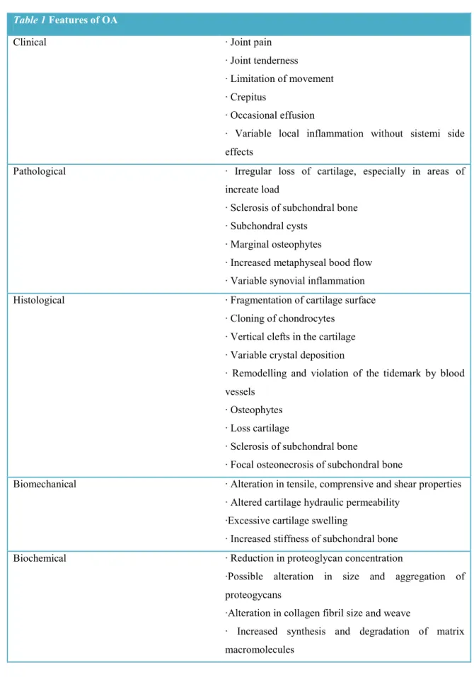
Articular cartilage is avascularized, aneural tissue, and is highly specialized tissue constituted by an extensive extracellular matrix (ECM), which is mainly composed of water, collagen type 2, collagen type 9, collagen type 11 and proteoglycan (aggrecan). (Figure 1)
The biophysical properties of cartilage derive from this highly organized fibrillar framework, that supplies shape, strength, tensile stiffness and compressive resistance to the tissue. The synthesis, maintenance and degradation of ECM proteins are coordinated by chondrocytes, the only resident cell type in cartilage. (Goldring and Marcu, 2009; Wang et al., 2011)
Figure 1: The extracellular matrix of cartilage is composed of proteoglycans attached to a backbone of hyaluronic acid that is intertwined among collagen fibrils. Proteoglycans have both chondroitin-sulfate- and keratin-sulfate-rich regions, and link proteins facilitate binding of aggrecan to hyaluronic acid.
Chondrocytes are the only cells type of the cartilage, encapsulated within a lacuna, which hinders their ability to migrate to the site of injury. Chondrocytes do not have the capacity for renewal, proliferation, or repair the damage tissue. (Wang et al., 2010)
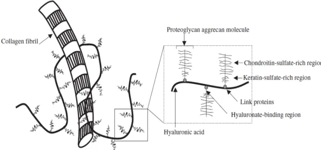
In normal cartilage, there are four zones of chondrocytes: resting cells orientating within the collagen fibers in the superficial zone, large and randomly distributed cells in the middle zone, columns of chondrocytes in the deep zone, and hypertrophic cells in the calcified zone (Figure 2). (Salminen et al.,2002)
Under normal conditions, articular chondrocytes maintain a dynamic equilibrium between synthesis and degradation of ECM components, including collagen type 2 and aggrecan, the most abundant proteoglycan in articular cartilage. (Sandell et al., 2001)
Figure 2: Zonal architecture in normal articular cartilage.
A series of catabolic and anabolic mediators have been found to play key roles in articular cartilage homeostasis. The balance between synthesis and degradation is affected by age and is regulated by several factors produced by the synovium and chondrocytes, including cytokines, growth factors, aggrecanases, and metalloproteinases (MMPs).
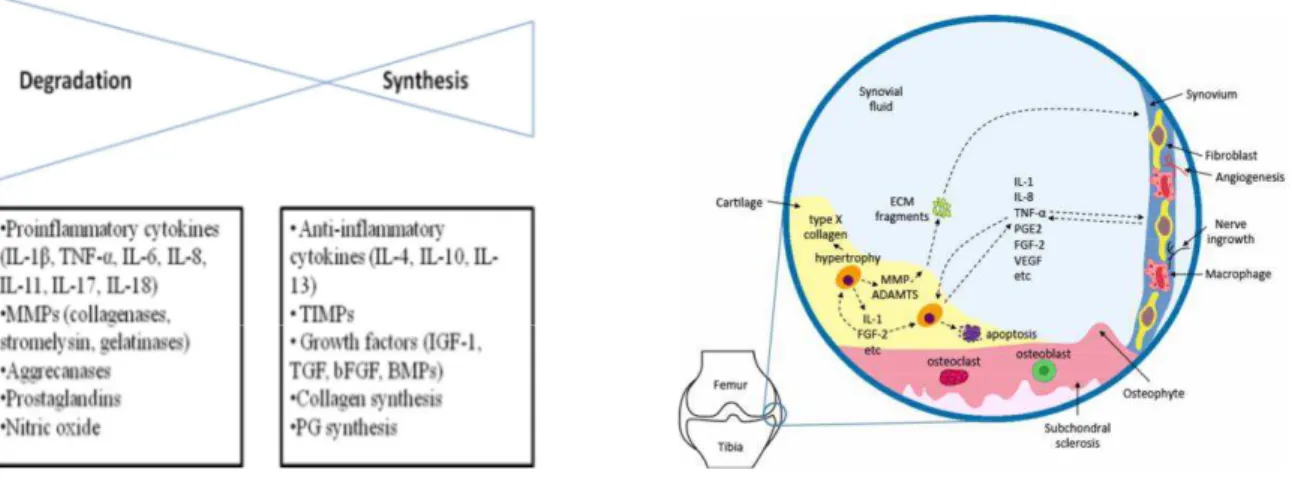
The pathogenesis of knee OA have been linked to biomechanical and biochemical changes in the cartilage of the joint mainly associated to inability to withstand normal mechanical stresses, limited supply of nutrients and oxygen, inadequate synthesis of extracellular matrix components, increased synthesis of tissue-destructive proteinases (matrix metalloproteinases and aggrecanases) and overall apoptosis of chondrocytes (Figure 3). (Buja and Krüger, 2014)
Figure 3: Main characteristics of normal and OA knee joint
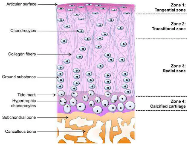
Chondrocytes can respond to direct biomechanical perturbation by up-regulating synthetic activity or by increasing the production of inflammatory cytokines (e.g. IL- -6, IL-8, TNF- , which are also produced by other joint tissues. This process causes depletion of proteoglycans and damage to the collagen network and decreases the synthesis of cartilage matrix proteins, whereas dynamic compression increases matrix synthetic activity. (Guilak et al., 2004) In response to traumatic injury, global gene expression is activated, resulting in increased expression of inflammatory mediators, cartilage-degrading proteinases, and stress response factors (Figure 4). (Goldring and Otero, 2011)
Figure 4: In osteoarthritis (OA), the balance between cartilage degradation and synthesis leans toward degradation. In osteoarthritic state, aberrantly activated chondrocytes produce ECM-degrading proteases (MMPs, aggrecanases) and pro-inflammatory cytokines (e.g. IL-1). Fragments derived from ECM degradation are also present in the synovial fluid as catabolic inducers. In OA, a subpopulation of chondrocytes undergoes hypertrophic changes, as manifested by their expression of type X collagen. Chondrocytes may also upregulate apoptosis, resulting in diminished local cellularity. In response to cartilage loss, pathological remodeling of subchondral bone gives rise to sclerosis and osteophyte formation.
Chondrocytes have receptors for responding to mechanical stimulation, many of which are also receptors for ECM components. (Millward-Sadler and Salter, 2004) The activation of these receptors can stimulate the production of matrix-degrading proteinases and inflammatory cytokines and chemokines. (Goldring MB and Goldring SR, 2007; Pulai et al., 2005)
The pathological changes of OA in articular cartilage are mainly due to an increase of catabolic factors, that cause the loss of ECM and cell apoptosis. Cartilage matrix degradation products like collagen type 2, proteoglycans, and fibronectin seem to favour cartilage destruction. (Santos et al., 2011)
A further hallmark of degenerated cartilage is the modification of chondrocyte differentiation stage, that switches them toward a hypertrophic phenotype (Goldring and Marcu, 2009; Fosang and Beier 2011), thus recapitulating some of the physiological differentiation steps occurring in growth plates and endochondral ossification.
Hypertrophic chondrocytes are characterized by the expression of terminal differentiation markers,
including Runt-related transcription factor 2 (RUNX-2), Collagen 10, MMP-13. (Van der Kraan and Van den Berg, 2012)
In fact, when OA occurs and proceeds, chondrocytes start to express several proteins such as biglycan, decorin, perlecan, and collagen type 1 and 10, so forming repaired
1.6.2 Subchondral bone
Subchondral bone consists of the subchondral bone plate and the underlying trabecular bone and bone marrow space. The subchondral bone plate consists of cortical bone and is separated from the articular cartilage by the zone of calcified cartilage.
Subchondral bone properties are modified through the cell mediated process of remodeling and modelling. Bone remodeling includes the coupling of mechanisms that reabsorb bone and form new bone on a previously reabsorbed surface, whereas bone modeling is a mechanism that drives changes in the architecture and volume of bone via direct apposition to existing bone surfaces. (Man and Mologhianu, 2014)
During OA process all of these mechanisms may be altered so resulting in subchondral bone structure changes.
Subchondral sclerosis is commonly considered an indisputable sign of OA. However, some studies suggest that different microarchitectural alterations of subchondral bone occur during different stages of OA; subchondral sclerosis may be observed only during more advanced stages of OA.
In early stages of OA, elevated bone remodeling and subchondral bone loss was observed, and was considered as a determinant of OA progression. (Li et al., 2013)
An incidence of microdamage (associated with subchondral bone stiffening and cartilage degeneration) was also reported in the subchondral bone of patients with early OA. Although the underlying mechanism for the increased bone turnover and structural deterioration in the early phase of OA is not completely understood, several factors have been implicated, including microdamage repair, increased vascularity stimulated by angiogenic factors and enhanced bone-cartilage crosstalk via increased subchondral plate pores. (Verborgt et al., 2000)
In the late stage of OA, subchondral bone microarchitectural is characterized by elevated apparent density, increased bone volume, thickening of subchondral bone plate, increased trabecular thickness, decrease of trabecular separation and bone marrow spacing. (Ding, 2010)
The overlying calcified cartilage is also thickened, with advancement and duplication of the tidemark, which contributes to articular cartilage thinning and deterioration. Despite increased bone volume density in the sclerotic subchondral bone, its mineralization is reduced and lower than that in normal or even osteoporotic joints. In OA subchondral
bone, collagen synthesis is elevated (especially collagen type 1), the deposited collagen is abnormal and this leads to abnormal mineralization. (Man and Mologhianu, 2014)
Moreover, in areas of the joint that not support the weight, usually develop further bone and cartilaginous tissue, better known as osteophytes.
The osteophytes are covered with a layer of hyaline and fibrous cartilage and blends with adjacent synovium.
The shape and position of osteophytes depends on: - the instability of the joint;
- the degree of subluxation.
However, the presence of osteophytes is not absolute proof of the OA development.
Summarizing features of OA in the subchondral bone are fibrillation, sclerosis, even collapse, together with bone cysts, thickened cortical plate, extensive remodelled trabeculae and osteophyte formation surrounding articular margins.
1.6.3 Synovial membrane
Clinical evidence shows that OA and synovitis (inflammation of the synovium) are closely related.
Abnormalities of the synovium are detectable in 50% of patients with OA; synovitis is detected by some osteoarthritic symptoms that characterize the disease, such as swelling, effusion, redness and pain.
The synovial inflammation is certainly more evident in patients who have pain, and the severity of the pain is associated with thickening of the synovial membrane. It remains unclear whether the morphological changes that occur in the osteoarthritic synovial membrane are primary or whether they are the result of joint inflammation, cartilage degradation and lesions of the subchondral bone. (Sutton et al., 2009)
Histologically, the synovial membrane of osteoarthritic joints commonly exhibits hyperplasia of the lining cell layer occasionally accompanied by focal infiltration of lymphocytes and monocytes in sublining layers. (Brandt et al., 2009)
Synovitis is believed to be induced at first by the cartilage matrix proteolytic degradation products that produce wear particles and soluble cartilage-specific neo-antigens, as well as other factors including microcrystals and abnormal mechanical stress. These components are released into the synovial fluid and are phagocyted by synovial lining macrophages,
perpetuating the inflammation of the synovial membrane through the synthesis of mediators, which in turn diffuse through the synovial fluid into the cartilage, and create a vicious circle, with increased cartilage degradation, and subsequently produce more inflammation. This also explains the increase in the amount of CD68-positive type A synoviocytes (macrophage-like), which have phagocytic capacity, in the synovial lining layer. (Martel-Pelletier and Pelletier, 2010)
The synovium produces some of the chemokines and metalloproteinases that degrade cartilage, even though the cartilage itself produces most of these destructive molecules. In turn, cartilage breakdown products, resulting from mechanical or enzymatic destruction, can induce the release of collagenase and other hydrolytic enzymes from synovial cells and lead to vascular hyperplasia in osteoarthritic synovial membranes. (Man and Mologhianu, 2014)
Synovial neovascularization may be largely driven by synovitis as inflammatory cells such as macrophages that can themselves secrete pro-angiogenic factors. These cells also secrete factors that stimulate other cells, such as endothelial cells and fibroblasts, to produce vascular endothelial growth factor (VEGF), basic fibroblast growth factor (bFGF) and other factors that further promote angiogenesis.
Angiogenesis in the synovium is closely associated with chronic synovitis and may occur at all stages of osteoarthritis.
1.7 Molecules involved in OA pathophysiology
Numerous mediators contribute to the progression of OA. Inflammatory stimuli initiate a cascade of events, including the release of cytokines by chondrocytes, leading to complex biochemical and mechanical interplay with other biological mediators to induce OA and promote pain. Particular catabolic mediators include pro-inflammatory members from the interleukin family (IL-1, IL-6, and IL-17), tumor necrosis factor- (TNF- ), and prostaglandin E2 (PGE2). Each of these mediators not only stimulates the production of cartilage-degrading proteases to induce ECM degradation, but also contributes to OA-associated pain pathways. (Bauer et al., 2006)
On the other hand a number of growth and differentiation factors that regulate cartilage development and homeostasis of mature articular cartilage have been identified. The most characterized factors which stimulate the anabolic activity in cartilage include transforming growth factor
(TGF-(FGF), insulin growth factor 1 (IGF-1). (Vinatier et al., 2009)
Moreover, since articular cartilage is an avascular tissue and, consequently, chondrocytes receive oxygen and nutrients via a passive diffusion from the synovial fluid, and their adaptation to low oxygen tension is mediated by transcription factors, such as hypoxia inducible factor (HIF). (Schipani et al., 2001)
1.7.1 Interleukin-1 (IL-1)
IL-1 is one of the most well-studied cytokines involved in OA.
IL-1 demonstrates potent bioactivities in inhibiting ECM synthesis and promoting cartilage breakdown, represses the expression of essential ECM components (e.g. aggrecan and collagen type II) in chondrocytes and induces proteolytic enzymes such as collagenases (MMP-1 and MMP-13) and ADAMTS-4, in both chondrocytes and synovial fibroblasts. In addition to these direct effects, IL-1 induces a variety of other cytokines, including IL-6, IL-8, and leukemia inducing factor (LIF), which interact to induce additive or synergistic effects in the catabolic cascade. (Goldring et al., 1988)
1.7.2 Interleukin-6 (IL-6)
IL-6 is a pro-inflammatory cytokine that is involved in cartilage degradation, and it has also been associated with hyperalgesia and hypersensitivity in joint tissues. IL-6 plays an important role in the pathogenesis of arthritis diseases, and its concentration is elevated in the serum and synovial fluid of arthritic patients. (Lee et al., 2013)
The production of IL-6 in the tissues of the affected joint is usually in response to IL-1 and TNF and is mainly implemented by chondrocytes, osteoblasts, fibroblast-like synoviocytes, macrophages, and adipocytes. (Wojdasiewicz et al., 2014)
The effect of IL-6 on joint cartilage is not different from other cytokines and, in synergy with them, causes a decrease in the production of type II collagen and increases the production of enzymes from the MMPs group. (Porée et al., 2008)
IL-6 is considered to be the key cytokine, which causes changes in the subchondral bone layer. Its effect is largely based on promoting the formation of osteoclasts and thus bone resorption while showing synergism with 1 and TNF . Osteoblasts stimulated by IL-1 , TNF , and IL-6 become a source thereof and may also produce MMPs by adversely affecting the cartilage located near it. (Wojdasiewicz et al., 2014)
1.7.3 Tumor necrosis factor- (TNF- )
TNF- is known for its powerful catabolic effects in the pathophysiology of OA. In OA patients,
TNF-subchondral bone and cartilage. TNF- inhibits synthesis of the major extracellular matrix (ECM) components: proteoglycans, collagen type II and cartilage link protein. Moreover, TNF- up-regulates expression and release of various cartilage-destructive MMPs, like MMP-1, MMP-3 and MMP-13.
It is an extremely potent pro-inflammatory cytokine that interacts with chondrocytes by binding to receptors on their surface. (Haseeb and Haqqi, 2013)
In fact, TNF- has an exacerbating effect on inflammation by inducing production of the pro-inflammatory cytokines and chemokines: IL-6, IL-8, IL-1 via the receptors TNFR1 and TNFR2. (Wojdasiewicz et al., 2014)
Systemic
1.7.4 Prostaglandin E2 (PGE2)
During pro-inflammatory states in articular cartilage, numerous enzyme products are produced and released, including PGE2, that is considered to be the major contributor to inflammatory pain in arthritic conditions. PGE2 exerts its effects via a variety of E prostanoid (EP) receptors (EP1, EP2, EP3, EP4), which are present in both peripheral sensory neurons and the spinal cord. Activation of these receptors induces a variety of effects, ranging from calcium influx to cAMP activation or inhibition
It has been shown that IL-1 stimulates and produces high levels of PGE2 that may induce pain and the degeneration in OA. Further, PGE2, when combined with the catabolic cytokine IL-1, synergistically up-regulates IL-6 mRNA levels. (Li et al., 2009)
1.7.5 Fibroblast growth factor (FGF-2)
FGF-2, a potent catabolic and anti-anabolic factor, play an important role in human cartilage homeostasis. FGF-2 is released in higher amounts during loading and/or injury of the cartilage matrix and activates multiple transduction signal pathways (MAPKs), such as ERK, p38, and JNK. These kinases in turn phosphorylate a set of transcription factors to regulate gene expression and modify cellular function, resulting in a decrease in proteoglycans synthesis and antagonism against anabolic growth factors, such as insulin-like growth factor 1 (IGF-1) and bone morphogenetic protein (BMP-7) in articular cartilage. FGF-2 potently stimulates MMP-13 expression, which is the major type II collagen-degrading enzyme. (Im et al., 2008)
However anabolic activity of FGF-2 in articular cartilage has been reported. (Ellman et al., 2008)
In adult cells, the chondrogenic effect of FGF has been confirmed in some studies. In adult chondrocytes, it has been shown that FGF2 has various property, in fact it has mainly a mitogenic, proliferative and chondrogenic factor. Furthermore, the contradictory results for the potential role of various FGFs in chondrogenesis highlight the need for a better characterization of the signalling pathways that are activated by FGFs to be able to fully understand how they affect FGF activity. (Vinatier et al., 2009)
1.7.6 Insulin-like growth factor (IGF)
The insulin-like growth factor (IGF) family comprises the ligands IGF-1 and IGF-2, the receptors IGF1R and IGF2R, at least six different IGF-binding proteins (IGFBPs) and multiple IGFBP proteases, which regulate IGF activity. IGF-2 mainly has a role in embryonic and foetal development, whereas IGF-1 is more relevant for cartilage repair. In adults, IGF-1 and IGF1R are expressed by chondrocytes, osteoblasts and osteoclasts. IGF-1 is considered an essential mediator of cartilage homeostasis through its capacity to stimulate proteoglycan synthesis and to promote chondrocyte survival and proliferation. IGF-1 was also able to induce migration of chondrocytes and, moreover, the combined use of chondrocytes and IGF-1 seemed to improve the overall consistency of the repair tissue. (Vinatier et al., 2009)
However, the ability of chondrocytes to respond to IGF-I decreases with age and in OA. Evidence suggests an uncoupling of IGF-I responsiveness in OA, indicating that, in OA cartilage IGF-I is able to robustly stimulate proteoglycan synthesis, but it is unable to modulate proteoglycan catabolism. (Martin et al., 1997)
1.7.7 Hypoxia inducible factor (HIF)
HIF is a heterodimer that consists of the subunit HIF-1 or -2 and the aryl hydrogen receptor nuclear translocator (ARNT) subunit, also known as HIF-1 .
Whereas HIF-1 is stable in normoxic conditions, HIF- 1 and -2 are unstable and are rapidly degraded through the ubiquitin proteasome pathway. Under hypoxic conditions, HIF-1 and -2 are stabilized and translocate from the cytoplasm to the nucleus, where they heterodimerize with ARNT to bind to the hypoxic responsive element (HRE), thereby initiating the transcription of hypoxia-specific genes. HIF-1 has been shown to be essential for growth arrest and survival of chondrocytes.
Hypoxia has also been shown to increase the synthesis of ECM proteins in cultured chondrocytes in vitro. These data suggest that low oxygen tension is a key regulatory factor of proliferation, differentiation and activity of chondrogenic cells. Interestingly, hypoxia has also recently been suggested to inhibit the expression of collagen type X, which is the major marker of chondrocyte hypertrophy. (Vinatier et al., 2009)
gene expression of two main matrix components: collagen II and aggrecan. (Duval et al., 2009)
An increasing transcription of HIF-1 in OA cartilage compared to normal samples was shown, particularly in the late-stage of the disease. Consistent with this evidence, subsequent studies reported a growing number of HIF-1 -positive chondrocytes during OA progression and a higher expression of HIF-1 mRNA in degenerated cartilage compared to uninjured cartilage.
In addition to hypoxic conditions, HIF-1 expression can be up-regulated by other factors, including inflammatory cytokines (IL-1 and TNF- ), reactive oxygen species and mechanical loading, which are all recognized as key players in cartilage damage. (Yudoh et al.,2005)
Since HIF-1 has a pivotal role in supporting chondrocyte survival and cartilage homeostasis, these characterize HIF-1 as a key factor for chondrocyte survival promoting compensatory mechanisms in response to catabolic modifications of OA cartilage.
2. Therapeutic treatments for OA
Treatments aim at educating the patient about OA, reducing pain, optimizing and maintaining physical function, and preventing or retarding progression of adverse structural damage affecting the joint tissues (cartilage, bone, ligament, muscle). Actually available treatments include a wide range of non-pharmacologic, pharmacologic and surgical modalities.
The basic premise for the management of patients with symptomatic OA is that it involves a combination of both non-pharmacologic and pharmacologic modalities that are
-pharmacologic modalities that can be added as necessary for additional symptom control, in some cases require the surgery. (Gossec et al., 2007)
In addition to non-surgical treatments (e.g. physiotherapy, diet rich in vitamin D and supportive sport, like swimming), there are several medicinal and homeopathic products on the market, which promise pain relief and a decrease in symptoms. However, researchers have a keen interest in investigating new treatments to cure OA of the knee. (Uth and Trifonov, 2014)
Currently available pharmacological therapies target palliation of pain and include analgesics (e.g., acetaminophen, cyclooxygenase-2-specific inhibitors, selective non-steroidal anti-inflammatory drugs, tramadol, opioids), intra-articular therapies (glucocorticoids and/or hyaluronic acid, HA), and topical treatments (i.e. capsaicin, methylsalicylate). (Moreland, 2003)
Treatments are individual for each patient and they are dependent by severity of pain and stiffness of affected joints and by specific response to the treatment. It is important to work with a healthcare provider to create an effective and acceptable plan in the long term for living with arthritis.
2.1 Non-pharmacological therapy of osteoarthritis
Non-pharmacological treatments can substantially improve OA symptoms, and they are usually the first treatments recommended.
They include:
Rest: OA symptoms are typically worsened by activity and are improved with rest. However, a complete lack of activity can lead to a loss of muscle and joint stiffness. If arthritis causes significant pain and inflammation, healthcare provider may recommend rest for 12 to 24 hours, followed by a return to usual activities. It would seem sensible if something hurts to rest it. This may only be true in acute situations and may not hold for chronic conditions.
Muscle loss is a feature of both rheumatoid arthritis and OA, and so pain is not an indicator of musculoskeletal damage. (Hochberg et al., 2012)
Weight loss: Obesity is strongly linked to the development of arthritis of the knee. Weight loss, even modest weight loss, appears to lower this risk. It is not known if weight loss slows the worsening of arthritis in joints that are already affected. However, weight loss may reduce joint pain in weight bearing joints, such as the hips and knees. (Bliddal et al., 2011)
Physical therapy and exercise programs: Physical therapy and exercise improve
flexibility and strengthen the muscles surrounding the joints. People who exercise regularly despite their arthritis will typically have less pain and better function than those who are inactive. Canes, walkers, electric-powered seat lifts, raised toilet seats, and tub and shower bars can reduce the stress on joints and can make it easier to perform daily tasks. A physical therapist may suggest these and other assistive devices, depending upon the severity and location of arthritis. (Fransen et al., 2001) Vitamins: Studies have linked certain vitamins to joint health, but the role of vitamins in arthritis treatment is uncertain. OA is less likely to worsen in people who have a high dietary intake of vitamin C (ascorbic acid) and a high dietary intake and high blood levels of vitamin D. However, it is unknown if supplementation with these vitamins has the same effects or if high dietary intakes of vitamins can prevent the onset of OA.
Thermoterapy: Thermotherapy has for many years been advocated as a useful adjunct to pharmacological therapies. Ice is used for acute injuries and warmth is
used for sprains and strains. It seems appropriate to use hot and cold packs in osteoarthritis knee.
Applying heat and cold to arthritic joints can help to control arthritis symptoms such as pain and stiffness.
1) Heat therapy: Heat relieves pain and stiffness in arthritic joints. Heat can be applied to the joints with hot packs, hot water bottles, heating pads, or electrically heated mittens. It is important to avoid burning the skin with heat therapy. To avoid burns, hot water bottles should be filled with warm, not boiling, water. Heating pads should be set on a timer and used for no more than 20 minutes at a time. The heating pad can be reapplied after 20 minutes of no use.
2) Cold therapy: Cold relieves pain in arthritic joints and reduces muscle spasms. Cold can be applied for short periods using ice packs or coolant sprays. People with certain medical conditions, such as the Raynaud phenomenon, should not use cold therapy. (Brosseau et al., 2003)
Transcutaneous electrical nerve stimulation (TENS): A TENS unit delivers a mild electrical current to the skin, stimulating nerve fibers in the skin that may interfere with the transmission of pain signals from the arthritic joint. The use of TENS as an arthritis treatment is controversial. Some studies have found that those who use TENS for arthritis of the knee have reduced knee pain, a greater ability to bend the knee, and a reduced duration of morning stiffness. However, another study found that TENS was no more effective for relieving pain than the drug naproxen (Aleve, Anaprox) or a placebo. (Hochberg et al., 2012)
Dietary supplements Glucosamine and chondroitin are dietary supplements that have received a lot of attention for their potential benefit in reducing pain and in slowing the progression of arthritis.
1) Glucosamine: Glucosamine was no more effective in relieving arthritis pain or in improving function than placebo in a well-designed, controlled trial; it is possible that other formulations may be effective. Glucosamine does not appear to slow the worsening of arthritis over the long term. There are few side effects of glucosamine; it should not be used by patients who are allergic to shellfish. 2) Chondroitin: Chondroitin used alone appears to provide little benefit for people
The combination of glucosamine and chondroitin sulfate has not proven to be better than placebo for pain relief or for functional improvement in patients with OA of the knee. (Clegg et al., 2006)
Traditional Chinese medicine: Several components of traditional Chinese medicine, including herbs and acupuncture, may help control the arthritis symptoms in some people, although the benefits of these therapies have not been confirmed in large, well-designed clinical studies. Reumalex, willow bark, stinging nettle, Articulin-F, d
(ASU), and Phytodolor may improve arthritis pain, while other herbs and combinations such as Eazmov, Gitadyl, or ginger extract are probably ineffective. (Vickers et al., 2012)
2.2 Pharmachological treatments of osteoarthritis
An appropriate pharmacological treatment forms is the main key for treating osteoarthritis when non-pharmacological therapy on its own is insufficient. The use of such analgesia may be use to cure different kind of pain, including night pain or exercise-associated pain. Oral analgesics, especially paracetamol, have been used for many years, with increasing use of opioid analgesics in recent years, partly fuelled by fears over the safety of NSAIDs. (NICE, 2014)
Pharmacologic modalities could potentially be divided into topical, intra-articular and oral (systemic) agents. Furthermore, those that relieve symptoms may have a rapid or slow onset of effect.
Topical agents: Some people experience relief of arthritis pain when they apply creams containing capsaicin, the active substance in hot chili peppers. Capsaicin depletes a pain-causing substance in nerve endings and lessens the arthritis pain by about 30 percent in some people. Forty percent of people experience side effects when using capsaicin cream, including burning, stinging, and redness of the skin and especially the eye. (Vaile and Davis, 1998) There are a variety of non-steroidal anti-inflammatory drug (NSAID) preparation that are available for topical application. Analgesics relieve pain but do not have any effect on inflammation. These drugs are often recommended when arthritis pain does not respond to non-pharmacologic measures. Drugs in this class include acetaminophen and opioid (narcotic) analgesics. Acetaminophen (Tylenol and others) can relieve mild to moderate arthritis pain. To avoid the serious but rare side effects of kidney and/or liver damage due to acetaminophen, it is important to follow dosing instructions and to avoid drinking excessive amounts of alcohol. The sudden pain, severe arthritis exacerbations may require treatment with narcotic analgesics such as codeine. Narcotics should be taken for only short periods of time because they can be addictive.
Intra-articular therapy: Two types of injections are used for people with arthritis pain: corticosteroid injections or hyaluronic acid injections.
1. Corticosteroid injections: Corticosteroid can suppress inflammation and can relieve arthritis symptoms when injected into arthritic joints. corticosteroid injections may be recommended for people who have OA confined to a few joints and who still have pain despite the use of NSAIDs. Corticosteroid injections may also be recommended for people with OA who cannot take NSAIDs. Joint injections have few side effects, but some people experience a brief flare of arthritis symptoms after an injection. There is also a small risk of joint infection. Glucocorticoids may damage certain joints when injected frequently. Therefore, clinicians recommend no more than three to four injections per year for each particular weight bearing joint such as a knee. Corticosteroids have not only anti-inflammatory effects but also immunosuppressive effect. The presence of an effusion is not in itself an indication for corticosteroid injection, unless there is significant restriction of function associated with the swelling. Rather, the indication should be based on severity of pain and disability. (Jevsevar, 2013)
2. Hyaluronic acid (HA) injections Normal joint fluid contains a large amount of hyaluronic acid, which allows the joint fluid to be slippery. Synthetic HA may be injected into the knee to treat arthritis. After the injection, pain relief may last for several months. HA is generally injected in the knee, but their use in other joints is being studied. Joint inflammation can occasionally occur after this type of injection and as with steroids, it is possible a small risk of infection. HA injections are generally reserved for people with OA who cannot take NSAIDs or who do not achieve adequate pain relief with them. People awaiting joint surgery may benefit from these injections. HA is administered as 3-5 intra-articular weekly injections and is generally well tolerated, with a low incidence of local adverse events (from 0% to 13% of patients) that was similar to that found with placebo.
Because the residence time of exogenously administered HA in the joint is relatively short (approximately less than 26 hours), a variety of chemical modifications of the molecule, such as coupling or cross-linking, have been tested
as a means of increasing both residence time and viscoelastic properties. (Borzacchiello et al., 2010; Larsen et al., 2008)
HA is a large, linear glycosaminoglycan and is a major non-structural component of both the synovial and cartilage extracellular matrix. It is also found in synovial fluid and is produced by the lining layer cells of the joint. HA is removed from the joint via the lymphatic circulation and degraded by hepatic endothelial cells. Its key functions in the joint are to confer viscoelasticity, lubrication and help maintain tissue hydration and protein homeostasis by preventing large fluid movements and
umbilical cord tissue and developed for clinical use in ophthalmic surgery and arthritis in the 1960s. The beneficial effects in ophthalmic surgery were followed by the use of HA in osteoarthritis: the rationale was to replace the properties lost by reduced HA production and quality as occurs in osteoarthritis joints, a concept known as viscosupplementation. Commercial preparations of HA have the same structure as endogenous HA although cross-linked HA molecules (known as hylans) were later engineered by linking HA molecules in order to obtain greater elastoviscosity and intra-articular dwell-time. However, the mechanism by which HA exerts its therapeutic effect, if any, is currently unknown, and evidence for restoration of rheological properties is lacking. It has been suggested that two stages might be involved; an initial biomechanical stage followed by a physiological stage. It is suggested that biomechanical mechanisms initially come into effect when the synovial fluid in the osteoarthritic joint is replaced by the higher molecular weight exogenous HA. Clinical studies report that exogenous HA contribute in restoring the elastoviscosity, and the lubricating and shock absorbing abilities, of synovial fluid. It is noted that physiological mechanisms may account for the clinical benefits of intra-articular administration of HA that persist beyond the residence time of HA, although evidence has largely been obtained from preclinical studies. Given the relatively short intra-articular residency (hours to days), any hypothesis for its mechanism of action must account for the sometime reported long-duration of clinical efficacy (months). (Lohmander et al., 1996; Altman and Moskowitz, 1998)
Nowadays the development of pharmacological treatments with the potential for structure-modifying activity in OA joint treatment has become a major focus in the field of OA research. Such compounds retard or stabilize the progression of
established OA by altering the underlying pathological processes. There is a growing body of preclinical and clinical data, which suggests that intra-articular injection of hyaluronan (HA) has a disease-modifying effect, in addition to its proven efficacy and safety in treating the OA patients, aimed at relieving pain and regaining function. (Grishko et al., 2009; Elron-Gross et al., 2008)
Oral pharmacological agents: This group of agents includes analgesics such as non-opioid analgesics (e.g. paracetamol) and opioid analgesics, or anti-inflammatory agents such as NSAIDs.
1. Non-opioid analgesics: for many patients with OA, relief of mild to moderate joint pain can be achieved with paracetamol. However, in several trials, NSAIDs have been more efficacious, particularly in those with moderate to severe symptoms from their OA. (Altman and Moskowitz 1998)
2. Opioid analgesics: there is limited published clinical research, weak opioid analgesics have been commonly administered for OA. Stronger narcotics or narcotic derivatives are useful in selected patients with OA. Narcotics are used most often as rescue medications for severe pain
3. Non- steroidal anti-inflammatory drugs: NSAIDs relieve pain and reduce inflammation. Many of the nonprescription products that are available for treating arthritis pain are NSAIDs. These drugs are often recommended before analgesics for people who have osteoarthritis and evidence of inflammation. They are also recommended for some people with non-inflammatory OA who do not get adequate pain relief with simple analgesics. Non-steroidal anti-inflammatory drugs (NSAIDs) have been available for many years and are thought to work by reducing the production of pro-inflammatory and pain-related prostaglandins. The discovery of different cyclooxygenase (COX) enzymes with different physiological actions brought with it the concept that differential blockade of COX-1 (important in normal regulation of the gastro-intestinal (GI) mucosa) and COX-2 (up-regulated at sites of inflammation amongst other functions and thought responsible for pro-inflammatory mediator production) may provide effective analgesic/anti-inflammatory actions without the common GI complications of traditional NSAIDs. These GI complications are well known to clinicians and include a spectrum of problems from
dyspepsia and ulcers to life-threatening ulcer perforations and bleeds. However the blocking of COX-2 always carried the potential for a pro-thrombotic effect, by changing the balance of pro- and anti-thrombotic mediators. (NICE, 2014; Camu et al., 2002)
2.3 Differents agents
Although research continues into symptom-modifying medications for OA, there is a new emphasis on the development of structure modifying agents. Structure modifying medications are intended to retard, prevent or reverse the progression of OA.
Several groups of agents are under investigation, mostly directed at cartilage repair: - Growth factors and cytokine
- Sulphated and non-sulfated sugars - Hormones and other steroids - Enzyme inhibitors
- Chondrocytes/stem cell transplantation - Hyaluronan with structure-modifying
2.4 Surgery
Surgery is usually reserved for severe arthritis that significantly limits your activities and that does not respond to other arthritis treatments.
Furthermore, those who undergo surgery should be in the best possible physical condition and should be prepared for rehabilitation after surgery.
Only 5% of OA patients need surgical treatment when conservative treatment displays no satisfactory effect. There are four categories of surgical procedures: osteotomy, arthroscopy, arthrodesis, and arthroplasty.
The purpose of the osteotomy is to transfer the load bearing from the pathologic to the normal compartments of the knee. Thirteen studies involving over 693 people indicated that valgus high tibial osteotomy is effective in improving knee function and relief of pain. However, it is uncertain which treatment, osteotomies or conservative treatment is more effective. Furthermore, a successful result of the osteotomy (about 60.3%) depends on
proper patient selection, stage of osteoarthritis, and achievement and maintenance of adequate operative correction.
Arthroscopy is an alternative procedure to osteotomy. It is not only useful for the treatment of the same symptoms, but also for diagnosis of the disease. Although arthroscopic methods are widely used, they are not suitable for patients who have displayed OA symptoms for more than 2 years, or who display tibial osteophytes and joint space narrowing of less than 5 mm. Arthroscopy is effective only temporarily in reducing the pain of mild to moderate hip OA.
Arthrodesis is an efficient procedure for OA of the hands, feet, ankles, and spine, but usually not for the hip and knees.
Arthroplasty refers to the insertion of an artificial joint in order to restore the integrity and the function of the joint withered by OA. Joints commonly deteriorate after more than ten years, depending on the composition. The perioperative morbidity of unicompartmental knee arthroplasty would be less than total knee arthroplasty; furthermore, this arthroplasty has been reported to be used in very elderly patients (79-94 years) with tricompartment OA. Therefore, age is not a limiting factor for this surgical treatment.
3. Hyaluronic acid (hyaluronan)
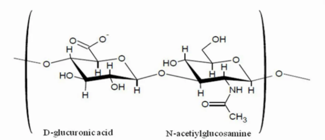
Hyaluronic acid, also called hyaluronan (HA) is a carbohydrate, more specifically a mucopolysaccharide, occurring naturally in all living organisms. It can be several thousands of sugars (carbohydrates) long. When not bound to other molecules, it binds to
extracellular matrix and pericellular matrix, but has also been shown to occur intracellularly. The biological functions of HA include maintenance of the elastoviscosity of liquid connective tissues such as joint synovial and eye vitreous fluid, control of tissue hydration and water transport, supramolecular assembly of proteoglycans in the extracellular matrix, and numerous receptor-mediated roles in cell detachment, mitosis, migration, tumor development and metastasis, and inflammation. (Toole and Hascall 2002; Turley et al., 2002)
The unique viscoelastic nature of HA along with its biocompatibility and non-immunogenicity has led to its use in a number of clinical applications, including the supplementation of joint fluid in arthritis (Medina et al., 2006), as a surgical aid in eye surgery, and to facilitate the healing and regeneration of surgical wounds. (Necas et al., 2008)
In addition the high exclusion properties of HA molecules restrict the entry of plasma proteins into the aqueous phase of synovial fluid. This exclusion effect is dependent on the molecular weight (MW) of the proteins.
By contrast HA facilitates the transport of water and smalls solutes through synovial fluid to articurlar cartilage from capillaries in the synovium and reduces fluid loss as intra-articular (IA) pressure is raised during joint flexion. These properties of HA are important for the nutrition of articular cartilage as well as for the elimination of metabolites and noxious substance from the joint cavity. (Gosh and Guidolin, 2002)
3.1 Biochemistry of hyaluronan
HA is a linear polysaccharide composed of repeating disaccharide units, 1,4-glucuronic acid (GlcUA) and 1,3-N-acetylglucosamine (GlcNAC). HA belongs to the group of glycosaminoglycans, but unlike chondroitin sulfate or keratan sulfate, HA is not sulfated. HA is synthesized by bioactivity of hyaluronan synthase (HAS), which has been reported to have three isoforms (HAS1, HAS2, and HAS3) in humans. (Masuko et al., 2009)
HA is characterizes by an high molecular weight (Figure 5) and its relatively simple structure is conserved throughout all mammals, suggesting that HA is a biomolecule of considerable importance. (Necas et al., 2008)
3.2 HA receptors
HA plays several important organizational roles in the extracellular matrix (ECM) by binding with cells and other components through specific and nonspecific interactions. Receptors are constituents of the extracellular matrix, and stabilize its integrity. Hyaluronan receptors are involved in cellular signal transduction. It is now widely accepted that HA binds to specific receptors, CD44, ICAM-1, LYVE-1 and RHAMM, that are expressed by a wide range of cells, including those implicated in the pathology of OA (e.g. inflammatory cells, synoviocytes and chondrocytes). (Banerji et al., 1999) The finding of specific cell receptors for HA, notably CD44, support a pharmacological mechanism of action for this GAG in OA, and because these receptors are widely distributed on the surfaces of many types of cells, it also accounts for the diversity of its effects.
CD44 is a structurally variable and multifunctional cell surface glycoprotein expressed on most cell types, including macrophages and hepatocytes, and has been implicated in many biological process. (Kang et al., 2013) CD44 has been reported to regulate a variety of inflammatory responses, including the induction of pro-inflammatory cytokines and the migration of macrophages and neutrophils. (Hollingsworth et al., 2007)
ICAM: also known as CD54 (Cluster of Differentiation 54) is a protein that in humans is encoded by the ICAM1 gene. This gene encodes a cell surface glycoprotein which is typically expressed on endothelial cells and cells of the immune system.
LYVE-1: the first identification of an HA receptor that is almost exclusively expressed on lymph vessels and is absent from blood vessels. (Banerji et al., 1999)
RHAMM (Receptor for HA-Mediated Mobility), has been found on cell surfaces, as well as in the cytosol and nucleus. (Leach et al., 2004) It has been implicated in regulating cellular responses to growth factors and plays a role in cell migration, particularly for fibroblasts and smooth cells. (Necas et al., 2008)
The binding of HA to CD44 and RHAMM and their various isoforms has been reported to trigger a variety of intracellular signal events, such as the protein phosphorylation cascades, cytokine release and stimulation of cell cycle proteins. (Gosh and Guidolin, 2002)
3.3 Mechanism of action
HA is highly hygroscopic and this property is believed to be important for modulating tissue hydration and osmotic balance. (Dechert et al., 2006) In addition to its function as a passive structural molecule, HA also acts as a signalling molecule by interacting with cell surface receptors and regulating cell proliferation, migration, and differentiation. (Necas et al., 2008)
Synovial cells, fibroblasts and chondrocytes synthesize and secrete HA into the joint, contributing to enhance viscosity and elastic nature of synovial fluid. In the osteoarthritic joint, synovial inflammation leads to increased permeability of the synovial membrane for HA. Also, the elevated synovial fluid levels of free radicals, inflammatory cytokines, and proteolytic enzymes in osteoarthritic knees impair HA function and contribute to the progression of OA. Therefore in OA, both the molecular weight and the concentration of HA are decreased. (Moreland, 2003)
The intra articular injection of HA is thought to restore normal viscoelastic properties of the pathologically altered synovial fluid, which explains why it was defined the term of the
. (Balazs and Denlinger, 1993)
Moreover, several studies suggest that viscosupplements also have disease modifying effects, such as reduction of synovial inflammation, protection against cartilage erosion (Amiel et al., 2003), and promotion of intra-articular HA production. (Ayhan et al., 2014) HA possesses a number of functions that may provide some additional chondroprotective effects and may explain its longer term effects on articular cartilage.
3.3.1 Condroprotective effects
The physical properties of HA are important but there is evidence to suggest that HA may provide both physiochemical and pharmacological advantages. Chondrocytes express the glycoprotein CD44 on their cell surface. This has the capacity to function as a HA receptor and so may be involved in biochemical interactions with chondrocytes. The effect of a HA injection may be mediated via CD44 interactions. (Necas et al., 2008)
Therefore, HA has important chondroprotective effects, indeed covers the articular surface and exerts mechanical protection on synoviocytes of the articular cartilage, preventing cell damage by mechanical stress. In articular cartilage HA allows the organization of proteoglycans in huge aggregates and act as a filter against the free diffusion of molecules through the synovial membrane. Maintenance of normal structure of HA is essential for the homeostasis of the environment and any event that could change the physiological characteristics of this polysaccharide can cause serious repercussions.
3.3.2 Effects of hyaluronan on the extracellular matrix
Beneficial effects on proteoglycans synthesis have also been demonstrated in vitro with HA. This glycosaminoglycan has been shown to increase proteoglycans synthesis in equine articular cartilage, rabbit chondrocytes, and bovine articular cartilage treated with IL-1, which has been shown to reduce proteoglycans synthesis in vitro. An increase in high-MW proteoglycans production was also demonstrated with HA in cells of rabbit ligament. (Ghosh et al., 1995)
HA also reduced the expression of IL-1 and stromelysin (MMP-3), two mediators known to play a role in cartilage degradation. (Takahashi et al., 1999)
A reduction in collagen gene expression induced by IL-1 in rabbit articular chondrocytes has also been suppressed by HA. In an in vivo model of canine OA, a reduced amount of glycosaminoglycan release was found in HA-treated joints compared with an increased release in untreated joints.
HA has also been shown to suppress cartilage damage by fibronectin fragments in vitro and in vivo. This protective effect was associated with its coating of the articular surface, suppression of fibronectin-fragment-enhanced stromelysin-1 release, increased